Hand Bone
Scaphoid fracture and nonunion
Scaphoid waist fracture diagnosis, screw fixation, nonunion and advanced collapse.
36 questions
4 source pages
2 images
1 fact-check flags
Images appear with the first question taken from each source page — tap a question to open it.
Q1Describe the X-ray findings of this scaphoid waist fracture.📷▸
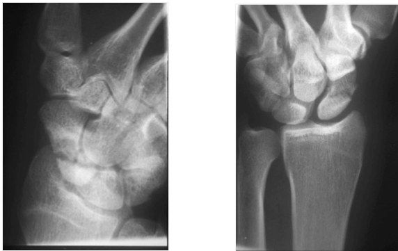
- Complete, displaced >1mm, horizontal oblique scaphoid waist fracture
- Unstable according to the Herbert classification (Herbert B)
- Acute - no cyst, sclerosis or arthritis
- Associated SL widening; no carpal collapse or other fracture
Q2Why are scaphoid fractures prone to nonunion?▸
- Biological: retrograde blood supply, no tendon attachment, 80% covered with cartilage, lacks osteogenic cambium layer, bathed in synovial fluid, small bone contact
- Mechanical: connects the two carpal rows, area of high stress; proximal fragment extended/radially deviated by SL and DIC, distal fragment flexed/ulnar deviated around the RSC
- Delayed presentation: 25% not seen on initial X-ray
Q3Describe the Herbert classification of scaphoid fractures.▸
- A: stable - tubercle, horizontal waist, treated conservatively
- B: unstable
- C: delayed (6 weeks)
- D: nonunion
- Russe adds orientation: horizontal oblique, transverse, vertical oblique
Q4What is the blood supply of the scaphoid?▸
- All supply is retrograde
- Proximal 80% via the dorsal carpal artery
- Distal 20% via the superficial palmar branch
- No anastomosis between them
Q5What is the conservative management and union rate for a scaphoid waist fracture?▸
- Short arm cast with thumb left free, wrist slightly extended, functional cup position
- Cast for 8 weeks, union rate 90-95%
- Scaphoid cast management is reliable in undisplaced stable waist fracture with union rate up to 90%
- Duration by site: 8/10/12 weeks and union 90, 80, 60% for distal/waist/proximal fractures
- No consensus on long versus short arm cast or thumb immobilisation
- Netherlands meta-analysis (J Trauma 2011, 500 patients): no difference in union, pain, grip, time to union or osteonecrosis
Q6What is the evidence for cast versus surgery in scaphoid waist fractures?▸
- SWIFT trial (Dias, Lancet 2020): fractures displaced 2mm or less should be treated with cast immobilisation, with early fixation of confirmed nonunion
- Surgery gives similar long-term outcomes but more complications
- McQueen (JBJS 2008): surgery shortens time to union (9 vs 14 weeks) and earlier return to work, but no difference in union rate, ROM or grip at 2 years
Q7What is the summary diagnosis and which further views are requested?▸
- Herbert B scaphoid waist fracture, suspicious of SL widening
- Better lateral view for displacement and humpback deformity
- Stress view for SL dissociation
Q8What history is taken in a scaphoid fracture?▸
- Age, hand dominance, occupation, functional demand
- Mechanism of injury, delayed presentation, symptoms
Q9What examination findings are sought in scaphoid fracture?▸
- Localised tenderness over the anatomical snuffbox and SL interval
- Rule out other injuries
Q10What is the acute first aid for a scaphoid fracture?▸
- Analgesics, rest, elevate
- Short arm thumb spica slab
Q11Which patient and fracture factors guide definitive scaphoid management?▸
- Timing, orientation, associated injuries
- Displacement, site
- Patient counselling
Q12What is the aim and technique of surgical fixation of a scaphoid waist fracture?▸
- Reduce with stable fixation and compression; union rate up to 97%
- Percutaneous approach with K wire as a joystick for reduction
- Headless compression screw with differential pitch targeting the central longitudinal axis
Q13How is a grossly displaced scaphoid fracture approached?▸
- Volar approach for ORIF - direct visualisation of the whole scaphoid, correct humpback deformity, preserve dorsal blood supply
- Dorsal approach if the fracture is proximal
Q14Describe the percutaneous approaches to the scaphoid.▸
- Volar: incision distal to STTJ, entry at scaphoid tubercle abutting STT; 12G hypodermic needle as guide; reduce by hyperextension and ulnar deviation; bean view - axis from middle third of scaphoid tubercle to distal half of proximal pole
- Dorsal: pronate and flex until the scaphoid is seen as a double ring; guidepin at centre; length 18-24mm (-4mm from measured)
Q15Describe the dorsal open approach to the scaphoid.▸
- Longitudinal incision over Lister tubercle, between the 3rd and 4th compartments, between EPL and EDC
- Incise extensor retinaculum and dorsal capsule up to the DIC; flex the wrist over a bolster to reveal the proximal pole
- Danger: superficial branch of radial nerve, dorsal carpal branch of radial artery
- Pros: can address SL, also good for perilunate dislocation
Q16Describe the volar (Russe) approach to the scaphoid.▸
- Incision along FCR then towards the scaphoid tubercle
- Incise RSC (preserve the RSC stump to prevent humpback) and long radiolunate ligament
- Pros: good exposure, correct humpback, avoids the dorsal carpal branch of the radial artery
- Cons: radial artery at risk
Q17What is a good lateral (SPC) view and how is displacement measured?▸
- SPC view: volar cortex of pisiform should lie between the middle 1/3 of the interval between volar cortex of scaphoid and capitate
- Displacement >1mm = displaced
- Radiolunate angle >15deg dorsal = DISI; capitolunate >15deg volar = VISI
- Intra-scaphoid angle >35deg = humpback deformity
Q18On PA and AP wrist views, where is the ulnar styloid seen?▸
- PA view: ulnar styloid is peripheral (ulnar)
- AP view: ulnar styloid is central
Q19If a scaphoid fracture is suspected but not seen on initial X-ray, what is the next step?▸
- Scaphoid series: PA, lateral, ulnar deviated (scaphoid view), oblique PA with wrist extension 20deg + ulnar deviation 20deg, 45deg pronation and supination
- If still negative: thumb spica cast and repeat scan in 2/52
- Consider CT and MRI
Q20Which approach is used for fixation of an undisplaced scaphoid fracture?▸
- Distal fracture: volar percutaneous
- Proximal fracture: dorsal percutaneous
Q21What is a Herbert screw?▸
- A headless compression screw used for scaphoid fixation
- It has a differential pitch
Q22How does the Herbert screw achieve interfragmentary compression?▸
- The lead of the leading (distal) thread is larger than the trailing (proximal) thread
Q23Describe the X-ray findings of scaphoid nonunion.📷▸
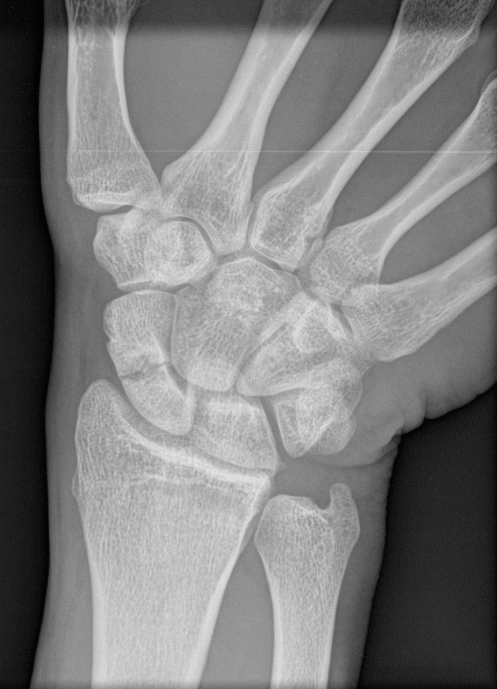
- Scaphoid fracture with sclerosis and cystic change
- Relevant negatives for malalignment/SNAC: no obvious radial styloid arthritis, no carpal collapse, Gilula lines intact
Q24What investigations are used in scaphoid nonunion and why?▸
- XR lateral and scaphoid series: humpback deformity, displacement, DISI
- CT 1mm cuts: cyst, bone stock, deformity
- MRI with contrast: proximal pole AVN
Q25What is the natural history and pathoanatomy of scaphoid nonunion?▸
- Natural history is predictable: 97% arthrosis in 5 years, 90% symptomatic
- Distal scaphoid flexes, capitate is pulled proximally, compression on scapholunate -> SC arthritis
Q26What are the principles of scaphoid nonunion surgery?▸
- Debridement of necrotic bone and exposure of a healthy vascularised bed
- Alignment: correction of scaphoid malalignment
- Biology: bone grafting
- Mechanical stability: stabilisation
Q27Which bone graft is chosen in scaphoid nonunion with AVN or deformity?▸
- AVN -> vascularised bone graft: dorsal 1,2 ICSRA, volar radial carpal artery
- Deformity -> opening wedge interposition graft (Fisk)
- No deformity -> cancellous graft (arthroscopic) or modified Russe inlay cortical graft
- OA/SNAC at presentation -> non-salvageable
Q28Which surgical approach is preferred for scaphoid nonunion?▸
- Mostly volar, as it causes less disruption to the dorsal carpal branch of the radial artery
Q29What history is important in scaphoid nonunion?▸
- Age, hand dominance, functional demand, occupation, smoking
- Mode and time of injury, treatment received
- Symptoms
Q30What is the surgical aim in scaphoid nonunion?▸
- Realign the fracture and promote union with bone graft + stable fixation
Q31Describe the X-ray findings of a stage III SNAC wrist.▸
- Scaphoid nonunion with OA change at the radial styloid and distal scaphoid, scaphocapitate and capitolunate joints
- Collapsed carpal height as indicated by Nattrass and Youm indices
- Lateral: increased SL angle (may be due to humpback deformity) and increased capitolunate angle -> DISI deformity
Q32How is SNAC wrist managed by stage?▸
- I: radial styloidectomy + excision of distal scaphoid
- II: excision of radial styloid + 4 corner fusion or PRC
- III: four corner fusion
- Pan arthritis: wrist fusion
Q33What is the aim and technique of four corner fusion?▸
- Eliminate the arthritic midcarpal joint (usually capitolunate)
- Maintain carpal height
- Also fuse hamate and triquetrum to increase fusion rate
- Fusion rate also depends on smoking status
Q34When can PRC be used in SNAC and what are its drawbacks?▸
- SNAC 2 with SC joint arthritis and intact midcarpal (LC) joint
- Decreased carpal height may cause decreased grip strength and risk of future OA
Q35What is the aim and what are the means of surgery in SNAC wrist?▸
- Aim: removal of the pain generator +/- maintain carpal height in 4 corner fusion
- Means: excisional arthroplasty / limited fusion / wrist fusion
Q36Why is the lunate relatively resistant to osteoarthritis?▸
- The lunate is spherical; even if orientation is disrupted it is unlikely to develop OA
Fact check
Untreated scaphoid nonunion causes arthrosis in 97% within 5 years — imprecise — The 97% figure (Ruby 1985) applied to nonunions already at least 5 years old in a symptomatic series; a 102-case series found 22% arthritis at <5 years, 75% at 5-9 years and 100% at >=10 years. It is not '97% by 5 years'. — (medium confidence) — source