Sports
Meniscal tears - patterns, imaging and management
Meniscal tear types, MRI features, clinical presentation and management in young athletes.
37 questions
4 source pages
4 images
Images appear with the first question taken from each source page — tap a question to open it.
Q1How does a meniscal tear present?📷▸
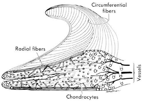
- Presents as delayed swelling
Q2What is the differential diagnosis of acute haemarthrosis (Frank Noyes JBJS 1980)?▸
- ACL tear 72%
- Meniscal injury 62%
- Osteochondral fragment 20%
- Patella dislocation
Q3What MRI signal changes suggest a meniscal tear?▸
- Globular signal is not specific
- Linear signal is more suggestive of a tear
Q4How do you describe a meniscal tear?▸
- Timing
- Morphology: radial, longitudinal, transverse, bucket handle, complex
- Zone (RR 3-4mm from rim, RW, WW) from rabbit study
- Site (root/body), stability, length
- Partial thickness/ full thickness
- These affect whether the tear can heal
Q5Which meniscal tears can heal by themselves?▸
- Stable longitudinal tear
- RR zone
- Partial thickness
- <5mm
Q6Which meniscal tears should undergo meniscectomy (classical teaching)?▸
- Degenerative/complex/radial tear - radial tear breaks many longitudinal fibres
- Chronic tear
- WW zone
- Irreducible tear
- Leave a stable rim to maintain hoop stress (may not apply now)
Q7Which meniscal tears go for repair?▸
- Acute, traumatic tear
- Vascular zone
- Reducible and stable
- Simple pattern (longitudinal/bucket handle)
- Root tear
Q8What other factors affect meniscal healing?▸
- Age (young is <40 years old)
- Smoking
- Stability of the joint, malalignment
Q9What disease and patient factors are considered when treating a meniscal tear?▸
- Disease factors: chronicity (<8/52 better healing), size, shape, location, displacement, associated injury
- Patient factors: age, functional demand, symptoms (pain/locking)
- Aim: relieve symptoms, restore the meniscus as much as possible
Q10Describe the inside-out meniscal repair technique.▸
- Inside-out with nonabsorbable suture, vertical mattress = gold standard
- Higher biomechanical strength
- Problems: need an assistant, risk of needle stick injury and NV injury (saphenous, CPN)
Q11What is the medial approach to the capsule for inside-out meniscal repair?▸
- Incision posterior to the MCL at the level of the joint line (one third above and two thirds below), knee in 90 degrees flexion
- Sartorial fascia incised, dissect anterior to semimembranosus, retract pes tendons anteriorly
- Saphenous nerve and vein lie posterior to the incision in this position
- Place a Henning retractor between the medial gastrocnemius and the capsule
Q12What is the lateral approach to the capsule for inside-out meniscal repair?▸
- Interval between the ITB and biceps femoris, just posterior to the LCL at the level of the joint line
- Flex the knee to 90 degrees to allow the peroneal nerve to fall posteriorly
- Then use the interval between the lateral head of gastrocnemius and the capsule
Q13What are the advantages and disadvantages of outside-in meniscal repair?▸
- For anterior 2/3 tears
- Pros: no assistant, no posterior incision, no needle stick, no NV injury
- Cons: Difficult reduction & oppose edge
Q14What are the advantages and disadvantages of all-inside meniscal repair?▸
- Pros: quick, easy, all arthroscopic, single surgeon
- Cons: reduced strength, limited compression, variable resorption profile
Q15What are the generations of all-inside repair techniques?▸
- 1st gen: described by Morgan - curved suture hooks through accessory posterior portals
- 2nd gen: T-fix - polyethylene bar with attached suture deployed through a sharp needle; inability to tension the knots
- 3rd gen: bioabsorbable meniscus repair device e.g. meniscus arrow
- 4th gen: Fastfix - 2 suture anchors connected by a nonabsorbable polyester suture with a pretied slip knot
Q16What must be preserved during meniscectomy and why?▸
- Keep the rim and root intact, otherwise there is no hoop stress
- Meniscus functions = shock absorption, stability, load bearing, lubrication, proprioception - all affected
- Medial side degeneration is more common due to roll back
Q17How can meniscal healing be improved?▸
- Trephination/shaving at the tear site to increase punctate bleeding; microfracture of the notch
- PRP: CORR 2015 no difference in reoperation rate; Orth J Sport Med systematic review found lower failure rates but no difference in PRO
- Exofibrin clot technique: chemotactic, mitogenic, acts as a scaffold to fill the defect -> inflammatory fibrovascular scar; 30-50ml blood stirred till clot, inserted under the repair site
Q18What are the options if the meniscus is not repairable?▸
- Allograft with bone graft
- Artificial meniscus
Q19What is the definition of the red-red zone?▸
- 3mm from the edge according to Warren and Arnoczky 1982 AJSM
Q20What are the pros and cons of acute one-stage bucket handle repair with ACL reconstruction?▸
- Pros: one OT
- Biological: drilling of tunnels allows stem cells to seep into the joint -> good healing
- Mechanical: stable environment for meniscal healing
- Cons: contradictory rehab -> arthrofibrosis
Q21List the meniscal tear patterns shown.📷▸
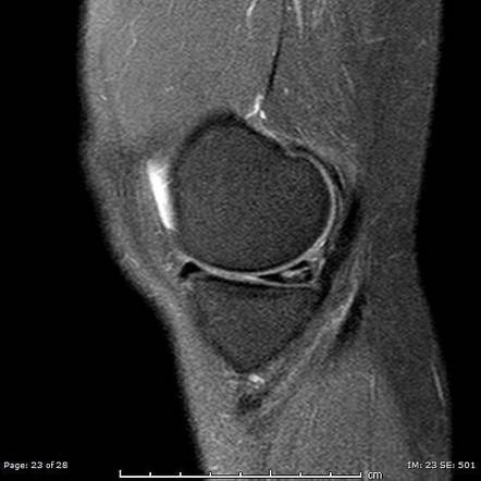
- Horizontal/cleavage tear
- Longitudinal/vertical tear
- Radial tear of the medial meniscus (body)
Q22What are the MRI findings of a radial tear of the medial meniscus body?▸
- Sagittal: T2W signal change
- Coronal: meniscal extrusion defined as 3mm
- Axial: radial tear
Q23Describe the MRI findings of a meniscal root tear.📷▸
- Coronal: extrusion of the medial meniscus (>3mm), vertical linear defect (truncation)
- Axial: fluid interposition at the meniscus root and posterior horn (high signal cleft)
- Sagittal: ghost sign - absence of the posterior horn of the meniscus
Q24What is the definition and incidence of a meniscal root tear?▸
- Avulsion of the meniscus insertion or radial tears within 9mm of the insertion
- Incidence 7-13%
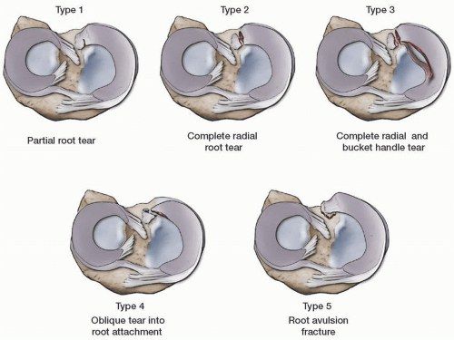
Q25What is the Laprade classification of meniscal root tears?▸
- 1: partial stable root tear
- 2: complete root tear
- 3: bucket handle tear with complete root detachment
- 4: complex oblique or longitudinal tear with complete root detachment
- 5: bony avulsion of the root attachment
Q26How is a meniscal root tear repaired?▸
- Restore the root in anatomical position by suture anchor or drill bone tunnel
Q27What are the consequences of meniscectomy?▸
- Medial meniscectomy: contact area decreases 50-70%, contact stress increases 100%
- Lateral meniscectomy: contact area decreases 40-50%, contact stress increases 200-300% (convex lateral tibial plateau)
- So lateral meniscectomy has a much worse outcome
- Lateral meniscus occupies ~80% vs medial ~60% of the articular surface
- ~70% of load in the lateral and 50% in the medial compartment is transmitted through the menisci
Q28What is a discoid meniscus and how does it develop?📷▸
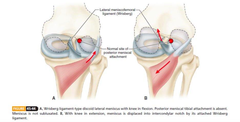
- Abnormal development of the meniscus causing a thickened, discoid-shaped meniscus
- Embryology: failed regression of tissue
- 25% bilateral
Q29What is the common presentation of a discoid meniscus?▸
- Often asymptomatic
- When symptomatic, usually secondary to a meniscal tear
- Knee clunk/click/lock after trivial trauma
Q30What are the X-ray findings of a discoid meniscus?▸
- Widened lateral joint space (11mm)
- Squaring of the lateral femoral condyle
- Hypoplastic lateral tibial spine
- Cupping/ flattening of lateral tibial plateau
Q31What are the MRI findings of a discoid meniscus?▸
- Sagittal: consecutive 3 (5mm) cuts with continuous meniscus (bow tie sign)
- Transverse: transverse diameter in the mid body >15mm
- Coronal: medial-lateral height difference >2mm; meniscal width to maximal tibial width >20%
- Sagittal: ratio of the sum of both lateral horns to maximal meniscal diameter >75%
- R/O tear; +/- coronary ligament integrity
Q32What is the Watanabe classification of a discoid meniscus?▸
- Incomplete (type 2)
- Complete (type 1)
- Wrisberg type: absent meniscotibial ligament, only the meniscofemoral ligament remains; displaces into the intercondylar notch during extension
- May alternatively be classified as stable or unstable
Q33How is a discoid meniscus treated?▸
- Surgery only for symptomatic patients
- Partial meniscectomy / meniscal repair for a tear
- Saucerization (5-8mm compared to the medial side) - reshapes to a semilunar shape, triangular cross-section, less click
- Repair of the meniscotibial ligament for peripheral attachment
- James Hui KSSTA 2021: meniscoplasty gives good mid- to long-term outcomes; concomitant repair/partial meniscectomy does not change outcomes
Q34What are the meniscofemoral ligaments?▸
- Anterior MFL (ligament of Humphrey) - anterior to the PCL
- Posterior MFL (ligament of Wrisberg) - behind the PCL
- 70% of knees have either one, 6% have both
Q35What are the dimensions and attachments of the meniscofemoral ligaments?▸
- Anterior MFL is thinner (<one third the diameter of the PCL), from the posterior horn of the LM to the distal edge of the femoral PCL attachment; can be mistaken for the PCL at arthroscopy
- Posterior MFL is usually larger (~half the diameter of the PCL), from the posterior horn of the LM to the medial femoral condyle, inserting near the posteromedial band of the PCL
Q36What is the function of the meniscofemoral ligaments?▸
- Stabilisers and protectors of the posterolateral femorotibial compartment
- Increase congruity between the mobile lateral meniscus and lateral femoral condyle, protecting the posterior horn of the LM
- Anterior MFL supplements the anterior band of the PCL; posterior MFL supplements the posterior band; they may act as a splint in PCL injuries
- Anterior MFL is taut in flexion and lax in extension; posterior MFL is taut in extension and lax in flexion
Q37What is a pseudotear and a Wrisberg rip?▸
- Pseudotear: the posterior MFL commonly causes a pseudotear of the posterior horn of the lateral meniscus on imaging - a normal vertical/oblique signal at the junction of the ligament of Wrisberg with the posterior horn
- Wrisberg rip: a longitudinal vertical tear of the posterior horn of the LM seen in association with ACL tears
- It is frequently missed and may be used as a secondary sign of ACL disruption