Short Stature
Paediatric knee - tibial eminence and femur
Tibial spine avulsion and ACL injury in children, alignment targets and distal femur fracture.
34 questions
5 source pages
1 images
Images appear with the first question taken from each source page — tap a question to open it.
Q1Describe the Meyers and McKeever classification of tibial eminence fractures.▸
- Type 1: undisplaced (<3mm)
- Type 2: hinged
- Type 3: displaced
- Type 4: displaced + comminuted
Q2What is the mechanism and typical age of a tibial eminence fracture?▸
- Common mechanism is hyperextension
- Age 8-14 years
- Displaced fracture in a skeletally immature patient, no hinge
Q3What clinical findings must be checked in a tibial eminence fracture?▸
- Look for an open wound
- Rule out other injuries
Q4What associated injuries occur with tibial eminence fractures?▸
- Association 40%: meniscal injury, collateral ligament injury, capsular damage, osteochondral fracture
Q5How are associated injuries investigated in a tibial eminence fracture?▸
- MRI to rule out associated injuries
- Look for interposed meniscus or intermeniscal ligament - needs OT
Q6How are tibial eminence fractures managed?▸
- Type 1 and reducible Type 2: immobilise at 20deg
- OT indications: type 3 or 4, mechanical block, entrapped soft tissue, other injuries
Q7What is used for fixation in a tibial eminence fracture?▸
- Young with pin, old with screw
Q8Describe the operative procedure for a tibial eminence fracture.▸
- Supine with tourniquet, AM and AL portal, diagnostic scope
- Debride haematoma, interposed meniscus and intermeniscal ligament
- 2 strand repair with nonabsorbable sutures, wrap around midsubstance of ACL
- Drill hole with an ACL tibial jig
- Before closing, ensure there is no impingement at full extension
Q9Describe the injury shown in the lateral knee X-ray.▸
- Skeletally immature patient
- Fracture over the proximal tibia involving the tibial tuberosity
- Fracture extension into the knee joint
- Ogden Type 3B fracture
Q10What are the clinical concerns and investigations for a tibial tuberosity fracture?▸
- Expect extension lag (may still be possible as the medial retinaculum extends beyond the proximal tibial physis)
- Rule out compartment syndrome (proximal tibial recurrent artery)
- Investigate with AP, lateral and oblique X-rays
- +/- CT; +/- MRI for an undisplaced Type 2
Q11What is the management of a tibial tuberosity fracture?▸
- Long leg cast in full knee extension for IA and IB with <2mm displacement
- >3 years from closure: smooth pin + TBW
- <3 years from closure: lag screw
- Type III: +/- arthroscopy; 5 soft tissue repair
Q12What are the complications of a tibial tuberosity fracture?▸
- Acute: compartment syndrome
- Chronic: recurvatum (anterior growth arrest), stiffness
- Bursitis related to the screw, patellar alta
Q13What are the ossification centres of the tibial tuberosity and which is at risk?▸
- Two ossification centres: primary (proximal tibial physis) and secondary (tibial tubercle physis)
- The secondary centre is at risk at 13-16 years
- Primary physis fuses posterior to anterior
- Secondary physis fuses proximal to distal
Q14What are the 4 stages of development in the proximal tibia?▸
- 1. Cartilaginous phase - no ossification centre
- 2. Apophyseal phase - 9 years, second ossification centre appears
- 3. Epiphyseal phase - fuses from posterior to anterior to the primary ossification centre
- 4. Bony phase - 16 years, closure of the physis
Q15What is the Ogden classification of tibial tuberosity fractures?▸
- Type 1: through the secondary ossification centre
- Type 2: between the primary and secondary centres
- Type 3: crosses the primary physis
- Type 4: through the entire physis
- Type 5: sleeve
- A undisplaced, B displaced
Q16What are the risk factors and mechanism of a tibial tuberosity fracture?▸
- Risk factors: patellar baja, tight hamstrings, Osgood-Schlatter disease
- Usually from eccentric contraction of the quadriceps as the patella inserts onto the secondary ossification centre
- Occurs near the end of growth
Q17Describe the fracture in this 3-10 year old with a valgus knee injury.▸
- AP knee X-ray of a skeletally immature patient
- Fracture over the proximal tibia
- Incomplete fracture over the medial metaphyseal area
- No plastic deformity of the fibula
Q18What is the mechanism of a proximal tibial metaphyseal fracture?▸
- Valgus bending -> the medial tibial metaphysis fails in tension
- Usually from a trampoline
Q19What must be ruled out clinically in a proximal tibial metaphyseal fracture?▸
- Open fracture
- Compartment syndrome
Q20How is an undisplaced proximal tibial metaphyseal fracture treated?▸
- Conservative: long leg cast with the knee in full extension and varus moulding
- Known to have overgrowth causing valgus deformity
- OT indication: torn pes anserinus
Q21What are the associated problems of a proximal tibial metaphyseal fracture?▸
- Compartment syndrome (recurrent anterior tibial artery)
- Vessel injury (popliteal artery tethered)
- Cozen phenomenon - progressive valgus from medial tibial overgrowth/fracture hyperaemia; 50-90%, most resolve in 24 months
- Recurvatum (anterior closure of physis)
- LLD (average 9mm)
Q22What are the deforming forces in a proximal tibia fracture?▸
- Proximal fragment: extension by patellar tendon, varus by pes anserinus
- Distal fragment: valgus by tibialis anterior, flexion by gastrocnemius
Q23What is acceptable alignment after a paediatric tibial fracture?📷▸
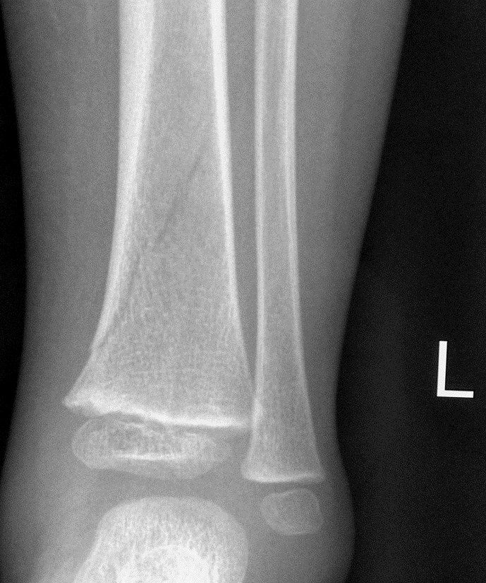
- >50% apposition of ends
- <1cm shortening
- <10 degrees angulation in all planes
- Comment on any fibular fracture
Q24What is a toddler's fracture?▸
- Undisplaced spiral fracture of the tibial shaft
- Mechanism: forceful foot external rotation with the knee fixed
- Age 1-2 years
Q25How is a stable toddler's fracture managed?▸
- Long leg cast for 3 weeks, then short leg cast for 3 weeks
- Adolescent: to a patellar bearing/Sarmiento brace at 8 weeks, for a total of 12 weeks
- Casting position: slight ankle plantarflexion (prevents apex posterior angulation) and knee flexion (controls rotation, prevents WB)
- Weekly follow-up for the first 3 weeks
Q26What are the fixation options for an unstable tibial shaft fracture?▸
- Too narrow canal: cross K wire
- >=6mm: elastic IMN
- Closed physis: locking nail
Q27What is the time to healing by age?▸
- Neonate: 2-3 weeks
- Children: 4-6 weeks
- Adolescent: 8-12 weeks
Q28What is the principle of the Sarmiento cast?▸
- Hydraulic mechanism - tissues and noncompressible fluid within a rigid container
- The incompressible fluid around a fracture prevents excessive shortening
- Substantial pressure develops in the soft tissues under load and supports the bone fragments
- Continued function is desirable for osteogenesis; motion between fragments may create an environment conducive to osteogenesis
- Muscle activity stimulates new blood supply for peripheral callus
Q29What is the design of the Sarmiento brace?▸
- Patellar tendon bearing for partial weight bearing
- Proximal ears continued as far posteriorly as possible opposite the tibial tubercle without impinging the hamstrings
- Triangular shape to fit the tibial contour
- Coronal plane: large anterior shell and small posterior shell
- Rotational plane: femoral condylar extension with precise infrapatellar moulding (patellar bearing) to hold the tibial plateau and enhance rotational (M-L) stability
- Distal flaring for both malleoli; plastic foot-insert with a hinged ankle component for pistoning
Q30Describe the Salter-Harris patterns of distal femur fractures.▸
- SH II - commonest; metaphyseal fragment at the compression side; risk of arrest
- SH III - distal femur (MFC most, close to physeal closure); lateral tibial plateau + intra-articular fracture + MCL injury -> OT
- SH IV - risk of partial physeal arrest -> angular deformity
Q31What must be assessed in a distal femur fracture?▸
- Popliteal artery injury
- Collateral and cruciate ligament injury
- Peroneal nerve injury
Q32How is closed reduction and casting performed for a distal femur fracture?▸
- Reverse the original mechanism
- Hyperextension injury: initial 90 degrees flexion then gradual extension
- Hyperflexion injury: full extension
- Valgus/varus injury: flexion 10-20 degrees
Q33What is the operative fixation for a distal femur fracture?▸
- Indication: failed CR, unstable in cast
- Screw parallel to the physis for a large epiphyseal/metaphyseal fragment
- Transphyseal pin for a small fragment or close to maturity (increases stability); bury under skin, otherwise risk of septic knee
Q34What are the complications of a distal femur fracture?▸
- 30-50% physeal arrest -> angular deformity
- Physeal arrest does not remodel well
- Tolerable if <2 years of growth remaining
- LLD