Short Stature
Paediatric hip - slipped capital epiphysis
Femoral head blood supply, age of onset and treatment principles in epiphyseal slip.
22 questions
3 source pages
2 images
Images appear with the first question taken from each source page — tap a question to open it.
Q1What is the retinaculum of Weitbrecht?📷▸
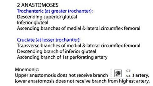
- It equals the ascending artery of the cervical retinacular artery
- It contributes to the blood supply of the femoral head and neck
Q2How is the blood supply of the femoral head described in the notes?▸
- As a 2 cruciate system
- Together with the ascending artery of the cervical retinacular artery (retinaculum of Weitbrecht)
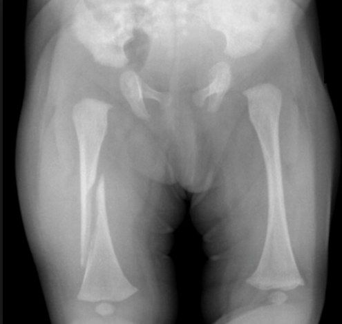
Q3Describe the fracture and radiographic findings in this skeletally immature femur.▸
- X-ray of a skeletally immature patient
- Mid-shaft femoral fracture, spiral with no comminution
- Shortened with some rotational deformity, no valgus/varus angulation
- Need lateral Xray to see AP (sagittal) angulation
Q4Why must non-accidental injury (NAI) be ruled out and how?▸
- Corroborate the history with other family members
- Observe interaction with others
- Look for evidence of neglect
- Ask for a family history of skeletal dysplasia as a cause of bone fragility
Q5What does treatment of a paediatric femoral shaft fracture depend on?▸
- Patient factors: age, weight, underlying bone disease
- Disease factors: location, deformity, length stable
- length stable is less relevant now with the advent of endcaps
Q6What are the effects of increasing age on a femoral shaft fracture?▸
- Less healing
- More muscle pull
- Less remodeling
- Less overgrowth
Q7What are the acceptable deformity limits by age (Rockwood 2010)?▸
- coronal sagittal shorten (Rockwood 2010)
- <2 30 30 20: coronal 30, sagittal 30, shortening 20
- 2-6 15 20 20: coronal 15, sagittal 20, shortening 20
- 6-11 10 15 15: coronal 10, sagittal 15, shortening 15
- >11 5 10 10: coronal 5, sagittal 10, shortening 10
- Will overgrow ~1.5cm, static 2 years from injury (Shapiro 2)
Q8What is the treatment of a femoral shaft fracture at <6 months and 6 months-6 years?▸
- <6 months: Pavlik harness
- 6m-6y: skeletal traction if needed then hip spica up to 8 weeks
- Surgery (ex fix/elastic nail if canal >7mm) if alignment unacceptable, open fracture or polytrauma
Q9What is the treatment of a femoral shaft fracture in a 6-10 year old?▸
- Flexible nail if not very proximal or very distal and not length unstable
- Length unstable is only relative because of endcaps
- Otherwise ex fix or submuscular plate
Q10What is the treatment of a femoral shaft fracture if >10 years, >100lb or length unstable?▸
- Submuscular bridging plating/nailing through a lateral entry
- Avoid piriformis entry - injures the medial circumflex artery -> AVN
- GT entry causes epiphysiodesis, acceptable if >10 years
Q11Describe Bryant traction.▸
- Indication: shortening >3cm and a decision for cast
- Overhead skin traction, knees in full extension
- 5-10lbs - just enough to lift the buttock off the mattress
- Until sticky callus seen on X-ray at ~2/52 (16-18kg)
Q12What are the key points of hip spica application?▸
- Never traction - muscle contraction after GA will cause a compartment
- Applied after traction and callus formed
- Duration Age + 3week/ most 8weeks; weekly X-ray in the first 3 weeks
- Place the child on a spica table; long leg cast with the knee in 90 deg flexion and generous padding over the popliteal fossa (protects popliteal vessels and peroneal nerve)
Q13How is the hip spica moulded and positioned?▸
- Three-point molding centred at the fracture site
- Proximal: may need ORIF due to strong muscle pull
- Middle: valgus moulding for varus displacement
- Distal: flex the knee 20 degrees to reduce gastrocnemius pull
Q14How is the trunk prepared during hip spica application?▸
- Folded towel on the anterior thorax and abdomen to create space for inspiration
- 2 layers of stockinette then cotton (Velband) or gortex material over the trunk
- Thick felt belt across the chest just below the nipple line, second felt belt over sacrum, PSIS and ASIS
Q15How is the hip spica completed?▸
- One and a half leg spica with hip flexed 90 degrees and 30 degrees abduction (human position), connected with a long leg cast
- Reinforce with a broomstick between the thighs
- Perineal window; double check the reduction on X-ray
Q16What is the evidence for management of femur fractures in 4-10 year olds?▸
- 4-10 yrs: Wright et al Lancet 2005 RCT: malunion rates after ex fix vs flexible IM nails
- Improved outcomes with nails
- AAOS guidelines: do not advocate routine use of ex fix in younger children
Q17What is the principle behind flexible intramedullary nailing?📷▸
- Based on the double arc secant in equilibrium principle
- load sharing device relying on 3-point fixation of two opposing nails
- The apex of each nail is at the fracture site
- Bending moments of the elastic nails counteract with equal and opposite force to create a stable construct
Q18What are the 4 biomechanical effects of symmetrical splinting (RAFT)?▸
- Rotational stability
- Axial stability
- Flexural stability
- Translational stability
Q19What is the indication for flexible IM nailing?▸
- Works best at the middle 1/3, length-stable fracture
- Body build <100 lb
Q20How is the nail chosen and bent in flexible IM nailing?▸
- Choose a nail 1/3 of the inner canal diameter
- Bend each nail 3 times the inner canal diameter
- Apex at the fracture site so the two nails are maximally separated from each other at the fracture
Q21Describe the flexible IM nailing procedure.▸
- Radiolucent table, reduce with the F tool
- Insert the nail retrograde at the metaphysis, then cap to prevent irritation and migration
- Flat tips at the nail end improve metaphyseal anchorage and rotational control; avoid the physis of the femur
Q22What is the post-operative protocol after flexible IM nailing?▸
- TDW as tolerated post-op with gentle mobilisation exercises
- Avoid overaggressive knee strengthening (may displace the fracture)
- Usually allow FWB after 6/52
- Follow up with XR for healing/loss of reduction; nail removal in 1 year