Short Stature
Acquired short stature - polio and trauma
Post-polio and post-traumatic causes of stature loss, plus acute arm pseudo-paralysis.
17 questions
3 source pages
2 images
Images appear with the first question taken from each source page — tap a question to open it.
Q1What is poliomyelitis and how is poliovirus transmitted?📷▸
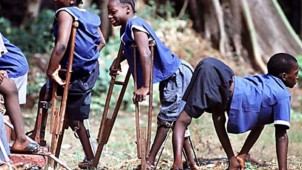
- Non-progressive asymmetrical motor paralysis with sensory sparing
- Poliovirus: non-enveloped single-stranded RNA virus, a picornavirus and enterovirus
- Destroys anterior horn cells in spinal cord and brainstem motor nuclei
- Faecal-oral route; major type (meningitis, encephalitis) and minor type (flu-like)
Q2Describe the four phases of poliomyelitis.▸
- Acute: prodromal flu-like, painful muscles, joints flexed
- Paralysis: after 2-3 days; limbs weak, swallowing/breathing affected; ends 7-10 days
- Recovery: within 6 months
- Residual: LMN flaccid residual paralysis with intact sensation, then deformity and growth disturbance
Q3What is post-polio syndrome and how is it managed?▸
- Aging phenomenon: remaining neurons degenerate after working extra hard to compensate
- NOT reactivation; affects up to 50%
- Exercise at sub-exhaustion levels to tone affected muscles + periods of rest + light weighted caliper
- Pacing: maintain but do not overuse
Q4What are the two scenarios of late polio presentation?▸
- New symptoms after a period of convalescence = post-polio syndrome
- Chronic problems: weakness/flail joint, deformity (posture, muscle imbalance, gravity)
- OA, LLD, vascular dysfunction
- Assessment for post-polio syndrome = rule out other medical, ortho and neuro causes
Q5How is chronic polio managed by region?▸
- Look at the following and treat accordingly
- Hip: FABER contracture (Ober procedure, Campbell procedure), Trendelenbeg gait (Trendelenburg gait) (EO transfer, iliopsoas to GT), dislocation, glut max weakness (glut max lurching gait)
- Knee: quads weakness (hand knee gait) (KAFO), FFC (serial casting, soft tissue release, extension osteotomy), recurvatum (triple tenodesis), flail knee (fusion, KAFO)
- Foot and ankle: foot drop and circumduction gait, cavovarus, equinovarus
- LLD (short limb gait); spine/pelvis - determine if pelvic obliquity is infra, supra or combined
Q6What is the most common weakness in polio and what adaptive mechanisms occur?▸
- Most common weakness is at the quads
- Weak quads cause knee flexion with CG behind the knee -> flexion moment
- Compensation: hip flexion brings CG forward, ankle plantarflexion (plantarflexion-knee extension couple)
- Alternatively knee recurvatum so ITB acts as extensor, counterbalanced by glut max and ankle dorsiflexors
- Weakness order LL: TA, quads, glut max, gastroc; UL: deltoid, biceps, opponens
Q7What is the vaccination history of polio?▸
- 1958: first general use of Sabin live attenuated vaccine by mouth (OPV)
- Salk = inactivated polio vaccine (IPV)
Q8How does the IT band contribute to deformity in polio?▸
- IT band is the culprit for many presentations
- Increased lumbar lordosis, pelvic obliquity, FABER hip
- Knee FFC + valgus, LLD, tibial ER
- Secondary foot and ankle problems
Q9What factors affect remodelling of a paediatric fracture?▸
- Age, with >=2 years of remaining growth
- Amount of deformity
- Distance from the physis
- Direction of deformity
Q10What structural differences make a paediatric fracture different from an adult fracture?▸
- Thick cartilage cap (need arthrogram to visualise articular fracture)
- Thick periosteum
- More collagen, more ductile
- More cancellous bone, less comminution
- Bone is weaker than ligament/tendon
Q11Why is an arthrogram needed to assess a paediatric articular fracture?▸
- The thick cartilage cap makes the articular fracture difficult to visualise
- Arthrogram is needed to visualise the articular fracture
Q12What are the healing differences between paediatric and adult fractures?▸
- Paediatric fractures have remodelling power
- They carry a risk of growth disturbance
Q13Describe the X-ray findings and likely diagnosis in this 2-year-old refusing to move the arm.📷▸
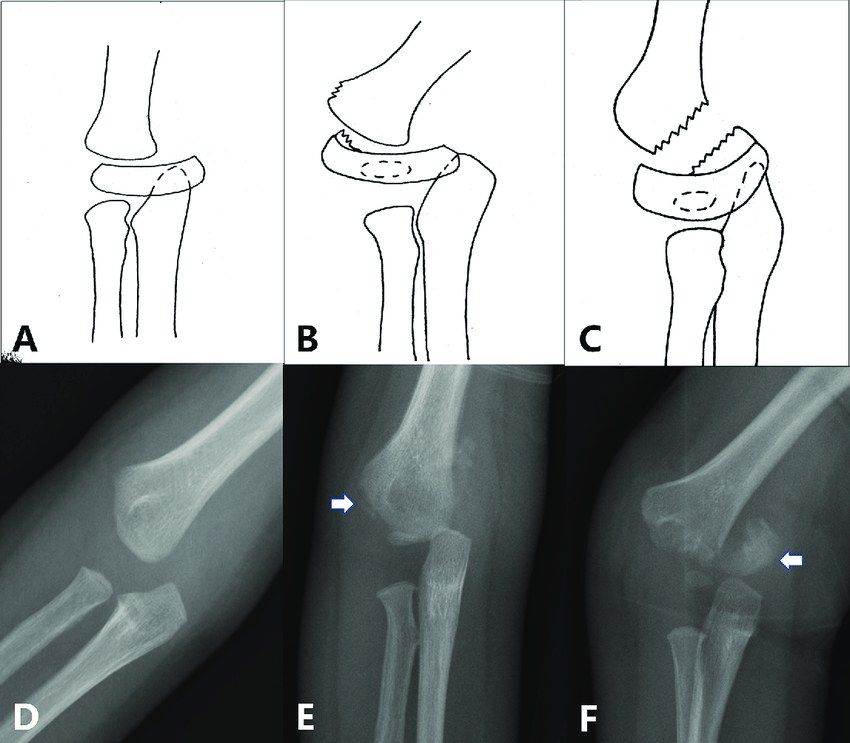
- Skeletally immature patient, bilateral elbow X-ray
- Proximal ulna and radius relationship maintained, but humerus-ulna/radius relationship disrupted with posteromedial displacement
- Likely transphyseal fracture of distal humerus (physis is biomechanically the weakest location)
Q14How do you differentiate a transphyseal distal humerus fracture from other injuries?▸
- Radiocapitellar line normal (proximal radius aligns with capitellum); proximal radius and ulna align with each other
- Elbow dislocation: posteromedial displacement favours transphyseal injury
- Lateral condyle fracture: assess lateral condyle-radial head relationship
- Low SCH fracture: transphyseal has wider bone contact and less displacement
Q15What is the DeLee classification?▸
- Type A: infant to 7 months; no lateral condyle ossification centre (SH 1)
- Type B: 7 months to 3 years; lateral condyle ossified (SH 1,2)
- Type C: 3-7 years; large metaphyseal fragment exiting laterally
- This fracture almost only occurs <7 years
Q16What is the management of a transphyseal distal humerus fracture?▸
- Depends on timing of injury, displacement and DeLee type
- Acute undisplaced: CR + cast in pronation x 3/52
- Acute displaced: CR +/- OR + KWF + cast in pronation x 3/52 (+/- arthrogram)
- Late (>1 week) + displaced: DO NOT manipulate
Q17What associated features and anatomy are important in transphyseal distal humerus fracture?▸
- Hyperextension injury (birth injury/abuse/trauma), usually <2 years
- Highly suspicious of NAI; further MRI
- Medial epicondyle involved at young age; physis moves distally with growth, V-shaped cleft
- Large fragment, usually less rotation and tilting; blood supply good but medial crista of trochlea may be disrupted -> cubital varus