Short Stature
Skeletal dysplasia - achondroplasia and Marfan
Radiographic features of achondroplasia and clinical features of Marfan syndrome.
24 questions
2 source pages
1 images
1 fact-check flags
Images appear with the first question taken from each source page — tap a question to open it.
Q1Describe the X-ray features of osteogenesis imperfecta.📷▸
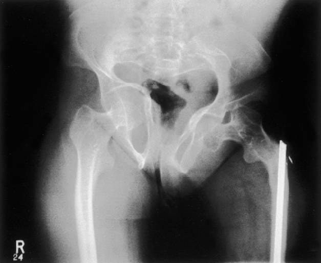
- Tri-foil shaped pelvis, protrusio, lateral bowing of femur shaft
- Erlenmeyer flask femur, gracile or overtubulated bones, anterior bowing of tibia
- Codfish vertebrae or kyphoscoliosis in the spine
- Skull XR: Wormian bones + basilar invagination
Q2What is the diagnosis and what are the differential diagnoses?▸
- Dx: OI
- Ddx: rickets/osteomalacia, idiopathic juvenile osteoporosis
- Ddx: non-accidental injury, fibrous dysplasia
Q3What clinical features would you look for in suspected osteogenesis imperfecta?▸
- Short stature (proportionate), thin skin
- Eyes: Blue sclera; Ears: Impaired hearing (in type 1); Mouth: Defective detinogenesis. High pitch voice
- Triangular shaped head with broad forehead
- Lateral bowing of femur, anterior bowing of tibia (saber shin), genu varum, acetabular protrusion
- Kyphoscoliosis, congenital radial head dislocation, basilar invagination (myelopathy), generalised ligamentous laxity
Q4How is osteogenesis imperfecta diagnosed and what is the treatment goal?▸
- Diagnosis based on radiological and clinical features +/- family history
- Skin biopsy is gold standard (culturing dermal fibroblasts)
- Goals: prevent fracture, deformity correction, optimise function
- Refer to geneticist to establish diagnosis
Q5What medical treatment is used in osteogenesis imperfecta?▸
- Bisphosphonates: decrease fracture, 90% increase in cortical diameter, 50% increase in cancellous bone, decrease pain, increase ambulation
- Growth hormone
- Calcium and vitamin D
- Gene therapy
Q6What are the Sillence types of osteogenesis imperfecta?▸
- Type I: mildest, AD, most common, blue sclera, abnormal dentinogenesis
- Type II: AD/AR, die, blue sclera
- Type III: AR, white sclera, fracture at birth, limb deformity, short stature, worst survivable type
- Type IV: AD, moderate severity, hearing normal, spine deformity, abnormal dentinogenesis
Q7What fracture-related findings may be seen in osteogenesis imperfecta?▸
- Hypertrophic tumoral callus
- Malunion
- Pseudoarthrosis
Q8What is the genetic basis of osteogenesis imperfecta?▸
- Hereditary disease with abnormal Type 1 collagen (quantitative/qualitative); 90% have an identifiable genetic mutation
- COL1A1 and COL1A2: substitution of glycine by a bulky amino acid (e.g. cystine) in the procollagen structure, unable to crosslink
- Reduced collagen secretion and abnormal collagen production -> insufficient osteoid production; unable to remodel normally, but initial fracture healing is normal
- Affects bone (woven), tendon, ligament, dentin, sclera
Q9What is the Rubin classification of osteogenesis imperfecta?▸
- Diaphyseal hypoplasia
Q10What are the Sillence types V-VII?▸
- No COL1 mutation but similar phenotype
Q11How do you monitor the effect of bisphosphonates in osteogenesis imperfecta?▸
- Clinical: decreased pain
- XR: vertebral remodelling / Zebra lines
- DEXA: any increase in total body and lumbar spine bone density
- Serum: ALP, C-telopeptide, N-telopeptide, TRAP5b (C telopeptide, N telopeptide , TRAP5b, produced during bone resorption); osteocalcin (synthesised by osteoblasts to control osteoclasts)
- Urine: deoxypyridinoline (a collagen breakdown product)
Q12What are the problems with bracing fractures in osteogenesis imperfecta?▸
- Normal healing rate but no remodelling
- Increased risk of further fracture due to immobilisation and/or the stress riser effect at the edges of the cast
- If recurrent fracture: CR + IMN +/- multiple osteotomies to correct deformity at the same time
Q13How is limb deformity corrected in osteogenesis imperfecta and why does it progress if untreated?▸
- Sofield-Miller procedure with telescoping IM system such as Bailey-Dubow rod (if medullary canal wide enough) or Rush pin
- Deformity will progress if left untreated due to the HV law (Hueter-Volkmann) leading to selective inhibition of physeal growth
Q14When is scoliosis surgery indicated in osteogenesis imperfecta and what are the growing rod generations?▸
- Scoliosis surgery if Cobb >45 (no brace due to rib fragility)
- 1st gen: Sheffield; 2nd gen: Bailey-Dubow; 3rd gen: Fassier-Duval
- 1st and 2nd gen need arthrotomy; 3rd gen allows one proximal insertion with a distal screw
Q15What are the anaesthetic and orthopaedic difficulties in osteogenesis imperfecta surgery?▸
- Anaesthesia: malignant hyperthermia, basilar invagination, mitral valve regurgitation
- Ortho: bleeding, abnormal bone quality, minimal autograft, ligamentous laxity
Q16What are the pros and cons of Rush pin vs telescoping rod in osteogenesis imperfecta?▸
- Telescoping rod: only a narrow rod can be inserted due to the narrow canal -> increased risk of rod fracture or trapped rod; need to change every 4 years
- Rush pin: easy exchange, reduces implant breakage; change over 2.5 years
Q17What are the physical examination features of Marfan syndrome?▸
- Tall stature, long narrow limbs, scoliosis
- Dolichostenomelia: arm span > height (>1.05)
- Arachnodactyly: Steinberg sign (thumb tip extends beyond small finger when clasped in palm) and Walker sign (thumb/index distal phalanges overlap around opposite wrist)
- Ligamentous hyperlaxity, pes planus
Q18What is the genetic basis of Marfan syndrome?▸
- Connective tissue disease, Chr 15 (chromosome 15)
- AD, fibrillin 1 (FBN1) - ECM glycoprotein for fibrinogenesis; abnormal mechanical and elastic properties of connective tissue
- 30% sporadic mutation
- Rubin classification: physeal hyperplasia
Q19What are the Ghent nosology criteria for Marfan syndrome when family history is negative?▸
- Aortic root dilatation AND ectopia lentis
- Aortic root dilatation AND FBN1 gene
- Aortic root dilatation AND systemic score >=7 points
- Ectopia lentis AND FBN1 gene associated with aortic dilatation
Q20What are the Ghent nosology criteria when family history is positive for Marfan syndrome?▸
- Ectopia lentis
- Aortic root dilatation
- Systemic score >=7 points
- Family history (FHx+) of Marfan syndrome
Q21What are the systemic features of Marfan syndrome?▸
- Ortho: dolichostenomelia, arachnodactyly, acetabular protrusio, ligamentous laxity (scoliosis, cervical instability, recurrent patella dislocation), dural ectasia
- Face: down-slanting palpebral fissures, malar hypoplasia, retrognathia, enophthalmos, dolichocephaly
- Ocular: lens dislocation; chest: carinatum, excavatum, pneumothorax
- Cardiac: valvular problem, aortic dissection; skin striae
Q22What imaging findings are expected in Marfan syndrome?▸
- XR: acetabular protrusion, scoliosis
- MRI: dural ectasia (>60%)
Q23What is the general management of Marfan syndrome?▸
- Refer to paediatrician - need monitoring of the cardiac condition
- Scoliosis treatment generally has a worse response than AIS
- Mild: bracing
- Rapidly progressing/large curve in an adult: surgical management with pre-, intra- and post-operative considerations
Q24What are the pre-, intra- and post-operative considerations for scoliosis surgery in Marfan syndrome?▸
- Preop: ectopia lentis -> AACG; cardiac -> MVP and aortic root dilatation; recurrent PTX; MRI to rule out dural ectasia
- Intraop: construct needs to span longer (prevent add-on phenomenon); narrow pedicles; higher complication (durotomy)
- Postop: add-on curves; fixation failure (thin laminae, thin pedicles, osteopenia); higher risk of infection
- Postop: pseudoarthrosis
Fact check
Skin biopsy is the gold standard for diagnosing OI (culturing dermal fibroblasts) — outdated — OI is now diagnosed by molecular genetic testing of COL1A1/COL1A2; cultured dermal fibroblast collagen analysis has fallen out of favour — source