Neuromuscular
Cerebral palsy gait analysis and treatment
GMFCS levels, crouch, recurvatum, jump and stiff knee gait patterns, gastrocnemius lengthening
22 questions
6 source pages
2 images
Images appear with the first question taken from each source page — tap a question to open it.
Q1What are the GMFCS levels?📷▸
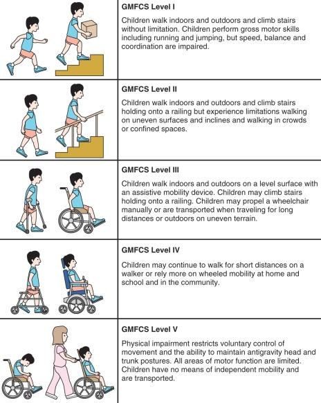
- Level 1: no restriction in walking, limited in advanced motor skills
- Level 2: no aids, some limitation outdoors or in the community
- Level 3: needs assistive aids, limitation outdoors or in the community
- Level 4: self mobility with limitation, needs transport or powered mobility outdoors
- Level 5: self mobility severely limited even with assistive technology
Q2What is the most predictive factor for becoming an independent walker?▸
- independent sitting at 2 y.o
Q3What are the goals for a non-ambulator with cerebral palsy?▸
- Good sitting balance, forward gaze, use UL for feeding
- Spine straight, shoulder and pelvis level
- Hip located, stable, pain free with good ROM
- Knee mobile, flexed for sitting, extends in brace for transfer
- Foot plantigrade for standing
Q4What are the Gage principles for an ambulator?▸
- Stability in stance
- Good clearance in swing
- Preposition of the foot at end of swing
- Adequate step length
- Energy conservation
Q5What are the midstance features of crouch gait?▸
- Excess ankle dorsiflexion
- Knee flexion
- Hip flexion
- Overall short stride length and muscle atrophy
Q6What are the causes of crouch gait?▸
- Weakness (triceps surae, quads)
- Spasticity (hamstrings, iliopsoas)
- Joint contracture (knee flexion contracture)
- Lever arm dysfunction (increased femoral anteversion, increased tibial torsion, collapse tripod)
Q7Briefly explain the mechanism of crouch gait.▸
- Weak gastrocsoleus / excessive ankle dorsiflexion means the knee extension / ankle plantarflexion couple cannot work
- GRF falls posterior to the knee, producing a knee flexion moment
Q8How is crouch gait managed?▸
- Weakness: avoid iatrogenic excessive gastrocsoleus release/recession; GRAFO for gastroc; hamstring transfer for quads
- Spasticity: hamstring lengthening (semiM + gracilis fractional +/- biceps femoris) and semiT to adductor tubercle transfer
- Psoas spasticity: release at the pelvic brim or LT
- Joint contracture: extension osteotomy
- Lever arm dysfunction: reconstruct hip, tibia or foot
Q9What are the sequelae of hamstring lengthening and why is the semiT transferred?▸
- Sequelae: weak hip extension + anterior pelvic tilt
- SemiT to adductor tubercle transfer converts a biarticular to monoarticular muscle
- Aim: prevent hip extension and decrease anterior pelvic tilt
Q10What compensatory mechanisms maintain balance in recurvatum gait?▸
- Knee recurvatum with the ankle in plantarflexion at midstance
- Hip flexion moves the CG anterior to balance the GRF
- Contralateral crouch occurs due to pelvic obliquity
Q11What is the cause of recurvatum gait and how does age guide treatment?▸
- Contracture of triceps surae
- <6 years / spasticity: botox, stretching, solid AFO
- Contracture + >6 years: assess with Silverskiold
Q12What operations are used for fixed triceps surae contracture in recurvatum gait?▸
- Gastroc only: gastroc recession
- Gastroc + soleus: ETA
- Applied when there is contracture and the patient is >6 years old
Q13What are the features of jump knee gait?▸
- Ankle equines in late stance
- Knee flexion in early stance and terminal swing
- Hip flexion in early stance
- Pelvis normal or anteriorly tilted
Q14What causes jump knee gait and how is it managed?▸
- Problem is spastic hamstring and triceps surae
- No OT
- Serial cast, botox, stretching, hinged AFO
Q15What is gait analysis?▸
- Systematic description, assessment and measurement of the quantities that characterise human locomotion
- Components: kinematic, kinetic, EMG, energy consumption
Q16What is the set-up for gait analysis?▸
- 2D video analysis
- 3D computer analysis using specialised markers on specific bony landmarks
- Force plates to measure GRF
- EMG to look at muscle firing patterns
Q17What causes stiff knee gait and when is knee flexion reduced?▸
- Spastic rectus femoris or rectus firing out of phase
- Decreased knee flexion in terminal stance and throughout the swing phase
Q18How is stiff knee gait managed in a child under 6 years?▸
- Botox and stretching
Q19What are the indications for rectus to pes transfer?▸
- Peak knee flexion <50 degrees
- Peak knee flexion later than 30% of swing
- Active rectus in the mid 3/5 of swing
- Toe dragging
Q20Describe the Strayer, Vulpius and Baker techniques.📷▸
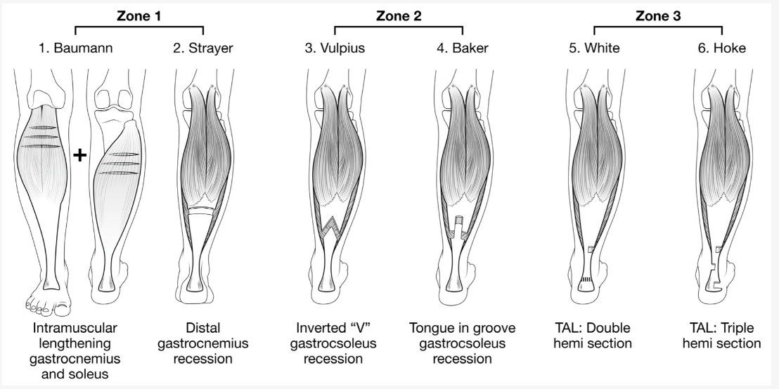
- Strayer: gastroc release from soleus proximal to the conjoint tendon -> 1 cm
- Vulpius: chevron at the MST junction -> 1.5 cm
- Baker: central fascial tongue
Q21Describe the White and Hoke PETA techniques.▸
- White PETA: 1 medial + 1 lateral incision -> 2-4 cm
- Hoke PETA: 2 medial + 1 lateral incision -> 4 cm
Q22Which zone is preferred in cerebral palsy and why?▸
- Zone 1 is more preferred in CP
- It allows a more selective release