Neuromuscular
Cerebral palsy hip - classification and dislocation
Hip surveillance radiographs, migration percentage and Winters/Sutherland classification of displacement
22 questions
3 source pages
2 images
1 fact-check flags
Images appear with the first question taken from each source page — tap a question to open it.
Q1What are the radiographic findings in this skeletally immature patient with a dislocated left hip?▸
- Broken Shenton's line and dislocated left hip
- Increased acetabular index and increased neck-shaft angle, perhaps with more anteversion
- Relatively well formed tear drop and femoral head suggest neuromuscular dislocation rather than DDH
- Pelvis looks like an inlet view due to pelvic anteversion from tight rectus; look for VP drain
- Triradiate cartilage fuses at 11-13 years old
Q2What history should be taken in a child with a neuromuscular hip dislocation?▸
- Perinatal and birth history
- Developmental milestones (age of sitting and walking)
- Problems faced by patient him/herself; Problems faced by cares (perineal hygiene, pain, transfer, sitting)
- Determine GMFCS level
- Other medical problems and social support
- Prognostication if very young - good: head control by 9 months, sit by 2 years, hemiplegic; bad prognosis: bleck, lack of head control by 20 months, non ambulatory by 7 yr
Q3How is function classified and prognosis assessed in cerebral palsy?▸
- GMFCS I no restriction, II stairs with rails, III wheelchair, IV powered wheelchair, V self mobility limited
- If non-ambulatory and >1 year old, test primitive reflexes; 2 or more = poor prognosis for walking (Bleck)
- Gait analysis lab for quantitative measurements
Q4What is cerebral palsy and how is it classified?▸
- Non-progressive insult to the immature brain; primary insult non-progressive but musculoskeletal features evolve with growth
- Topographically: diplegic, hemiplegic, quadriplegic
- Neurologically: spastic, hypotonic, mixed, athetoid, ataxic
- Functionally: GMFCS
- Deviations: primary (loss of selective motor control), secondary (growth related), tertiary (compensatory)
- Secondary deviation examples: Anatomic shortening of muscle tendon unit (e.g. myostatic contractures); Persistent bone deformities due to HV principle (e.g. femoral anteversion); Joint subluxations/ dislocations
Q5What are the principles of managing cerebral palsy?▸
- Multi-disciplinary approach (MDT); orthopaedics has a role only in spastic type and cannot reverse the primary insult
- Overall aim: maximize patient’s function consistent with the current disease status and prevent deterioration
- Walker aims (Gage) and prevention of early OA; sitter aims: spinopelvic balance, perineal hygiene, mobile knees, plantigrade feet
- Medical spasticity: physio, analgesia, oral or intrathecal baclofen, botox (3-6 months)
- Surgery: delay until >6 years but before tertiary issues; SEMLS vs OSSCS (release/lengthen biarticular muscles, preserve monoarticular antigravity muscles)
Q6How is hip dysplasia in cerebral palsy prevented and treated?▸
- Spastic biarticular adductors overpower monoarticular abductors, so the hip displaces; hip dysplasia is preventable
- Surveillance per NICE guidelines and the Swedish CPUP study: X-ray before 3 years; repeat one in 6 y.o; monitor Reimer migration index, hip abduction; refer if abduction <30 degrees or RMI >33%
- Early subluxation RMI 33-50%: botox/soft tissue release (adductor tenotomy, iliopsoas +/- hamstring lengthening) and night abduction splint
- Moderate subluxation RMI >50%: VDRO/pelvic osteotomy; dislocation: <1yr reconstruct, >1 year observe if painless, salvage if painful (Girdlestone, capsule interpositional arthroplasty, McHale)
- Evidence suggests less than 50% of asymptomatic dislocated hips will develop pain in the long run, so observation can be supported
- Psoas spastic, hip flexed, reduced torque on proximal femur, femur cannot derotate, so anteversion; Valgus as non weight bearing
Q7What does the physical examination include in neuromuscular disease?▸
- General: IQ, sitting balance, gait (stance and swing phase), orthosis
- Hip: tone, ROM, Thomas or Staheli test for FFC, Phelps test for abduction tightness, Duncan Ely for rectus, Ober for ITB
- Knee: tone, ROM, popliteal angle (hamstring shift more accurate to see if psoas contracture contributes)
- Ankle: tone, ROM, Silverskiold test, bony impingement or impending ulcers
- Spine for scoliosis, pelvic obliquity, LLD
Q8How is spasticity defined and treated globally vs focally?▸
- Velocity dependent increase in tone due to loss of supraspinal inhibition
- Global: selective dorsal rhizotomy - decrease afferent input from muscle spindle
- Global: baclofen - GABA agonist, increase K channel opening --> easy hyperpolarization
- Global: gabapentin - VGCC deactivator, decrease nerve excitation and decrease neurotransmitter release
- Focal: botox
Q9What is a contracture and how is it treated?▸
- Shortening of the musculotendinous unit due to slowing of growth from lack of tensile loading
- Muscle: release or transfer; joint: release; bone: osteotomy
Q10What are the four types of lever arm dysfunction?▸
- Unstable fulcrum (hip dislocation)
- Floppy lever (pes planus)
- Short lever (coxa valga)
- Malrotation (tibial ER)
Q11What are the hemiplegic gait patterns and their management?▸
- Foot drop -> hinged AFO / solid AFO
- True equinus (gastroc soleus contracture) -> hinged AFO -> heel cord lengthening
- Jump knee (spastic hamstring + triceps surae) -> hinged AFO -> no OT
- Apparent equinus (iliopsoas contracture + gastroc weakness) -> solid AFO -> psoas lengthening
Q12What are the diplegic gait patterns?▸
- True equinus
- Jump knee
- Apparent equinus
- Crouch knee
- Recurvatum knee
- Stiff knee (swing phase problem)
Q13What are the timing and components of surgery in cerebral palsy?▸
- Timing: delay OT as long as possible (>6 years) but before tertiary issues - contain the hip before dislocation, modify gait before deviation
- SEMLS vs OSSCS (orthopaedic selective spasticity control surgery)
- OSSCS: correct lever arm dysfunction by releasing or lengthening biarticular muscles and preserving monoarticular antigravity muscles
- Components: balance spasticity, prevent contractures, correct lever arm dysfunction
Q14What soft tissue releases are used for early hip subluxation in CP?▸
- Adductor longus transect - protect the anterior branch of the obturator nerve
- Gracilis myotomy and adductor brevis lengthening
- Iliopsoas lengthening +/- hamstring lengthening
- Nighttime abduction splint
Q15What is Winters type 1 gait?📷▸
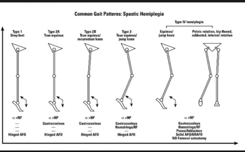
- Type 1 = 'drop foot' = equinus in swing phase only
Q16What is Winters type 2 gait?▸
- Type 2a = 'true equinus' = equinus in the whole gait cycle
- Type 2B = true equinus + recurvatum knee
Q17What is Winters type 3 gait?▸
- 'Jump knee' = equinus + tight hamstrings
Q18What is Winters type 4 gait?▸
- Equinus + tight hamstrings + tight flexors and adductors
- Results in adduction and internal rotation deformity
Q19What is true equinus and how is it managed?📷▸
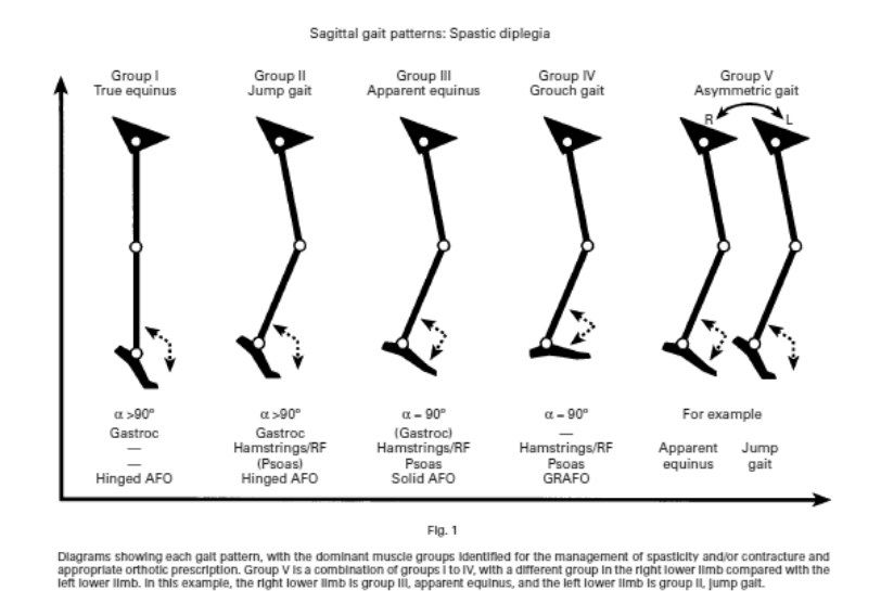
- Tight gastrocsoleus complex
- Mx: hinged AFO (allows dorsiflexion while blocking plantarflexion)
Q20What is apparent equinus and how is it managed?▸
- Ankle goes into dorsiflexion due to persistent loading with lengthening of the Achilles tendon
- Ankle ROM is still normal
- Mx: solid AFO
Q21What is crouch gait (equinoplanovalgus) and how is it managed?▸
- Excessive dorsiflexion due to an overlengthened Achilles
- Can be caused iatrogenically by releasing the Achilles while ignoring hamstring tightness
- Mx: GRAFO
Q22What is jump gait in the Sutherland classification and how is it managed?▸
- Tight hamstrings flex the knee and the hip flexes to push the CG in front, with progressively worse equinus
- Mx: hinge AFO
Fact check
Triradiate cartilage fuses at 11-13 years old — imprecise age range — Triradiate cartilage closes at about 12 years bone age in girls and 14 in boys (radiographic closure roughly 13-14 years in females and 15-16 in males); overall quoted range is about 12-16 years. — (medium confidence) — source