Dysplasia Syndromes
Rickets and metabolic bone disease
Vitamin D and PTH physiology with X-ray and histology of rickets and osteomalacia.
20 questions
4 source pages
2 images
Images appear with the first question taken from each source page — tap a question to open it.
Q1What are the actions of vitamin D3 on the GI tract?📷▸
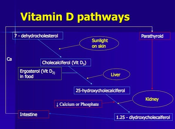
- Absorbs Ca
- Absorbs PO4
Q2What are the actions of vitamin D3 on bone?▸
- Activates osteoblast matrix production
- Increased osteoclast action through RANK-RANKL
Q3What is the effect of vitamin D3 on PTH and what upregulates vitamin D3?▸
- Inhibit PTH
- Increased PTH (hydroxylation in the kidney)
- Low calcium level, low PO4 level
- Decreased vitamin D3 itself (auto-regulation)
Q4What is the effect of PTH on calcium reabsorption?▸
- Increases Ca reabsorption at the bone
- Increases Ca reabsorption at the kidney
Q5What is the effect of PTH on phosphate reabsorption?▸
- Increases PO4 reabsorption at the bone
- Decreases PO4 reabsorption at the kidney
Q6What is the net effect of PTH on serum calcium and phosphate?▸
- Increase serum Ca
- Decrease serum PO4
Q7Describe the X-ray findings in this skeletally immature patient.📷▸
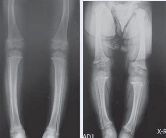
- Increased physeal width in general, cupping of the metaphyseal area
- Bowing of bilateral tibia
- No coxa vara
- Systemic disease affecting the physis; Ddx MED, SED
Q8What is rickets / osteomalacia?▸
- Defect in mineralization of osteoid matrix and chondroid (in the zone of calcification)
- Caused by inadequate calcium and phosphate
- Impaired calcification of the cartilage matrix of growing long bones -> increased physeal width, cortical thinning and bowing
Q9In a child with widened physes and bowed legs, what history and examination findings should be sought?▸
- History: birth and family history, diet
- PE: hyperlaxity, generalised weakness
Q10What are the clinical features of rickets?▸
- Short stature (proportionate)
- Frontal bossing + caput quadratum
- Pigeon chest
- Rachitic rosary
- Harrison's sulcus
- Cat back kyphosis
- LL alignment abnormalities
Q11What are the biochemical features of rickets?▸
- Decreased Ca and PO4
- Increased ALP and PTH (except in familial hypophosphatemic rickets and hypophosphatasia)
- Vitamin D level depends on the cause
Q12Describe the X-ray and histological findings of rickets.▸
- Long bone bowing
- looser’s zone/ pseudofracture (Looser's zone / pseudofracture): transverse radiolucency from unmineralized osteoid seam; weight-bearing bones on the compression side, non-weight-bearing at the nutrient foramen
- Metaphyseal flaring in paintbrush pattern and cupping; widened physis at the zone of hypertrophy
- +/- osteopenia; cod-fish vertebrae
- Histology: unmineralized osteoid seam
Q13What are the causes of rickets by category?▸
- Intake: intake problem, lack of sunlight exposure, GI problem
- Processing: vitamin D dependent rickets
- Recycling: vitamin D independent rickets (X-linked dominant familial hypophosphatemic rickets), hypophosphatasia
Q14What is the defect in vitamin D dependent rickets type I and type II?▸
- Type I: defective 1-alpha hydroxylase
- Type II: end organ insensitivity to 1,25-(OH)2 vitamin D
Q15Describe X-linked dominant familial hypophosphatemic rickets.▸
- PHEX encodes an endopeptidase cleaving FGF23; cannot chelate FGFR23
- Cannot reabsorb PO4 in the proximal tubules, cannot mineralize
- More diaphyseal bowing rather than metaphyseal bowing
- New drug: Burosumab
Q16What is hypophosphatasia?▸
- Mutation in the tissue nonspecific isoenzyme of ALP (tissue non-specific isoenzyme of ALP)
- Reduced ALP, decreased synthesis of phosphate, decreased mineralization
- Increase in urine phosphoethanol-amine (urine phosphoethanolamine)
Q17How is rickets treated?▸
- Treat the underlying cause
- Dietary input -> vitamin D3 5000 IU/day
- Vitamin D dependent type 1 -> calcitriol 1-2 micrograms/day; type 2 -> high dose vitamin D
- Hypophosphataemic syndrome -> calcitriol +/- phosphate 1g
- Mild deformity -> watchful observation for remodelling; severe deformity affecting ADL -> deformity correction
Q18What is the histology of osteomalacia / rickets?▸
- Widened osteoid seams (large amount of unmineralized osteoid)
- Abnormal tetracycline labelling (tetracycline binds the mineralized surface of bone)
Q19What is the histology of osteoporosis?▸
- Thinned trabeculae
- Decreased osteon size
- Enlarged Haversian and marrow spaces
- Normal tetracycline labelling
Q20How does tetracycline labelling differ between osteomalacia and osteoporosis?▸
- Tetracycline binds to the mineralized surface of bone
- Abnormal in osteomalacia/rickets
- Normal in osteoporosis