Limbs Dysplasia Deformity
Paediatric foot deformity and flatfoot
Metatarsus adductus, skew foot, tarsal coalition and accessory navicular causing painful flatfoot.
25 questions
4 source pages
2 images
Images appear with the first question taken from each source page — tap a question to open it.
Q1What conditions are associated with 'packaging'?▸
- Torticollis
- DDH
- Knee dislocation
- CTEV
Q2How does metatarsus adductus present and what is the natural history?▸
- Intoeing - differential includes femoral anteversion and tibial intorsion
- Must rule out DDH and Blount's
- Natural history (Ponsetti) – 88% spontaneous resolution
Q3Describe the Berg classification.▸
- 1. Simple MTA
- 2. Complex MTA = MTA + midfoot lateral shift
- 3. Simple skew = MTA + hindfoot valgus
- 4. Complex skew = simple skew + midfoot lateral shaft
Q4What is the Bleck classification of metatarsus adductus?▸
- Bleck classification for MTA uses the values 2,3 / 3 / 3,4 / 4,5
- Berg classification additionally includes skew foot (4 types)
Q5How is metatarsus adductus managed according to severity?▸
- Mild (corrects beyond 2nd web): observe
- Moderate (corrects to 2nd web): stretch
- Severe (correct not to 2nd web): casting
- Failed: OT at 4 years - cuboid shortening + 1st MC lengthening
Q6What is skew foot and how is it treated?▸
- Deformity: hindfoot valgus, midfoot abduction (TNJ lateral subluxation), forefoot adduction
- Causes: neuromuscular; iatrogenic - improper casting for metatarsus adductus
- Management: cast first
- Refractory: calcaneus lateral opening wedge, medial cuneiform medial closing wedge
Q7What is the diagnosis and the differential?📷▸
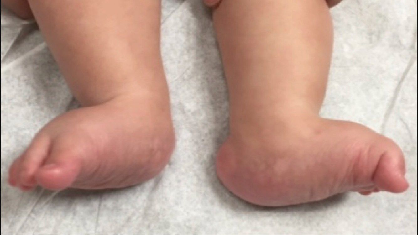
- Diagnosis: congenital vertical talus (CVT)
- Differential: congenital oblique talus, calcaneovalgus, tarsal coalition, paralytic pes planus
Q8Describe the clinical photograph.▸
- Midfoot: rocker bottom deformity
- Equinus hindfoot
- Dorsiflexion and abduction of the forefoot
Q9How would you assess this child?▸
- Screen systemically: 50% +ve neuromusclular disease (spina bifida, arthrogryposis), syndromal (Larsen), maltreated CTEV, look for DDH
- 20% positive family history
- Locally confirm a rigid deformity: rocker bottom foot, equinovalgus hindfoot with tight TA and peroneus
- Chopart joint: dorsiflexion with navicular dorsal dislocation; forefoot abduction/dorsiflexion (EDL/EHL, tib ant contractures)
- Prominent talar head plantarly; check lower limb neurology; peg leg gait
Q10What are the X-ray findings and how do you differentiate CVT from oblique talus?▸
- Lateral: vertically pointed talus, long axis of the talus below the 1st ray axis (TAMBA - talus axis MT bone angle)
- Dorsally dislocated navicular (navicular ossifies at 3 years old)
- AP: increase in talocalcaneal angle >40 degrees
- Plantarflexion view: if the TNJ reduces it is a congenital oblique talus, not CVT
Q11What is the pathoanatomy of vertical talus?▸
- Irreducible dorsal dislocation of the navicular on the talus, producing a rigid flatfoot deformity
- Soft tissue: tight TA; displaced PT and peroneals act as dorsiflexors; attenuated spring ligament
- Bony: dorsally dislocated navicular
- Oblique talus is also treated with TA release
Q12How is congenital vertical talus managed?▸
- Counsel: reverse Ponseti may be tried, but the majority eventually need surgery
- Pre-op stretching of dorsolateral structures + reverse Ponseti for 3 months (push forefoot plantar-medially)
- After reduction: talonavicular pinning (MIS) + peroneus/extensor release +/- lengthening +/- spring ligament recon + percutaneous Achilles tenotomy 6-12 months after casting
- Presenting late: talus + navicular excision and lateral column lengthening
- Salvage: triple arthrodesis; Dobbs (JBJS Am 2006) reported good results in 11 patients with stretching, reverse Ponseti, pinning and tenotomy
Q13Describe the clinical photo and give the diagnosis.📷▸
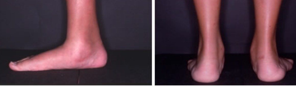
- Bilateral flatfoot with reduced medial arch
- Medial bulge suggests middle facet coalition
- From behind: hindfoot valgus, too many toes sign positive on the right
- Diagnosis: pes planus
- Must differentiate flexible from rigid
Q14What is the differential diagnosis for a painful flatfoot in a child?▸
- Tarsal coalition
- Painful flexible flatfoot (tight Achilles)
- JIA
Q15What are the general causes of flatfoot by laterality?▸
- Unilateral: post-traumatic, degenerative, Charcot
- Bilateral: ligamentous laxity, RA, neuromuscular (spastic diplegic, spina bifida)
- Both: idiopathic, PTTD, congenital vertical talus, tarsal coalition
Q16What is the genetics and pathology of tarsal coalition?▸
- Autosomal dominant with a high level of penetrance
- NOG (Noggin) gene
- Bilateral in 50%
- Failure of mesenchymal segmentation
Q17Outline your assessment of a child with a painful flatfoot.▸
- History: onset, site of pain, trauma, footwear
- Exclude obvious syndromic or neurological causes
- Tenderness: CN - lateral pain at sinus tarsi; TC - medial pain (microfracture of coalition interface, ossification of previously fibrous or cartilaginous coalition, chondral injury, peroneal spasm, subfibular impingement)
- Painful inversion against resistance (peroneal spasm)
- Deformity (hindfoot valgus, forefoot abduction, rigid flatfoot); PTT function
- Limited subtalar movement - reverse Coleman block; examine the contralateral side (50% bilateral, can be associated with fibular hemimelia/ PFFD, carpal coalition, Apert syndrome)
Q18What imaging would you request?▸
- CN: oblique view - anteater sign
- TC: lateral view - C sign (subtalar/middle facet coalition), talar neck traction spur
- Harris view may also show coalition
- CT/MRI for size and location of bony/fibrous connection, r/o additional coalitions (5%)
Q19How is tarsal coalition treated?▸
- Conservative: arch support/orthosis moulded at current position, walking cast boot, temporary immobilization
- CN coalition: excision via Olliers approach, EDB interposition +/- deformity correction and TA release
- TC coalition: bony union >50% -> fusion; <50% -> excision + interposition (split FHL graft, fat) via medial incision between FDL and PT
- Indications for fusion: malalignment, >50% involvement, degeneration, older patient
- Salvage: triple arthrodesis + deformity correction
Q20What does peroneal spasm mean and what is the epidemiology of flatfoot?▸
- Peroneal spasm is a protective mechanism to reduce pain; if absent the subtalar joint is likely okay
- Canadian army foot study: 1/5 have flatfoot; 67% asx (asymptomatic)
- 25% symptomatic flexible flatfoot with tight TA; 8% symptomatic rigid (accessory navicular, tarsal coalition, CVT)
- Wagner JBJS 1989: improvement not related to orthosis; increased laxity showed more improvement
Q21What is an accessory navicular?▸
- A secondary ossification centre that failed to unite in childhood (usually ossifies at 9 years old)
- Mostly asymptomatic
Q22What causes pain from an accessory navicular?▸
- Repeated microfracture of the synchondrosis
- PTT insertion inflammation
Q23How does an accessory navicular cause rigid flatfoot?▸
- PTT inserts into the accessory navicular -> change in PTT pull
- Midfoot adduction -> accessory navicular impinges onto the medial malleolus -> PTT pain
- Patient adopts forefoot abduction
Q24What is the Ray and Goldberg classification?▸
- Type I: ossicle in PTT
- Type II: synchondrosis
- Type III: synostosis
Q25What is the management?▸
- Asymptomatic: no treatment
- Mild: donut-shaped moleskin
- Severe: cast for 6 weeks then UCBL
- OT indication: refractory to cast/brace - Kidner procedure
- Kidner: remove accessory navicular at the synchondrosis + reattach PTT (advance distal and plantar - controversial)