Limbs Dysplasia Deformity
PFFD - classification and management
Proximal femoral focal deficiency: Birch classification, clinical and radiographic features, treatment.
25 questions
4 source pages
2 images
Images appear with the first question taken from each source page — tap a question to open it.
Q1Describe the clinical and radiographic features of this child with LLD.📷▸
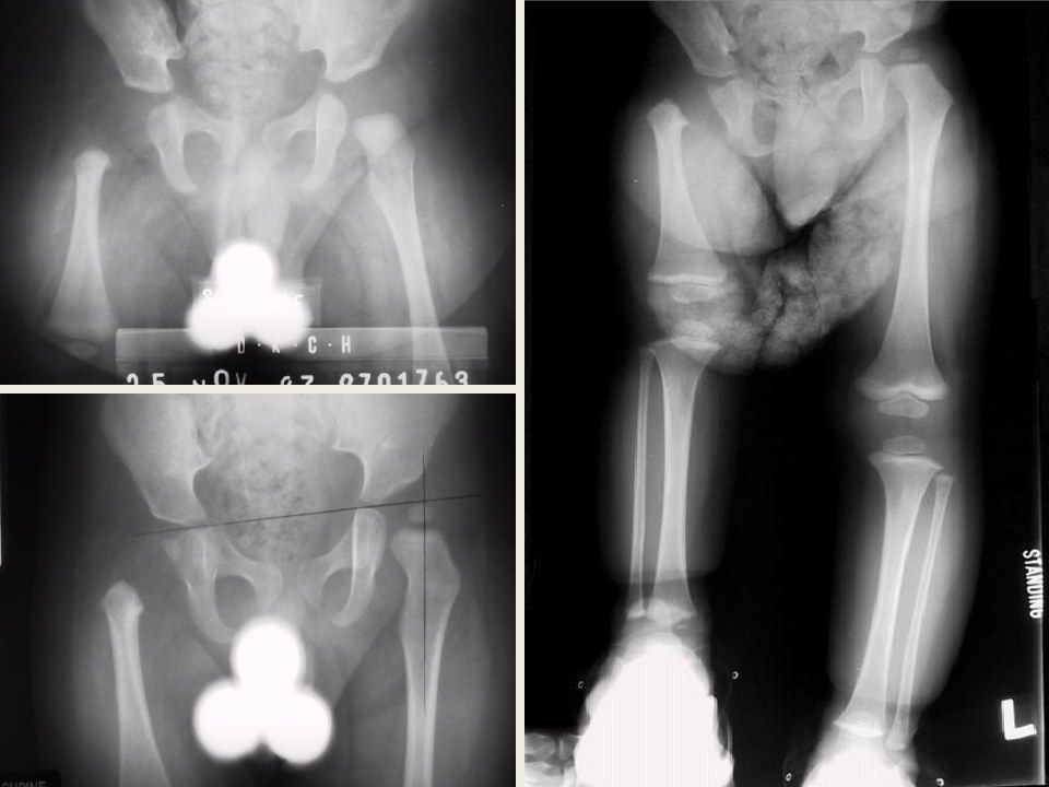
- Femur appears short, flexed + abducted + externally rotated (FABER)
- Knee and foot abnormalities; facial features in the AD type
- X-ray: hip abnormalities + short femur + coxa vara, ?epiphysis
- Acetabular (AI) / tear drop abnormalities; look for fibular hemimelia
- Impression: PFFD - MRI needed to see if the femoral ossification centre is present
Q2What history and imaging are important in PFFD?▸
- Birth history, including maternal thalidomide exposure (the classic teratologic agent)
- MRI to see whether the femoral ossification centre is present
- MRI to assess development and version of the femur
Q3What examination findings are important in PFFD?▸
- Assess the hip and knee stability
- Is the foot functional?
- Note leg length - at least to the mid tibia for the limb to be salvageable/reconstructable
Q4What is the pathology and aetiology of PFFD?▸
- Congenital defect of the proximal femur (dysplasia, coxa vara, retroversion, +/- absent head)
- Defect in the primary ossification centre - an intercalated transverse limb deficiency
- Mainly sporadic; occasional AD inheritance; bilateral in 15%
- Pathology: sclerotome subtraction
Q5Describe the Aitken and Gillespie classifications of PFFD.▸
- Aitken classification A-D: based on femoral and acetabular involvement
- AB: femoral head present; CD: femoral head absent
- A to D femur: head present, subtrochanteric pseudoarthrosis, absent head, absent head and proximal femur
- A to D acetabulum: normal, mild dysplasia, severe dysplasia, absent
- Gillespie: I congenital short femur (40-60%, functional hip/knee); II true PFFD (dysplastic hip, non-functional knee)
Q6What is the significance of the acetabular index and epiphysis in PFFD?▸
- Look for the femoral epiphysis (ossification centre) on X-ray
- A normal acetabular index suggests the femoral head is present
- Look for acetabular index/tear drop abnormalities
- The defect involves the femoral head, neck and acetabulum
Q7What associated anatomical anomalies accompany PFFD?▸
- Hip: coxa vara + retroversion, dysplasia
- Knee: LFC hypoplasia, genu valgum, patellar instability, cruciate (ACL) insufficiency, knee contracture
- Tibia: short + anteromedial bowing
- Fibula: fibular hemimelia
- Ankle: ankle valgus + ball and socket joint
- Foot: tarsal coalition, absent fibular rays
Q8What are the treatment aims and factors guiding management?▸
- Aim: allow the patient to ambulate, via the native limb or a prosthesis
- Factors: LLD, Aitken classification, foot stability
- Conservative: shoe raise
- First determine if LLD is too significant for reconstruction: predicted >20cm, foot to mid tibia, femur <50% of the other leg
Q9How does the Aitken classification guide reconstruction versus prosthesis?▸
- A/B (femoral head present): reconstruct the hip - valgus derotation osteotomy, PAO, ITB release, excise pseudoarthrosis + bone graft
- C/D (femoral head absent): iliofemoral fusion, then limb lengthening procedures
- Significant LLD: aim for prosthesis; functional foot -> iliofemoral fusion + Van Nes rotationplasty
- Non-functional foot -> knee fusion + amputation
Q10What are the principles and complications of limb lengthening?▸
- Roughly 1cm /month; may start around 5 years (consider psychological factors)
- Maximum 20-30% lengthening; related to complications
- Soft tissue: neurovascular; bone: non-union
- Joint: contracture; general: infection
Q11Describe the abnormalities seen in this standing patient.▸
- Abnormalities over the left lower limb; knee in valgus
- Anterior scars over tibia (?external fixator); medial and lateral scars over proximal tibia and distal femur (?previous epiphysiodesis)
- Missing 5th toe; +/- tibial anteromedial bowing; +/- patella subluxed
- Foot is hypoplastic with missing lateral rays
- Suspect fibular hemimelia; differential includes trauma (tumour and infection unlikely)
Q12What history would you take in this child with a suspected limb deficiency?▸
- Establish the diagnosis and rule out associated problems
- Delineate current symptoms
- Find out about previous treatment
Q13What associated abnormalities would you look for on examination?▸
- LLD
- Hip: PFFD, coxa vara
- Knee: hypoplastic lateral femoral condyle, genu valgum, patella instability, ACL deficiency
- Tibia: anteromedial bowing
- Foot/ankle: instability from ball and socket ankle, talipes equinovalgus, tarsal coalition, missing lateral toes
Q14What is the treatment goal in fibular hemimelia?▸
- Provide a plantigrade foot and a stable ankle
- Achieve equality of leg length
- Correct the tibial deformity to allow more efficient gait
Q15How does the Birch classification guide management?▸
- Type 1 = functional foot; type 2 = non-functional foot
- Functional foot (=>3rays, stable ankle) with mild LLD -> shoe raise and brace
- LLD <5cm -> contralateral epiphysiodesis
- LLD >5cm but <30% -> ipsilateral tibial lengthening
- Non-functional foot, unreconstructable ankle or significant LLD -> Syme amputation
Q16What determines ablation rather than reconstruction?▸
- Predicted LLD >20cm at maturity
- Foot to the level of mid tibia
- Femur <50% of the other leg
- Radiographic classification by Achterman
Q17How do you work up fibular hemimelia?▸
- Scannogram for LLD
- Examination for associated anomalies: hip, knee, tibia, foot/ankle
- Radiographic classification by Achterman
- Treatment depends on LLD at maturity and ankle stability
Q18What are the components of reconstruction in fibular hemimelia?▸
- 1. LLD
- 2. Tibial bow correction
- 3. Knee reconstruction
- 4. Ankle reconstruction
Q19How is the ankle reconstructed in fibular hemimelia?▸
- Soft tissue: peroneus longus release
- Mild: medial malleolar epiphysiodesis
- Moderate: supramalleolar osteotomy
- Severe: Gruca reconstruction - oblique sliding osteotomy of the tibia to make the lateral fragment the new lateral malleolus
Q20Tell me about fibular hemimelia.▸
- Most common congenital long bone deficiency; post-axial longitudinal deficiency (Swanson: failure of formation, problem with AED), occurs in the 1st trimester
- Achterman and Kalamchi classification based on the amount of fibula present
- Type 1A: part of fibula present but proximal fibular epiphysis distal to the proximal tibial physis, distal fibula proximal to the talus
- Type 1B: partial absence (30-50%), distal fibula unable to support the ankle; type 2: absent fibula
- If LLD >30% or unstable ankle -> Symes amputation (88% satisfactory vs ~50% lengthening)
Q21What is the Birch classification?📷▸
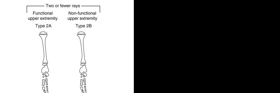
- A radiological classification of fibular deficiency
- Used to guide management
- Type 1 = functional foot
- Type 2 = non-functional foot
Q22How is a functional foot defined in the Birch classification?▸
- 3 or more rays
- Can provide a stable weight-bearing platform
Q23How do you recognise a leg length discrepancy and fibular hypoplasia on this radiograph?▸
- Pelvis not level despite a block under the short leg -> recognise LLD
- Fibular hypoplasia - the fibula is short
Q24What radiographic associations of fibular deficiency should you look for?▸
- Foot: tarsal coalition, absent lateral rays
- Ankle: ball and socket joint, ankle valgus
- Tibia: short + anteromedial bowing
- Knee: LFC hypoplasia, genu valgum, patellar subluxation
- Hip: PFFD
Q25Why is the fibula short? Describe the normal relationships.▸
- Normally the fibular head is level with the proximal tibial physis
- The distal fibular physis is level with the tibial plafond
- In fibular deficiency the proximal fibular epiphysis lies distal to the proximal tibial physis