Spine Non Trauma
Spondylolysis and isthmic spondylolisthesis
Pars interarticularis defects and SDSG classification of L5-S1 slip
7 questions
2 source pages
1 images
1 fact-check flags
Images appear with the first question taken from each source page — tap a question to open it.
Q1What is spondylolysis and what is spondylolisthesis?▸
- Spondylolysis: defect in the pars interarticularis
- Spondylolisthesis: forward slippage of one vertebra over another
Q2What are the radiographic findings of spondylolysis?▸
- Defect in the neck of the 'Scottie dog'
- Notes state 80% seen on lateral, 15% on oblique, 5% subtle defect on CT/SPECT
Q3What is the pathophysiology and who is affected?▸
- Fatigue fracture from repetitive hyperextension stress
- Examples: gymnasts, football linemen
Q4What is the treatment and prognosis of spondylolysis without listhesis?▸
- Activity restriction, flexion exercise, bracing
- Nonunion is common
- Unilateral defect never progresses to listhesis
Q5What is the SDSG L5-S1 slip classification used for?📷▸
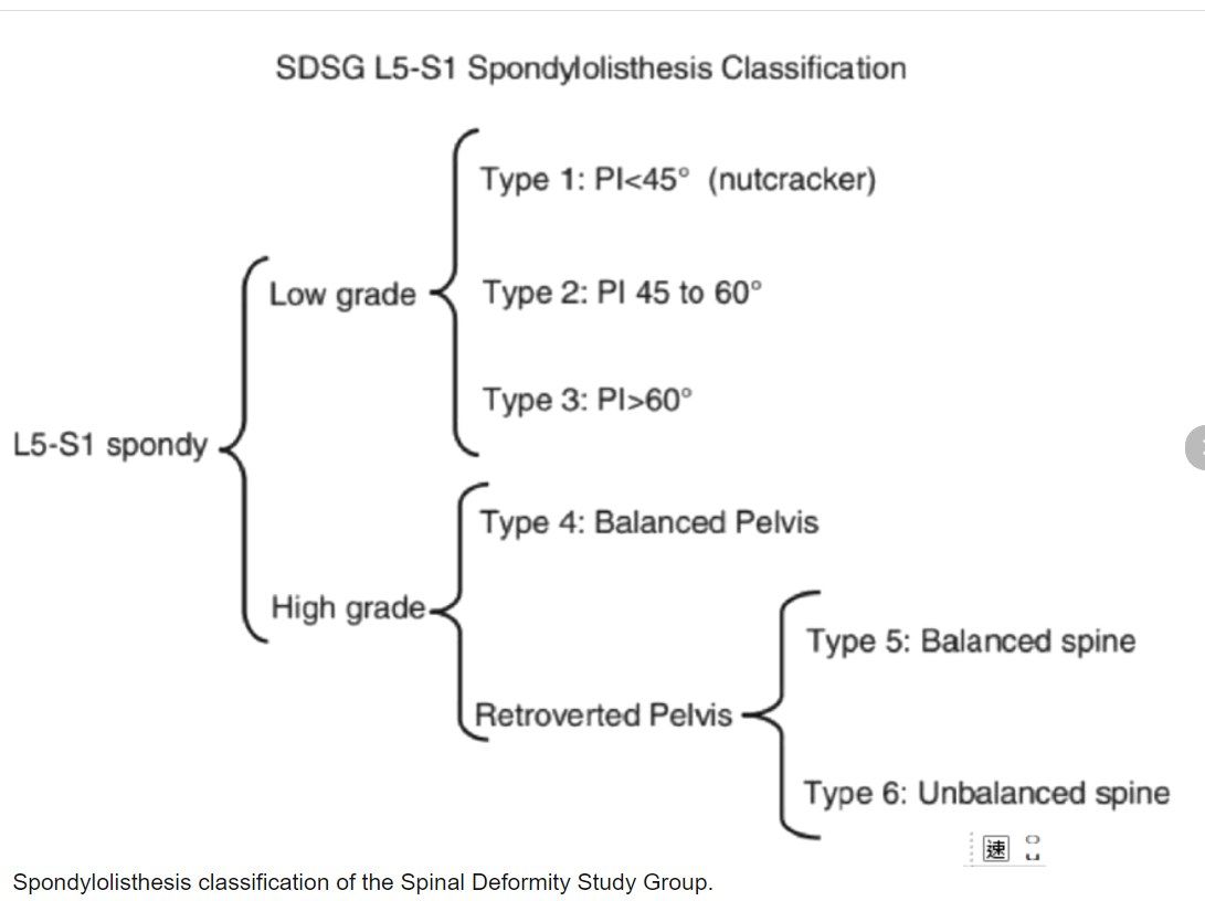
- The current recommended simple classification for dysplastic slip
- Balanced pelvis = low pelvic tilt, high sacral slope
Q6How are high and low grade slips defined in the SDSG classification?▸
- High grade = >50% slip
- Low grade = <50% slip
Q7What is the management principle for high grade dysplastic slip?▸
- Consider partial reduction in high grade slip
- Restore normal lumbosacral/pelvis relationship
Fact check
Spondylolysis on XR: 80% seen on lateral, 15% on oblique, 5% are subtle defects on CT/SPECT — contested — The oblique 'Scotty dog' view is classically the standard projection for spondylolysis, though sensitivity of lateral vs oblique is debated (coned lateral may detect ~85% of bilateral L5 defects). Early stress reactions are commonly invisible on plain XR (detection ~11-23% in early lesions), so more than 5% require CT/SPECT/MRI — (medium confidence) — source