Spine Non Trauma
Cervical cord myelopathy and rehabilitation
Cervical radiographs, OPLL and pannus, myelopathy classification, cord injury rehab
25 questions
6 source pages
4 images
1 fact-check flags
Images appear with the first question taken from each source page — tap a question to open it.
Q1Describe the lateral flexion/extension cervical X-ray findings.▸
- C1/2 subluxation with increased ADI and decreased PADI
- No associated cranial settling
- No subaxial cervical spine abnormalities
- Cervical height index measured
Q2What are the risk factors/causes and symptoms of C1/2 instability?▸
- Inflammatory joint disease or crystal arthropathy; signs of infection
- Previous trauma (odontoid nonunion / os odontoideum)
- Ligamentous laxity (Down's syndrome, MPS)
- Symptoms: pain (neck and occiput), myelopathy, vertebrobasilar insufficiency (vertigo, diplopia, drop attacks)
- Functional grading (Ranawat): I normal, II subjective weakness, IIIA objective weakness (ambulatory), IIIB no longer ambulatory
Q3What do you examine and what further investigations are required?▸
- Examination: neurological exam, cranial nerves, RA hand signs, Syndromal features (down, MED, morquio) - e.g. Down's syndrome, MPS
- Open mouth C-spine XR for rotatory instability: rule of Spence >8mm = TAL incompetence; controlled F/E to check if reducible
- CT: bony anatomy for surgical planning
- MRI: compressive pathology; cervicomedullary angle <135deg highly correlated with paralysis; myelomalacia
- Bloods to rule out RA if undiagnosed (RF, anti-CCP, EULAR or ACR criteria)
Q4What is the natural history of C1/2 instability (Boden's classic paper)?▸
- ~70 patients with C1/2 instability reviewed
- 60% developed symptoms; best predictor PADI <13mm
- Non-operated: 86% neuro deterioration, all died within 4 years
- Operated: 70% symptom relief, none died
Q5What lines and measurements are used to assess cranial settling?▸
- McRae's line: from basion or opisthion; dens tip should be below it
- Ranawat line: centre of C2 pedicle should lie below mid transverse axis of C1 (M 17, F 15; abnormal <13)
- Redlund-Johnell: C2 base to McGregor line (34/30mm)
- Chamberlain's line: hard palate to opisthion; dens tip should be <3mm above
- McGregor's line: hard palate to external occipital protuberance; dens tip <4.5mm above; Clark station: anterior C1 ring level with first third of odontoid
- Pannus: soft tissue mass encircling the eroded dens; stabilization leads to decrease in pannus
Q6When do you operate for cranial settling versus C1/2 subluxation?▸
- Cranial settling: neurological compromise, progressive cranial migration >5mm, cephalomedullary angle <135deg -> C0-C2 fusion +/- transoral odontoid resection
- C1/2 subluxation: neurology, SAC <13, ADI >10mm, cord diameter <6mm
- Reducible: C1/2 fusion with transarticular screw, wiring, pedicle/rod or segmental screws; non-reducible: C1 posterior arch excision + O-C2 fusion, or C1 laminoplasty + C1/2 fusion (less morbidity)
- Subaxial: subluxation >4mm or >20% = cord compression; cervical height index <2.0 almost 100% sensitive/specific for neuro compromise; translation >3.5mm, angulation >11deg; operate if >4mm or neurology
Q7What is the pattern of cervical spine involvement in rheumatoid arthritis?▸
- Cervical involvement in 90% of RA patients
- Atlantoaxial subluxation 80%: anterior subluxation of C1 on C2 from pannus between dens and C1 ring, destroying the transverse ligament and dens
- Basilar invagination 40%: cranial migration of dens from erosion and bone loss between occiput, C1 and C2
- Subaxial subluxation 20%: pannus and soft tissue instability of facet joints and Luschka joints
Q8What is pannus and how does it destroy structures?▸
- Latin for 'cloth'
- Hypertrophied synovium with fibrovascular tissue and inflammatory cells releasing collagenolytic enzymes
- Destroys cartilage, bone, tendons, ligaments and blood vessels
- Stabilization leads to decrease in pannus
Q9What is pannus?📷▸
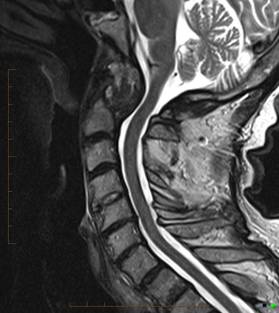
- A soft tissue mass
- It encircles the eroded dens
Q10Where does the term pannus come from and what is its composition?▸
- Latin for 'cloth'
- Hypertrophied synovium with fibrovascular tissue and inflammatory cells releasing collagenolytic enzymes
- Leads to destruction of cartilage, bone, tendons, ligaments and blood vessels
- Stabilization decreases pannus
Q11What are the classifications of OPLL?📷▸
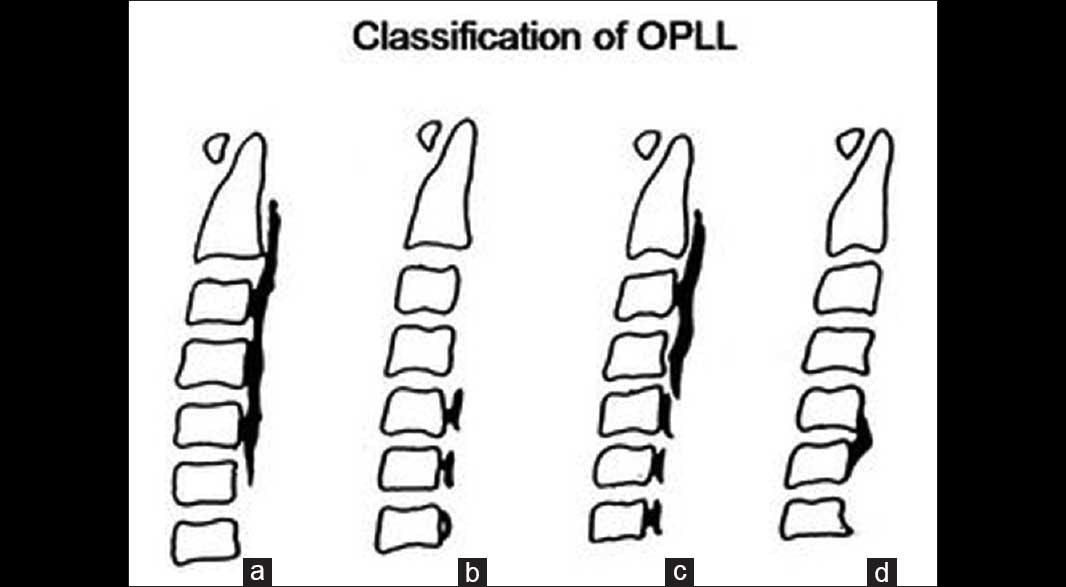
- XR (Mizuno): focal, segmental, continuous, mixed
- CT (Hirabayashi): square, mushroom, hill
Q12Why is OPLL difficult to treat, and what is the anterior strategy?▸
- Difficult: multi-level, at the level of the vertebral body, high chance of dural tear
- If anterior approach needed: decompress the surrounding bone; may need to leave OPLL on the dura (floating technique)
- Corpectomy may be required
Q13What is the indication for treatment of OPLL?▸
- Indication for treatment is the same as for cervical myelopathy
- No separate OPLL-specific indications are given in the notes
Q14Which functional classifications of cervical myelopathy are shown?📷▸
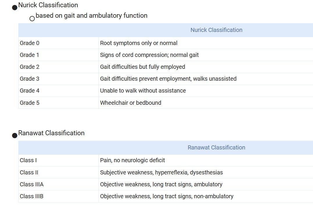
- Nurick classification - based on gait and ambulatory function
- Ranawat classification - based on pain, weakness and ambulatory status
Q15Describe the Nurick classification (grades 0-5).▸
- Grade 0: root symptoms only or normal
- Grade 1: signs of cord compression, normal gait
- Grade 2: gait difficulties but fully employed
- Grade 3: gait difficulties prevent employment, walks unassisted
- Grade 4: unable to walk without assistance
- Grade 5: wheelchair or bedbound
Q16Describe the Ranawat classification.▸
- Class I: pain, no neurologic deficit
- Class II: subjective weakness, hyperreflexia, dysesthesias
- Class IIIA: objective weakness, long tract signs, ambulatory
- Class IIIB: objective weakness, long tract signs, non-ambulatory
Q17What is the finger escape sign classification (Ono, JBJS 1987)?📷▸
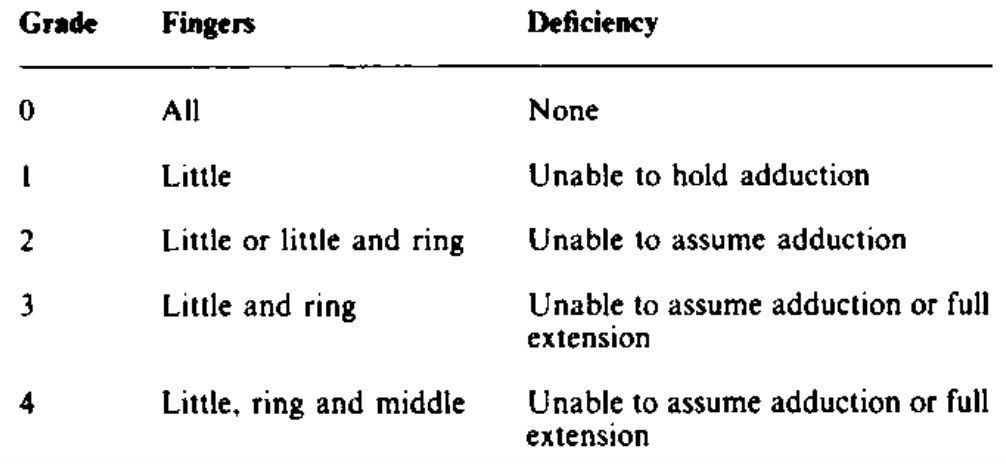
- Cannot hold
- Cannot do
- 2 finger / cannot do full extension
- 3 finger
- Closing the eyes is not necessary for the test
Q18What is the source of this classification and how is the test performed?▸
- JBJS 1987 by Ono
- Finger escape sign classification
- Patient does not need to close the eyes
Q19What is the ventilator weaning success rate after cervical spinal cord injury?▸
- C3: 51%
- C4: 78%
Q20Why does vital capacity decrease on sitting in cervical cord injury?▸
- Increased dead space as the abdominal organs drop down when sitting
- Can be improved with a corset
Q21What is the motor recovery prognosis after traumatic spinal cord injury?▸
- Muscles with some motor power below an antigravity muscle have a better prognosis than muscles with no motor power
- Grade 1 or 2: 90% reach grade 3 by 1 year
- Grade 0: 64% reach grade 3 by 2 years
- Recovery is faster in incomplete injuries
- Median time to antigravity strength: 2 months motor complete vs 2 weeks motor incomplete (Ditunno 1992)
Q22What is the ambulatory potential by ASIA grade?▸
- 3% / 50% / 75% / 95% in ASIA A / B / C / D (van Middendorp, Global Spine J 2011)
Q23What are the functional goals at C5 and C6?▸
- C5: use of power wheelchair
- C6: manual wheelchair, feeding well
- Consider tenodesis grasp/transfer at C6
Q24How does sphincter function differ in suprasacral vs cauda equina lesions?▸
- Suprasacral: detrusor-sphincter dyssynergia, spastic bladder, reduced volume, may develop reflux/HN, finally incontinence
- Cauda equina: flaccid bladder that stretches out, can develop stress incontinence
Q25Which 1999 consensus published the traumatic spinal cord injury rehabilitation potential data?▸
- Consortium of Spinal Cord Medicine 1999
Fact check
90% of RA patients have cervical spine involvement; atlantoaxial subluxation in 80%, basilar invagination 40%, subaxial subluxation 20% — misleading precision — Prevalence varies widely by cohort and imaging: cervical involvement reported up to ~86%, AAS is the most common form (radiographic rates roughly 16-96%), basilar invagination ~5-34% and subaxial subluxation ~10-44%; fixed 80/40/20 figures overstate certainty — (medium confidence) — source