P and O
Amputation indications and levels
Indications for amputation and levels including foot amputation and hip disarticulation
27 questions
3 source pages
1 images
1 fact-check flags
Images appear with the first question taken from each source page — tap a question to open it.
Q1What are the indications for lower limb amputation?▸
- DM/PVD
- Trauma
- Tumour
- Paediatric malformation/infection
Q2How is the level of amputation chosen?▸
- Balance between pathology, wound healing and energy expenditure
- Ideal level: best for prosthesis fitting while minimising energy expenditure
Q3What is the increase in energy consumption at each amputation level?▸
- Generally inversely proportional to remaining length
- Symes 15; BKA traumatic 25, vascular 40; through knee 50; AKA traumatic 70, vascular 100
- Bilateral BKA 40; BKA + AKA 120; bilateral AKA >200
Q4What makes a good amputation stump?▸
- Ideal stump: mobile nonadherent muscle mass covered by full thickness skin tolerating weightbearing and shear
- Skin: scar mobile and away from bony edges, as sensate as possible
- Soft tissue: cylindrical or conical stump without excessive tissue
- Muscle: myodesis (muscle to bone) versus myoplasty (muscle to fascia)
- Nerve: clean section and retraction into soft tissue; bone ends bevelled and contoured
Q5How do you perform a below-knee amputation (BKA)?▸
- Decide level (15cm below medial joint line); check pulse/healing and no contracture
- Bone cut 10-15cm from medial joint line; skin incision 15cm distal to tibial tubercle, posterior flap ~1.5x anterior flap (anterior 2/3, posterior 1/3)
- Section tibia, fibula 1cm above; bevel cut; Ertle osteomyoplasty fibula strut (paediatric)
- Ligate anterior and posterior tibial vessels and peroneal nerves; meticulous soft tissue balance; U slab to avoid knee contracture
Q6What are the complications of amputation and the differential for pain in the missing limb?▸
- Unfit prosthesis - sore; neuroma; bone impingement; phantom limb
- Phantom limb risk factors: traumatic, young patients, upper limb, bilateral
- Ddx primary: neuroma, phantom limb pain, HO, bone impingement
- Ddx secondary: recurrence of primary pathology (tumour), CRPS, radiation pain from elsewhere
Q7What are the key steps after the skin incision in a below-knee amputation?▸
- Position supine with tourniquet and hip support
- Divide anterolateral muscle down to intermuscular septum; ligate anterior tibial vessels and peroneal nerves
- Section tibia, and fibula 1cm above
- Dissect posterior soft tissues distally to the posterior transverse incision
- Ligate posterior tibial and peroneal vessels and the posterior tibial nerve
- Bevel cut; meticulous soft tissue balancing; do not trim dog-ear; U slab to avoid knee contracture
Q8Compare the long posterior flap and sagittal flap for BKA.▸
- LPF: better flap blood supply from gastrocnemius; limited by proximal extent of infection/tumour
- SF: versatile, less infection in DM (Christensen JBJS 1989)
- SF disadvantage: tibial bone close to wound
- Ruckley J Vascular Surgery 1991: no difference
Q9What are the length guidelines for transtibial and transfemoral amputation?▸
- Transtibial optimal 8cm for every 1m of height
- Transtibial shortest 7.5cm below medial joint line
- Transfemoral shortest 8cm below pubic rami
- Transfemoral longest 15cm above medial joint line
Q10How is an above-knee amputation (AKA) performed?▸
- Bone: level of cut 8cm below pubic ramus to 15cm above joint line, smoothened and bevelled end
- Skin: AP flap
- Soft tissue: myodesis to counterbalance muscle pull and prevent contracture
- Myodesis of adductor and hamstring to prevent flexion and abduction deformity
- Not to do myodesis in vascular compromise
- Nerve: clear cut under gentle tension and allow retraction to prevent neuroma
Q11What are the important considerations in the length of an AKA?▸
- Longer gives a better lever arm for control of the prosthesis
- Balance accommodation of the prosthesis
- Myoplasty/myodesis of adductor preserves 70% adductor power --> decreases AL drift
Q12What are the indications and technique of through-knee amputation?▸
- Life saving
- Paediatric: preserve distal femur physis for longer lever arm
- Non-ambulatory patients
- Technique: suture patellar tendon to cruciate ligament
- Gastroc muscle (gastrocnemius) used as soft tissue envelope
Q13What are the advantages of through-knee amputation?▸
- Less bleeding, faster OT
- Most thigh muscle preserved
- Long lever arm
- Broad end bearing surface
- Stable suspension
- No bony outgrowth
Q14What are the disadvantages of through-knee amputation?▸
- Different levels of knee joint, less modularity
- Bulbous shape, less cosmetic
- Less room for error for flap (cannot cut more bone)
Q15What did the LEAP study show for through-knee amputation?▸
- Similar pain to AKA/BKA
- Reduces walking speed and return to work
- Asymmetric knee + limited knee component choices
- Reduces satisfaction
Q16What is direct versus indirect load transfer in an amputation stump?▸
- Direct - through knee/through ankle: wide metaphysis distributes the force of weight bearing
- Direct: less stiff bone at metaphysis dampens GRF at heel strike
- Indirect - BKA/AKA: terminal surface incapable of weight bearing; stiff bone
Q17What is the pathophysiology and treatment of phantom limb pain?▸
- Pathophysiology: corresponding primary somatosensory cortex deprived of sensory input --> reorganization via increased expression of previously marked inputs - maladaptive plasticity model
- Physio: mirroring, USG, TENS, massage, immersive virtual reality
- Pharmacological: opioids, anticonvulsants, antidepressants, botulinum agents
- Surgical: stump revision, intrathecal implants, trigger point injections, dorsal column tractotomy
Q18What are the types of foot amputation?▸
- Transmetatarsal
- Lisfranc (TMTJ disarticulation)
- Chopart
- Syme
- Boyd
Q19Describe transmetatarsal amputation and its problems.▸
- Lowest energy expenditure; no tendon transfer
- Bone cut >=3cm to MTB; preserve MT cascade; 5MTB
- Problem: equinus from loss of extensor lever arm; solution TA lengthening
Q20How are Lisfranc and Chopart amputations balanced?▸
- Lisfranc: PB to cuboid transfer to reduce varus / shell out 5MTB to preserve PB insertion
- Lisfranc: +/- TA lengthening to reduce equinus
- Chopart: tib ant to talus transfer to reduce equinus and cavus; +/- TA lengthening
- Chopart problem: unstable hindfoot
Q21What are the prerequisites and indications for Syme amputation?▸
- Patent posterior tibial artery (ischemic index >0.5)
- Healthy heel pad
- Indication: forefoot pathology - tumour/trauma/congenital; DM/infection
- Contraindicated in PVD
Q22Describe the Syme amputation technique.▸
- Fishmouth flap 1cm distal + anterior to intermalleolar line; anterior through ankle joint, posterior through CCJ
- Ligate PTA as distal as possible
- Malleoli cut at 6cm proximal to joint line (see factcheck)
- Posterior flap anchored to distal tibia through drill holes to prevent heel pad posterior migration
- Preserve tibia articular surface for a resilient limb; avoid vigorous trimming of dogears
Q23What are the advantages and disadvantages of Syme amputation, and what is Boyd amputation?▸
- Adv: end bearing stump (walk on unsupported stump); Self regulated walking speed, more natural gait pattern
- Adv: longer lever arm --> reduce energy expenditure
- Disadv: technically difficult; limited indication; fat pad migration; socket fabrication challenging
- Boyd = modification of Syme: talectomy + calcaneal tibial fusion; no heel pad migration
- Boyd disadv: bulky stump increases rehab time and nonunion
Q24Describe the racquet-shaped incision for hip disarticulation.📷▸
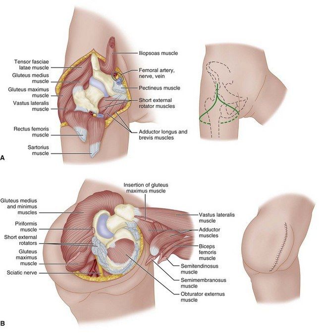
- Anterior: ASIS to hip
- Medial: parallel to inguinal ligament to inferior pubic rami (adductor origin)
- Posterior: transverse 5cm distal to ischial tuberosity to 8cm distal to GT
- Lateral: curve up to anterior incision
Q25What are the anterior dissection steps in hip disarticulation?▸
- ASIS: detach sartorius; AIIS: detach rectus femoris
- Divide femoral NAV at femoral triangle
- LT: detach iliopsoas (hip ER) and obturator externus
- Superior PR: detach pectineus (0.6cm residual cuff), divide obturator AVN underneath
- Inferior PR: detach adductors + gracilis; ischial tuberosity: detach hamstring
Q26What are the posterior dissection steps in hip disarticulation?▸
- Divide fascia lata and glut max along posterior incision
- GT: detach glut med and min
- Trochanteric ridge: detach short ER
- Divide sciatic nerve
Q27How is the stump covered after hip disarticulation?▸
- Deep myoplasty: obturator externus to glut med at acetabulum (GMOE)
- Superficial myoplasty: iliopsoas to quadratus femoris (IQ)
- Fascia: glut fascia --> inguinal ligament
Fact check
In Syme amputation the malleoli are cut at 6cm proximal to the joint line — incorrect level — Classic Syme divides the tibia and fibula ~1-1.5cm above the ankle joint (or flush with the plafond) so the distal articular surface is preserved; 6cm would be a transtibial level — source