Statistics
Metabolic and systemic bone disease
Osteopetrosis, hyperparathyroidism, renal osteodystrophy and related radiographic findings.
28 questions
5 source pages
1 images
2 fact-check flags
Images appear with the first question taken from each source page — tap a question to open it.
Q1Describe the X-ray findings in osteopetrosis.▸
- Skeletally immature patient with generalised increased bone density
- Canal not well seen; radiolucency at the distal femur
- Erlenmeyer flask appearance over the distal femur
- Dense endplate with rugger jersey appearance
- Wormian bones; Ddx: hypoparathyroidism, pyknodysostosis, lead poisoning
Q2What is the pathophysiology of osteopetrosis?▸
- Congenital disease causing dysfunction of the osteoclast
- Defect in proton pump, carbonic anhydrase 2, or chloride channel
Q3What are the orthopaedic and non-orthopaedic features of osteopetrosis?▸
- Ortho: usually asymptomatic in AD type except fracture, OA, higher risk of osteomyelitis
- X-ray: coxa vara due to repeated fracture, bone-in-bone appearance, Erlenmeyer flask, rugger jersey spine
- Non ortho: blindness/ deafness in AR type, anemia, cranial nerve dysfunction
- Bloods usually normal (AR type may show increased acid phosphatase/PTH)
Q4What is the treatment of osteopetrosis?▸
- AR: bone marrow transplant, high-dose vitamin D
- AD: interferon gamma-1b (interferon gamma -1 beta) (Key et al NEJM 1995)
- Surgery for proximal femur fracture: use a plate (marrow space obliterated)
Q5What is the histology of osteopetrosis?▸
- Osteoclasts lack a ruffle border and clear zone
- Empty lacunae
- Plugging of the haversian canals
Q6What are the types of osteopetrosis?▸
- Malignant (severe): AR, may be fatal in childhood without transfusion
- Intermediate: AR, can survive to adulthood
- Benign (mild): AD, most common, 2 types
- Type I: no increased fracture risk
- Type II (Albers-Schonberg disease): anaemia, pathological fractures, premature osteoarthritis; first presentation is fracture
Q7What are the X-ray features of hyperparathyroidism?▸
- Metastatic calcification: nephrocalcinosis, lungs, blood vessels, synovium/chondrocalcinosis, calcinosis cutis
- Subperiosteal resorption: salt and pepper skull, middle phalanx and ulna (increased concavity)
- Brown tumour (osteitis fibrosa cystica): lytic and expansile
- Rugger jersey spine with sclerotic endplates
Q8What is the clinical presentation of hyperparathyroidism?▸
- Bones, groans, stones, psychic moans
- PE: signs of CKD (oedema, cafe au lait appearance), vitamin D deficiency, renal transplant scar/RLQ mass
Q9What is a brown tumour?▸
- Osteitis fibrosa cystica
- Lytic and expansile; rapid bone loss -> hypervascular granulation -> fibrous tissue
- Histology = GCT
- Other Ddx: metastasis, multiple myeloma, GCT/ABC
Q10What is the histology and management of hyperparathyroidism?▸
- Focal area of increased osteoclasts amidst fibrous stroma
- Peripheral rim of osteoid
- Treat the underlying cause (parathyroidectomy / renal replacement therapy)
Q11What is metastatic calcification in hyperparathyroidism?▸
- Calcification in normal soft tissue due to a local increase in calcium -> calcium phosphate deposition
- Sites: nephrocalcinosis, lungs, blood vessels, synovium/chondrocalcinosis, calcinosis cutis
Q12What is renal osteodystrophy and what are the XR findings?📷▸
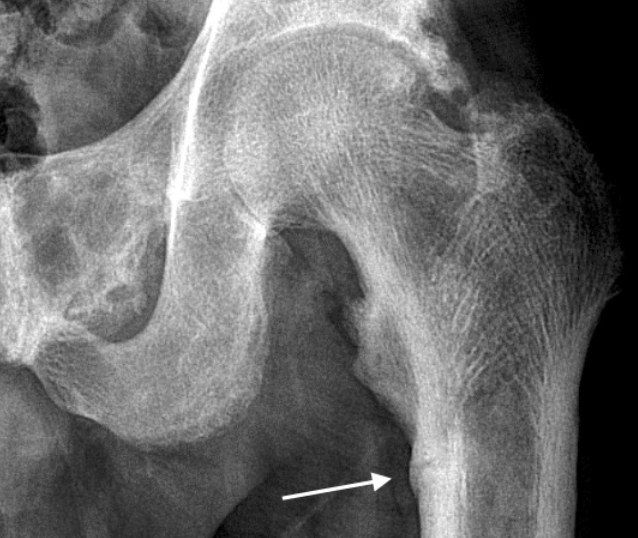
- Failure of bone mineralisation due to chronic renal failure
- XR: brown tumour and looser zone at the compression side
Q13What is the pathophysiology of low-turnover renal osteodystrophy?▸
- Aluminium toxicity
- Decreases PTH release
- Decreases osteoblast differentiation and proliferation
- Decreased mineralisation -> osteomalacia (adynamic bone secondary to Al toxicity)
Q14What other bone disease occurs in chronic renal failure?▸
- Osteoporosis
- Amyloid bone disease: B2 macroglobulin deposits in the juxta-articular region, especially ball-and-socket joints
- XR: bone cyst with sclerotic edge causing erosive arthritis
- Other sites: carpal tunnel syndrome + carpal cyst
Q15What is the pathophysiology of high-turnover renal osteodystrophy?▸
- Phosphate retention increases PTH by 3 mechanisms
- 1. Direct decrease in serum calcium, stimulating PTH
- 2. Decrease in renal 1-alpha hydroxylation -> less serum calcium
- 3. Direct increase in synthesis of PTH
- Secondary hyperPTH raises serum Ca, which binds phosphate -> ectopic calcification
Q16Describe the imaging findings of loose bodies in the elbow.▸
- Well-circumscribed radio-opacity
- Clustered ossification/calcification
- Mainly over the anterior elbow, also around the trochlear groove
- Minimal OA; no varus/valgus malalignment; no stippled calcification
Q17What is the clinical presentation and what is it associated with?▸
- Age 30-40 years
- Pain, loose body sensation, locking
- Association: OA, OCD, osteochondroma
Q18What is the pathogenesis of loose bodies?▸
- Synovial cell metaplasia into chondrocytes (primary spontaneous; secondary incited by loose bodies)
- Nutrition from synovial fluid
- Cartilage shell: increased nourishment -> increase in size
- Calcified core: decreased nourishment, apoptosis, calcium salt remains
Q19What is the classification of loose bodies?▸
- Primary: synovial metaplasia; XR smaller, similar in size
- Secondary: OCD, OA, AVN, osteochondroma; XR larger, varied
Q20What are the phases of synovial osteochondromatosis?▸
- Phase 1: active synovitis without loose bodies
- Phase 2: nodular synovitis along with loose bodies
- Phase 3: loose bodies present with resolution of synovitis
Q21What are the MRI findings and management of loose bodies?▸
- Cartilage shell: decrease T1, increase T2
- No cartilage shell: decrease in both T1 and T2
- Mx: symptom relief, prevent secondary OA
- Synovectomy + removal of loose bodies
Q22What are the three main categories of soft tissue calcification?▸
- Dystrophic
- Metastatic
- Calcinosis (cutaneous, subcutaneous, deep connective tissue)
Q23What are the causes of dystrophic soft tissue calcification?▸
- Vascular: phlebolith
- Infection: TB
- Tumour: synovial sarcoma, osteosarcoma, chondrosarcoma, synovial chondromatosis, haemangioma
- Haematoma
- Calcific tendinitis
Q24What are the types and causes of metastatic soft tissue calcification?▸
- Increased calcium - hyperPTH
- Increased phosphate - CKD
- Gout
- May resemble tumoral calcinosis
Q25How is calcinosis classified by extent?▸
- Focal: hand - calcinosis circumscripta; joint - chondrocalcinosis, tumoral calcinosis
- Diffuse: calcinosis universalis, myositis ossificans progressiva
Q26Where can calcinosis be deposited anatomically?▸
- Cutaneous
- Subcutaneous
- Deep connective tissue
Q27What are the radiological features of tumoral calcinosis?▸
- Amorphous, lobulated
- Chicken-wire lucencies (thin fibrous septae)
- Cobblestone appearance
- Cystic lesions with fluid level = calcium hydroxyapatite precipitation
Q28What is calcinosis circumscripta and how does it present clinically?▸
- Well defined subcutaneous nodules
- Ulcerating with chalky hydroxyapatite discharge
Fact check
Intermediate osteopetrosis is autosomal recessive and is called Albers-Schonberg disease — Misleading / internally contradictory — Albers-Schonberg disease is the autosomal dominant type II (mild) form, as the same page later states; intermediate osteopetrosis can be AR or AD — source
Autosomal dominant osteopetrosis is treated with interferon gamma-1b (Key et al NEJM 1995) — Misleading - Key 1995 studied severe infantile (recessive) disease — IFN-gamma-1b is FDA approved for severe malignant osteopetrosis (autosomal recessive), often as a bridge to HSCT; evidence in ADO is insufficient and it was poorly tolerated — source