Statistics
Primary bone malignancy differentials
Named primary tumour diagnoses including Ewing sarcoma, myeloma and telangiectatic osteosarcoma.
21 questions
3 source pages
1 images
1 fact-check flags
Images appear with the first question taken from each source page — tap a question to open it.
Q1Where does Ewing sarcoma occur and what are the X-ray features?▸
- Typically lower limb, pelvis, proximal humerus
- Destructive permeative, moth eaten, lytic lesion in the metaphysis and diaphysis with variable new bone formation
- Onion-skin periosteal lifting (characteristic but uncommon)
- Soft tissue component is often large
Q2What is the clinical presentation and DDx of Ewing sarcoma?▸
- Fever + pain - thus mimics infection
- DDx: osteosarcoma, haematological, osteomyelitis, eosinophilic granuloma
Q3What investigations confirm Ewing sarcoma?▸
- Bloods: raised ESR, anaemia, increased WBC, LDH (prognostic >200IUml / >200 IU/mL)
- MRI: soft tissue component, reduced T1 due to marrow invasion, increased with contrast
- Biopsy: monotonous small blue round cells, high N:C ratio, CD99+
- Bone marrow biopsy to rule out metastasis to the marrow: consistent chromosomal translocation (11;22) with EWS-FLI 1 fusion protein
- CT/CXR: metastases in 30% at presentation
Q4What is the treatment principle in Ewing sarcoma?▸
- Oncologic clearance with preservation of limb and function: save life (systemic control), save limb (local control)
- Multimodality approach: multiagent chemotherapy, irradiation and surgical resection
- Pre-op chemotherapy 2-3 months, then resection, maintenance chemo 6-12 months
Q5What is the local control strategy and when is radiotherapy used?▸
- Trend towards surgical resection (wide margin excision) for all surgically accessible sites; assess tumour necrosis
- RT indications: positive margin, metastasis, poor response to chemo
- Chemo + OT +/- adjuvant RT = chemo + RT
Q6What are the RT complications, poor prognostic factors and survival?▸
- RT complications: 10-20% secondary sarcoma, fracture, physeal damage/LLD, osteonecrosis, muscle atrophy, stiffness, Joint: contracture
- Poor prognostic factors: metastases (1/4), >14yo, male, spine/pelvic tumours, >100cm3, poor chemo response (<90% necrosis), raised LDH/WCC, low Hb, p53 mutation
- 5-year disease-free survival ~65% (~half with mets); lung mets 30% cure, marrow 30% cure, both 10-15% cure
Q7What is the epidemiology and origin of Ewing sarcoma?▸
- Distinctive small round cell sarcoma occurring most often in children and young adults (5-25)
- 2nd most common malignant tumour in children
- Neuroectodermal cells arise from the medullary canal and invade the haversian system
Q8What is the epidemiology and immunoglobulin profile of multiple myeloma?▸
- Most common primary bone malignancy; 1% of all malignancies; males > females
- Neoplastic plasma cells produce immunoglobulins
- Heavy chains: IgG 52%, IgA 21%, IgM 12%
- Light chains: kappa and lambda (Bence Jones proteins)
Q9Why are osteolytic lesions seen in multiple myeloma?▸
- Osteoclastic stimulation via RANKL and pro-osteoclastic mediators (M-CSF, IL-6, IL-11)
- Osteoprotegerin (OPG) synthesis is suppressed, causing further osteoclast activation
- Osteoblastic differentiation inhibited by TNF and Dickkopf-1 (DKK-1)
Q10What are the diagnostic criteria and CRAB features of multiple myeloma?▸
- >=10% monoclonal plasma cells on bone marrow biopsy (or biopsy-proven plasmacytoma) plus >=1 CRAB feature/MDE
- Calcium: >0.25 mmol/L above upper limit of normal or >2.75 mmol/L (>11mg/dL)
- Renal: creatinine clearance <40mL/min or creatinine >177umol/L (>2mg/dL)
- Anaemia: Hb <100g/L or >20g/L below the lowest limit of normal
- Bone: >=1 osteolytic lesion on radiographs, CT or PET/CT
- If marrow has <10% clonal plasma cells, >1 bone lesion is required to distinguish from solitary plasmacytoma with minimal marrow involvement
Q11What are the myeloma-defining events (MDEs) and ISS staging?▸
- MDE: >=60% clonal plasma cells on bone marrow biopsy
- MDE: involved:uninvolved free light chain ratio >=100 (involved light chain >=100mg/L)
- MDE: >=1 focal lesion on MRI >=5mm
- ISS stage I: beta-2 microglobulin <3.5mg/L, albumin >=3.5g/dL (62 months); II: 3.5-5.4mg/L (44 months); III: >=5.5mg/L (29 months)
Q12What are the XR and imaging findings in myeloma and the role of bone marrow aspirate?▸
- Punched-out lytic lesions, only visible once >50% destruction has occurred
- No sclerotic border - due to lack of osteoblastic activity
- Bone scans are cold in 30%; skeletal survey is recommended
- FDG-PET 93% sensitivity; may uncover additional sites in 'solitary' plasmacytoma
- Marrow aspirate: plasmacytoma 10-30% vs myeloma >30% plasma cells (normal <2%); CD138+, Hoffa clear zone
Q13What are the types, treatment and prognosis of plasma cell neoplasms?▸
- Solitary plasmacytoma: single skeletal site, radiosensitive, progresses to myeloma in >50%; MRI/FDG-PET finds additional lesions in 33%
- Osteosclerotic myeloma: rare POEMS - polyneuropathy, organomegaly, endocrinopathy, sclerotic bone lesions, M protein, skin changes
- Treatment: asymptomatic/MGUS - annual surveillance; solitary plasmacytoma - external beam irradiation alone; myeloma - multiagent chemo +/- stem cell transplant +/- bisphosphonates
- Fractures: surgical stabilisation + RT (life expectancy >3 months); kyphoplasty for vertebral compression with instability/neural compression resistant to radiation
- Prognosis: 5-year survival 30%, 10-year 11%, median 3 years; shortest survival with renal failure; solitary plasmacytoma has the best prognosis
Q14What histological features are seen in myeloma plasma cells?▸
- Round plasma cells with eccentric nucleus and prominent nucleolus
- Clock-face organisation of chromatin
Q15What are the diagnostic criteria for solitary plasmacytoma?▸
- Solitary lesion on skeletal survey
- Histologic biopsy confirmation of plasmacytoma
- Negative bone marrow biopsy (no plasma cells in bone marrow)
Q16What are the poor prognostic factors in multiple myeloma?▸
- Chromosome 13 deletion or translocations t(4;14), t(14;16)
- Circulating plasma cells
- Increased beta-2 microglobulin (elevated tumour burden); decreased serum albumin
- Increased marrow microvessels
- Overall survival relates to stage and secondary factors like renal failure or hypercalcaemia
Q17Describe the X-ray findings of telangiectatic osteosarcoma.📷▸
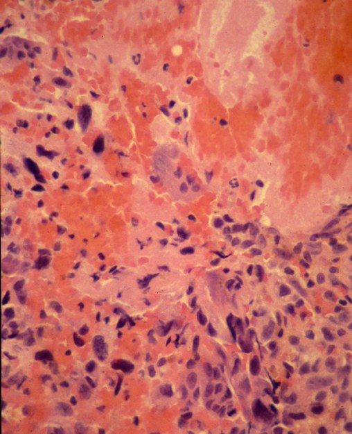
- Skeletally mature patient
- Eccentric lytic lesion in the metaphyseal region of the medial distal femoral metaphysis
- Cortical destruction; no extension into the joint
- Periosteal reaction with Codman triangle; matrix is osseous
- Significant soft tissue component
Q18What is the typical presentation of telangiectatic osteosarcoma?▸
- Age 15-25, male
- Previous radiation
- Pain; pathological fracture in 25%
Q19How does the MRI of telangiectatic osteosarcoma differ from ABC?▸
- Less defined fluid levels compared with ABC
- Extensive soft tissue oedema
Q20What are the differentials and management of telangiectatic osteosarcoma?▸
- Ddx: GCT, ABC
- Mx as per conventional osteosarcoma
Q21What features favour telangiectatic osteosarcoma over an aneurysmal bone cyst?▸
- Codman triangle and periosteal reaction
- Osseous matrix
- Significant soft tissue component
- Less defined fluid levels and extensive soft tissue oedema on MRI
Fact check
Multiple myeloma 5-year survival is 30%, 10-year survival 11%, median survival 3 years — outdated — With modern therapy, SEER data (2016-2022) report 5-year relative survival around 62-64%; the 30%/11% figures are historical and should not be quoted as current — source