Statistics
Bone tumour radiograph spotter cases
Radiograph and clinical cases of lytic, sclerotic and mixed bone lesions to identify the diagnosis.
61 questions
8 source pages
2 images
1 fact-check flags
Images appear with the first question taken from each source page — tap a question to open it.
Q1What is the likely diagnosis and which conditions must be excluded?📷▸
- Large juxtaarticular (juxta-articular), eccentric, lytic expansile lesion with geographic destruction of the lateral condyle
- No cortical break, no matrix, no sclerotic margins, narrow zone of transition
- Likely an aggressive benign lesion such as GCT
- Tumourous DDx: ABC, clear cell chondrosarcoma, telangiectatic osteosarcoma
- Non-tumourous DDx: brown’s tumor (brown's tumour), infection
Q2What history, examination and investigations are needed?▸
- History: onset, progression, pain, constitutional symptoms
- PE: knee tenderness, AROM, effusion; systemic exam for lymph nodes/masses
- Bloods to r/o infection/metabolic cause: CBC, CRP, ESR, Ca/PO4 (brown's tumour), bone profile
- MRI (T1 hypointense lesion); confirm with image-guided biopsy
- Systemic staging: CT thorax (2% pulmonary met) + bone scan
Q3What is the histological appearance and the Campanacci classification?▸
- Mononuclear cells (monocytes), spindle cells and multinucleated giant cells
- Campanacci 1-3: cortex undisrupted, thinning, disrupted
- Guides recurrence rate and local progressiveness
Q4What are the management principles and recurrence rates?▸
- Reduce local recurrence and preserve function
- Discuss pros and cons of resection vs curettage with the patient
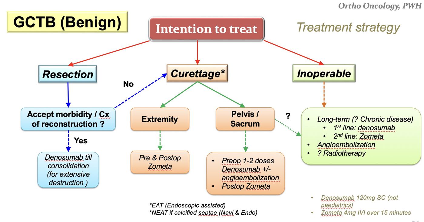
- Resection 5% recurrence, extended curettage 10%, curettage 20-50%
- Overall 5-year disease-free survival 75%, mortality 15%
Q5Why is cement inserted after extended curettage and what does extended curettage mean?▸
- Cement provides thermal ablation of tumour, immediate mechanical support and easy detection of recurrence
- Recurrence decreased from 10% to 5% with adjuvant therapy
- Extended curettage = curettage (extensive exterioration) + adjuncts: mechanical burring, thermal cement, cryo nitrogen, chemical phenol, osmosis water
- 2014 paper: curettage + nitrogen + cement recurrence rate 6%, similar to resection
Q6What adjunct medical treatments are used and what are their caveats?▸
- Bisphosphonate (Shi et al 2019 meta-analysis: reduces local recurrence)
- Denosumab 120mg once/week x3, then once/2 weeks, then monthly (Lancet 2019)
- Denosumab is very potent at stopping all osteoclastic activity but has no anti-tumour effect; stopping may cause 44% recurrence (JBJS 2021) and it makes curettage harder
- Zoledronic acid has dose-dependent cell inhibition and apoptosis in stromal cells
- Denosumab may be neoadjuvant in extensive bone destruction to consolidate tumour before resection; shown to reduce angiogenesis and microvessel density
- RT for unfit/multiple recurrence: 15% malignant transformation risk
Q7What other surgical option exists for GCT besides extended curettage?▸
- Resection + reconstruction with graft / prosthesis / both
Q8Describe the X-ray findings and impression.▸
- Left humerus: metaphyseal, intramedullary, centrally located bone lesion with flocculent calcification
- Narrow zone of transition; no cortical erosion, no periosteal reaction, no soft tissue swelling
- Impression: enchondroma
Q9What is the DDx and what is the role of MRI?▸
- Bone infarct
- Chondrosarcoma
- MRI to rule out chondrosarcoma: increase in T2 signal, no marrow oedema/periosteal reaction
Q10What is the management of enchondroma and the indications for surgery?▸
- Observe if asymptomatic or static in size
- Curettage + bone graft + fixation
- Indications: symptoms, increase in size/>5cm, pathological fracture
Q11Immediate vs delayed surgery for a pathological fracture through an enchondroma?▸
- Immediate: one stage, immediate rehab; disadvantage - difficult to maintain stability
- Delayed: bone healing increases stability; disadvantage - more stiffness
- Outcomes in terms of union are equal, therefore immediate is preferred
Q12What is the pathogenesis and biopsy appearance of enchondroma?▸
- Incomplete enchondroma ossification
- Escape of chondroblasts and epiphyseal cartilage proliferation at the metaphysis
- Beware chondrosarcoma if in pelvis, rib or scapula
- Biopsy: hypocellular, mature hyaline cartilage, chondrocytes with small single nuclei in lacunar space
Q13What are the XR differences between enchondroma and chondrosarcoma?▸
- Enchondroma: chondroid/lytic, <5cm, cortical expansion, <50% canal width
- Chondrosarcoma: lytic + blastic, >5cm, cortical erosion + reactive thickening, >50% canal width
Q14What is the diagnosis and what differentials must be considered for this humeral lesion?📷▸
- Dx: bone metastasis - poorly defined lytic lesion over the distal 1/3 of the humerus with endosteal scalloping; no fracture
- Request whole-length X-ray to screen the rest of the bone
- DDx: haematological (myeloma), infection, brown's tumour
Q15What history and examination findings are important in this patient with a suspected bone metastasis?▸
- Premorbid status + ECOG (0 fully active, 1 strenuous activity restricted, 2 self care, 3 limited self care, 4 completely disabled)
- Pain; Sx of hypercalcemia (moans, groans, stones etc); LL weakness or numbness; constitutional symptoms
- Local: NV status, soft tissue
- Systems examination: thyroid, breast, PR
Q16How would you investigate a suspected bone metastasis?▸
- Acute: hypercalcaemia; inflammatory markers (ESR - MM), LDH (lymphoma), tumour markers
- Haematology: serum electrophoresis, A/G ratio (normal 1.1-2.5)
- Find primary: bone scan or PET-CT
- No primary found -> biopsy to rule out primary bone tumour
- Image the whole bone
Q17What is the management aim and plan for bone metastasis?▸
- MDT approach; Take into account patient factors, and also disease factors (primary prognosis <3 months -> conservative; local lesion Mirel's score)
- Aim: single operation to restore function, improve quality of life/pain and facilitate nursing care
- Construct should allow immediate full weight bearing and be durable enough to outlast the patient's remaining lifespan
- Non-op: RT, bisphosphonate, denosumab (superior effect decreasing fractures)
- Peri-op: embolization to reduce bleeding (RCC, thyroid, HCC), consent: perioperative mortality >10% (1/2 due to tumor emboli), antibiotics/cement, post-op RT + bisphosphonate
Q18What is the Mirel's score and how does it guide management?▸
- Prognostic tool predicting risk of pathological fracture (retrospective study of fractures 6 months post RT)
- Components: site (UL, LL, peritrochanteric), size (<1/3, 1/3-2/3, >2/3), lesion (blastic, mixed, lytic), pain (mild, moderate, severe)
- <=7: 4% risk (RT); 8: 15%; >=9: 33% (OT)
- Sensitivity 91%, specificity 35%
- Rule of thumb: >50% of a single cortex of a long bone = significant fracture risk; avulsion of the lesser trochanter = imminent hip fracture
Q19Describe the pathogenesis of bone metastasis and osteolysis.▸
- Metastasis: intravasation (E cadherin, PDGF), immunological evasion, location (integrins), extravasation (matrix metalloproteinase (MMP)), proliferation (VEGF), reduced apoptosis (decrease thrombospondin)
- Osteolysis: tumour cytokines (IL-6, IL-11, PTHrP, TGFbeta / TGF-beta) activate osteoblasts
- Osteoblasts secrete RANKL, which binds RANK and activates osteoclasts
- Endothelin-1: osteoblast activation (osteoblastic lesions of breast, prostate)
Q20What newer radiological and oncological assessment scores exist for bone metastases?▸
- Tatar (J Radiol Oncol 2014): circumferential involvement >30% was the only predictive factor for pathological fracture
- Scandinavian Sarcoma Group score (2013) predicts survival to tailor treatment
- Takes into account: number of skeletal mets, presence of organ mets, type of cancer, Karnofsky score
Q21What is the DDx for proximal humeral head flattening with surgical clips in the axilla?▸
- Post-radiotherapy AVN
- Tumour recurrence with metastasis
- Post RT osteosarcoma (post-radiotherapy osteosarcoma)
- Came back to be metastasis
Q22What history is relevant in this patient?▸
- History of malignancy (surgical clips in axilla)
Q23What are the problems with using a nail in this case?▸
- Humeral head already collapsed - potential pain generator
- Difficulty in getting the entry site in a stiff shoulder
Q24What conservative options may be tried?▸
- Bisphosphonates
- Radiotherapy
Q25Describe the X-ray findings of a unicameral bone cyst in this patient.▸
- Centrally located lytic lesion in the metaphysis, abutting the physis
- Narrow zone of transition - favours a nonaggressive lesion
- Fallen leaf sign+ with pathological fracture
- Dx: UBC with pathological fracture
Q26What is a unicameral (simple) bone cyst?▸
- Benign serous fluid-filled bone lesion with a fibrous lining
- Temporary failure of medullary bone formation near the physis
- Theories: synovial remnant, traumatic haematoma, venous occlusion
- Common in children <20 years; central metaphyseal lesion abutting the physis
- Often presents with pathological fracture (50%)
Q27What is the modified Neer classification of a UBC?▸
- I healed: cyst filled with new bone, radiolucent area <1cm
- II healed with defect: radiolucent area <50% of the bone diameter
- III persistent cyst: radiolucent area >50% of the bone diameter with a thin cortical rim
- IV recurrent cyst: reappearance or increase in size of the radiolucent area
Q28What are the risk factors for pathological fracture in a UBC?▸
- Peritrochanteric region
- Cyst transverse diameter ballooning >85% of the affected bone
- Cyst wall <0.5mm thick
- Active phase, male, multilocuated
- Curettage + bone grafting +/- internal fixation for fracture/AVN (structural concern)
Q29Describe the Scaglietti injection technique for a UBC.▸
- Technique: (Scaglietti method)
- Insert the needle and aspirate
- Yellow serous fluid + complete contrast filling -> second needle for irrigation; no suction (increases bleeding), then steroid injection
- Bloody or incomplete contrast filling -> biopsy
- Mechanism of steroid: exerts a destructive action on pathological tissue and thus favoured a progressive process of repair and avoid surgery (80-200mg)
- Inhibit prostagalndin E
Q30What are the management options for a UBC?▸
- Conservative first; if 6/52 not heal then likely ABC - go for MRI
- Intervene if high risk of fracture regardless of symptoms, or symptomatic + active cyst
- Injection (steroid, autogenous bone marrow aspirate +/- DBM/calcium phosphate), decompression, combined techniques
- Decompression had better healing than steroid or bone marrow aspirate (JPO 2011)
- Calcium sulfate pellets 66%; allograft only 25-36%; combined healing >95%
- Humerus fracture: immobilise; femur fracture: curettage + BG + ORIF
Q31What are the differentials for a centrally located metaphyseal lytic lesion in a child?▸
- Aneurysmal bone cyst (ABC)
- Fibrous dysplasia
- Non-ossifying fibroma (NOF)
- Telangiectatic osteosarcoma
Q32What history should be elicited in a suspected UBC?▸
- Pain?
- Previous fracture?
- Infection?
Q33What is the histology of a UBC?▸
- Thin, fibrous lining containing fibrous tissue
- Giant cells
- Haemosiderin pigment
- A few chronic inflammatory cells
Q34What is the role of MRI in a UBC?▸
- Not necessary for diagnosis
- T1 dark, T2 bright
- Gadolinium shows classic rim enhancement of a cystic lesion
- May look for a solid component
Q35What factors determine the management of a UBC?▸
- 1. Symptoms? 2. Active? 3. Location? 4. Fracture?
- Aim: promote healing and prevent complications
- Conservative first
Q36What is the difference between an active and a latent UBC?▸
- Active: touching the physis, increasing in size, thin cortex
- Latent: grows with increasing distance between the lesion and physis, intact cortex
- UBC and ABC abut the physeal scar; GCT can cross the physis
Q37What is the fallen leaf sign?▸
- Pathological fracture with a fallen cortical fragment in the base of a fluid-filled cyst
- Pathognomonic of a unicameral bone cyst
Q38What are the common sites and natural history of a UBC?▸
- Children <20 years; proximal humerus (also proximal femur, distal tibia, ilium, calcaneum)
- Symmetric cystic expansion with thinning of the cortices; becomes Trabaculated after multiple # (fractures)
- Often decrease/heal as the patient approaches skeletal maturity
- Fracture healing leads to cyst resolution in 15%
- If active -> close follow-up for recurrence, fracture and growth arrest
Q39What is the combined technique for a UBC and its healing rate?▸
- 1. Cystography with aspiration of cyst fluid + injection of dye
- 2. Percutaneous curettage of the cyst lining
- 3. IM decompression with angle curet or flexible IM nail
- 4. Calcium sulfate pellets
- Healing rate >95%
Q40Describe the X-ray findings of an aneurysmal bone cyst.▸
- Lytic expansile lesion with internal septae and a sclerotic rim in the metaphyseal region abutting the physis
- Sclerotic rim; narrow zone of transition
- No periosteal reaction
- Eccentric metaphyseal lesion with bone destruction, a classic rim of new bone and bony septae (bubbly appearance)
Q41What are the differentials for an expansile metaphyseal lytic lesion in a young patient?▸
- Chondromyxoid fibroma (if young), UBC, GCT
- Telangiectatic osteosarcoma
- Brown tumour
Q42What are the key features of an aneurysmal bone cyst?▸
- Non-neoplastic reactive lesion able to destroy local bone and extend into soft tissue
- Multiple blood-filled cavities
- 75% <20 years; long bone >60% (usually metaphysis), spine 15%
- Gene: USP-6 ubiquitin specific protease (Paget = UBPS/sequestosome)
Q43What are the histology and MRI findings in an ABC?▸
- Cavernous blood-filled spaces without an endothelial lining (essential)
- Thin bony septa with numerous giant cells
- MRI: T2 fluid-fluid levels (characteristic) +/- periosteal layer surrounding the lesion
Q44What is the management of an aneurysmal bone cyst?▸
- Rule out a secondary cause, control symptoms, prevent progression
- Always look for a primary tumour (30%): GCT, chondroblastoma, FD, CMF, NOF
- Fracture: immobilise and let the fracture heal first, then treat as no fracture
- No fracture: curettage +/- adjunct (phenol, argon beam, liquid nitrogen) + bone graft +/- OIRF if high stress area
- 25% recurrence if the physis is still open
Q45What are the clinical features and complications of an aneurysmal bone cyst?▸
- Pain from expansion
- Pathological fracture
- May be secondary to a primary tumour
Q46What laboratory tests are done in an aneurysmal bone cyst?▸
- Inflammatory markers
- CaPO4, ALP, LDH
Q47Why must every aneurysmal bone cyst be biopsied?▸
- It can be secondary to GCT, chondroblastoma, chondromyxoid fibroma or fibrous dysplasia
- Always look for a primary tumour (30%)
Q48What are the typical sites of an aneurysmal bone cyst?▸
- Spine 15%
- Long bones >60% (usually metaphysis)
- Posterior pelvis; calcaneum
Q49Describe the X-ray findings in polyostotic Paget's disease.▸
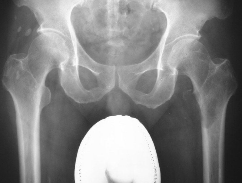
- Asymmetry of the pelvis; left hemipelvis larger with diffuse osteosclerosis and coarse trabeculae
- Right proximal femur: thickened cortex, loss of corticomedullary junction, coxa vara
- Brim sign: thickened iliopectineal line
- Dx: Polyosteotic Paget's disease
- Ddx: bone metastases, fibrous dysplasia, myelofibrosis
Q50What are the causes of pain in Paget's disease?▸
- Compression neuropathy (spinal stenosis, hearing loss, basilar invagination)
- 2.Arthritis; 3.Blood flow/bone enlargement
- Pathological fracture (fissure or chalk-stick transverse fracture)
- Malignant transformation
Q51What is the risk and prognosis of malignant transformation in Paget's disease?▸
- Paget's osteosarcoma <1%, usually >70 years
- Poor prognosis: 5% survival at 5 years
Q52What investigations are used in Paget's disease?▸
- Raised ALP (osteoblast activity), raised serum acid phosphatase (osteoclast activity); calcium normal
- Raised urine hydroxyproline and collagen cross-linked peptides (collagen turnover)
- X-ray: deformity/enlarged bone, coarse trabeculae, loss of corticomedullary differentiation
- Osteoporosis circumscripta; flame sign (advancing V-shaped lytic lesion)
- Bone scan (80% polyostotic); echo
Q53When is medical treatment indicated in Paget's disease?▸
- Pain, before operation, high-risk lesion, or medical complication (high-output failure)
- Aim: retard osteoclast activity
- Bisphosphonate (oral/IV) first line; calcitonin (SC/IM)
- Teriparatide contraindicated (increases Paget's sarcoma)
Q54What are the operative concerns in Paget's disease?▸
- General: high bleeding risk, post-op HO, hypercalcaemia with immobilisation (hypercalcemia with immobilization)
- Fracture: flexible or stiff locked IM nail with osteotomy; post-op bracing due to slow healing
- TJR: malalignment from very soft and patchy very hard bone
- Causes of hypercalcaemia: secondary hyperPTH, immobilisation, neoplastic change
Q55What is Paget's disease?▸
- Disorder of high bone turnover causing distortion of architecture and abnormal lamellar bone formation
- Increased osteoclastic bone resorption is the primary cellular abnormality
- Mono-ostotic or poly-ostotic (80%)
- Most are sporadic; some AD inheritance; paramyxovirus infection implicated
Q56What is the histology and what are the phases of Paget's disease?▸
- Histology: mosaic pattern of disorganized lamellar bone
- Phases: lytic, mixed, blastic - can co-exist in the same bone
Q57What deformities and gait changes occur in Paget's disease?▸
- Saber tibia; LLD (leg length discrepancy)
- Gait: stooped posture, lumbar kyphosis, hips in fixed flexion contracture (FFC)
- History should include premorbid status and family history
Q58What is the role of NSAIDs and monitoring in Paget's disease?▸
- NSAIDs for symptom control
- Monitor for complications
Q59How is a fracture in Paget's disease managed?▸
- Avoid immobilisation; prefer flexible IM fixation with bracing afterwards
Q60Why is immobilisation detrimental in Paget's disease?▸
- Causes osteopenia
- Causes hypercalcaemia
Q61What are the pre-, intra- and post-operative considerations?▸
- Pre-op: bisphosphonate, cardiac condition optimisation
- Intra-op: deformity, bleeding
- Post-op: heterotopic ossification (HO), slow healing
Fact check
Denosumab for GCT is 120mg once/week x 3 then once/2 weeks then monthly — dosing schedule imprecise — Approved GCTB dosing is 120 mg SC every 4 weeks with additional 120 mg loading doses on days 8 and 15 of the first month (weekly for the first 3 doses, then every 4 weeks - not 2-weekly) — source