Bone and Joint
Bone remodelling and fracture healing
Cutting cone and remodelling cycle, osteoclast-osteoblast coupling and stages of fracture healing
14 questions
2 source pages
1 images
Images appear with the first question taken from each source page — tap a question to open it.
Q1What is the sequence of cellular events in a cutting cone?📷▸
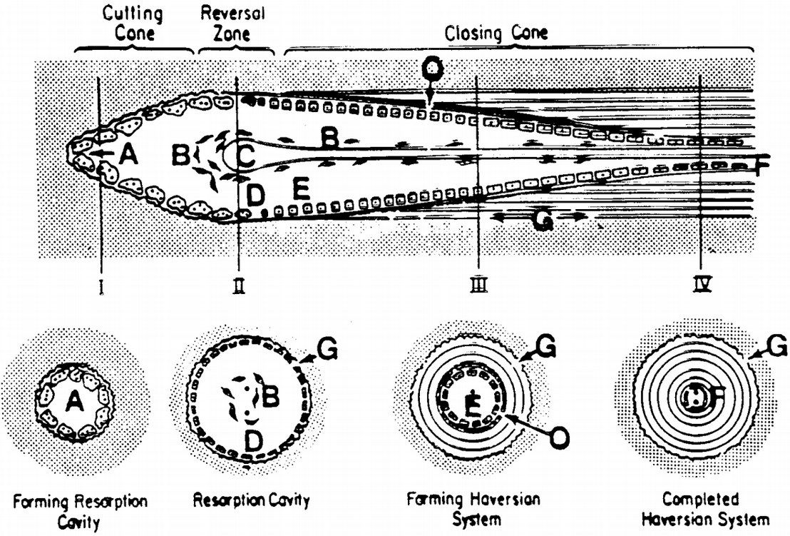
- Osteoclasts at the front to remove bone
- Followed by a layer of osteoblasts depositing successive centripetal layers of lamellae and osteoid
- Tunnel size narrows to a haversian canal
Q2What are the labelled components of a cutting cone?▸
- A osteoclast
- B osteoprogenitor cells
- C capillary
- D mononuclear cell lining reversal zone
- E osteoblast apposing bone centripetally
- F flattened osteoblast/osteoclast lining haversian canal
- G osteoid
Q3How does the cutting cone tunnel change as it advances?▸
- Tunnel size narrows
- Eventually forms the haversian canal
Q4From which direction do osteoblasts deposit new bone in a cutting cone?▸
- Centripetally (towards the centre of the tunnel)
- Deposit successive layers of lamellae and osteoid
- Following behind the osteoclast front
Q5Which cells line the haversian canal after the cutting cone has passed?▸
- Flattened osteoblast/osteoclast lining the haversian canal
- Mononuclear cells lining the reversal zone
Q6What is Perren's strain theory?▸
- The degree of interfragmentary strain governs the cellular response and therefore the tissue that forms
- <2% = primary bone healing
- 2-10% = secondary bone healing
- >10% = nonunion / fibrous union
Q7Describe primary bone healing.▸
- It is actually bone remodelling - haversian cutting cones cross the fracture site and directly lay down osteons
- It is a process of intramembranous ossification where there is no intermediate step of cartilage formation, therefore no callus formation
- Contact healing: direct bone apposition with gap <0.01mm
- Gap healing: gap <1mm; vessels grow into the gap and osteoblasts lay lamellar or woven bone
- Followed by haviersian remodelling by cutting cones (osteoclast reams, vessel grows in, trailing osteoblasts lay new bone)
Q8Describe the stages of secondary bone healing.▸
- Hematoma and inflammation (<1wk): haematoma brings in haemtopoetic cells. Inflammatory mediators lead to granulation and angiogenesis; osteoclasts and macrophages remove debris
- Soft callus (1 weeks to 1 month): endochondral ossification at intermediate strain/low O2; intramembranous ossification at low strain/high O2
- Hard callus (1-4 months): soft calcified chondral callus remodels into hard mineralised osteoid callus
- Remodelling: Wolff's law and piezoelectric charges dictate distribution of remodelling
Q9What adjuncts aid fracture healing?▸
- Low intensity pulsed USG increases nanomotion across the fracture site, increasing healing and callus strength
- Bone stimulator (electromagnetic stimulation) increases BMP concentration and osteoblastic differentiation
Q10Differentiate bone modelling from remodelling.▸
- Modelling: change of shape or growth by independent, simultaneous actions of osteoblast and osteoclast
- Remodelling: replacement of the same amount of matrix, sequential osteoblast then osteoclast within the same BMU
- Cancellous remodelling: osteoclastic resorption + osteoblastic deposition of lamellar bone
- Cortical remodelling: cutting cone
Q11What is the role of fibrinolysis in fracture healing?▸
- Plays a key transition step from haematoma formation to angiogenesis and fracture healing
- Cited from NEJM 2015 O'Keiffe
Q12Describe enchondral ossification during soft callus formation.▸
- Occurs in areas of intermediate strain and low O2 tension
- Stem cells differentiate into chondrocytes, laying down type I collagen and fibrocartilaginous matrix
- Followed by vascularisation and osteoblasts laying down bone on the cartilaginous scaffold
- Forms the interfragmentary callus and external callus
Q13Where does intramembranous ossification occur in secondary bone healing?▸
- At sites of low strain and high O2 tension (when fracture strain decreases)
- Osteoblasts from the cambrium layer of periosteum directly lay down woven bone
- Woven bone is later remodelled to lamellar bone
- Forms the medullary callus and periosteal callus
Q14What is the bone healing nonunion (BHN) model?▸
- Described by Elliot, JBJS 2016
- Unifies Perren's strain theory, Wolff's law and Frost's concept of mechanostat
- Tissue around a fracture is called a bone healing unit, producing a physiological response to its biological and mechanical environment
- The lecturer suggests drawing the graph