Bone and Joint
Calcium metabolism and metabolic bone disease
Calcium and phosphate homeostasis, PTH and vitamin D, plus renal osteodystrophy changes
11 questions
2 source pages
1 images
2 fact-check flags
Images appear with the first question taken from each source page — tap a question to open it.
Q1What is the distribution of calcium in the body?▸
- ~99% in bone
- ~1% extracellular, <1% intracellular
- Normal level 2.2-2.6 mmol/L
Q2What are the functions of calcium?▸
- Neuromuscular function
- Clotting/metabolic function
- Bone mineralisation
Q3Where and how is calcium taken up?▸
- Duodenum: active transport
- Jejunum: passive diffusion
Q4How is calcium excreted?▸
- Kidney: 98% reabsorbed at proximal tubules (per speaker notes)
- Stool
Q5What is the distribution and function of phosphate?▸
- 86% in bone, 14% intracellular, <1% plasma
- Bone mineralisation and enzyme/molecular interaction
- Uptake from food; kidney 100% resorb (per speaker notes)
Q6What are the X-ray features of renal osteodystrophy?📷▸
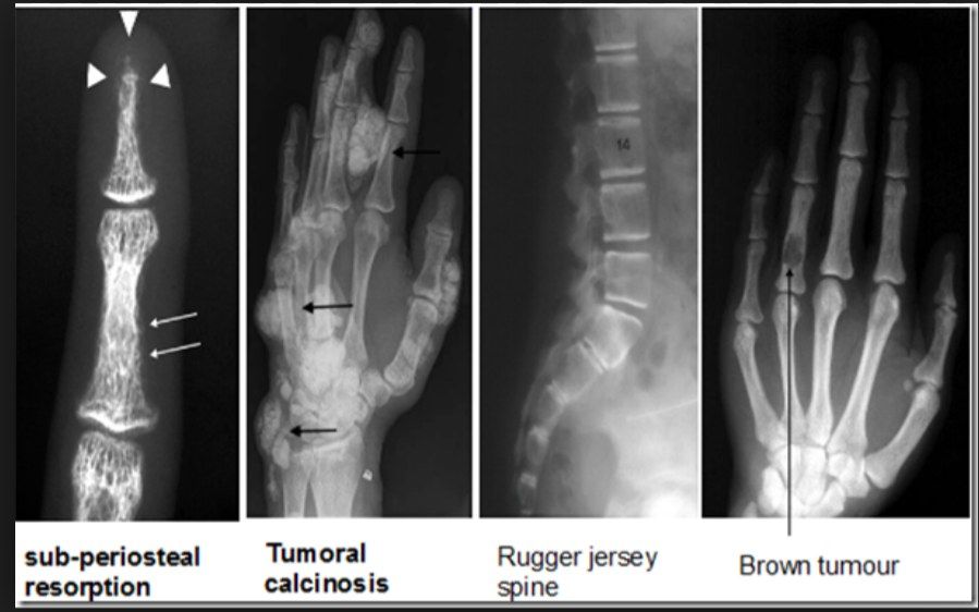
- Subperiosteal resorption
- Rugger jersey spine
- Brown tumour
- Tumoral calcinosis (lecturer advises avoiding this term)
- All are features of high bone turnover disease
Q7How is renal osteodystrophy classified and what causes the bone mineralisation deficiency?▸
- High turnover disease with elevated PTH vs low turnover disease with normal PTH
- Bone mineralisation deficiency due to electrolyte and endocrine abnormalities
- X-ray features of both osteomalacia (impaired mineralisation) and hyperparathyroidism
Q8What causes low-turnover renal osteodystrophy?▸
- Aluminium accumulation in renal failure
- Excess deposition into bone -> toxic to osteoblasts (differentiation and proliferation)
- Also impairs PTH release
- PTH is normal
Q9How does renal osteodystrophy present?▸
- Bony problem: pathological fracture
- Soft tissue: tendinitis, tendon rupture, carpal tunnel syndrome
- Growth retardation, deformity, SCFE in children
Q10What is the management of renal osteodystrophy?▸
- Treat the underlying cause, multidisciplinary approach
- Aluminium chelator
- Calcium supplement
- Vitamin D supplement
- Bisphosphonates
Q11Describe the pathophysiology of high-turnover renal osteodystrophy.▸
- Uraemia -> abnormal PO4 excretion -> PO4 retention (glomerular) + kidney unable to convert vitamin D to active form (tubules) -> hypocalcaemia
- Failure of 1-alpha hydroxylase -> hypocalcaemia
- High PO4 and low Ca -> increased PTH -> hyperplasia of chief cells of the parathyroid gland
Fact check
Kidney reabsorbs 98% of calcium at the proximal tubules — imprecise — 98-99% of filtered calcium is reabsorbed overall; the proximal tubule accounts for only ~60-70%, with the rest reabsorbed in the loop of Henle and distal nephron — source
Kidney 100% resorbs phosphate — misleading — About 80-90% of filtered phosphate is normally reabsorbed (mostly in the proximal tubule); reabsorption approaches 100% only with very low dietary phosphate intake — source