9 slides
▸ Slide 805 · elbowElbow · 2 questions expand
Slide render
Question list
Q1-Q22 questions — tap to reveal all answerslist
- What elbow condition or anatomy does this slide illustrate?
- What are the key points in assessment of the elbow?
Answers · Q & A
Q1.What elbow condition or anatomy does this slide illustrate?
- Not covered in the speaker notes
Q2.What are the key points in assessment of the elbow?
- Not covered in the speaker notes
▸ Slide 806 · Lateral elbow pain ddx:Elbow · 7 questions 1 check expand
Question list
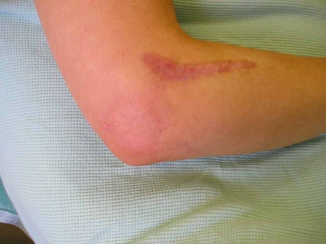
Q1-Q77 questions — tap to reveal all answerslist
- List the differential diagnosis of lateral elbow pain.
- What is lateral epicondylitis and its pathophysiology?
- Which muscles originate from the lateral epicondyle and supracondylar ridge?
- What are the symptoms and signs of lateral epicondylitis?
- Describe the non-operative management of lateral epicondylitis.
- When is surgery indicated in lateral epicondylitis and what are the results?
- What is plica syndrome of the elbow?
Answers · Q & A
Q1.List the differential diagnosis of lateral elbow pain.
- Tennis elbow (lateral epicondylitis)
- Radial tunnel syndrome (4-5cm distal to lateral epicondyle)
- Plica syndrome (most common is posterolateral fold)
- RCJ arthritis, OCD of lateral condyle, PLRI
Q2.What is lateral epicondylitis and its pathophysiology?
- Overuse injury with eccentric overload at origin of common extensor tendon
- Tendinosis and inflammation at origin of ECRB
- Concomitant radial tunnel syndrome in 5%
- Patho: angiofibroblastic hyperplasia + disorganized collagen
Q3.Which muscles originate from the lateral epicondyle and supracondylar ridge?
- ECRB, ED, EDM, ECU and anconeus all originate from the lateral epicondyle
- ECRL and brachioradialis arise from the supracondylar ridge
Q4.What are the symptoms and signs of lateral epicondylitis?
- Sx: lateral elbow pain, weak grip, pain on resisted wrist extension
- PE: tenderness over lateral epicondyle
- Pain on resisted wrist/MF extension with elbow extended; pain on wrist flexion
- Imaging to rule out other causes
Q5.Describe the non-operative management of lateral epicondylitis.
- 95% effective; lifestyle modification, physio, NSAID, steroid (<20mg steroid)
- Counterforce brace (5cm wide non elastic support distal to elbow, does not allow full muscle expansion, thereby decreased stretch on lateral epicondyle)
- BMJ Bisset 2006: steroids better effects at 6 weeks but with high recurrence rate, poorer outcomes c/w physio in the long term
- PRP: 2020 meta-analysis favoured PRP long term, steroid best short term; Cochrane 2021 does not support PRP
Q6.When is surgery indicated in lateral epicondylitis and what are the results?
- After failed 9-12 months of non-operative management
- Release and debridement of ECRB origin (open/arthroscopic/percutaneous equivalent)
- Results: 85% complete relief, 5% no benefit, 10% some improvement
Q7.What is plica syndrome of the elbow?
- Part of the ddx of lateral elbow pain
- Normally 4 folds: anterior, posterior, posterolateral, lateral
- Most common symptomatic plica is the posterolateral fold
Fact check
PRP has no evidence for lateral epicondylitis (2021 Cochrane does not support it) — contested/oversimplified — Cochrane 2021: PRP or autologous blood probably gives no clinically significant benefit vs placebo at 3 months, but Hohmann 2023 found PRP superior to steroid at 3-6 months; steroid benefit is short term. — medium confidence — source
▸ Slide 807 · Cubital varus/ valgusElbow · 6 questions 1 check expand
Slide render
Question list
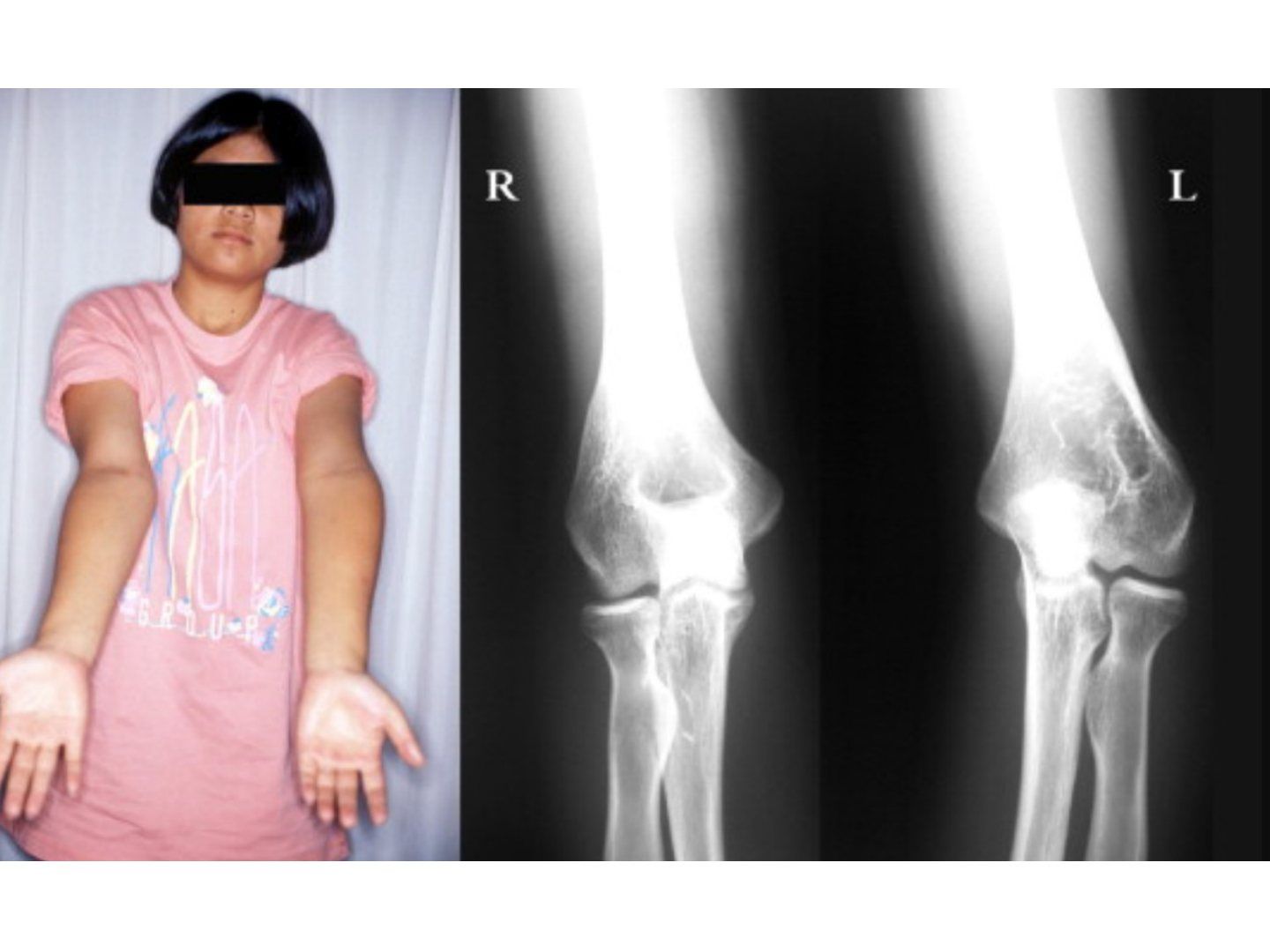
Q1-Q66 questions — tap to reveal all answerslist
- What is the normal carrying angle of the elbow?
- What are the causes of cubitus varus?
- What is the incidence of cubitus varus after supracondylar fracture and how does the patient present?
- What is a fishtail deformity after lateral condyle fracture?
- What corrective osteotomy options are available for cubitus varus?
- Describe the French osteotomy for cubitus varus.
Answers · Q & A
Q1.What is the normal carrying angle of the elbow?
- Male: 5-10 degrees
- Female: 15-20 degrees
Q2.What are the causes of cubitus varus?
- Static (malreduction) or dynamic (growth) causes
- Supracondylar fracture malunion (varus, IR, extension)
- Lateral condyle fracture - fishtail deformity, overgrowth of lateral condyle (50% regardless of treatment)
- Transphyseal injury; bilateral causes include HME
Q3.What is the incidence of cubitus varus after supracondylar fracture and how does the patient present?
- 3% after pinning, 14% after casting
- Complaint is cosmetic; seldom causes functional deficit
Q4.What is a fishtail deformity after lateral condyle fracture?
- Type 1: malunion of lateral condyle causing under development of lateral crista
- Type 2: AVN of the medial crista of the trochlea
- One cause of cubitus varus
Q5.What corrective osteotomy options are available for cubitus varus?
- Timing: till skeletal maturity
- French osteotomy, lateral closing wedge with step cut, medial opening wedge, computer assisted
Q6.Describe the French osteotomy for cubitus varus.
- Lateral closing wedge with correction of rotation
- Screws inserted from lateral: proximal screw posterior, distal screw anterior
- Figure-of-8 wire loop tightening
Fact check
Normal carrying angle is 5-10 degrees in males and 15-20 degrees in females — imprecise - values vary widely — Commonly quoted normal values are about 11 degrees in men and 13 degrees in women (Orthobullets); studies report means of roughly 9-14 degrees in men and 11-16 degrees in women. — medium confidence — source
▸ Slide 808 · Cubitus valgus (in lateral condyle #)Elbow · 4 questions expand
Slide render
Question list
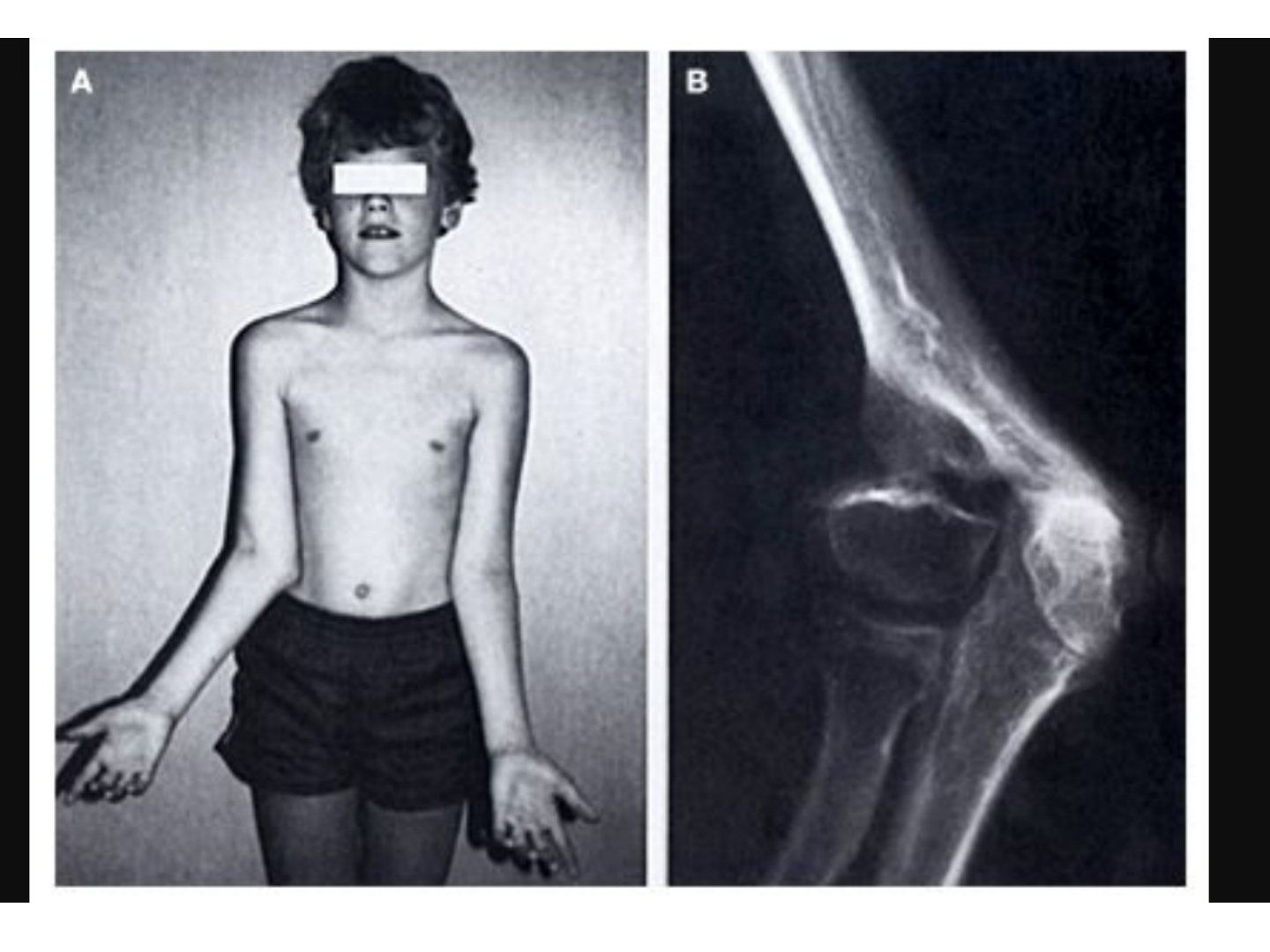
Q1-Q44 questions — tap to reveal all answerslist
- A child develops cubitus valgus after a lateral condyle fracture. What is the mechanism, and what other causes should be considered?
- List the causes of radial head subluxation.
- Describe the blood supply of the distal humerus.
- What is the complication of cubitus valgus and how is it treated?
Answers · Q & A
Q1.A child develops cubitus valgus after a lateral condyle fracture. What is the mechanism, and what other causes should be considered?
- AVN with physeal arrest of the lateral condyle after fracture
- Other unilateral causes: OA, HME
- Bilateral causes: congenital dislocation of the radial head, HME
Q2.List the causes of radial head subluxation.
- Congenital (nail-patella syndrome)
- Synostosis
- MHE
- Arthrogryposis; achondroplasia
- Post-traumatic
Q3.Describe the blood supply of the distal humerus.
- Lateral condyle: from posterior, just lateral to origin of the capsule
- Trochlea: lateral crista supplied from the lateral condyle; medial crista has dual supply
- Lateral supply crosses the posterior surface of the distal humeral metaphysis and is an end artery; medial supply comes from multiple vessels
Q4.What is the complication of cubitus valgus and how is it treated?
- Tardy ulnar nerve palsy
- Treatment: supracondylar osteotomy after skeletal maturity
▸ Slide 809 · Total elbow replacementElbow · 7 questions expand
Question list
Q1-Q77 questions — tap to reveal all answerslist
- What is the functional range of the elbow and the anatomy of the distal humerus?
- What is the differential diagnosis of elbow arthritis?
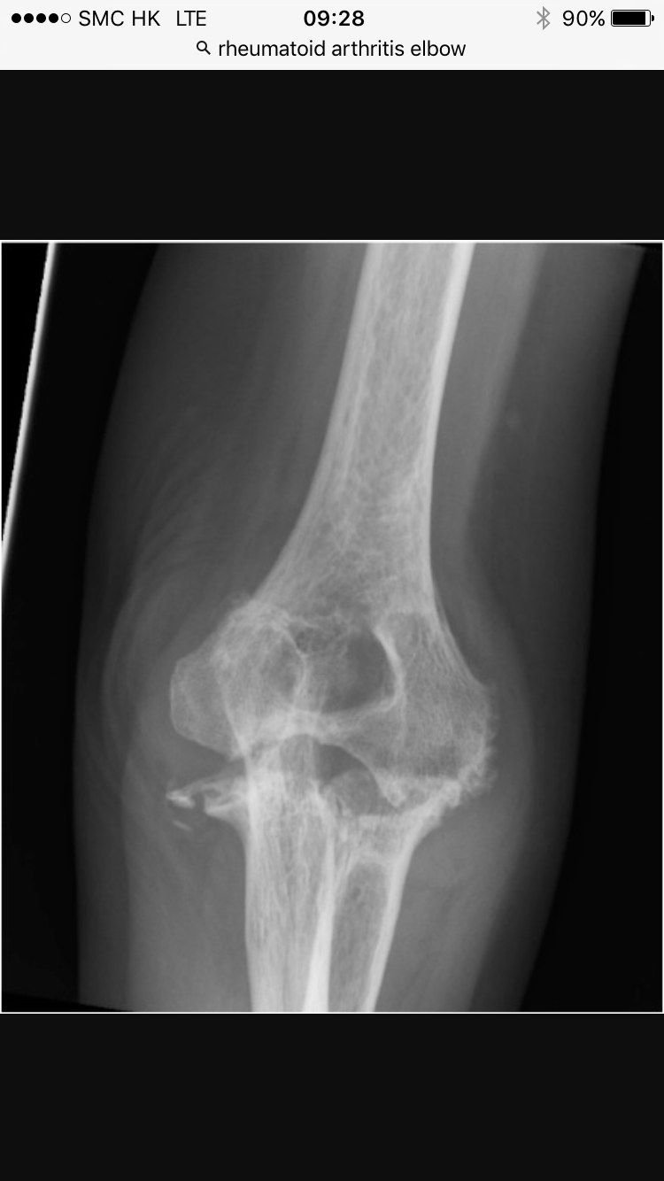
- What are the problems in the RA elbow and describe the Larsen classification.
- Describe the operations available for the RA elbow.
- What are the problems and operations in OA elbow?
- How is elbow arthroscopy performed?
- What are the complications of elbow arthroscopy?
Answers · Q & A
Q1.What is the functional range of the elbow and the anatomy of the distal humerus?
- Elbow functional range: F/E 30-130, S/P 60/40
- Distal humerus: 30 degrees flexion, 5 IR, 5 degrees valgus
Q2.What is the differential diagnosis of elbow arthritis?
- Primary OA
- Post-traumatic arthritis
- RA
- Infection
Q3.What are the problems in the RA elbow and describe the Larsen classification.
- Problems: osteopenia, ligamentous laxity with instability, contracture
- Larsen 1: swelling + osteoporosis; 2: mild joint space narrowing + erosion; 3: significant joint space narrowing
- Larsen 4: advanced erosion and laxity, subchondral bone breaching; 5: ankylosis
Q4.Describe the operations available for the RA elbow.
- Synovectomy - indications: pain, PIN compression by synovitis; prerequisite: stable, F/E combined >90
- Radial head excision - for painful rotation, PIN compression from radial head synovitis; cons: valgus instability, caput ulnare
- Ulnohumeral distraction interpositional arthroplasty (tensor fascia lata)
- TER (total elbow replacement)
Q5.What are the problems and operations in OA elbow?
- Problems: hard bone, osteophytes, capsule contracture, loose bodies
- Arthroscopic debridement (CI: previous ulnar nerve transposition)
- OK (Outerbridge-Kashiwagi) procedure
- Ulnohumeral distraction interposition arthroplasty; TER
Q6.How is elbow arthroscopy performed?
- Patient selection (CI: previous ulnar nerve transposition, severe trauma with distorted anatomy, scarring); GA; supine/prone/lateral decubitus
- Distend the joint through the lateral soft spot (olecranon, LE, radial head) with 20ml NS before placing portals
- Anterior portal in elbow F90, posterior portal in slight flexion; open portal under direct vision
- 1st portal supine: distal anterolateral (1cm anterior and distal to LE); anteromedial 2cm proximal to medial epicondyle, anterior to intermuscular septum
- Post-op: immobilise in extension for 1 day, physio, CPM
Q7.What are the complications of elbow arthroscopy?
- Portals: persistent drainage, fistula
- Nerves: radial (most at risk), PIN, median, ulnar, antebrachial cutaneous nerves
- Soft tissues: compartment syndrome
- Contracture
▸ Slide 810Elbow · 3 questions expand
Question list
Q1-Q33 questions — tap to reveal all answerslist
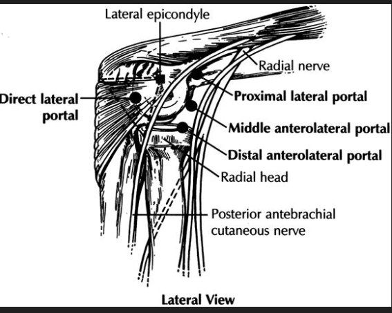
- Which procedure does this slide illustrate, and which portal views are shown?
- Which portals are shown in the lateral view, and which structures must be avoided?
- Which portals are shown in the medial view, and which nerves are at risk?
Answers · Q & A
Q1.Which procedure does this slide illustrate, and which portal views are shown?
- Elbow arthroscopy portal anatomy — image-only slide (no speaker notes)
- Lateral view: direct lateral portal; proximal lateral, middle anterolateral and distal anterolateral portals
- Medial view: proximal medial and anteromedial portals
Q2.Which portals are shown in the lateral view, and which structures must be avoided?
- Portals: direct lateral, proximal lateral, middle anterolateral, distal anterolateral
- Related structures: lateral epicondyle, radial head, radial nerve
- Posterior antebrachial cutaneous nerve also runs in the field
Q3.Which portals are shown in the medial view, and which nerves are at risk?
- Proximal medial portal and anteromedial portal
- Ulnar nerve lies posterior to the medial epicondyle; intermuscular septum is a key landmark
- Median nerve and medial antebrachial cutaneous nerve are also shown
▸ Slide 811 · Coonrad MoreyElbow · 7 questions expand

Question list
Q1-Q77 questions — tap to reveal all answerslist
- Describe the Coonrad-Morrey total elbow prosthesis.
- What is the role of the anterior flange and bone graft in the Coonrad-Morrey prosthesis?
- What is the survivorship of the Coonrad-Morrey prosthesis?
- What are the indications/contraindications and approach for total elbow replacement?
- Describe the surgical steps of total elbow replacement.
- What are the postoperative restrictions after total elbow replacement?
- Compare linked, unlinked and semi-constrained total elbow replacements.
Answers · Q & A
Q1.Describe the Coonrad-Morrey total elbow prosthesis.
- Titanium alloy (Ti6Al4V)
- Cemented, linked, semi-constrained with a floppy hinge
- Floppy hinge allows varus/valgus so load is distributed to soft tissue
- Displacement force of the humeral implant is anterior
Q2.What is the role of the anterior flange and bone graft in the Coonrad-Morrey prosthesis?
- Bone graft increases rotational stability and shares torsional forces, protecting the bone-cement-implant interface
- Controls posterior displacement
- Allows thickening of bone stock at the site of maximal stress
Q3.What is the survivorship of the Coonrad-Morrey prosthesis?
- Highest survivorship for RA
- 92.4% at 10-12 years
Q4.What are the indications/contraindications and approach for total elbow replacement?
- Indications: RA, complex distal humerus fracture in the elderly, OA
- CI: infection, Charcot, young and active patient needing heavy lifting
- 15cm incision just lateral to the medial epicondyle and medial to the tip of the olecranon
- Bryan-Morrey approach (elevate triceps with periosteum of olecranon and proximal ulna, ulnar nerve transposition)
Q5.Describe the surgical steps of total elbow replacement.
- Humerus: remove midportion of trochlea to enter canal; remove trochlea/capitellum level with epicondylar margins, preserve supracondylar columns; rasp canal
- Ulnar: remove tip of olecranon to enter canal, serial rasping
- Insert ulnar first (align center of ulnar component with centre of greater sigmoid fossa), then humerus; first generation cementation technique
- Articulate by placing the axle; autograft at anterior flange; preserve radial head if possible
Q6.What are the postoperative restrictions after total elbow replacement?
- Post op need immobilization (locked in 30 deg elbow flexion for 4 weeks)
- Life-long weight lifting limit 10 lbs
Q7.Compare linked, unlinked and semi-constrained total elbow replacements.
- Linked fully constrained: loosening; indication marginal bone stock and stability
- Semi-constrained (Coonrad-Morrey): sloppy/floppy hinge, load distributed by soft tissue; anterior flange decreases posterior migration and rotation
- Unlinked (unconstrained): unstable; prerequisite good soft tissue tension and bone stock
- Semi-constrained (Souter): deep groove for stability; short humeral stem (stirrup) decreases rotational stress; RA laxity causes anterior tilt of humeral stem
▸ Slide 812 · SouterElbow · 3 questions expand

Question list
Q1-Q33 questions — tap to reveal all answerslist
- Describe the Souter total elbow prosthesis.
- What is the advantage of the short humeral stem in the Souter prosthesis?
- What design features improve stability in the Souter prosthesis?
Answers · Q & A
Q1.Describe the Souter total elbow prosthesis.
- Cemented, unlinked, semi-constrained
- Broad metallic plate acts against deforming force
Q2.What is the advantage of the short humeral stem in the Souter prosthesis?
- Maintains humeral bone stock
Q3.What design features improve stability in the Souter prosthesis?
- Deep groove for better stability
- Ulnar stem made of UHMWPE
▸ Slide 813 · GSB IIIElbow · 3 questions expand
Question list
Q1-Q33 questions — tap to reveal all answerslist
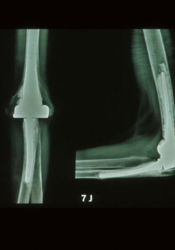
- What is the linkage mechanism of the GSB III total elbow prosthesis?
- How does the GSB III resist deforming forces?
- What is the complication of the GSB III prosthesis?
Answers · Q & A
Q1.What is the linkage mechanism of the GSB III total elbow prosthesis?
- Linked semi-constrained with a floppy hinge
- Large joint surface on the humeral component
Q2.How does the GSB III resist deforming forces?
- Flange on the condyle acts against deforming force
- Difficult in RA patients with deformed anatomy
Q3.What is the complication of the GSB III prosthesis?
- Dislodged component