Hand Soft Tissue
Cysts, tumours and nail lesions
Ganglion and digital cysts, carpal boss, glomus and giant cell tumours, nail melanoma and deformity
32 questions
8 source pages
3 images
2 fact-check flags
Images appear with the first question taken from each source page — tap a question to open it.
Q1How do you assess a dorsal radial or volar radial wrist mass?▸
- Look: location, skin change, sinus
- Feel: temperature, tenderness, surface, border, size, consistency, fluctuance, layer, attachment, pulsatile, thrill, Tinel
- Transillumination if fluctuant
- Allen test if a volar radial wrist ganglion
Q2What is the pathology and origin of a ganglion cyst?▸
- Mucin-containing fibrous cyst with no epithelium (glucosamine, albumin, hyaluronic acid)
- Caused by trauma or mucoid degeneration of collagen, synovial herniation
- Dorsal radial wrist: from the SL ligament; volar radial wrist: radiocarpal, STT, RSC or LT ligament
- Flexor sheath ganglion: usually between A1 and A2 pulleys (does not move with the tendon)
Q3What is the prevalence and non-operative treatment of ganglion cysts?▸
- Dorsal carpal 70%, volar carpal 20%, volar flexor sheath 10%
- Conservative management as spontaneous resolution may occur
- Aspiration has a 50% recurrence rate; radial artery at risk in volar wrist ganglion
Q4How is a dorsal or volar wrist ganglion resected and what is the recurrence?▸
- Identify origin, resect and cauterise the stalk + PIN end branch (decreases neuroma)
- Dorsal approach between the 3rd/4th compartments
- Volar ganglion has higher recurrence (15-20%)
- Arthroscopic excision is an option
Q5What is a mucoid cyst and how is it managed?▸
- A ganglion cyst associated with Heberden's nodules (1st DIPJ osteophytes)
- Affects the germinal matrix causing nail deformity
- Options: aspiration, steroid injection, excision
- Excision: complete cyst and stalk, protect germinal matrix, resect osteophytes, skin reconstruction (rotational/bilobed flap)
- Complications: terminal extensor lag 17%, recurrence 3%, nail deformity
Q6What is carpal boss syndrome?▸
- A dorsal bony osteophyte over the base of the 2nd or 3rd metacarpal
- Causes reduced range of motion
- Related to degeneration or Os styloideum; may be associated with a ganglion
Q7How is carpal boss syndrome managed?▸
- Mainly conservative management
Q8What is an inclusion cyst and what is its pathogenesis?📷▸
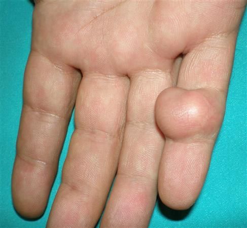
- Epithelial-lined cyst filled with keratin
- Pathogenesis: penetrating injury drives keratinised epithelium into soft tissue or bone
Q9What are the differentials for an inclusion cyst?▸
- Skin lesion (tethers skin)
- Ganglion (mobile ML>PD, transilluminates)
- Calcifying aponeurotic fibroma
- Fibroma or schwannoma, haemangioma
- Giant cell tumour
Q10What is the management of an inclusion cyst?▸
- Marginal excision
- Curette if bone is involved
- Amputation if advanced bone destruction
- Low recurrence if the capsule is excised en bloc
Q11What is a glomus tumour?▸
- Tumour of the glomus body, a perivascular temperature-regulating structure
- Neuromyoarterial neoplasm of smooth muscle with eponychium metaplasia
- 75% in the hand, 50% subungual
Q12What history is taken for a suspected glomus tumour?▸
- Age and premorbid status
- Ask about the triad of paroxysmal pain, exquisite tenderness and cold intolerance
Q13How does a glomus tumour present?▸
- Subungual mass with bluish discolouration
- Nail ridging
- Triad of paroxysmal pain, exquisite tenderness and cold intolerance
- 50% have bone erosion
Q14What special tests are used for a glomus tumour?▸
- Love's test: pressure to a pinhead causes exquisite pain (sens 100%, spec 78%)
- Hildreth test: inflating a tourniquet abolishes the pain with Love's test (sen 92%, spec 91%)
Q15What investigations are used for a glomus tumour?▸
- XR: bone erosion from pressure effect
- MRI: T1 isodense, T2 hyperintense with strong gadolinium enhancement
- Histology: 3 subtypes - mucoid hyaline, solid and angiomatous; round cells with dark nuclei
Q16What is the differential diagnosis of a glomus tumour?▸
- Dupuytren's exostosis
- Gout
- Inclusion cyst
- Osteomyelitis
- Amelanotic melanoma
Q17What is the management of a glomus tumour?▸
- Marginal excision
- Low recurrence (20% quoted in the notes)
- Counsel for nail ridging and nailbed grafting
Q18What is giant cell tumour of tendon sheath?▸
- 2nd most common hand soft tissue tumour
- Arises from the flexor tendon sheath, usually between A1 and A2
- PVNS of the tendon sheath
- Examine for NV and bone involvement
Q19What investigations and histology are seen?▸
- XR: bone indentation up to 5%
- MRI: T1 and T2 hypodense
- Histology: multinucleated cells with haemosiderin staining, vascular villi with stromal cell infiltration, xanthoma
Q20How is giant cell tumour of tendon sheath managed?▸
- Excision + H2O2 irrigation (to oxidise residual cells)
- High recurrence up to 50%, locally aggressive
Q21Describe the presentation of subungual melanoma.📷▸
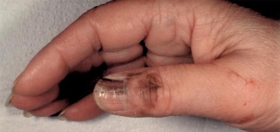
- Pigmented band from the germinal matrix
- Nail fold involvement = Hutchinson sign
- DDx: subungual haematoma, onychomycosis, benign longitudinal melonychia (straight border, single band)
Q22What history is important in subungual melanoma?▸
- Age and premorbid status
- Current disability and complications, chronicity, history of trauma
- Then take the ABCDEF history and ask about metastatic symptoms
Q23What does the ABCDEF history cover in melanoma?▸
- Age 50-70 years
- Band + breadth >3mm, irregular border
- Coloured + change (rapid increase in size, unhealing dystrophic nail)
- Digit (thumb > big toe > index finger)
- Extension (Hutchinson sign) and FHx
Q24What risk factors and symptoms of metastasis are asked about?▸
- Risk factors: sun exposure, family history, fair skin, multiple naevi, immunosuppression
- Metastatic symptoms: LN, lung, liver, brain
Q25What investigations are performed?▸
- LFT and LDH
- CXR for lung mets
- MRI
- PET-CT
Q26What is the Breslow classification and how does it guide treatment?▸
- I <0.75mm (survival >90%); II 0.76-1.5mm; III 1.6-4mm; IV >4mm (survival 50%)
- Sentinel LN biopsy with planning for radical LND and adjuvant chemo
- WLE: <1mm -> 1cm margin; 1mm or more -> 2cm margin + SLN biopsy
Q27What are the poor prognostic factors?▸
- Male
- Ulceration
- Metastases
- Subungual 5yr 40%, H&N lesion
- Basic science: melanocytes from neural crest, separate from dermis by basement membrane; Clark level 1-5; acral lentiginous subtype starts at nailfold/germinal matrix
Q28What history is important in a ridged nail?📷▸
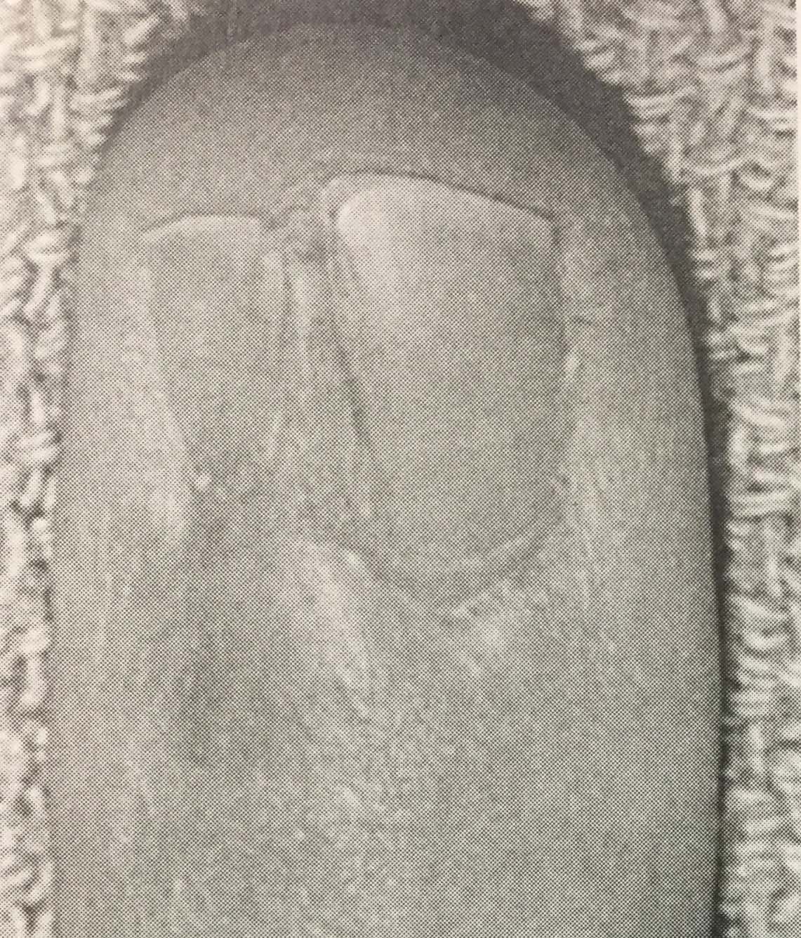
- Age and premorbid status
- History of infection or trauma (poor suture technique, missed germinal matrix injury)
- Current disability: cosmesis, hypersensitive scar, previous paronychia or osteomyelitis
Q29How is a ridged nail managed?▸
- Observation
- <2mm: scar resection + primary closure
- 2mm or more: scar resection + 2nd toe nailbed graft
Q30What is a hook nail and what causes it?▸
- Volar curving of the nail matrix
- Cause: curved matrix upon suture
- Or a shortened bone
Q31What history and investigation are needed for a hook nail?▸
- Age and premorbid status
- Current disability: cosmesis, pain from ingrown nail
- XR to check residual distal phalanx length
Q32What is the management of a hook nail?▸
- Observation
- Antenna procedure
- If due to tight suture: soft tissue procedure to reconstruct the volar tip defect (VY or cross finger flap)
- If shortened bone: VBG from 2nd toe or K wire to support the nail bed
Fact check
Breslow classification: I <0.75mm, II 0.76-1.5mm, III 1.6-4mm, IV >4mm — outdated — This is Breslow's original prognostic classification; current AJCC 8th edition T categories use 1/2/4mm thresholds (T1 <1.0mm, T2 >1.0-2.0mm, T3 >2.0-4.0mm, T4 >4.0mm), with T1 subdivided at 0.8mm and thickness rounded to the nearest 0.1mm — source
Subungual melanoma 5-year survival 40% — contested — Survival varies widely by study and stage; 5-year overall survival ~40% in an MD Anderson cohort but 5-year melanoma-specific survival 56-82% in other series, so a single 40% figure is potentially misleading — (medium confidence) — source