▸ Slide 740 · Hand soft tissueHand Soft Tissue · 3 questions expand
Q1-Q33 questions — tap to reveal all answerslist
- Which hand soft-tissue conditions are covered in this lecture?
- Which nerve injuries are covered under hand soft tissue?
- Which infection, emergency and reconstructive topics are covered?
Q1.Which hand soft-tissue conditions are covered in this lecture?
- Contractures and masses: Dupuytren contracture (palmar fascial cord causing progressive finger flexion deformity), De Quervain, wrist ganglion
- Tendon problems: flexor tendon injury, Quadriga effect
- Pain syndrome: CRPS
Q2.Which nerve injuries are covered under hand soft tissue?
- Median, ulnar and radial nerve injuries
- Brachial plexus injuries
Q3.Which infection, emergency and reconstructive topics are covered?
- Hand infection; finger tip injury
- Compartment release
- Replantation
- Reconstruction ladder
- Hand splint for rehabilitation
▸ Slide 741 · After 6 weeks of casting for # DRHand Soft Tissue · 8 questions 1 check expand

Q1-Q88 questions — tap to reveal all answerslist
- How would you describe this clinical photo and what is your differential?
- What is complex regional pain syndrome (CRPS)?
- What are the Budapest criteria for diagnosing CRPS?
- What is the differential diagnosis of CRPS?
- How is CRPS classified and staged?
- What investigations are used in CRPS?
- How do you prevent CRPS after a distal radius fracture?
- What is the treatment of CRPS?
Q1.How would you describe this clinical photo and what is your differential?
- Red and swollen right hand after 6 weeks of casting for a distal radius fracture
- Ddx: complex regional pain syndrome (CRPS)
- Also consider infection and DVT
Q2.What is complex regional pain syndrome (CRPS)?
- Sustained sympathetic activity in a perpetuated reflex arc, characterised by pain out of proportion to physical findings
- Previously known as RSD (reflex sympathetic dystrophy)
Q3.What are the Budapest criteria for diagnosing CRPS?
- Exclude other diagnoses
- Continuing pain disproportionate to any inciting event
- >=1 symptom in >=3 categories, or >=1 sign in >=2 categories
- Categories: sensory (hyperaesthesia/allodynia), vasomotor (temperature/colour asymmetry), oedema/sweating (asymmetry, hyperhidrosis), motor/trophic (weakness, tremor, trophic hair/skin/nail changes)
Q4.What is the differential diagnosis of CRPS?
- Neuropathic: post-injury neuralgia
- Inflammatory: RA/SLE, soft tissue inflammation
- Vascular: PVD
- Psychiatric: malingering, somatization
Q5.How is CRPS classified and staged?
- International association for the study of pain classification - type 1: without demonstrable nerve lesion; type 2: identifiable nerve damage
- Lankford stage: acute (<3 months) - inflammation (pain, red, sweaty, swollen), XR normal
- Dystrophic (3-12 months): worse pain, cyanosis, dry skin
- Atrophic (>12 months): decreased pain, stiffness
Q6.What investigations are used in CRPS?
- Bone scan has high negative predictive value
- Positive scan phase pattern: 2 min arteriogram, 5-10 min blood pool (cellulitis, synovitis), 2-3 hr bone phase
Q7.How do you prevent CRPS after a distal radius fracture?
- Vitamin C 500mg daily x 50 days in distal radius fractures treated conservatively
- Avoid prolonged immobilisation
Q8.What is the treatment of CRPS?
- Prevention: vitamin C 500mg daily x 50 days in distal radius fractures treated conservatively; avoid prolonged immobilisation
- Refer pain specialist; aim symptom control and improve ROM
- Conservative: physiotherapy (tactile discrimination training, contrast bath, stress loading exercise, TENS), medication (NSAID, alpha blocker, CCB, antidepressant, GABA agonist)
- Operative: surgical sympathectomy (type 1), surgical decompression (type 2)
Prevention: vitamin C 500mg daily for 50 days in distal radius fractures treated conservatively — contested — The Zollinger RCT and a 2017 meta-analysis support 500mg x 50 days, but a 2015 meta-analysis found no significant reduction in CRPS (RR 0.45, 95% CI 0.18-1.13) and graded the evidence low quality — medium confidence — source
▸ Slide 742 · After injury->triggering sensed by brain->send sym signal to limb->trigger inflaHand Soft Tissue · 2 questions expand

Q1-Q22 questions — tap to reveal all answerslist
- Describe the proposed cycle in RSD.
- What is the role of the sympathetic nervous system in this cycle?
Q1.Describe the proposed cycle in RSD.
- Injury -> triggering sensed by brain -> sympathetic signal sent to limb -> inflammatory response -> triggers another response (cycle) -> RSD
Q2.What is the role of the sympathetic nervous system in this cycle?
- After the injury is sensed by the brain, it sends a sympathetic signal to the limb, which triggers an inflammatory response that perpetuates the cycle
▸ Slide 743 · Clinical photo showing the patients palmHand Soft Tissue · 16 questions 2 check expand

Q1-Q1616 questions — tap to reveal all answerslist
- How would you describe this clinical photo?
- What is Dupuytren's disease?
- What is the pathophysiology of Dupuytren's disease?
- What is the anatomy of the palmar fascia?
- Which cord is responsible for contracture at each site in Dupuytren's?
- What classifications are used in Dupuytren's disease?
- What are the risk factors and features of Dupuytren's diathesis?
- What do you examine locally in Dupuytren's disease?
- When do you intervene and what are the operative options?
- What is the aim of surgery in Dupuytren's disease?
- What is the escalating ladder of surgical options for Dupuytren's disease?
- Define limited and segmental fasciectomy.
- What is the principle of Z-plasty and the gain at each angle?
- How is collagenase injection performed?
- What non-operative options exist for Dupuytren's and what is the evidence?
- What are the recurrence rates after different Dupuytren's treatments?
Q1.How would you describe this clinical photo?
- Visible cord with flexion deformity of the ring finger MCPJ and PIPJ
- Flexion deformity of the little finger
- Thickened nodule over dorsoradial aspect of the PIPJ of the index finger (Garrod's knuckle)
- Concern: Dupuytren's contracture with diathesis
Q2.What is Dupuytren's disease?
- Fibroproliferative disorder of the palmar fascia producing nodules/cords and causing flexion contracture of fingers
- Nodules: fibroblasts and myofibroblasts (intracellular actin filaments along long axis; extracellular fibronectin connects adjacent myofibroblasts)
- Cords: highly organised collagen, no myofibroblasts
Q3.What is the pathophysiology of Dupuytren's disease?
- Murrell theory: local microvascular ischaemia produces free radicles -> fibroblast proliferation -> type 3 collagen (thicker than type 1) and fibrosis
- Strangulation and deficient microcirculation -> self-perpetuating cycle due to ischaemia
- Luck stages: proliferative -> involutional -> residual
Q4.What is the anatomy of the palmar fascia?
- 3 groups of fibres based on orientation: longitudinal (pretendinous band), vertical (septum of Legueu and Juvara, separates lumbrical and flexor tendon), transverse
- Longitudinal layer has 3 layers: superficial, spiral, deep
- Spiral layer forms the pretendinous band; when pathological: pretendinous cord -> lateral digital cord -> spiral cord -> Grayson cord
Q5.Which cord is responsible for contracture at each site in Dupuytren's?
- MCPJ: pretendinous cord
- PIPJ: spiral cord
- DIPJ: retrovascular cord
- Web: natatory cord
- 1st web: commissure cord; little finger: abductor digiti minimi cord
Q6.What classifications are used in Dupuytren's disease?
- Woodruff: 1 no FFC, 2 MCPJ, 3 MCPJ + PIPJ, 4 more than one finger, 5 finger on palm
- Tubiana combined flexion contracture: I 0-45, II 45-90, III 90-135, IV >135
- Differentials: scars, burns, trigger finger, tendon adhesions, joint contracture, ulnar nerve palsy
Q7.What are the risk factors and features of Dupuytren's diathesis?
- Patient factors: male, family history, Celtic/Viking, bilateral
- Disease: HIV, TB, COPD, DM; drugs: alcohol, anti-epileptics
- Diathesis BERYS: Bilateral, Ectopic (Ledderhose, Peyronie, Garrod's knuckles), Radial digit, Young <50, Sex: males
- Incidence: ring > small > middle > index
Q8.What do you examine locally in Dupuytren's disease?
- Volar: scars, pits, confirm cord, extension of cord
- Dorsal: Garrod's knuckle pads
- Range: table top test (Hueston's test), differential ROM
- Contralateral hand; digital Allen's test + sensation
Q9.When do you intervene and what are the operative options?
- Intervene if PIPJ FFC >20deg or MCPJ FFC >30deg
- Offer limited fasciectomy (alternatives: conservative, collagenase, percutaneous fasciotomy, segmental fasciectomy, dermofasciectomy and skin grafting)
- Counsel: recurrence ~50% at 5-10 years, possible skin graft, Long rehab (need up to 6 months of night splintage), chance of amputation
- Incision: longitudinal then multiple Z plasties for finger, transverse for palm; early mobilization; complications include recurrence, NV injury, infection, CRPS, wound dehiscence
Q10.What is the aim of surgery in Dupuytren's disease?
- To improve the flexion contracture and to improve range of motion in the finger
Q11.What is the escalating ladder of surgical options for Dupuytren's disease?
- Percutaneous fasciotomy -> segmental fasciectomy -> limited fasciectomy -> total fasciectomy -> dermatofasciectomy
Q12.Define limited and segmental fasciectomy.
- Limited fasciectomy: removal of only the macroscopically diseased fascial tissue from the palm (vs abandoned total fasciectomy, where the entire palmar fascia was exenterated)
- Segmental fasciectomy: multiple small incisions targeting segments of cord most responsible for contracture, leaving intercalary fragments behind
- Surgical objective is the degree of contracture correction, not the extent of tissue removal
Q13.What is the principle of Z-plasty and the gain at each angle?
- Local transposition flap bringing skin from an adjacent area to release contracture
- Central limb along the line of contracture and tension; peripheral limb same length at 60deg
- 30deg = 25%, 45deg = 50%, 60deg = 75% gain in length
- Gain in length = 2 x cos30 = 1.73
Q14.How is collagenase injection performed?
- Enzymatic percutaneous fasciotomy
- 10,000 U collagenase diluted in CaCl injected percutaneously into the cord
- May inject up to 1.6mg for multiple digits
- Gentle manipulation -> extension splint for 4 months + home PT
Q15.What non-operative options exist for Dupuytren's and what is the evidence?
- Needle aponeurectomy: perforate the cord repeatedly under tension; avoid LA to retain feedback if digital nerve encroached
- Collagenase: FDA approved (licensed for MCPJ disease), injected along the cord, manual extension 24hr-7 days later
- Hurst NEJM 2009: 60% correction to within 5deg of full extension; PIPJ disease more recurrence; Leafbald 2019: collagenase highest reintervention at 2 years
- Cochrane review 2015: insufficient evidence for relative superiority of different surgical procedures
Q16.What are the recurrence rates after different Dupuytren's treatments?
- Post grad course figures: 80% collagenase, 60% fasciotomy, 40% selected fasciectomy, 20% open fasciectomy
- Leafbald 2019 (848 cases): collagenase had the highest reintervention rate at 2 years
- Fasciectomy has a high initial cost but the lowest reintervention rate
Collagenase: may inject up to 1.6mg for multiple digits — incorrect dose — FDA label: 0.58 mg per palpable cord, up to 2 joints in the same hand per treatment visit (maximum 1.16 mg), not 1.6 mg — source
Dupuytren recurrence: 80% collagenase, 60% fasciotomy, 40% selected fasciectomy, 20% open fasciectomy — imprecise/overstated — 5-year series report lower rates: CORDLESS collagenase overall ~47% (MCP ~40%, PIP ~66%); van Rijssen RCT needle fasciotomy 85% vs limited fasciectomy 21% — medium confidence — source
▸ Slide 744 · rehab programHand Soft Tissue · 14 questions expand
Q1-Q1414 questions — tap to reveal all answerslist
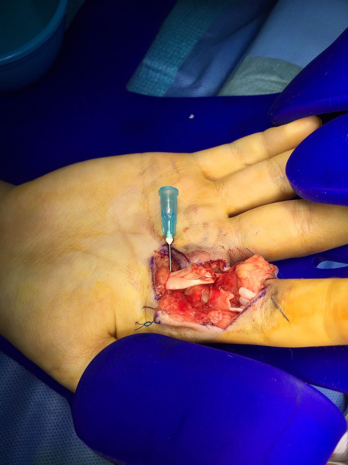
- How do you describe and assess this flexor tendon injury?
- What is the aim and the principles of an ideal flexor tendon repair?
- How do you retrieve and tunnel the flexor tendons?
- What are the key points of flexor tendon suture technique?
- What is the blood supply and healing of flexor tendons?
- Should you repair both FDS and FDP, and which pulleys are retained?
- What are the flexor tendon rehabilitation options?
- Describe the Kleinert rehabilitation program.
- Describe the Duran rehabilitation program.
- Describe the Strickland rehabilitation program.
- Describe the Belfast early active rehabilitation program.
- When do you use a static rehabilitation program?
- Why use a dynamic rehabilitation program?
- What are the complications of flexor tendon repair and rehabilitation?
Q1.How do you describe and assess this flexor tendon injury?
- I/F laceration with complete cut of FDS and FDP at zone 2, just at Camper's chiasma; contamination; check distal circulation
- Hx: mechanism of injury, occupation, functional demands
- PE: tenodesis, wound contamination, digital nerve 2PD, digital Allen, XR for FB/fracture
Q2.What is the aim and the principles of an ideal flexor tendon repair?
- Aim: secure repair to allow early mobilisation for intrinsic tendon healing
- Prep: smooth tendon ends, minimal disruption of blood supply, easy passage of suture
- Repair: minimal gap, secure knot, sufficient strength for early mobilisation
Q3.How do you retrieve and tunnel the flexor tendons?
- Retrieval: milking, reverse Esmarch, suction, tendon retriever, proximal skin incision
- Temporary locking suture to hold both ends, pass suture through pulley
- Preserve A2 (chiasma just distal to A2 at resting position); flex wrist and fingers and temporarily pin down tendon
Q4.What are the key points of flexor tendon suture technique?
- Use non absorbable suture at least 4-0 or thicker
- Kessler (locking) better than Bunnell - superior pull-out strength when core sutures are in the dorsal half of the tendon
- At least 4 core strands (active flexion ~20N; 2 core ~34N, 4 core ~43N, 6 core ~78N)
- At least 10mm from the edge; epitendinous suture +20% strength, reduces gap and improves gliding
Q5.What is the blood supply and healing of flexor tendons?
- Vascularised via paratenon -> travels longitudinally within endotenon; non-vascularised via synovial sheath -> mesotenon in the vincula
- Intrinsic healing from epitenon/endotenon (less adhesion) vs extrinsic healing from paratenon (promoted by immobilisation) - avoid extrinsic
- Phases: inflammatory <2 weeks (fibrin clot, strength decreases), reparative 2-6 weeks (collagen 3), remodelling >6 weeks (collagen 1, strength increases) - protect before 6 weeks
Q6.Should you repair both FDS and FDP, and which pulleys are retained?
- Repair both for independent DIPJ and PIPJ function; FDS one slip only can prevent swan neck yet minimise adhesions
- Retain A2 and A4
- Cutting A2 causes bowstringing -> need more excursion to produce same angular movement, result in less angular movement
Q7.What are the flexor tendon rehabilitation options?
- Static vs. dynamic program
- Dynamic: controlled passive (Kleinert/ Duran/ strictland) vs early active (Belfast) movement
Q8.Describe the Kleinert rehabilitation program.
- Dynamic splint allowing active extension and passive flexion
- Low force, low excursion (2.5cm)
- Wrist/MCPJ flexed 30/30; rubber band and pulley system; 1 pulley at midpalm to increase DIPJ flexion for better differential gliding; release at night with finger strap to prevent PIPJ contracture
- Change to night extension splint at 4 weeks, gentle active fisting; resistive exercises at 6 weeks
Q9.Describe the Duran rehabilitation program.
- Static wrist splint (wrist 20deg, MCPJ 50deg)
- Patient alternately passively extends DIPJ and PIPJ with other joints flexed to allow differential gliding
- Moderate force, low excursion
- Off splint at 4-6 weeks to wrist cuff with elastic traction (active extension, passive flexion); off cuff at 6 weeks, active fisting
Q10.Describe the Strickland rehabilitation program.
- Adds a tenodesis splint (wrist 30deg, MCPJ 50deg)
- Couples active wrist extension with passive digital flexion, and wrist flexion with digital extension
- Low force, high excursion (9cm)
Q11.Describe the Belfast early active rehabilitation program.
- Early active movement; dorsal blocking splint limiting wrist extension (wrist 40deg flexion, MCPJ 90deg flexion, IPJ 0)
- Place and hold exercises every 2 hours, started immediately
- 3 active flexion (palm + hook + flat fist), 2 active extension (PIPJ + wrist), 2 passive motion exercises (into palm + hook)
- Supported by Star et al Am J Hand Surg 2013 meta-analysis: active motion had significantly fewer flexion contractures, better satisfaction and ROM, no difference in re-rupture rates
Q12.When do you use a static rehabilitation program?
- Children, mental deficit / abnormal muscle tone
Q13.Why use a dynamic rehabilitation program?
- Stressed tendon heals faster, with better strength, less adhesion and better excursion
- Principle: provide motion but protect against excessive stress load which may cause rupture
Q14.What are the complications of flexor tendon repair and rehabilitation?
- Re-rupture (15-25%)
- Contracture / adhesions
- Quadriga / lumbrical
- Swan neck
▸ Slide 745 · Laceration at PIPJ 6 weeks ago sutured in GPHand Soft Tissue · 9 questions expand

Q1-Q99 questions — tap to reveal all answerslist
- What is the presentation and differential diagnosis after a neglected flexor tendon laceration?
- What history is important in a neglected flexor tendon injury?
- How do you assess a neglected flexor tendon injury?
- How is an isolated FDS or FDP injury managed?
- How do you decide between one-stage and two-stage flexor reconstruction?
- How do you choose between graft and tendon transfer?
- How does proximal stump excursion guide graft versus transfer?
- When is the timing for tendon transfer?
- What are the prerequisites of tendon transfer?
Q1.What is the presentation and differential diagnosis after a neglected flexor tendon laceration?
- Failed flexion over DIPJ/PIPJ, wound over volar side of MCPJ; suspected neglected FDP and FDS injury
- Ddx for decreased ROM: adhesion, re-rupture, joint contracture, bowstringing, triggering, quadriga effect
Q2.What history is important in a neglected flexor tendon injury?
- Mechanism of injury
- Timing
- Occupation
- PMHx
- Smoking
Q3.How do you assess a neglected flexor tendon injury?
- Supple skin; full passive ROM and stable joint; Sensate digit. Adequate vascularity
- Check for palmaris longus; test individual FDS and FDP function
- XR for fracture; USG for degree of retraction - >1cm gap cannot go for primary repair
Q4.How is an isolated FDS or FDP injury managed?
- Isolated FDS: conservative
- Isolated FDP options: DIPJ fusion, flexor reconstruction, FDP stump tenodesis to FDS (or MP neck), tendon transfer for non-functioning motor unit
- If both involved, choose one as donor: FDS better independent motion but weaker (60% of FDP strength); FDP less independent motion but stronger
Q5.How do you decide between one-stage and two-stage flexor reconstruction?
- Intraop decision after debriding scarred ends: assess gap, tendon quality, proximal stump excursion
- One stage if good pulley, good skin, good joint, good patient
- If staged, graft PL; Hunter-Salisbury (suture stump and distal to rod to induce sheath; anastomosis at DP base and zone 3 or 5 - zone 3 preferred to preserve lumbrical, zone 5 fewer adhesions)
- Paneva-Hollovich: suture FDP and FDS, rod induces pseudosheath, only one anastomosis in second stage; pulley with belt loop (PL) or FDS tail
Q6.How do you choose between graft and tendon transfer?
- How to compare and contrast graft vs tendon transfer? Decision based on a few factors, has to be individualized
- Individualise by number of tendons involved and chronicity
- Graft has 2 healing sites but uses native muscle; transfer has 1 healing site but donor muscle downgrades one motor grade
- Graft uses native muscle; transfer uses a donor motor unit
Q7.How does proximal stump excursion guide graft versus transfer?
- >2cm: graft
- <1cm: transfer
- 1-2cm: controversial
Q8.When is the timing for tendon transfer?
- When tissue equilibrium has been achieved: bone stability, joint supple, soft tissue envelope satisfactory
Q9.What are the prerequisites of tendon transfer?
- Patient: compliance, disease non-progressive
- Donor muscle: expendable, adequate strength (proportional to cross-section), expect one grade loss, synergistic
- Donor tendon: adequate excursion (Smith 357 rule: 3cm wrist, 5cm finger extensor, 7cm finger flexor), normal tension (actually want more), same line of pull, one tendon one function
- Recipient: tissue equilibrium
▸ Slide 746 · Quadriga effectHand Soft Tissue · 2 questions expand

Q1-Q22 questions — tap to reveal all answerslist
- What term is associated with the quadriga effect on this slide?
- Explain the quadriga effect and lumbrical plus.
Q1.What term is associated with the quadriga effect on this slide?
- Lumbrical plus
Q2.Explain the quadriga effect and lumbrical plus.
- Not covered in the speaker notes beyond the term lumbrical plus
▸ Slide 747 · What is this?Hand Soft Tissue · 5 questions expand
Q1-Q55 questions — tap to reveal all answerslist
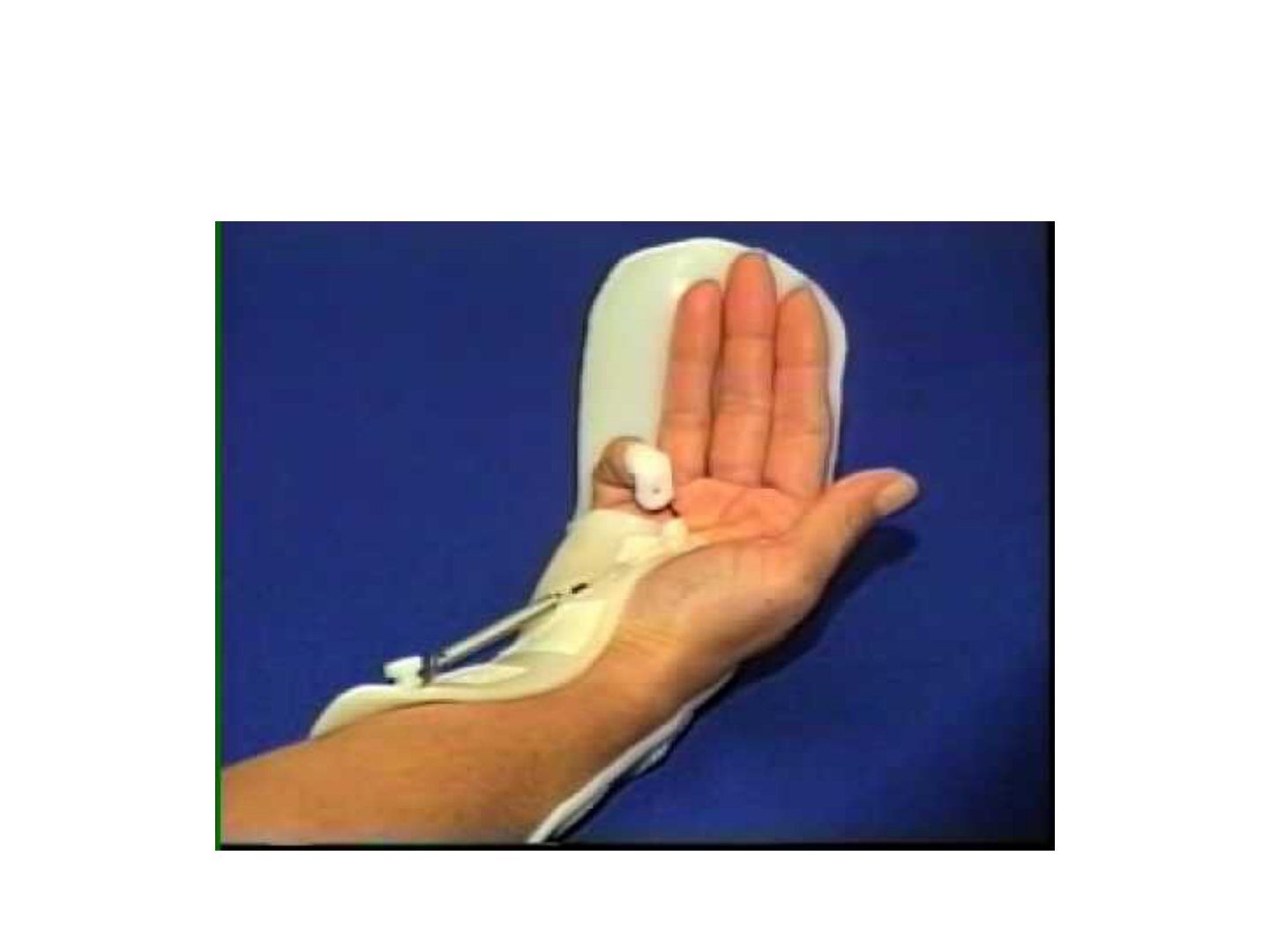
- What is this splint and how does it work?
- What other flexor tendon rehab protocols do you know?
- What is the rationale of a dynamic splint?
- How do you choose the flexor rehab protocol?
- What are the practical points about the Kleinert splint used in the centre?
Q1.What is this splint and how does it work?
- Dynamic controlled passive movement splint for flexor tendon repair rehab - Kleinert splint
- Blocks MCPJ extension; string and pulley system attached to rubber band and injured finger allows passive flexion and active extension
- Wrist 30deg flexion, MCPJ 30deg flexion; low excursion low force program
- Kept 4-6 weeks postop; at night rubber band released and IPJ kept in extension to prevent flexion contracture
Q2.What other flexor tendon rehab protocols do you know?
- Controlled passive: Kleinert / Duran / Strickland
- Early active: Belfast
Q3.What is the rationale of a dynamic splint?
- Prevent extrinsic healing which causes scar formation
- Encourage intrinsic tendon healing which aligns collagen fibres -> stronger tendon, more excursion, fewer adhesions
Q4.How do you choose the flexor rehab protocol?
- Patient factor: compliance (non-cooperative/children -> static splint)
- Injury factor: zone of injury (zone 2 needs dynamic splint to prevent adhesion); multiple/bilateral finger injury -> Kleinert instead of Duran
- Surgery factor: repair strength
Q5.What are the practical points about the Kleinert splint used in the centre?
- Difficult to instruct patient for active movement, but low re-rupture rate + good range
- During active extension there is reciprocal flexor relaxation (no co-contraction/active tension)
- 3-4mm motion is adequate to prevent adhesions
▸ Slide 748 · Position of safe immobilizationHand Soft Tissue · 3 questions expand
Q1-Q33 questions — tap to reveal all answerslist
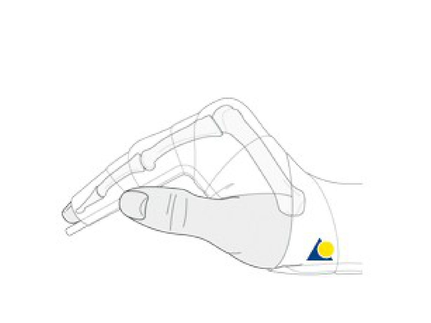
- What is the position of safe immobilisation of the hand?
- Why are the MCPJ flexed but IPJ extended in the safe position?
- Why is the wrist held in 30deg extension in the safe position?
Q1.What is the position of safe immobilisation of the hand?
- Wrist 30deg extension, MCPJ 70-90deg flexion, IPJ extended
Q2.Why are the MCPJ flexed but IPJ extended in the safe position?
- MC head is cam shaped (ellipsoid joint) with collaterals tight in flexion - maintains the collateral (proper) ligament at maximal length
- IPJ hinged like, collaterals tight in extension, flexor stronger than extensor - maintains the volar plate at maximal length
- Goal: tighten collaterals in MCPJ/PIPJ and maintain volar plate length
Q3.Why is the wrist held in 30deg extension in the safe position?
- Wrist 30deg extension is functional as flexors are stronger than extensors
▸ Slide 749 · UL orthosisHand Soft Tissue · 4 questions expand
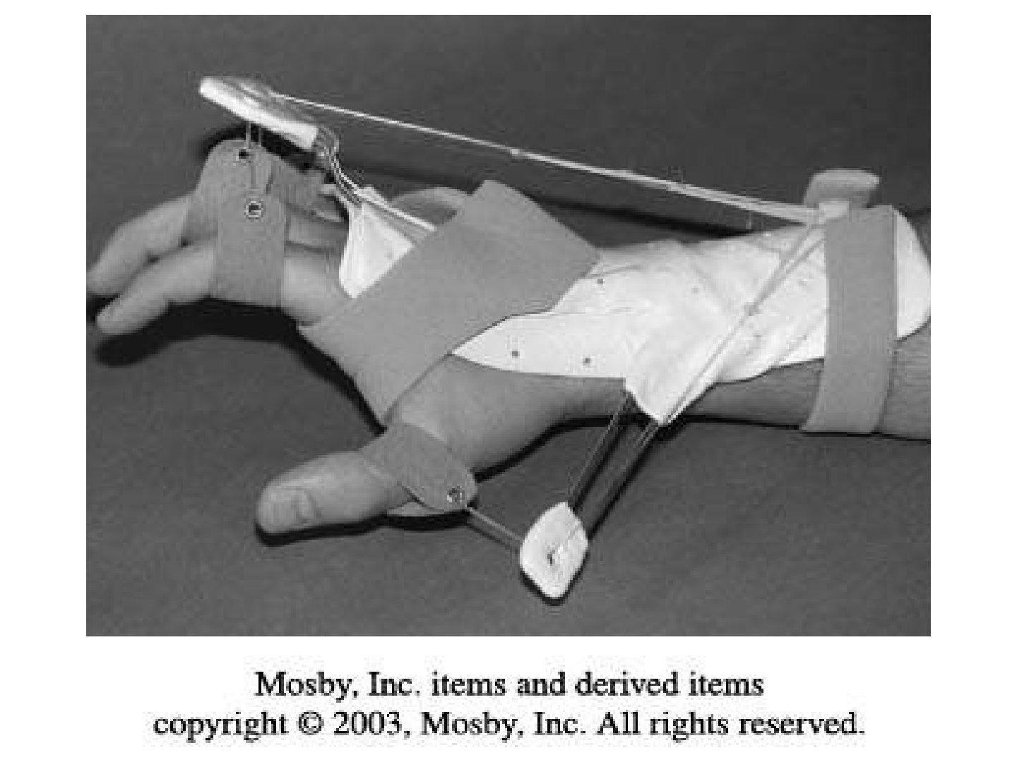
Q1-Q44 questions — tap to reveal all answerslist
- What is the purpose of an upper limb orthosis / outrigger splint?
- How do you approach a patient with wrist drop?
- What history and examination findings are relevant in wrist drop?
- What is the management of radial nerve palsy / wrist drop?
Q1.What is the purpose of an upper limb orthosis / outrigger splint?
- Promote function
- Immobilise to promote healing and prevent deformity
- Position the hand weakened by weakness
- Correct an existing deformity
Q2.How do you approach a patient with wrist drop?
- Confirm wrist drop (not joint/bony problem)
- Rule out systemic problem (CVA, MND)
- Localise the pathology
Q3.What history and examination findings are relevant in wrist drop?
- Hx: onset/progression, sensory deficit, closed/open injury, previous OT, rule out other nerve root involvement
- Inspect: posture, wasting, scar, radial deviation of wrist
- Palpate along radial nerve and sensation; passive wrist range and all radial-innervated muscles; Tinel
- Examine shoulder and C spine
Q4.What is the management of radial nerve palsy / wrist drop?
- Dynamic splint, NCT (nerve conduction test)
- Jones tendon transfer: PT to ECRB, PL to EPL, FCU to EDC
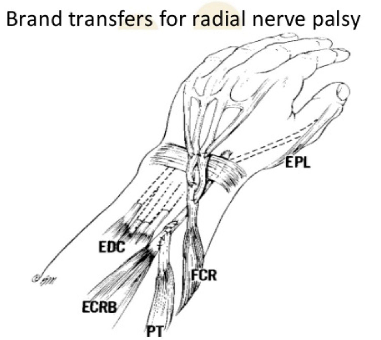
- Brand's transfer (modification of Jones): PL, PT, FCR (not FCU, to maintain dart-throwing motion of wrist)
▸ Slide 750 · Tendon transfer pre requisite:Hand Soft Tissue · 7 questions expand
Q1-Q77 questions — tap to reveal all answerslist
- What are the prerequisites for tendon transfer?
- What is the memorised summary of tendon transfer prerequisites?
- What is the aim of tendon transfer?
- What is Brand's transfer for radial nerve palsy?
- What tendon transfers are used for ulnar nerve palsy?
- What tendon transfers are used for median nerve (AIN) palsy?
- What opponensplasties are used in carpal tunnel syndrome?
Q1.What are the prerequisites for tendon transfer?
- Patient: good motivation, willing to train
- Receiving site: tissue equilibrium (Boyes) - no infection, healthy bed, no joint contracture, joint stable
- Donor muscle FESS: strength, one transfer one function, synergistic, expendable
- Donor tendon LEP: adequate length and excursion, straight line of pull, correct tension
Q2.What is the memorised summary of tendon transfer prerequisites?
- Transfer an expendable and good strength tendon to another synergistic action
- With straight line of pull under adequate excursion and tension
- And good soft tissue across a supple joint
- To perform single function in a compliant patient
Q3.What is the aim of tendon transfer?
- To concentrate the available motor unit into the best working combination
Q4.What is Brand's transfer for radial nerve palsy?
- Wrist extension: PT to ECRB
- Thumb extension: PL to EPL
- Finger extension: FCR to EDC
Q5.What tendon transfers are used for ulnar nerve palsy?
- Thumb adduction: ECRL/FDS4 to adductor pollicis
- 1st dorsal interosseous: EI to 1st dorsal interossei
- Little finger abduction: EDM to ADM
- Claw hand: Bouvier test - if positive, capsulodesis; if negative, Zancolli lasso (split FDS to lateral bands of ulnar digits)
Q6.What tendon transfers are used for median nerve (AIN) palsy?
- Thumb flexion: brachioradialis to FPL
- Thumb opposition: EI to APB
- Finger flexion: FDP of R and L/F to FDP of index and middle finger (side-to-side transfer)
Q7.What opponensplasties are used in carpal tunnel syndrome?
- Transfers to APB at MCPJ level
- Camitz (PL + palmar aponeurosis)
- Huber (ADM)
- Riordan opponensplasty (ring/little FDS, FCU slip as pulley)
- EI transfer
▸ Slide 751 · Radial nerve palsyHand Soft Tissue · 11 questions expand
Q1-Q1111 questions — tap to reveal all answerslist
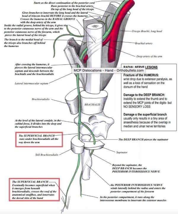
- Which muscles are supplied by the proper radial nerve?
- What is Saturday night palsy and how is it managed?
- How is radial nerve palsy classified from proximal to distal?
- What are the causes of posterior interosseous nerve palsy?
- What are the sites of compression in radial tunnel syndrome (FREAS)?
- What are the symptoms of radial tunnel syndrome (PIN entrapment)?
- What are the signs of PIN palsy?
- Which muscles does the PIN innervate?
- What is the nonoperative treatment and outcome of PIN palsy?
- What is cheiralgia paresthetica (Wartenberg syndrome)?
- How is cheiralgia paresthetica examined and what is the outcome?
Q1.Which muscles are supplied by the proper radial nerve?
- Triceps, brachioradialis, 2/3 of brachialis, anconeus and ECRL
- Its palsy is caused by trauma or is iatrogenic
Q2.What is Saturday night palsy and how is it managed?
- Compression of the proper radial nerve
- Involves triceps, brachioradialis and ECRL + PIN + sensory deficit
- Managed conservatively
Q3.How is radial nerve palsy classified from proximal to distal?
- Proper radial nerve palsy
- Posterior interosseous nerve (PIN) palsy
- Cheiralgia paresthetica (Wartenberg syndrome) - superficial sensory branch
Q4.What are the causes of posterior interosseous nerve palsy?
- Trauma: radial head fracture, Monteggia, elbow dislocation
- Space-occupying lesion: lipoma, ganglion, synovitis in RA
- Iatrogenic: after radial head replacement
- Entrapment (radial tunnel syndrome)
Q5.What are the sites of compression in radial tunnel syndrome (FREAS)?
- Fascial band at the radial head
- Recurrent leash of Henry
- Edge of the ECRB
- Arcade of Frohse (most common site, proximal edge of the supinator)
- Supinator distal edge
Q6.What are the symptoms of radial tunnel syndrome (PIN entrapment)?
- Lateral elbow pain
- Distal muscle weakness
- +/- dorsal wrist pain (dorsal wrist capsule)
Q7.What are the signs of PIN palsy?
- Radial deviation with active wrist extension (ECRL is innervated by the proper radial nerve more proximally)
- Weakness of PIN-innervated thumb and finger extension
Q8.Which muscles does the PIN innervate?
- 9 muscles: supinator + APL, EPB, ECRB, EPL, EI, EDC, EDM, ECU
Q9.What is the nonoperative treatment and outcome of PIN palsy?
- Activity modification, splinting and NSAIDs
- Outcome at 3 months (OT@ 3m): 85% good to excellent result
Q10.What is cheiralgia paresthetica (Wartenberg syndrome)?
- Compressive neuropathy of the superficial sensory branch of the radial nerve
- Compressed between brachioradialis and ECRL with forearm pronation (scissor-like action between the tendons)
- Symptoms: pain, numbness and paresthesias over the dorsoradial hand
Q11.How is cheiralgia paresthetica examined and what is the outcome?
- Forceful forearm pronation for 60 seconds
- Tinel sign over the nerve
- Outcome at 6 months
▸ Slide 752 · Describe photo:Hand Soft Tissue · 20 questions 1 check expand

Q1-Q2020 questions — tap to reveal all answerslist
- What does the clinical photo show and what is being assessed?
- What are the boundaries of the carpal tunnel?
- What structures pass through the carpal tunnel?
- What is the diagnostic approach to carpal tunnel syndrome (PC Ho)?
- What are the typical history features of carpal tunnel syndrome?
- What are the clinical tests for carpal tunnel syndrome?
- What are the NCV findings in carpal tunnel syndrome?
- What is the conservative management of carpal tunnel syndrome?
- What predicts a poor response to nonoperative management of carpal tunnel syndrome?
- What are the operative options for carpal tunnel syndrome?
- What are the efficacy and complications of endoscopic carpal tunnel release (ECTR)?
- What is required before performing a 2-portal ECTR?
- Where are the incisions placed for a 2-portal ECTR?
- Which structures must be avoided during a 2-portal ECTR?
- How is a 2-portal ECTR performed?
- What are the named nerve anastomoses in the hand?
- How is an open carpal tunnel release performed?
- What are the intra-operative complications of carpal tunnel release?
- What are the post-operative complications of carpal tunnel release?
- What are the causes of symptoms persisting after carpal tunnel release?
Q1.What does the clinical photo show and what is being assessed?
- Thenar wasting
- Carpal tunnel anatomy
- NCV result
- Carpal tunnel view to look for osteophytes
Q2.What are the boundaries of the carpal tunnel?
- Scaphoid tubercle, trapezium ridge, pisiform, hamate hook
- Transverse carpal ligament (proximally the volar carpal ligament)
- Carpal row
Q3.What structures pass through the carpal tunnel?
- 9 tendons: FDS, FDP and FPL
- 1 nerve: the median nerve
Q4.What is the diagnostic approach to carpal tunnel syndrome (PC Ho)?
- Clinical diagnosis (2/3): Phalen, DCT, Tinel + NCV +/- USG
- Look for risk factors, secondary causes, and rule out other diagnoses
Q5.What are the typical history features of carpal tunnel syndrome?
- Nocturnal numbness over the radial 3 and a half fingers
- Improves with shaking the hand
- Look for cause: mass (ganglion), inflammation (gout, RA), fluid (hypothyroidism, renal failure), post-trauma
- R/O DDx: high median nerve palsy, brachial plexus, C5/6 nerve root; double crush syndrome
Q6.What are the clinical tests for carpal tunnel syndrome?
- Phalen, direct compression, Tinel
- High specificity: sensory loss, CT compression (Durkan's test, press over CT x 30 sec)
- High sensitivity: Phalen (flexion 90 x 60 sec), CT compression
- Thenar wasting + weak APB
Q7.What are the NCV findings in carpal tunnel syndrome?
- Early: decreased velocity <50 m/sec, increased distal motor latency >4.2 ms
- Late: conduction block, decreased amplitude
- Sensitivity and specificity ~80%
- USG for space-occupying lesions
Q8.What is the conservative management of carpal tunnel syndrome?
- Night splint, nerve gliding exercises
- Steroid injection: transient relief in 80%, 20% symptom free after 1 year
- INSTINCTS trial (Lancet 2018): steroid superior clinical effectiveness vs splintage at 6 weeks
Q9.What predicts a poor response to nonoperative management of carpal tunnel syndrome?
- Age >50
- Symptoms >10 months
- Persistent paresthesia
- Trigger finger
- +ve Phalen test after 30 seconds
Q10.What are the operative options for carpal tunnel syndrome?
- Open carpal tunnel release
- ECTR (endoscopic carpal tunnel release): 1 portal or 2 portal
Q11.What are the efficacy and complications of endoscopic carpal tunnel release (ECTR)?
- Similar symptom relief, electrophysiology result and carpal tunnel pressure relief
- Earlier return to work (CORR 2015 Sayegh meta-analysis), no long term difference
- Higher nerve injury (2x open; open release risk 0.16%)
- Inadequate release
Q12.What is required before performing a 2-portal ECTR?
- Patient selection, confirmation of diagnosis of carpal tunnel syndrome and r/o other DDx and secondary causes
- R/O contraindications: inflammatory cause, limited wrist extension, SOL
- Consent, Bier's block with double cuff
Q13.Where are the incisions placed for a 2-portal ECTR?
- Distal incision: Kaplan's line (RMB) / hyperabduction thumb line (SPA), line extrapolated from the 3rd finger web
- Proximal incision: just ulnar to PL, at the proximal palmar crease
Q14.Which structures must be avoided during a 2-portal ECTR?
- Palmar cutaneous nerve (branch of median nerve 5cm from wrist): stay ulnar to PL
- Recurrent motor branch (extra-ligamentous 50%, sub-ligamentous 30%, trans-ligamentous 20%): incise TCL on the ulnar side
- Guyon's canal (hook of hamate and pisiform): stay radial to the line from the 4th webspace
- Superficial palmar arch (average 15mm distal to Kaplan line; DPA 6mm): stay proximal to Kaplan's line
- Riche-Cannieu median-to-ulnar connection at the distal portal: blunt dissection
Q15.How is a 2-portal ECTR performed?
- Open proximal incision, blunt dissect forearm fascia, insert McDonald to feel the washboard texture of TCL
- Hyperextend wrist, insert T&C and open the distal incision; visualise TCL and free soft tissue
- Incise TCL from distal to proximal with a retrograde knife, staying ulnar
- Success confirmed by prolapse of fat and transillumination seen on the skin
Q16.What are the named nerve anastomoses in the hand?
- Berrettini: digital nerve anastomosis
- Martin-Gruber: median to ulnar
- Marinacci: reverse Martin-Gruber
- Riche-Cannieu anastomosis: motor ulnar and median nerve anastomosis in the thenar region
Q17.How is an open carpal tunnel release performed?
- Confirm diagnosis, assess secondary causes; Bier's block
- Long incision: distal end proximal to Kaplan's line, just ulnar to thenar crease, curve ulnar slightly distal to the distal palmar crease, cross the crease at an angle
- Proximal end at proximal palmar crease, just ulnar to PL
- Structures to release: deep forearm fascia, TCL, volar carpal ligament
- Short incision: difficult to tackle the volar carpal ligament, but less hypertrophic scar
Q18.What are the intra-operative complications of carpal tunnel release?
- Anaesthesia related
- Injury to median nerve, palmar cutaneous nerve, recurrent motor branch
- Injury to superficial palmar arch
- Injury to ulnar nerve and artery
- Injury to tendon
Q19.What are the post-operative complications of carpal tunnel release?
- Pillar pain, scar pain
- Infection
- Recurrence of symptoms
- Stiffness, bowstringing of tendon
- Painful scar = injury to communicating branch of ulnar/median nerve (retract and cut)
Q20.What are the causes of symptoms persisting after carpal tunnel release?
- Wrong diagnosis
- Double crush
- Inadequate release
- Injury to nerve
- SOL
In carpal tunnel syndrome NCV shows increased distal motor latency >4.2s — unit error — distal motor latency is measured in milliseconds; normal median DML is <=4.2 ms and >4.2 ms is abnormal — source
▸ Slide 753 · Pronator syndrome compression of median nerve at elbow levelHand Soft Tissue · 5 questions expand

Q1-Q55 questions — tap to reveal all answerslist
- What are the causes of compression in pronator syndrome?
- What is pronator syndrome and how does it present?
- What are the causes of compression in AIN (anterior interosseous nerve) syndrome?
- What is the presentation of AIN syndrome?
- How do you distinguish pronator quadratus from pronator teres weakness?
Q1.What are the causes of compression in pronator syndrome?
- Supracondylar process
- Ligament of Struthers
- Lacertus fibrosus
- Deep head of pronator teres
- FDS arch
Q2.What is pronator syndrome and how does it present?
- Compression of the median nerve at the elbow level
- Numbness of the medial 3 and a half fingers
- Numbness of the thenar region
Q3.What are the causes of compression in AIN (anterior interosseous nerve) syndrome?
- Lacertus fibrosus
- Deep head of pronator teres
- FDS arch
- Accessory head of FPL
Q4.What is the presentation of AIN syndrome?
- Weakness of FPL
- Weakness of index and middle FDP
- Weakness of pronator quadratus
Q5.How do you distinguish pronator quadratus from pronator teres weakness?
- PQ: resisted pronation with elbow in full flexion (least mechanical advantage to PT)
- PT: resisted pronation with elbow slightly flexed
▸ Slide 754 · Cubital tunnel syndromeHand Soft Tissue · 11 questions expand
Q1-Q1111 questions — tap to reveal all answerslist
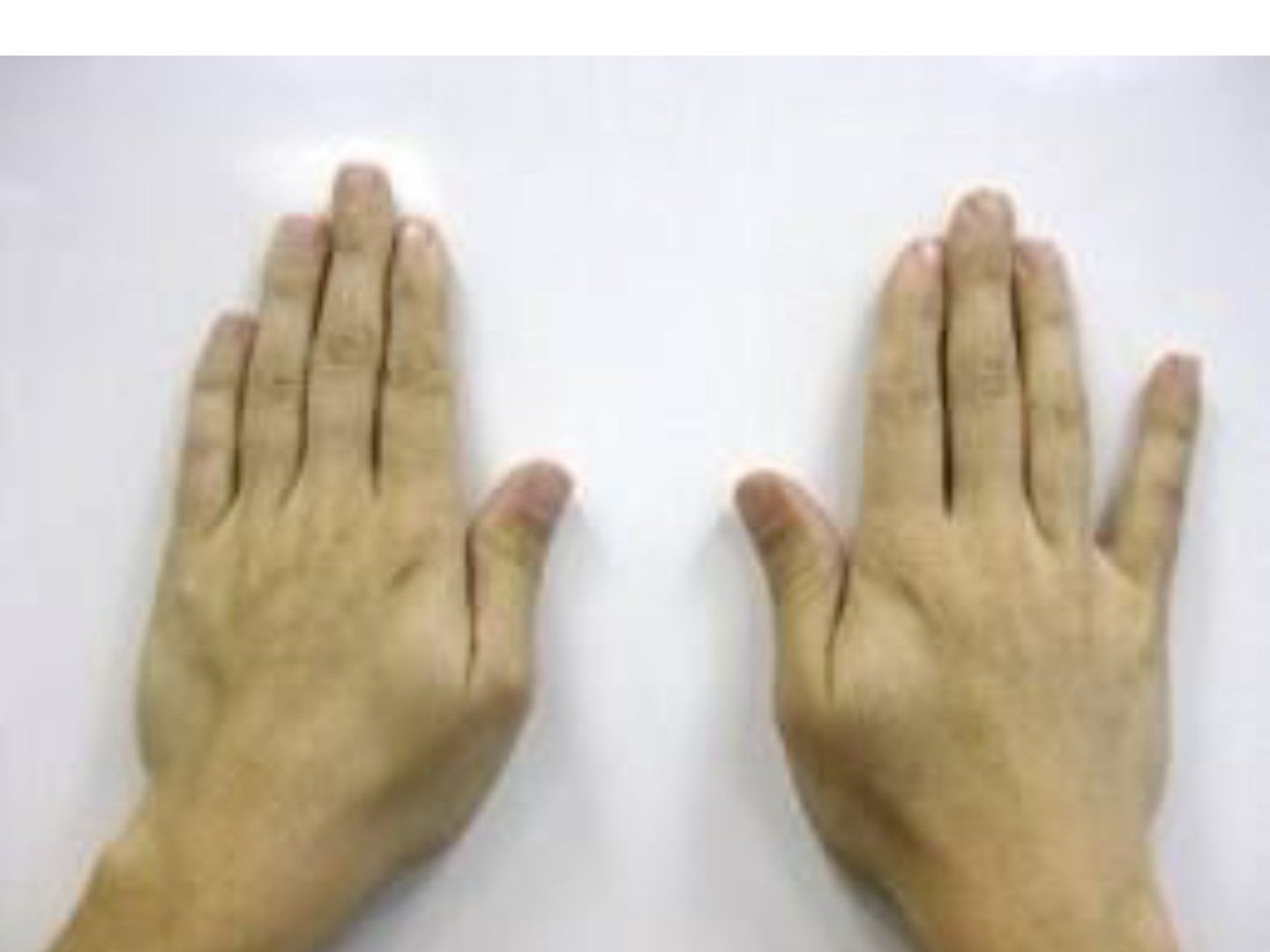
- What does the photo show and what is the differential diagnosis?
- What is Wartenberg's sign?
- How do you assess suspected cubital tunnel syndrome?
- List the sites of ulnar nerve compression around the elbow.
- Outline the conservative management of cubital tunnel syndrome.
- What are the indications and pros/cons of ulnar nerve decompression in situ?
- What are the indications and pros/cons of medial epicondylectomy?
- How do you perform a medial epicondylectomy?
- Compare the types of anterior transposition of the ulnar nerve.
- What is the evidence for in-situ decompression versus transposition?
- How is an anterior transposition of the ulnar nerve performed?
Q1.What does the photo show and what is the differential diagnosis?
- Failed abduction of the little finger (unilateral)
- No claw hand, no interossei muscle wasting
- DDx: Wartenberg sign, cervical myelopathy (bilateral)
Q2.What is Wartenberg's sign?
- Weak interossei
- Ulnarly deviated pull of EDM + EDC
Q3.How do you assess suspected cubital tunnel syndrome?
- Delineate high vs low ulnar nerve palsy
- R/O other causes with full neuro exam: C8/T1 root (I/F FDP, APB), sensory, myelopathic hand signs, LL involvement
- High: cubital valgum (malunited lateral condyle fracture), limited elbow range (OA elbow)
- Check Tinel and elbow flexion test (>60 sec)
- McGowan grading: 1 sensory, 2 weakness, 3 wasting
Q4.List the sites of ulnar nerve compression around the elbow.
- Around the medial intermuscular septum and arcade of Struthers (medial intermuscular septum to medial head of triceps)
- Medial epicondyle in cubital valgum
- Epicondylar groove
- Cubital tunnel: SOL, Osborne ligament (medial epicondyle to olecranon), FCU aponeurosis between the two heads
- The flexor pronator aponeurosis (fascia btw FDP and FDS), anconeus epitrochlearis
Q5.Outline the conservative management of cubital tunnel syndrome.
- Splint with elbow in 30 degrees flexion, wrist in neutral to relax FCU
- NSAIDs
Q6.What are the indications and pros/cons of ulnar nerve decompression in situ?
- CI: subluxable nerve, SOL, scar, severe elbow deformity
- Adv: minimal dissection, can do under LA, protects the vascular mesoneurium, avoids scarring
- Disadv: if released more proximal to the epicondylar groove, risk of nerve subluxation and a new site of compression
Q7.What are the indications and pros/cons of medial epicondylectomy?
- Releases compression against the epicondyle
- For visible and symptomatic subluxating nerve, or thin patients where subcutaneous anterior transposition may not be feasible
- Pros: smaller wound, nerve can seek its own course of least resistance, less ischaemic injury, no need to sacrifice the articular branch
- No need to immobilize elbow
- Cons: injury to MCL (prevent by not going too basally, <70%), pain, weakness of flexor origin, too little resection may not fully relieve traction
Q8.How do you perform a medial epicondylectomy?
- GA, supine, shoulder abducted and externally rotated; incision centred on medial epicondyle; protect the medial antebrachial nerve
- Proximally release arcade of Struthers and medial intermuscular septum, then Osborne ligament, distally the FCU aponeurosis and deep flexor pronator aponeurosis
- Expose the medial epicondyle subperiosteally and remove 3-4mm
- Reattach periosteum in 'pant over vest' manner to the CFP
- Identify the ulnar nerve posteriorly in its groove
Q9.Compare the types of anterior transposition of the ulnar nerve.
- Subcutaneous: technically easiest; nerve becomes superficial, more dissection of blood supply
- Submuscular: least recurrence, good protection and vascular bed; needs 3 weeks immobilisation, risk of recompression (improve with Z-plasty), not good in severe OA, too close to joint may cause impingement
- Intramuscular: nerve buried in a groove within flexor-pronator muscle; risk of recurrence, more bleeding, better preserves nerve blood supply
Q10.What is the evidence for in-situ decompression versus transposition?
- Ilyas JHS 2019 meta-analysis: functionally in-situ decompression = transposition; transposition has more complications
- JAMA Network 2020 (Wade): in-situ decompression associated with lower risk than any form of transposition
- Addition of epicondylectomy associated with increased probability of symptomatic cure without increasing complication risk
Q11.How is an anterior transposition of the ulnar nerve performed?
- GA, supine, shoulder abducted and ER; incision centred on medial epicondyle; avoid the medial antebrachial nerve (pierces brachial fascia adjacent to basilic vein)
- Anteriorly develop skin flap to expose the CFO; posteriorly identify the ulnar nerve in its groove
- Proximally release arcade of Struthers and medial intermuscular septum, then Osborne ligament; distally release FCU aponeurosis and deep flexor pronator aponeurosis
- For submuscular: sharply divide the tendinous origin of the medial epicondyle, retract the flexor mass distally, place nerve deep to the fascial sling
- Flex and extend the elbow to ensure no kinking; release tourniquet before closure for haemostasis; immobilise elbow 3 weeks
▸ Slide 755Hand Soft Tissue · 2 questions expand

Q1-Q22 questions — tap to reveal all answerslist
- What is the topic of this slide?
- What are the key learning points of this slide?
Q1.What is the topic of this slide?
- Hand Soft Tissue
Q2.What are the key learning points of this slide?
- Not covered in the speaker notes
▸ Slide 756 · Lower ulnar nerve palsyHand Soft Tissue · 9 questions expand
Q1-Q99 questions — tap to reveal all answerslist
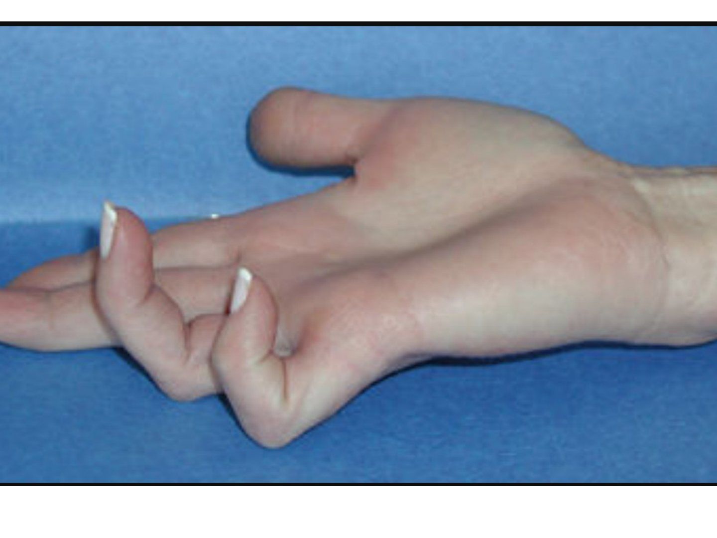
- Describe the claw hand deformity and its differential diagnosis.
- What is the pathomechanism of claw hand in ulnar nerve palsy?
- Why is the little finger more clawed than the others?
- Describe the anatomy and contents of Guyon's canal.
- What are the three zones of Guyon's canal and their causes?
- How is lower ulnar nerve palsy investigated?
- What is the conservative management of Guyon canal syndrome?
- How is claw hand managed?
- What is the tendon transfer strategy for combined ulnar and median nerve palsy?
Q1.Describe the claw hand deformity and its differential diagnosis.
- Little finger clawing with flexed IPJs and extended MCPJs
- Unilateral DDx: ulnar nerve palsy, Volkmann's contracture (volkman’s contracture), thoracic outlet syndrome
- Bilateral DDx: CMT, cervical myelopathy
Q2.What is the pathomechanism of claw hand in ulnar nerve palsy?
- Imbalance of intrinsic and extrinsic muscles
- Unopposed MCPJ extension due to weak intrinsic muscles
- Used up excursion of long extensors + weak intrinsics, so cannot extend IPJ
- Unopposed long flexors
Q3.Why is the little finger more clawed than the others?
- MCPJ of L/F more hyperextendable (the MCPJ of the little finger is more hyperextendable)
- The lumbrical of the ring finger has a median nerve contribution
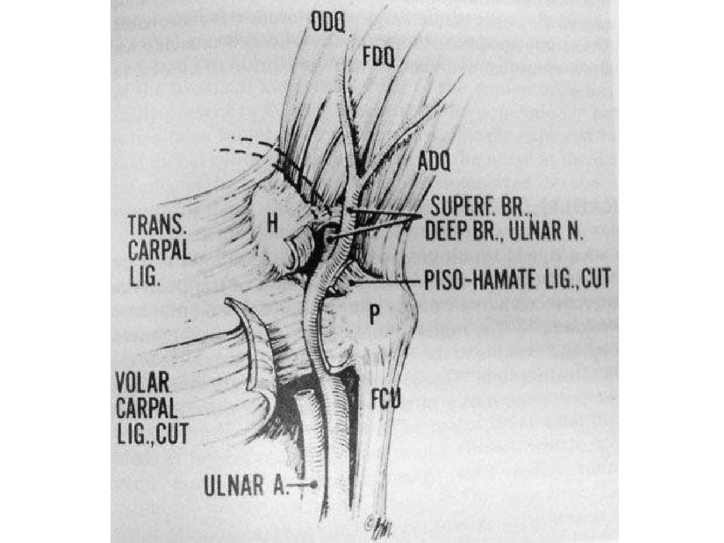
Q4.Describe the anatomy and contents of Guyon's canal.
- 4 cm long tunnel
- Floor: transverse carpal and pisohamate ligaments
- Roof: volar carpal ligament
- Walls: pisiform and hook of hamate
- Contains ulnar artery and nerve; nerve bifurcates into sensory (superficial/ulnar) and motor (deep/radial)
Q5.What are the three zones of Guyon's canal and their causes?
- Zone 1 (proximal to bifurcation): ganglion, fracture hook of hamate; contains both motor and sensory
- Zone 2: motor branch (ganglion, fracture hook of hamate)
- Zone 3: sensory branch (ulnar artery aneurysm)
- Operative treatment: decompress all three zones and address the primary cause
Q6.How is lower ulnar nerve palsy investigated?
- Investigation: thorough examination
- NCV
- CT/MRI for the underlying cause
Q7.What is the conservative management of Guyon canal syndrome?
- NSAIDs
- Splint
- Activity modification
Q8.How is claw hand managed?
- Claw hand splint: prevent MCPJ extension so extensor force is transmitted to the digits
- Dynamic: Zancolli lasso (FDS around A1 and A2 then onto itself), Bunnell (FDS into 4 slips inserted into lateral bands)
- Static: volar capsulodesis
Q9.What is the tendon transfer strategy for combined ulnar and median nerve palsy?
- Most common is combined low palsy
- Low: ECRB or FDS for key pinch; FDS or EIP opponensplasty; ECRL or BR for finger flexion
- High: ECRB, BR or EIP for key pinch; ECRL to FDP for finger flexion and grip; EIP, EPL, ECU for opponensplasty
▸ Slide 757 · Guyon canalHand Soft Tissue · 3 questions expand
Q1-Q33 questions — tap to reveal all answerslist
- What are the boundaries of Guyon's canal?
- What does Guyon's canal contain?
- Describe the three zones of Guyon's canal.
Q1.What are the boundaries of Guyon's canal?
- Ulnar: pisiform
- Radial: hook of hamate
- Floor: transverse carpal ligament
- Roof: volar palmar ligament and palmaris brevis
Q2.What does Guyon's canal contain?
- Ulnar nerve and ulnar vessels (ulnar NV)
Q3.Describe the three zones of Guyon's canal.
- 1. Before bifurcation: contains both motor and sensory
- 2. Motor, radial
- 3. Sensory, ulnar
▸ Slide 758 · Red and whealHand Soft Tissue · 15 questions expand

Q1-Q1515 questions — tap to reveal all answerslist
- How are brachial plexus injuries classified anatomically?
- What history is important in a brachial plexus injury?
- How do you differentiate a preganglionic from a postganglionic brachial plexus injury clinically?
- What is the role of nerve conduction studies in brachial plexus injury?
- What imaging and electrodiagnostic tests are used in brachial plexus injury?
- Describe the Leffert classification of brachial plexus injuries.
- What are the indications and timing for surgery in brachial plexus injury?
- What is the reconstructive priority in brachial plexus injury?
- What is the role of physiotherapy in brachial plexus injury?
- What are the primary nerve surgery options for brachial plexus injury?
- What is neurotization?
- What nerve transfers are used for elbow and shoulder reconstruction in brachial plexus injury?
- What secondary reconstructive options exist for brachial plexus injury?
- What are the poor prognostic features in brachial plexus injury?
- Describe Doi's functional free muscle transplantation (FFMT) for brachial plexus injury.
Q1.How are brachial plexus injuries classified anatomically?
- Preganglionic versus postganglionic
- Supraclavicular: root / trunk
- Infraclavicular: cord / terminal branch
Q2.What history is important in a brachial plexus injury?
- Premorbid status, occupation, past medical history
- Mechanism of trauma
- Associated injuries and area of sensory deficit
- Chronicity and current functional status
Q3.How do you differentiate a preganglionic from a postganglionic brachial plexus injury clinically?
- Horner's syndrome
- Scapular winging: medial - serratus anterior, lateral - rhomboids
- Histamine test: red and wheal occur locally, but the flare needs a reflex from the cell body; no flare = postganglionic
- Long tract signs
- Trapezius weakness (CN XI is in close proximity to the root)
Q4.What is the role of nerve conduction studies in brachial plexus injury?
- Delay 4-6 weeks for Wallerian degeneration to occur
- Normal sensory conduction indicates a preganglionic injury
Q5.What imaging and electrodiagnostic tests are used in brachial plexus injury?
- CXR: raised hemidiaphragm, transverse process fracture, clavicle fracture, scapulothoracic dissociation
- EMG: decreased recruitment, increased amplitude and duration; denervation in segmental paraspinal muscles (posterior rami) = preganglionic
- XR C-spine: transverse process fracture
- CT myelogram 3-4 weeks after injury
- MRI (after 6 weeks) for empty nerve root sign + meningocele
Q6.Describe the Leffert classification of brachial plexus injuries.
- Type 1 = open
- Type 2 = closed: 2a supraclavicular, 2b infraclavicular (usually involves branches from the trunks)
- Type 3 = radiation/iatrogenic
- Type 4 = obstetric: 4a Erb's, 4b Klumpke, 4c mixed
Q7.What are the indications and timing for surgery in brachial plexus injury?
- Indications: no clinical or electrodiagnostic signs of recovery, open injury, vascular injury, deteriorating neurology
- Timing: open or vascular injury - immediate; preganglionic - 3 weeks; postganglionic - 3-6 months
- Aim: restore upper limb function and protective sensation
Q8.What is the reconstructive priority in brachial plexus injury?
- Elbow flexion > shoulder abduction/ER stabilization > shoulder adduction
- > sensation below elbow > wrist extension and finger flexion > intrinsics
Q9.What is the role of physiotherapy in brachial plexus injury?
- Physiotherapy to maintain range of motion while waiting for nerve recovery or reconstruction
Q10.What are the primary nerve surgery options for brachial plexus injury?
- Nerve repair
- Neurolysis
- Nerve graft for postganglionic injuries
- Neurotization for preganglionic injuries
Q11.What is neurotization?
- Transfer of a functional but dispensable nerve to the distal end of a denervated nerve
- Aim: maximise the number of myelinated axons per target function
Q12.What nerve transfers are used for elbow and shoulder reconstruction in brachial plexus injury?
- Elbow: Oberlin (fascicle from the ulnar nerve to supply the biceps)
- Shoulder: Somsak, CN XI, contralateral C7, 3/4 intercostal nerves, phrenic nerve
- Phrenic nerve: lung function recovery in 2 years; C/I in paediatric patients (elevated hemidiaphragm)
Q13.What secondary reconstructive options exist for brachial plexus injury?
- Tendon transfer and arthrodesis
- Elbow: Steindler flexoplasty (proximalise FPO), triceps to biceps, pectoralis major transfer
- Shoulder: FF - LHB to acromion, anterior transfer of posterior deltoid; abduction - latissimus dorsi + teres major to GT (Lepiscopo), arthrodesis
Q14.What are the poor prognostic features in brachial plexus injury?
- Delayed presentation, preganglionic injury
- Old age, high-energy injury
- Anaesthetic limb, flaccid limb
Q15.Describe Doi's functional free muscle transplantation (FFMT) for brachial plexus injury.
- 2-stage reconstruction using gracilis
- Stage 1: 1st gracilis to restore elbow flexion and wrist extension (spinal accessory nerve neurotization)
- Stage 2: 2nd gracilis to restore finger flexion (intercostal nerves neurotization)
▸ Slide 759 · Describe deformityHand Soft Tissue · 10 questions expand
Q1-Q1010 questions — tap to reveal all answerslist
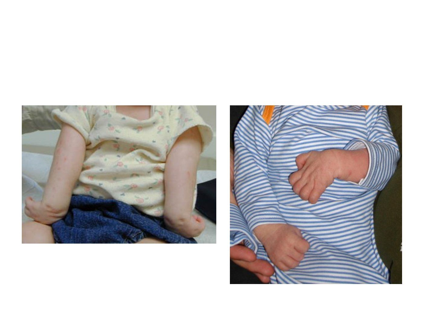
- What risk factors suggest obstetric brachial plexus palsy?
- What examinations and investigations are used in obstetric brachial plexus palsy?
- Describe the Narakas classification and prognosis.
- What is the Mallet score used for?
- What is the initial management of obstetric brachial plexus palsy?
- What are the indications for primary surgery (before 6 months) in obstetric brachial plexus palsy?
- What primary nerve surgery is used for obstetric brachial plexus palsy?
- What is the natural history of obstetric brachial plexus palsy?
- How is a shoulder posterior dislocation in obstetric brachial plexus palsy managed?
- What secondary procedures are used for shoulder internal rotation contracture?
Q1.What risk factors suggest obstetric brachial plexus palsy?
- High birth weight
- Forceps delivery
- Breech presentation
- Shoulder dystocia
Q2.What examinations and investigations are used in obstetric brachial plexus palsy?
- Examine to locate the injury; Moro or ATR reflex can be done
- XR, MRI
- NCT, EMG
- Toronto score (depends on ROM): <3.5 indicates surgery
Q3.Describe the Narakas classification and prognosis.
- I: C5-6 Erb - 90%
- II: C5-7 Erb + wrist drop - 60%
- III: C5-T1 total - 23%
- IV: total + Horner - <10%
Q4.What is the Mallet score used for?
- To monitor progress in obstetric brachial plexus palsy
- Assesses 5 major functions: abduction, ER, hand behind head, hand behind back, hand to mouth
Q5.What is the initial management of obstetric brachial plexus palsy?
- Maintain passive motion while the nerve recovers
- Observe for 3 months initially
Q6.What are the indications for primary surgery (before 6 months) in obstetric brachial plexus palsy?
- C8-T1 with Horner
- Total palsy with no biceps function by 3 months
- Toronto score <3.5
- EMG criteria by Smith; failure to progress after 1 year
- Erb's with no biceps by 3 months; shoulder abduction by 2 months
Q7.What primary nerve surgery is used for obstetric brachial plexus palsy?
- Nerve graft / nerve transfer
- Performed as primary surgery before 6 months
Q8.What is the natural history of obstetric brachial plexus palsy?
- >90% resolve
- Prognosis follows the Narakas classification
- Biceps absent at 3 months = poor prognosis
- Borderline case: explore the brachial plexus earlier for a better outcome
- Recovery usually up to 1 year
Q9.How is a shoulder posterior dislocation in obstetric brachial plexus palsy managed?
- Relocation + capsulorrhaphy
- Derotational osteotomy if older
Q10.What secondary procedures are used for shoulder internal rotation contracture?
- <2 years = soft tissue procedure (subscapularis release)
- >4 years (glenoid dysplasia) = bony procedure
- Lepiscopo procedure: teres major + latissimus dorsi transfer to GT
- Derotational osteotomy (Wickstrom procedure)
- Elbow supination contracture: flexible = biceps rerouting, rigid = osteotomy
▸ Slide 760 · Brachial NeuritisHand Soft Tissue · 10 questions 1 check expand

Q1-Q1010 questions — tap to reveal all answerslist
- What is the epidemiology of Parsonage-Turner syndrome?
- What are the causes and risk factors of brachial neuritis?
- Which nerves are most commonly affected in brachial neuritis?
- What is the proposed pathogenesis of idiopathic neuralgic amyotrophy?
- Describe the three phases of brachial neuritis.
- What are the physical examination features of brachial neuritis?
- What investigations are used in brachial neuritis?
- What is the differential diagnosis of brachial neuritis?
- What is the treatment and prognosis of brachial neuritis?
- What is the operative outcome in brachial neuritis?
Q1.What is the epidemiology of Parsonage-Turner syndrome?
- Uncommon: 1.6-3 cases per 100,000 persons per year
- Males, middle-age (4th decade; 20-60 most common, average 41)
- Usually unilateral; bilateral in 10-30% (16% simultaneously)
Q2.What are the causes and risk factors of brachial neuritis?
- Idiopathic (INA) or hereditary (HNA) neuralgic amyotrophy
- Multifactorial: autoimmune / genetic / infectious / environmental / biomechanical
- Viral infections 25-55% (EBV, VZV, Coxsackie B, parvovirus B19, CMV, mumps, smallpox, HIV); bacterial (Leptospira, TB, Yersinia, Salmonella, Borrelia)
- Immunizations 15% (tetanus, hepatitis B); perioperative and peripartum 14%; strenuous exercise 8%
- Drugs (abacavir, streptokinase, heroin, infliximab, interferon); iatrogenic (interscalene block, surgery, lumbar puncture, irradiation)
Q3.Which nerves are most commonly affected in brachial neuritis?
- Any nerve or branch within the brachial plexus; extra-plexus ~17%
- Most common: long thoracic, suprascapular, axillary, musculocutaneous, radial nerves
- Other: cervical roots, AIN, PIN, lateral antebrachial cutaneous nerve (LABC)
Q4.What is the proposed pathogenesis of idiopathic neuralgic amyotrophy?
- Autoimmune: increased blastogenic activity of lymphocytes and increased complement-fixing antibodies to peripheral nerve myelin
- Biomechanical: mobility of the upper trunk predisposes to wear-and-tear on the blood-nerve barrier
- Multifactorial: autoimmune / genetic / infectious / environmental / biomechanical
Q5.Describe the three phases of brachial neuritis.
- Phase I: sudden severe shoulder pain (90%), radiates to proximal arm/neck; lasts days to weeks
- Phase II: painless flaccid paralysis - 24 hours (33%) to 4 weeks (80%); patchy paresis is highly characteristic
- Phase III: slow recovery of motor function over 6-18 months; duration proportional to duration of pain phase
Q6.What are the physical examination features of brachial neuritis?
- Signs of denervation: fasciculations and atrophy
- Pain not affected by motion or palpation
- Severe weakness of shoulder, especially external rotation and abduction
- LMN signs: hypotonia and areflexia; medial scapular winging
- Sensory change in 78%; autonomic dysregulation in 15%
Q7.What investigations are used in brachial neuritis?
- Bloods: CBC and ESR usually normal, elevated liver enzymes; positive antiganglioside antibodies and ANA
- MRI: early T2 diffuse signal hyperintensity (edema) in affected muscles; late T1 fatty infiltration and atrophy
- EMG: early PSW/fibrillation; late (3-4 months) chronic denervation and early reinnervation with polyphasic MUAPs
- NCS: reduced amplitude with preserved conduction velocity and distal latency (less useful than EMG)
- CSF: usually normal, mildly elevated protein, slight pleocytosis and oligoclonal bands
Q8.What is the differential diagnosis of brachial neuritis?
- Cervical spine radiculopathy
- Rotator cuff pathology
- Entrapment neuropathy
- Idiopathic hypertrophic brachial neuritis (IHBN): painless upper limb weakness with brachial plexus hypertrophy
Q9.What is the treatment and prognosis of brachial neuritis?
- Nonoperative: observation and pain control (NSAIDs/physio); oral corticosteroids may hasten pain resolution but do not affect progression or prognosis
- Operative if no regeneration or early recovery by 6-9 months on exam and EMG: neurolysis, neurorrhaphy, grafting, nerve or tendon transfers
- Recurrence rare in non-hereditary cases; poor prognosis: female, lower trunk, persistent pain and no motor recovery by 3 months, hereditary cases; age has no effect
- Recovery: 66% show motor recovery within 1 month; excellent in 36% at 1 year, 75% at 2 years, 89% at 3 years; may take up to 8 years
Q10.What is the operative outcome in brachial neuritis?
- Exploration of patients without neurologic recovery reveals hourglass-like constrictions in peripheral nerves with no external compression
- Neurolysis alone was superior to neurorrhaphy and nerve grafting
66% have recovery of motor function within 1 month in Parsonage-Turner syndrome — misleading/unsupported — secondary sources cite 66% early partial or any recovery at 1 month; validated functional recovery is 36% at 1 year, 75% at 2 years and 89% at 3 years — medium confidence — source
▸ Slide 761 · Deep space infectionHand Soft Tissue · 12 questions 1 check expand
Q1-Q1212 questions — tap to reveal all answerslist
- How are felon and paronychia managed?
- What is a collar button infection and how is it treated?
- What is the anatomy of the webspace (volar to dorsal)?
- What are Kanavel's four cardinal signs of flexor tenosynovitis?
- What are the systemic features and initial treatment of flexor tenosynovitis?
- How is flexor tenosynovitis drained and what is the Michon grading?
- What are the routes of spread of flexor tenosynovitis?
- Describe the radial and ulnar bursae and their drainage.
- What are the boundaries and management of thenar and midpalmar space infections?
- What are the spread patterns of a thenar space infection?
- What are the spread patterns and risks of a midpalmar space infection?
- What are the boundaries and management of a hypothenar space infection?
Q1.How are felon and paronychia managed?
- Felon: multiple septal compartments; midaxial incision and disrupt all septation
- Paronychia: infection beneath the eponychial fold; needs elevation of the eponychium
Q2.What is a collar button infection and how is it treated?
- Webspace infection; clinically the finger is abducted
- 2 cavities connected by a narrow isthmus
- Dorsal spread through the deep transverse MC ligament causes digital artery thrombosis / numbness; proximal spread via lumbrical canal to midpalmar space
- Rx: both dorsal (longitudinal) and volar (zig-zag) incisions with no connection (avoid neurovascular structures)
Q3.What is the anatomy of the webspace (volar to dorsal)?
- 1. Natatory ligament (superficial transverse metacarpal ligament)
- 2. Neurovascular bundle
- 3. Lumbrical
- 4. Deep transverse metacarpal ligament
- 5. Dorsal interossei; laterally bounded by the MCPJ capsule
Q4.What are Kanavel's four cardinal signs of flexor tenosynovitis?
- Finger held in a flexed position
- Fusiform, sausage-like swelling
- Tenderness along the flexor sheath
- Pain with passive extension of the finger
- Classically quoted: all four positive is >90% sensitive
Q5.What are the systemic features and initial treatment of flexor tenosynovitis?
- Systemic: only 17% have fever
- Splinting, IV antibiotics and close observation for 24 hours
- Operative treatment if failed
Q6.How is flexor tenosynovitis drained and what is the Michon grading?
- Close sheath irrigation (proximal to A1 and distal to A4) or open drainage (midaxial scar)
- Open if Michon II or III; Brunner or midaxial incision
- Michon: 1 = serous, 2 = pus, 3 = necrosis of tendon/pulley
Q7.What are the routes of spread of flexor tenosynovitis?
- Thumb --> radial bursa
- Index, middle and ring --> midpalmar space
- Little --> ulnar bursa
- Radial/ulnar bursa --> horseshoe abscess / Parona's space (between PQ and FDP)
- Thenar --> hypothenar (through radial bursa, Parona's space, ulnar bursa)
Q8.Describe the radial and ulnar bursae and their drainage.
- Ulnar bursa: MCPJ, widening 3rd and 4th MC (50% contiguous with FDP and FDS of little finger)
- Radial bursa: MCPJ to proximal edge of TCL (95% contiguous with FPL sheath)
- Rx: close irrigation 2-incision approach
- Radial: incision adjacent to flexor crease of thumb MCPJ + ulnar to FCR
- Ulnar: incision at A1 pulley and radial to FCU
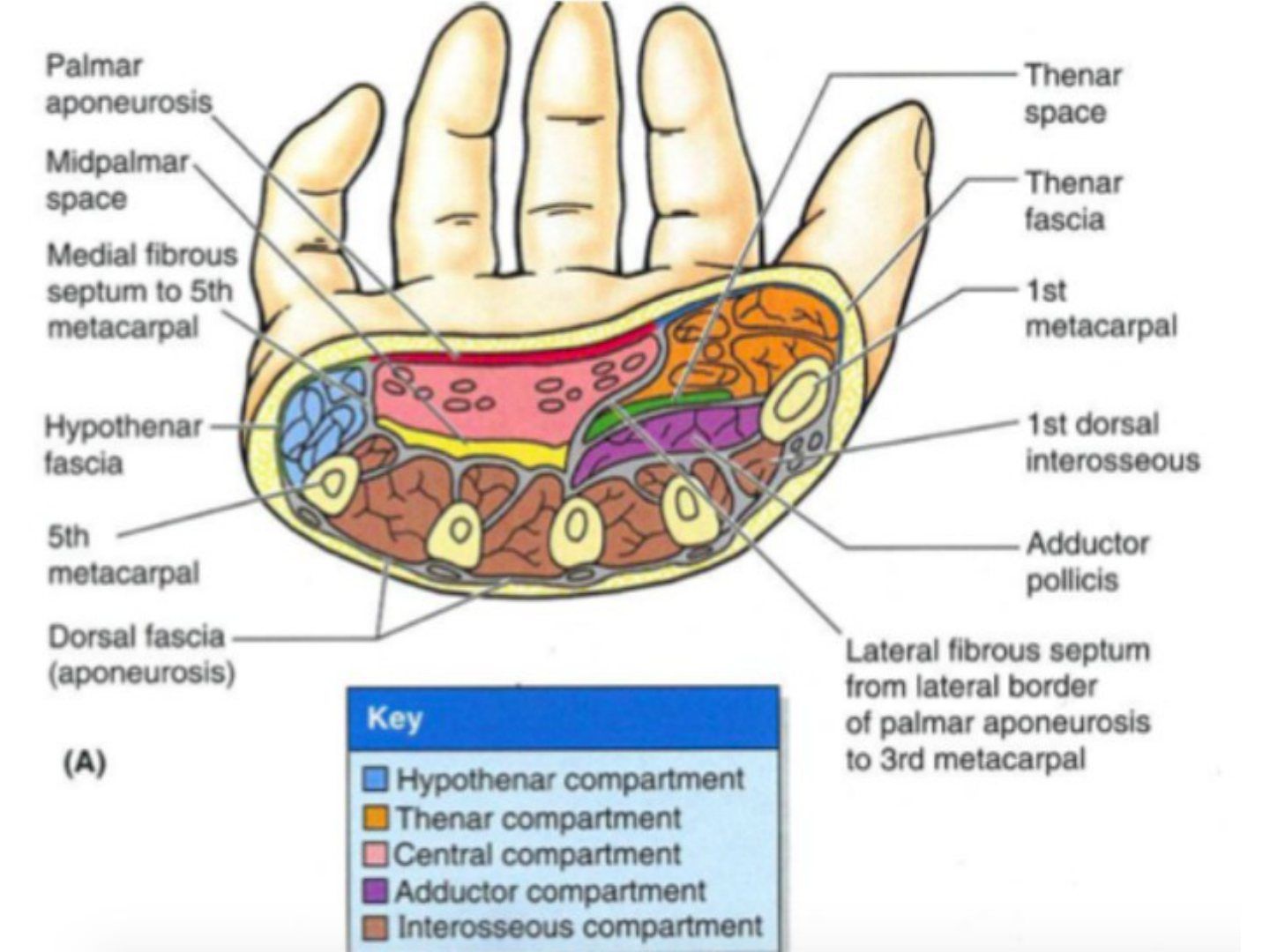
Q9.What are the boundaries and management of thenar and midpalmar space infections?
- Thenar space (most common): thenar muscles (superficial), adductor pollicis (deep), 1st MC (radial), midpalmar septum (ulnar)
- Rx thenar: volar incision along thenar crease (stay 1.5 cm from web) +/- dorsal between 1st and 2nd MC; risk to recurrent motor branch, superficial palmar arch, 1st/2nd digital NV
- Midpalmar space: MF-LF flexors (superficial), interossei and 3-5 MC (deep), midpalmar septum (radial), hypothenar septum (ulnar)
- Rx midpalmar: L-shaped incision (transverse distally then along radial border of hypothenar eminence)
Q10.What are the spread patterns of a thenar space infection?
- Can spread to the 1st web as a dumbbell infection
- Palpate along FPL as it may be associated with FPL tenosynovitis
- Dorsal extension through adductor pollicis and 1st DI
- Can spread to the FPL/radial bursa
Q11.What are the spread patterns and risks of a midpalmar space infection?
- Communicates with the flexor sheath
- Proximal extension to Parona's space; diffuse spread from Parona's space to radial/ulnar bursae, hypothenar and thenar spaces
- Can cause median nerve compromise
Q12.What are the boundaries and management of a hypothenar space infection?
- Rare
- Boundaries: palmar fascia and superficial hypothenar muscle fascia (superficial), 5th MC and deep hypothenar muscle fascia (deep), hypothenar septum (radial)
- Rx: incision along the radial border of the hypothenar eminence
All four Kanavel signs positive gives >90% sensitivity for flexor tenosynovitis — conflates individual sign sensitivity with all four; misleading — individual Kanavel signs have 91-97% sensitivity but poor specificity (51-69%); all four are present in only a minority and are specific rather than >90% sensitive — source
▸ Slide 762 · Septic arthritis approachHand Soft Tissue · 5 questions expand
Q1-Q55 questions — tap to reveal all answerslist
- How is wrist septic arthritis drained?
- What are the advantages and disadvantages of arthroscopic wrist drainage?
- How is MCPJ septic arthritis drained?
- How is PIPJ septic arthritis drained?
- How is DIPJ septic arthritis drained?
Q1.How is wrist septic arthritis drained?
- Longitudinal incision between the 3rd and 4th compartments (gold standard)
- Arthroscopic: 2/3 + 4/5 portals (viewing + working), 6U/6R for water out
Q2.What are the advantages and disadvantages of arthroscopic wrist drainage?
- Advantages: small wound, preserves the dorsal ligament
- Disadvantages: incomplete lavage, time-consuming
Q3.How is MCPJ septic arthritis drained?
- Dorsal approach, adjacent to the extensor tendon
- Split the sagittal band
Q4.How is PIPJ septic arthritis drained?
- Dorsal: between the central slip and lateral band
- Mid-axial: through accessory collateral
Q5.How is DIPJ septic arthritis drained?
- Dorsal: adjacent to the terminal extensor
- Mid-axial: through accessory collateral
▸ Slide 763 · Clinical photo of patients handHand Soft Tissue · 4 questions expand

Q1-Q44 questions — tap to reveal all answerslist
- What does the clinical photo show?
- Define a high-pressure injection injury and describe its pathogenesis.
- What history and examination findings are important in an injection injury?
- What is the management and outcome of a high-pressure injection injury?
Q1.What does the clinical photo show?
- Wounds over the thumb, index and middle fingers with mottling
- Another open wound over the palm --> possibility of an exit site
Q2.Define a high-pressure injection injury and describe its pathogenesis.
- Puncture of the epidermis by a jet of fluid/air under pressure
- Usually the index finger of the non-dominant hand
- Pressurised fluid passes through skin; kinetic energy dissipates in hand soft tissue
- Microscopic destruction --> vascular occlusion; skin degloving + soft tissue necrosis
Q3.What history and examination findings are important in an injection injury?
- Mechanism of injury; nature of fluid (oil or water based), volume injected, force of injection
- Time of injury; age, premorbid status, dominance; patient expectations
- PE: compartments, NV status
Q4.What is the management and outcome of a high-pressure injection injury?
- Debridement (non-viable skin, subcutaneous fat, fascia, muscle/tendon, bone; Debride til punctate bleeding; may need multiple surgeries)
- Compartment release
- Broad spectrum antibiotics
- Outcome: increase amputation if attend >10 hrs post injury
▸ Slide 764 · Clinical photo showing patients left hand with multiple finger amputation at leHand Soft Tissue · 11 questions expand

Q1-Q1111 questions — tap to reveal all answerslist
- What are the clinical photo findings and injury characteristics?
- How do you initially manage an amputation and preserve the amputated part?
- What factors determine whether to replant?
- What are the contraindications to replantation?
- What is the operation setup and order of replantation?
- Describe the sequence and technique of replantation.
- How do you induce flow intra-operatively during replantation?
- How do you monitor a replant and distinguish arterial from venous failure?
- What are the late complications and functional results of replantation?
- How does management differ for proximal amputations and ring avulsion injuries?
- How is the nerve managed in replantation?
Q1.What are the clinical photo findings and injury characteristics?
- Left hand with multiple finger amputations at the level of the middle phalanx, amputated parts retrieved
- Distal amputation = distal to FDS insertion
- Likely a clean cut injury with no gross contamination
- Distally I do not see any ribbon sign or red line sign signifying an avlusion type injury (i.e. no ribbon sign or red line sign, which signify an avulsion injury)
Q2.How do you initially manage an amputation and preserve the amputated part?
- ATLS; AMPLE history including smoking, hand dominance, job requirements and how the stump was preserved
- Rule out other injuries, stabilise patient, good storage of amputated part, Clinical photos: moist dressing to the proximal stump, IV antibiotics, tetanus
- Part: remove contamination, wrap in moist gauze (NS or lactated Ringer), place in sealed plastic bag, then bag in ice water bath
Q3.What factors determine whether to replant?
- Injury: mechanism, level, timing
- Patient: age, smoker, comorbidities, hand dominance, demand
- Amputated part: storage
- Indications: child, above wrist, multiple fingers, thumb; relative indication: zone 1
Q4.What are the contraindications to replantation?
- Crush injury/mangled, severe contamination, segmental injury
- Warm ischaemic time limits: finger 12/24, wrist 6/12
- Single digit especially index finger, zone 2
- Polytrauma / poor premorbid status
Q5.What is the operation setup and order of replantation?
- Warm OT room, GA, 2 teams
- Order: thumb, long, ring, little, index (1,3,4,5,2)
Q6.Describe the sequence and technique of replantation.
- Sequence: BE FAN V (bone, extensor, flexor, artery, nerve, vein) then skin; allow 1 cm shortening
- Artery first: earlier revascularisation and decrease cold ischemic time, washes out metabolic waste, Easier to identify and repair vein (not collapsed); downside = more blood loss
- Technique: remove adventitia to avoid unfolding into lumen, remove clot, cut sharp, heparin saline injection, Approximation of intima with 10/0 nylon interrupted
Q7.How do you induce flow intra-operatively during replantation?
- Systemic: warmth, blood pressure, dependent position, correct metabolic abnormalities
- Local: no tourniquet, relieve tension/pressure, resect to normal, flush lumen (heparin saline 10U in 1ml), papaverine
Q8.How do you monitor a replant and distinguish arterial from venous failure?
- Warm environment (26 degrees); hydration (hypervolemia), pain relief, anxiolytic (Thorazine), aspirin; NO smoking, caffeine, vasoconstrictive agents
- Monitor 5/7 days: colour, CR, turgor; skin temp (drop 2C in 1 hr / <30 degrees) is most reliable; pulse oximeter
- <12 hours = usually arterial (persistent vasospasm, pale): remove bandage, dependent position, heparin 3000-5000U, stellate ganglion block, re-explore in 6 hrs, needle prick on nail
- >12 hours = venous congestion/thrombosis (ruborous): remove bandage, elevate, heparin soaked gauze, medicinal leech (hirudin 8-12 hrs, need antibiotics), reoperate
- Always assess environment and patient first when a replant fails
Q9.What are the late complications and functional results of replantation?
- Late complications: stiffness (tenolysis) 50%, bone and nerve problems
- Results: universal cold intolerance, usually 50% function, 10mm 2PD
- Factors most predictive of digit survival: mechanism of injury and ischemia time
Q10.How does management differ for proximal amputations and ring avulsion injuries?
- Proximal: debridement + fasciotomy, arterial shunt, BAVNM with shortening (digit <1cm, hand 1.5cm, forearm 2-4cm); fixation with K-wire or ex-fix
- Post-op: compartment syndrome, reperfusion --> renal failure
- Ring avulsion (Urbaniak): I adequate circulation = soft tissue repair; II inadequate circulation = revascularisation if bone/tendon/nerve intact; III complete degloving/amputation = amputation
- Index finger can be considered for ray amputation at the MC neck level
Q11.How is the nerve managed in replantation?
- Aim: protective sensation (<15mm 2PD)
- Median and ulnar nerve gap <2cm: nerve/ vein conduit
- Gap >2cm: delayed nerve graft, or tendon/nerve transfer for a motor nerve
- Donor nerve: sural, MACN
▸ Slide 765 · Clinical photo showing left hand with finger tip injury with tissue loss over I/Hand Soft Tissue · 10 questions expand

Q1-Q1010 questions — tap to reveal all answerslist
- How do you assess a fingertip injury?
- What history and examination are needed for a fingertip injury?
- What are the goals and reconstruction ladder for fingertip injuries?
- What factors guide flap design?
- What is the reconstruction ladder and how are flaps classified?
- What are the prerequisites and indications for island flaps in fingertip reconstruction?
- What are the options for volar fingertip tissue loss?
- What are the options for dorsal/transverse and thumb tip tissue loss?
- What are the options for proximal finger tissue loss?
- What are the complications of fingertip reconstruction?
Q1.How do you assess a fingertip injury?
- Is bone exposed?
- Pattern/orientation (e.g. volar oblique); flexor or extensor involvement; contamination
- Allen's classification: distal to nailbed, distal to distal phalanx, distal to lunula, distal to DIPJ
- Also Tamai and Ishikawa classifications
Q2.What history and examination are needed for a fingertip injury?
- History: mechanism (avulsion, crush, sharp laceration), premorbid status, smoking, hand dominance, expectation and rehab motivation
- PE: check flexor and extensor function
- Digital Allen test
Q3.What are the goals and reconstruction ladder for fingertip injuries?
- Goals: function (sensate, durable tip, bone support for nail growth), Appearance: cosmesis, Time: early return to work, minimise hospital stay
- Consider wound factors (pattern, level, size), patient factors, surgeon expertise
- Follow reconstruction ladder: heal by secondary intention (3-5 weeks)
- Reamputation
- Reconstruction (graft, local, regional, distant pedicle, distant free)
Q4.What factors guide flap design?
- Donor site
- Texture / colour / volume
- Hairiness
- Sensation
Q5.What is the reconstruction ladder and how are flaps classified?
- Ladder: secondary healing -> primary closure -> skin graft (partial/full) -> local flap -> regional flap -> free flap
- By location: local, distant, free
- By blood supply: random, axial
- By composition: cutaneous, fasciocutaneous, myocutaneous, osteocutaneous
Q6.What are the prerequisites and indications for island flaps in fingertip reconstruction?
- Prerequisite: dual digital artery supply
- Indications: volar PP to DP large defect; heterodigital for a 2-4 cm thumb volar oblique defect
- Homodigital and heterodigital flaps are sensate; reverse homodigital is insensate; the heterodigital flap is from the Non opposing side of other finger
- Advantages: best axial pattern flap, single stage; disadvantages: technically difficult, loss of donor sensation
Q7.What are the options for volar fingertip tissue loss?
- Same finger: homodigital flap (Joshi) - local pedicle flap based on one digital NV bundle; reverse homodigital (insensate)
- Different finger: cross finger (donor adjacent dorsal PP/MP, needs FTSG; insensate, contracture, skin mismatch) and heterodigital
- Same hand: thenar flap (H-shaped incision, <30 yo, IF to RF; adv skin match/good fat pad; disadv PIPJ contracture, donor hyperaesthesia, risk thumb digital NV)
- Distant: pec major type 5 flap (pectoral branch of thoracoacromial artery); Free: pulp transfer
Q8.What are the options for dorsal/transverse and thumb tip tissue loss?
- Dorsal and transverse: volar VY / lateral VY flap (Kutler flap = bilateral VY); digital island flap if big
- Thumb dorsal: FDMA (Kite/Foucher) flap
- Thumb volar: Moberg advancement flap (defect <2cm, IPJ slightly flexed), heterodigital flap (ulnar side of M/F or R/F), FDMA flap
- Moberg: skin match and sensate; disadvantage is flexion contracture. Kite: skin mismatch for volar defect
- Moberg is only used in the thumb because of its isolated dorsal blood supply
Q9.What are the options for proximal finger tissue loss?
- Dorsal: reverse crossfinger flap (only deep dermis and subcutaneous layer transferred)
- Proximal: axial flag flap (adjacent PP), cross finger flap, homodigital arterial flap, reverse homodigital arterial flap
Q10.What are the complications of fingertip reconstruction?
- Insensate tip
- Hyperaesthesia
- Cold intolerance
- Hook nail
- DIPJ stiffness
▸ Slide 766 · Finger replantationHand Soft Tissue · 4 questions expand

Q1-Q44 questions — tap to reveal all answerslist
- What factors are assessed when considering replantation in a zone 2 injury?
- What are the treatment options for an exposed proximal phalanx?
- What are the pros and cons of replantation in a zone 2 PIPJ injury?
- What are the advantages of ray amputation?
Q1.What factors are assessed when considering replantation in a zone 2 injury?
- Patient: hand dominance, smoker, functional status, medical comorbidity
- Finger: timing of injury, injury mechanism, site (which finger and level)
- Injury is an avulsion with avulsed flexor and extensor tendons, avulsed digital artery/nerve and exposed proximal phalanx
Q2.What are the treatment options for an exposed proximal phalanx?
- Local flap to cover bone
- Ray amputation
- Replantation
Q3.What are the pros and cons of replantation in a zone 2 PIPJ injury?
- Pros: maintain length, hopefully function
- Cons: prolongs surgery (higher chance of infection), failure of revascularisation, may need multiple surgeries/2nd reamputation
- Prolonged rehab and stiffness; tendon adhesion (may affect other finger motion as FDP share the same belly)
Q4.What are the advantages of ray amputation?
- Quick operation, quick rehab
- Less stump impingement
- Cosmesis ok for ray amputation
▸ Slide 767 · Another option: jumping man flapHand Soft Tissue · 3 questions expand
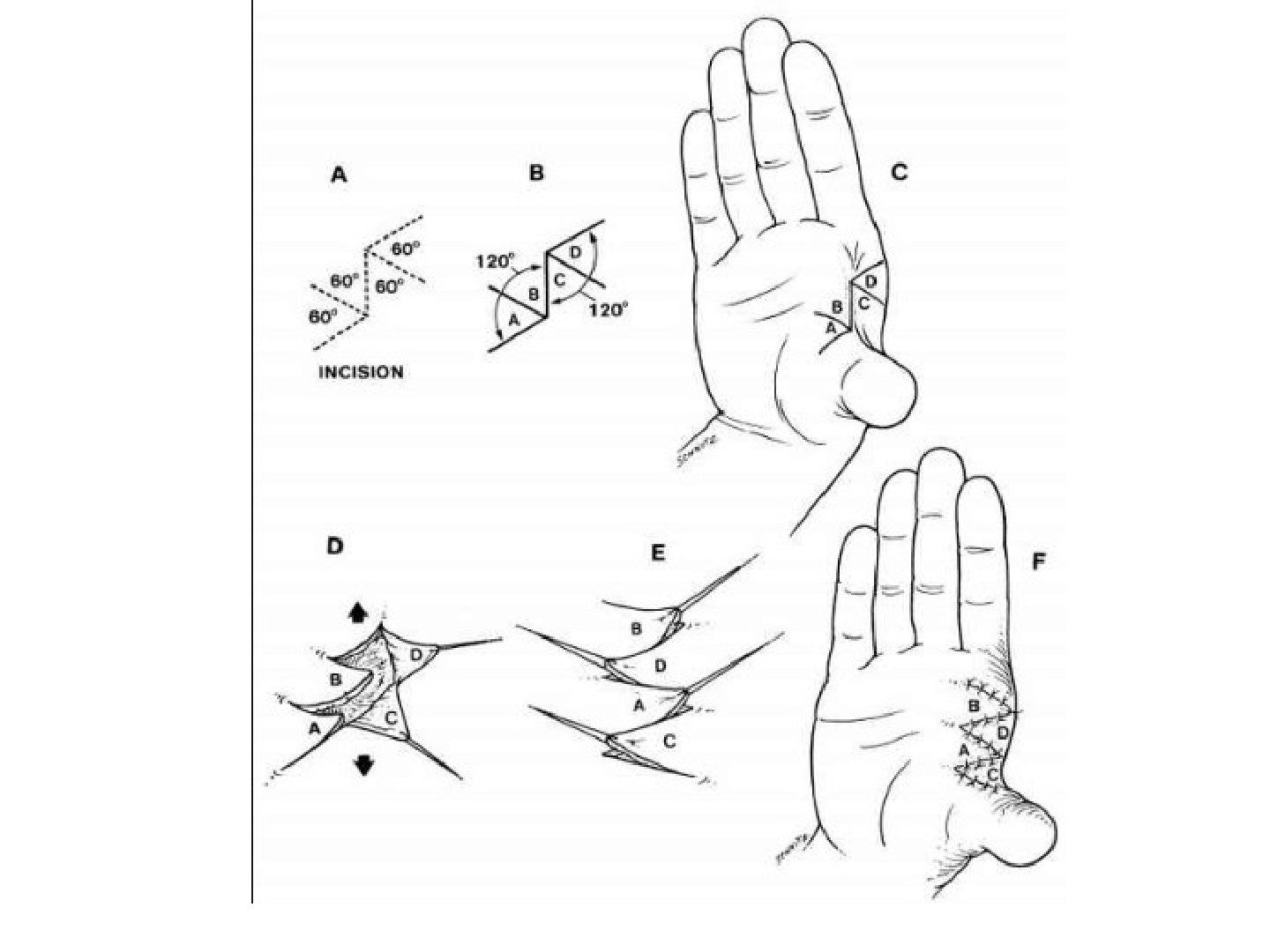
Q1-Q33 questions — tap to reveal all answerslist
- What is a jumping man flap used for?
- What is a Z-plasty?
- How much length can a Z-plasty gain and what are the options for long contractures?
Q1.What is a jumping man flap used for?
- Planned skin incision for release of a 1st web contracture
- Triangular shaped skip flaps are transposed to increase length along the first web
Q2.What is a Z-plasty?
- A transpositional flap using adjacent tissues near the incision to release a contracture
- Central limb is planned along the contracture, with two oblique wounds at 60 degrees to the main wound, same length and parallel
Q3.How much length can a Z-plasty gain and what are the options for long contractures?
- Theoretical gain in length along the contracture = 1.73 (practically 1.5)
- Options: one large Z-plasty or multiple small Z-plasties
- Pros of multiple Z plasty is that the ‘width’ reduction is reduced and stress is less concentrated, but more technically demanding especially tip necrosis
▸ Slide 768 · Describe clinical photoHand Soft Tissue · 4 questions expand
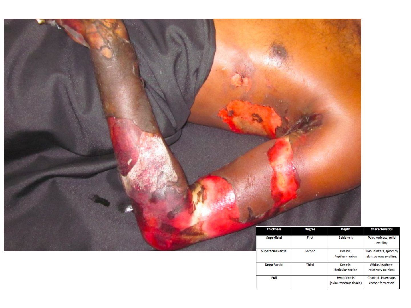
Q1-Q44 questions — tap to reveal all answerslist
- Describe the burn in the photo.
- What is the systemic management of a burn?
- What is the local management of a burn?
- How does PTSG compare with FTSG?
Q1.Describe the burn in the photo.
- Burn over the elbow extending to mid arm and forearm
- Assess whether it is circumferential
- Likely third degree (1st erythema, 2nd blister, 3rd waxy with no sensation)
Q2.What is the systemic management of a burn?
- Rehydration
- Analgesics
- Prevent infection (antibiotics)
- Close monitoring
Q3.What is the local management of a burn?
- Acute: consider escharotomy if circumferential --> emergency OT; dressing to prevent infection
- Definitive: coverage, prefer PTSG
Q4.How does PTSG compare with FTSG?
- PTSG: can obtain a large graft, easier to take
- PTSG: more fragile, more contracture than FTSG
▸ Slide 769 · Burn caseHand Soft Tissue · 3 questions expand

Q1-Q33 questions — tap to reveal all answerslist
- What do you look for when describing a burn?
- What is the emergency concern with a circumferential burn and how is it managed?
- How is this burn definitively covered and what is the main limitation of the graft?
Q1.What do you look for when describing a burn?
- Area of burn
- Degree of burn
- Is it circumferential?
Q2.What is the emergency concern with a circumferential burn and how is it managed?
- Circumferential eschar can cause compartment syndrome
- Requires emergency escharotomy
Q3.How is this burn definitively covered and what is the main limitation of the graft?
- Debride and achieve coverage with PTSG (pros: large size, easy to take)
- Con: texture
- NO SENSATION in FTSG/PTSG
▸ Slide 770 · Describe:Hand Soft Tissue · 10 questions expand

Q1-Q1010 questions — tap to reveal all answerslist
- Define a flap.
- Describe the reconstructive ladder for a hand soft tissue defect and the rationale for each step.
- What are the indications for a flap?
- What systemic and local factors are optimised before flap surgery?
- What equipment is prepared before flap surgery?
- How do you classify flaps?
- What are the four principles of the perforasome theory?
- What are the key steps in flap elevation and insetting?
- Why is a radial forearm flap used in this case and what are its pros and cons?
- What are the key postoperative measures after flap surgery?
Q1.Define a flap.
- A unit of tissue transfer from donor to recipient site
- It maintains its own vascular supply
Q2.Describe the reconstructive ladder for a hand soft tissue defect and the rationale for each step.
- Secondary intention: slow, and tendon may be exposed; may suit traumatic wounds as more scarring means less finger drop
- Primary or delayed primary closure: only if the defect is not too large
- Skin graft: not possible when tendon is exposed
- Flap when critical structures are exposed
Q3.What are the indications for a flap?
- Tissue loss with exposed bone (no periosteum)
- Exposed tendon (no paratenon)
- Exposed cartilage
- Exposed implant
Q4.What systemic and local factors are optimised before flap surgery?
- Systemic: smoking (PVD), immunocompromise (delays healing), nutrition and medical optimisation (long GA)
- Local design: point of pivot, axis of rotation, size of plane
- Composition and pedicle: fasciocutaneous or myocutaneous
- Donor site: scar, previous irradiation; recipient site: ensure no infection
Q5.What equipment is prepared before flap surgery?
- Prepare equipment: Microsurgery instruments and microscope
- Hand-held doppler
- Heparin and vein dilator
Q6.How do you classify flaps?
- By composition: cutaneous, fasciocutaneous, myocutaneous, osteocutaneous
- By blood supply: axial, random (length:width ratio 1:1)
- By location: local (advancement, transposition, rotation), regional (radial forearm), distant pedicle (groin), distant free (LD)
- Mathes classification applies to fasciocutaneous and myocutaneous flaps
Q7.What are the four principles of the perforasome theory?
- Each perforasome links to adjacent ones by direct linking vessels (suprafascial/adipose) and indirect subdermal plexus (protective if one is damaged)
- Flap orientation follows direct linking vessels: axial in extremities, perpendicular to trunk midline
- Preferential filling: same source artery first, then adjacent source arteries (e.g. ALT from LFCA then SFA/MCFA)
- Mass vascularity: perforator found adjacent to an articulation is directed away from that same articulation; perforators at a midpoint between two articulations have multidirectional flow (propeller flap)
Q8.What are the key steps in flap elevation and insetting?
- Elevation: preserve perforators; pedicle skeletonisation NOT required if local flap; tension free
- Insetting: tunnel size = flap base x 2
- Tension-free rotation and careful haemostasis
Q9.Why is a radial forearm flap used in this case and what are its pros and cons?
- Based on the radial artery, with retrograde flow from the ulnar artery
- Preop Allen test is done
- Advantages: good skin and hairless
- Disadvantage: single vascular supply to hand
Q10.What are the key postoperative measures after flap surgery?
- Warm room
- Pressure free, no constrictive bandage
- Splint to immobilise
- Give heparin
▸ Slide 771 · Summary of flapsHand Soft Tissue · 6 questions expand
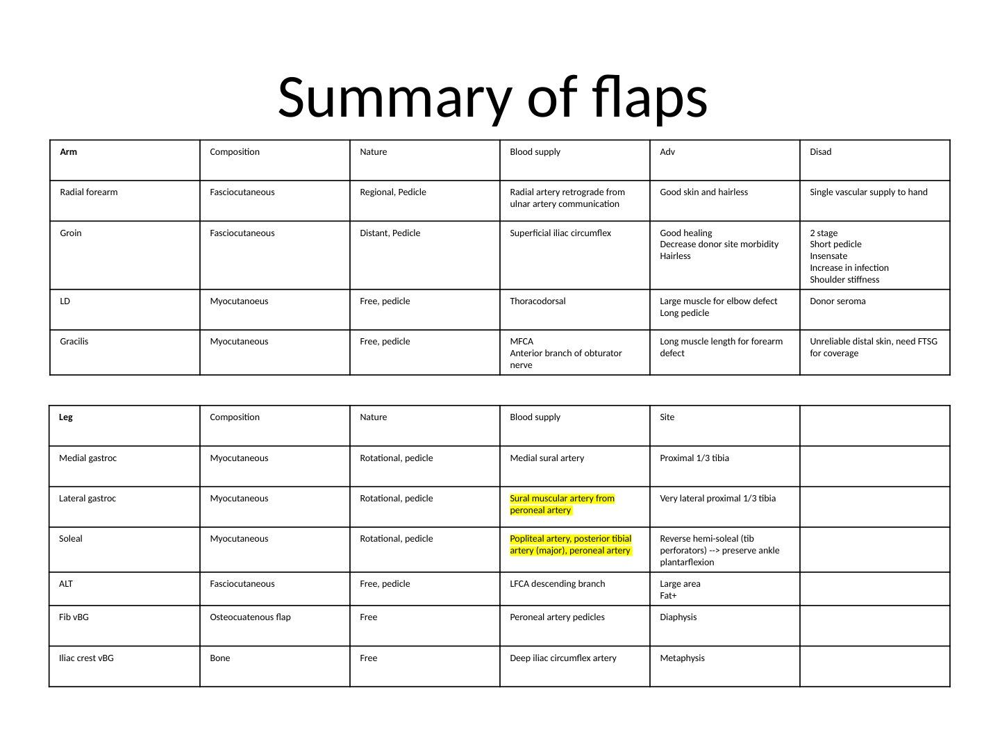
Q1-Q66 questions — tap to reveal all answerslist
- What is the note attached to this summary of flaps?
- Radial forearm flap: composition, nature, blood supply, advantages and disadvantages?
- Groin flap: composition, nature, blood supply, advantages and disadvantages?
- LD and gracilis flaps: composition, nature, blood supply, advantages and disadvantages?
- Leg muscle flaps (medial gastroc, lateral gastroc, soleal): composition, nature, blood supply and site?
- ALT, fibular vBG and iliac crest vBG flaps: composition, nature and blood supply?
Q1.What is the note attached to this summary of flaps?
- Sartorius (notes spell 'Sartoris') is paired with the superficial femoral vessel
- Recall flaps are classified by composition, blood supply and location
Q2.Radial forearm flap: composition, nature, blood supply, advantages and disadvantages?
- Composition: fasciocutaneous
- Nature: regional, pedicle flap
- Blood supply: radial artery retrograde from ulnar artery communication
- Advantages: good skin and hairless
- Disadvantage: single vascular supply to hand
Q3.Groin flap: composition, nature, blood supply, advantages and disadvantages?
- Composition: fasciocutaneous
- Nature: distant, pedicle flap
- Blood supply: superficial iliac circumflex
- Advantages: good healing, decrease donor site morbidity, hairless
- Disadvantages: 2 stage, short pedicle, insensate, increase in infection, shoulder stiffness
Q4.LD and gracilis flaps: composition, nature, blood supply, advantages and disadvantages?
- LD: myocutaneous (printed 'myocutanoeus'), free and pedicle; blood supply thoracodorsal; advantages large muscle for elbow defect, long pedicle; disadvantage donor seroma
- Gracilis: myocutaneous, free and pedicle; blood supply MFCA, nerve anterior branch of obturator nerve; advantage long muscle length for forearm defect; disadvantage unreliable distal skin, need FTSG for coverage
Q5.Leg muscle flaps (medial gastroc, lateral gastroc, soleal): composition, nature, blood supply and site?
- All are myocutaneous, rotational, pedicle flaps
- Medial gastroc: medial sural artery; site proximal 1/3 tibia
- Lateral gastroc: sural muscular artery from peroneal artery; site very lateral proximal 1/3 tibia
- Soleal: popliteal artery, posterior tibial artery (major), peroneal artery; reverse hemi-soleal (tib perforators) preserves ankle plantarflexion
Q6.ALT, fibular vBG and iliac crest vBG flaps: composition, nature and blood supply?
- ALT: fasciocutaneous, free and pedicle; LFCA descending branch; large area, fat+
- Fib vBG: osteocutaneous flap, free; peroneal artery pedicles; used for diaphysis
- Iliac crest vBG: bone, free; deep iliac circumflex artery; used for metaphysis
▸ Slide 772 · Describe clinical photo (Hx of restrain)Hand Soft Tissue · 3 questions expand
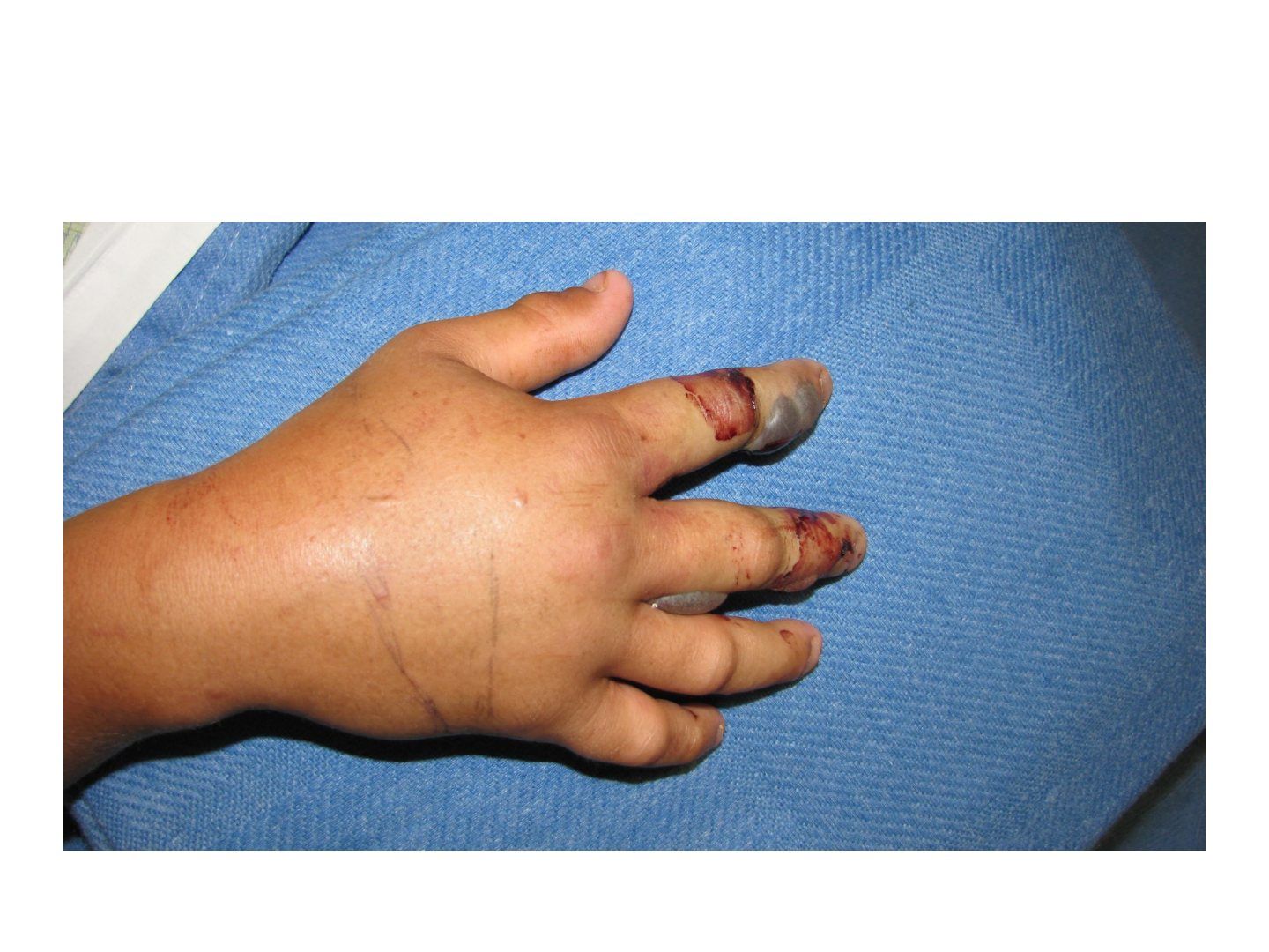
Q1-Q33 questions — tap to reveal all answerslist
- Describe the clinical photo and the likely diagnosis after a history of restraint.
- How many compartments are in the hand and how are they released?
- How is the forearm released in compartment syndrome?
Q1.Describe the clinical photo and the likely diagnosis after a history of restraint.
- Swollen hand with tense palm and fingertip
- Consistent with compartment syndrome of the hand
- Requires urgent assessment and release
Q2.How many compartments are in the hand and how are they released?
- 7 interossei released through 2 dorsal incisions
- Thenar and adductor released through 1 incision similar to a Wagner approach
- Hypothenar released through 1 incision at the ulnar palmar-dorsal skin junction
Q3.How is the forearm released in compartment syndrome?
- Release the carpal tunnel, volar compartment, mobile wad and lacertus fibrosus
- Carpal tunnel incision with a large radially based flap at the distal 1/3 forearm covering median nerve and tendons
- Curve radially to release the mobile wad, then back ulnar when crossing the elbow to release lacertus fibrosus
▸ Slide 773 · Describe photo:Hand Soft Tissue · 7 questions expand
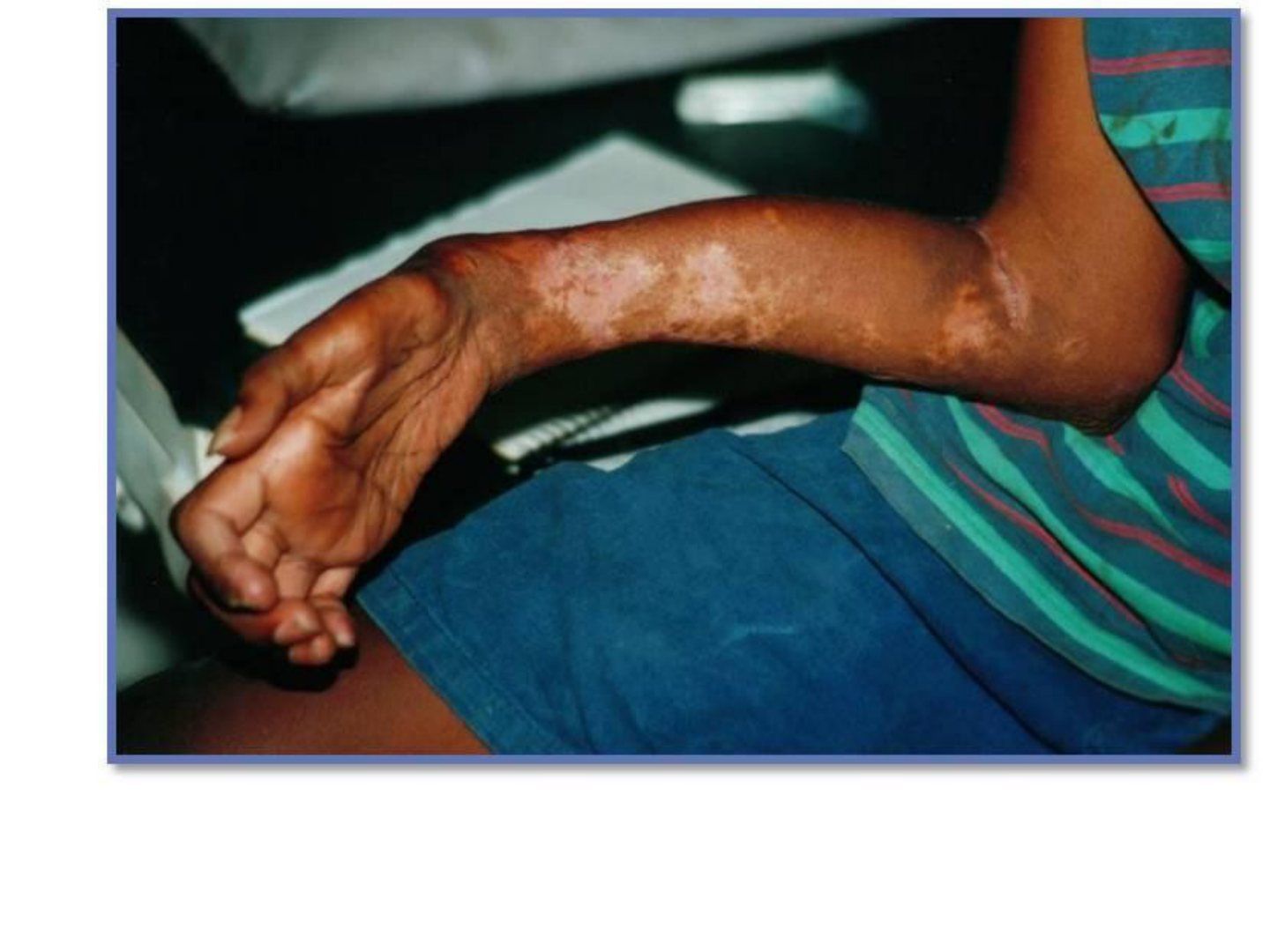
Q1-Q77 questions — tap to reveal all answerslist
- Describe the clinical photo and give the likely diagnosis with differentials.
- What is Volkmann's contracture?
- What history is important in Volkmann's contracture?
- What is the Tsuge classification of Volkmann's contracture severity?
- What are the treatment principles for Volkmann's contracture?
- What is syringomyelia and how does it present?
- How is syringomyelia treated?
Q1.Describe the clinical photo and give the likely diagnosis with differentials.
- Unilateral involvement with a possible surgical scar
- Gross wasting of the right upper limb over hand and forearm
- Elbow flexed, forearm pronated, wrist flexed, thumb adducted, intrinsic minus hand
- Impression: Volkmann's contracture; DDx nerve (polio), spine (syringomyelia), systemic (CMT if bilateral)
Q2.What is Volkmann's contracture?
- Contracture of the forearm, wrist and hand
- Results from muscle necrosis
Q3.What history is important in Volkmann's contracture?
- Previous injury and treatment
- Hand function
Q4.What is the Tsuge classification of Volkmann's contracture severity?
- Mild: finger flexor involvement
- Moderate: finger and wrist flexors
- Severe: finger and wrist flexors plus extensors
Q5.What are the treatment principles for Volkmann's contracture?
- Depends on hand function and functional demands
- r/o infection if previously performed ORIF
- Physiotherapy for stretching and dynamic splinting
- Tendon lengthening/transfer (for FPL/FDP)
Q6.What is syringomyelia and how does it present?
- A syrinx (fluid-filled cavity) within the spinal cord that progressively expands and causes neurological deficits
- Secondary to tumour, trauma or previous infection (meningitis)
- Presentation: scoliosis (25-80%), pain and temperature affected first (decussation), anterior horn involvement gives a lower motor neuron syndrome, cranial nerves if it extends proximally
- Charcot joint, often at the shoulder
Q7.How is syringomyelia treated?
- Observe
- Operate if severe neurological deficit (motor weakness, sensory deficit, cerebellar or cranial nerve signs)
- Operate if the curve is expected to progress (large curve, young onset)
- Decompression then +/- fusion
▸ Slide 774 · Ganglion and mucoid cystHand Soft Tissue · 5 questions expand
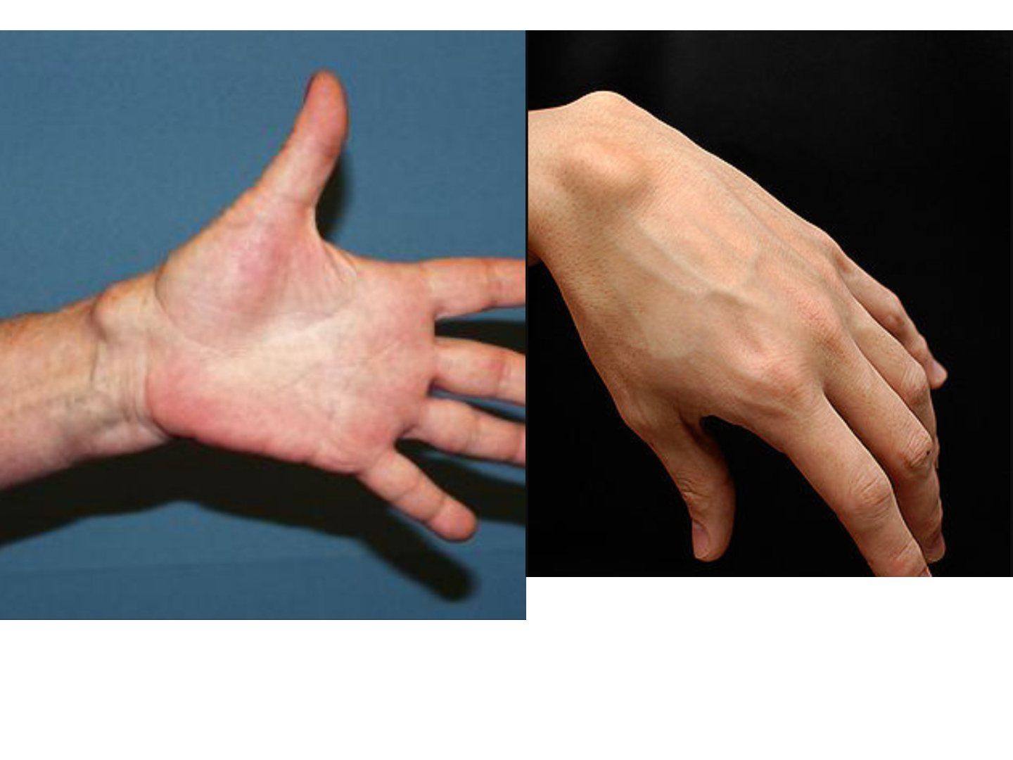
Q1-Q55 questions — tap to reveal all answerslist
- How do you assess a dorsal radial or volar radial wrist mass?
- What is the pathology and origin of a ganglion cyst?
- What is the prevalence and non-operative treatment of ganglion cysts?
- How is a dorsal or volar wrist ganglion resected and what is the recurrence?
- What is a mucoid cyst and how is it managed?
Q1.How do you assess a dorsal radial or volar radial wrist mass?
- Look: location, skin change, sinus
- Feel: temperature, tenderness, surface, border, size, consistency, fluctuance, layer, attachment, pulsatile, thrill, Tinel
- Transillumination if fluctuant
- Allen test if a volar radial wrist ganglion
Q2.What is the pathology and origin of a ganglion cyst?
- Mucin-containing fibrous cyst with no epithelium (glucosamine, albumin, hyaluronic acid)
- Caused by trauma or mucoid degeneration of collagen, synovial herniation
- Dorsal radial wrist: from the SL ligament; volar radial wrist: radiocarpal, STT, RSC or LT ligament
- Flexor sheath ganglion: usually between A1 and A2 pulleys (does not move with the tendon)
Q3.What is the prevalence and non-operative treatment of ganglion cysts?
- Dorsal carpal 70%, volar carpal 20%, volar flexor sheath 10%
- Conservative management as spontaneous resolution may occur
- Aspiration has a 50% recurrence rate; radial artery at risk in volar wrist ganglion
Q4.How is a dorsal or volar wrist ganglion resected and what is the recurrence?
- Identify origin, resect and cauterise the stalk + PIN end branch (decreases neuroma)
- Dorsal approach between the 3rd/4th compartments
- Volar ganglion has higher recurrence (15-20%)
- Arthroscopic excision is an option
Q5.What is a mucoid cyst and how is it managed?
- A ganglion cyst associated with Heberden's nodules (1st DIPJ osteophytes)
- Affects the germinal matrix causing nail deformity
- Options: aspiration, steroid injection, excision
- Excision: complete cyst and stalk, protect germinal matrix, resect osteophytes, skin reconstruction (rotational/bilobed flap)
- Complications: terminal extensor lag 17%, recurrence 3%, nail deformity
▸ Slide 775 · Carpal boss syndromeHand Soft Tissue · 2 questions expand
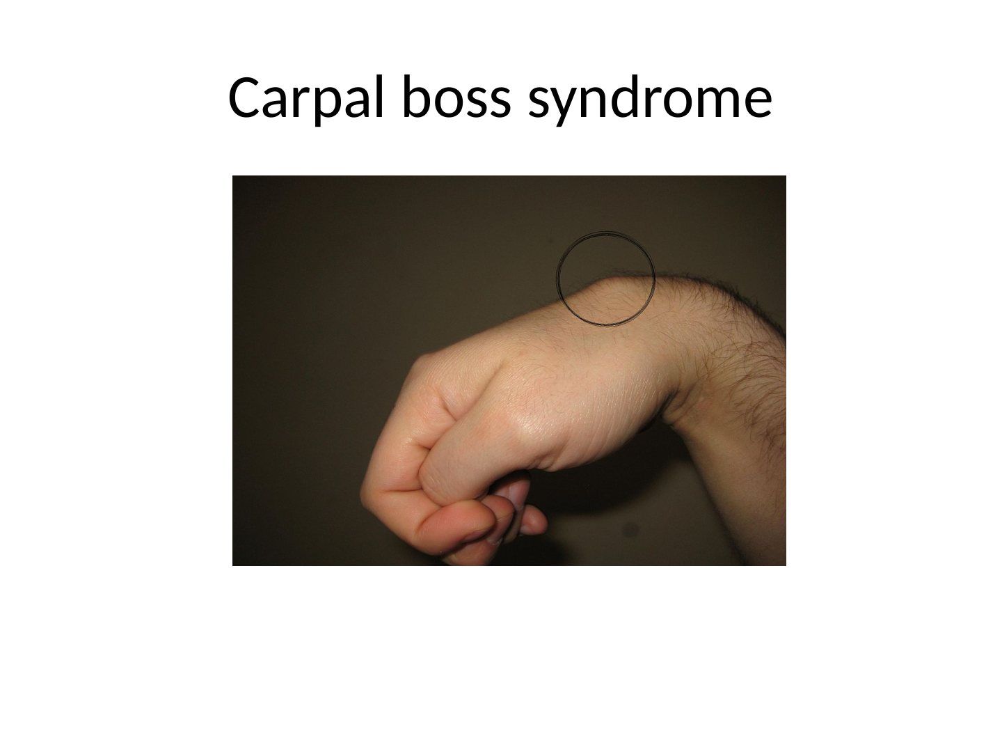
Q1-Q22 questions — tap to reveal all answerslist
- What is carpal boss syndrome?
- How is carpal boss syndrome managed?
Q1.What is carpal boss syndrome?
- A dorsal bony osteophyte over the base of the 2nd or 3rd metacarpal
- Causes reduced range of motion
- Related to degeneration or Os styloideum; may be associated with a ganglion
Q2.How is carpal boss syndrome managed?
- Mainly conservative management
▸ Slide 776 · Inclusion cyst (sebaceous cust)Hand Soft Tissue · 3 questions expand
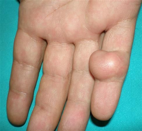
Q1-Q33 questions — tap to reveal all answerslist
- What is an inclusion cyst and what is its pathogenesis?
- What are the differentials for an inclusion cyst?
- What is the management of an inclusion cyst?
Q1.What is an inclusion cyst and what is its pathogenesis?
- Epithelial-lined cyst filled with keratin
- Pathogenesis: penetrating injury drives keratinised epithelium into soft tissue or bone
Q2.What are the differentials for an inclusion cyst?
- Skin lesion (tethers skin)
- Ganglion (mobile ML>PD, transilluminates)
- Calcifying aponeurotic fibroma
- Fibroma or schwannoma, haemangioma
- Giant cell tumour
Q3.What is the management of an inclusion cyst?
- Marginal excision
- Curette if bone is involved
- Amputation if advanced bone destruction
- Low recurrence if the capsule is excised en bloc
▸ Slide 777 · Wound over MPCJHand Soft Tissue · 3 questions expand
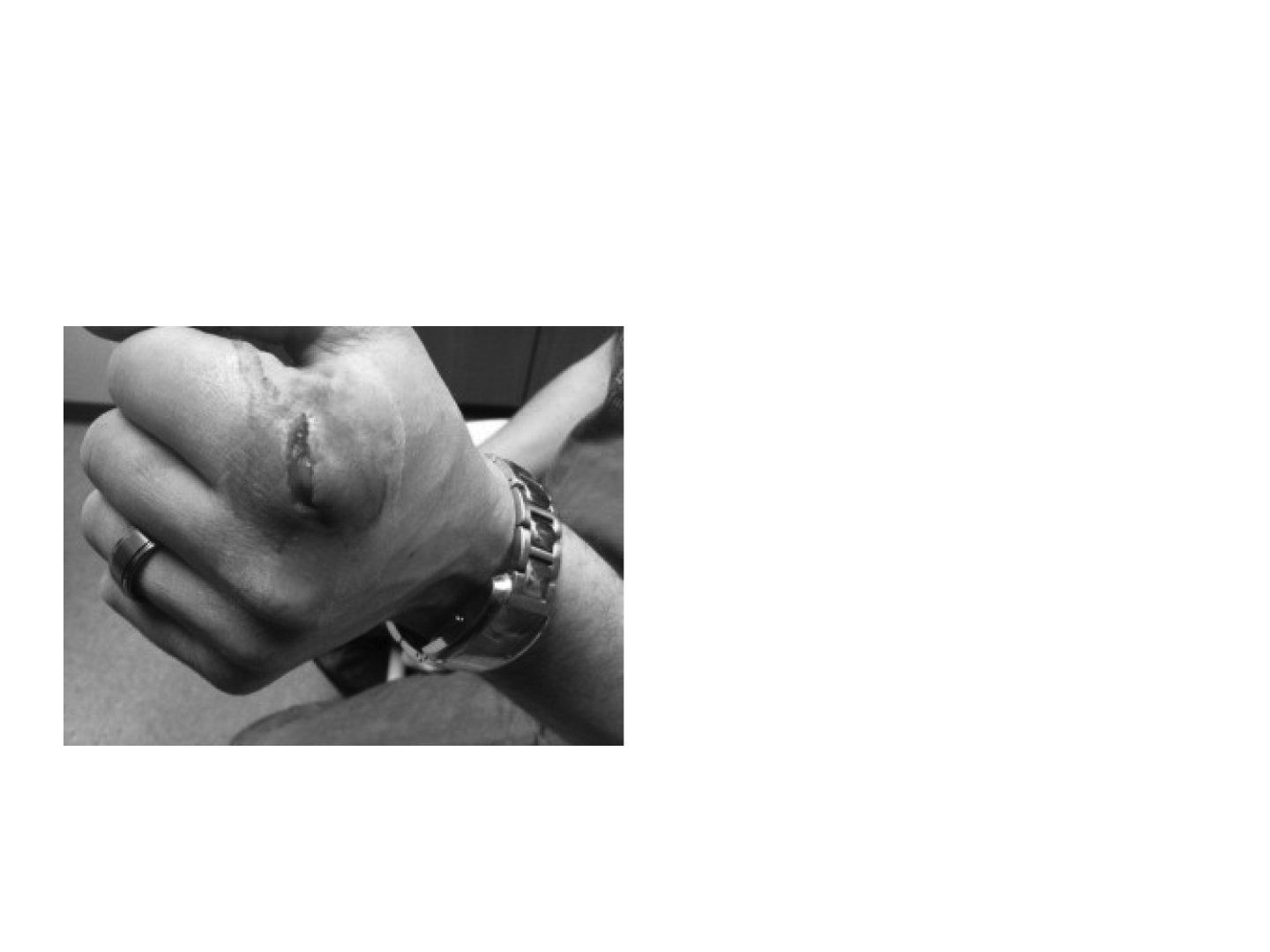
Q1-Q33 questions — tap to reveal all answerslist
- Why is a wound over the MCPJ suspicious and what must be excluded?
- How do you examine a suspected fight bite?
- What history is important in a fight bite?
Q1.Why is a wound over the MCPJ suspicious and what must be excluded?
- Suspicious of a fight bite
- Needs urgent exploration to rule out extension into the joint
- Also exclude extensor tendon and osteochondral injuries
- Neglected injuries are associated with fracture and osteomyelitis
Q2.How do you examine a suspected fight bite?
- Ask the patient to flex the MCPJ (mimic a punch)
- This reveals the true tendon cut position
Q3.What history is important in a fight bite?
- Mechanism and chronicity
- Occupation, hand dominance
- Ask if the assailant has a history of blood-borne virus
▸ Slide 778 · What is this test?Hand Soft Tissue · 5 questions 1 check expand
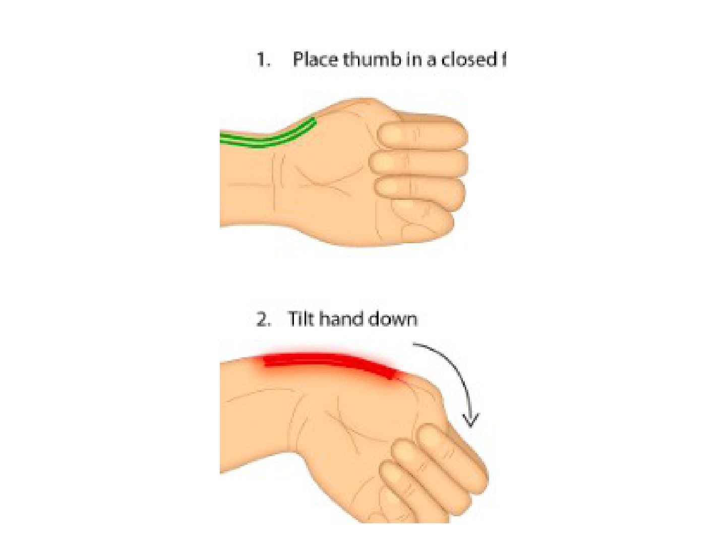
Q1-Q55 questions — tap to reveal all answerslist
- What is this test and how is it performed?
- What is de Quervain disease?
- What are the clinical features of de Quervain disease?
- What is the non-operative treatment of de Quervain disease?
- What are the operative steps and complications of de Quervain release?
Q1.What is this test and how is it performed?
- Eichhoff test (often misnamed Finkelstein's): ulnar deviation of the wrist with the thumb in the palm reproduces symptoms
- Finkelstein's test: examiner pulls the thumb into ulnar deviation
- Used for the diagnosis of de Quervain disease
Q2.What is de Quervain disease?
- Stenosing tenosynovitis of the first dorsal wrist compartment (APL and EPB)
- Not truly inflammatory: mucopolysaccharide accumulation and a thickened retinaculum
- Caused by attrition and degeneration
- Common in racquet sports and golfers, more common in women (postpartum mothers)
Q3.What are the clinical features of de Quervain disease?
- Dorsoradial wrist tenderness
- Swelling
- Crepitus
Q4.What is the non-operative treatment of de Quervain disease?
- Activity modification and splinting
- Local corticosteroid injection (steroid successful in >80%)
- Concomitant splint/NSAID after injection does not improve outcome
Q5.What are the operative steps and complications of de Quervain release?
- Oblique incision proximal to the wrist: longitudinal better cosmesis, transverse more complicated
- Release the dorsal side of the 1st compartment to prevent volar tendon subluxation
- Beware anatomic variation: APL multiple slips (2-4), EPB may have its own separate compartment
- Complications: injury to the superficial sensory radial nerve, tendon subluxation, CRPS, recurrence from incomplete release
The thumb-in-palm with ulnar deviation manoeuvre is Finkelstein's test — naming conflation — The thumb-in-palm then ulnar deviation manoeuvre is the **Eichhoff test**, commonly misnamed Finkelstein's test; the true Finkelstein test has the examiner grasp the thumb and ulnar-deviate the wrist and is more specific with fewer false positives — source
▸ Slide 779 · Extensor tendon problemsHand Soft Tissue · 6 questions expand
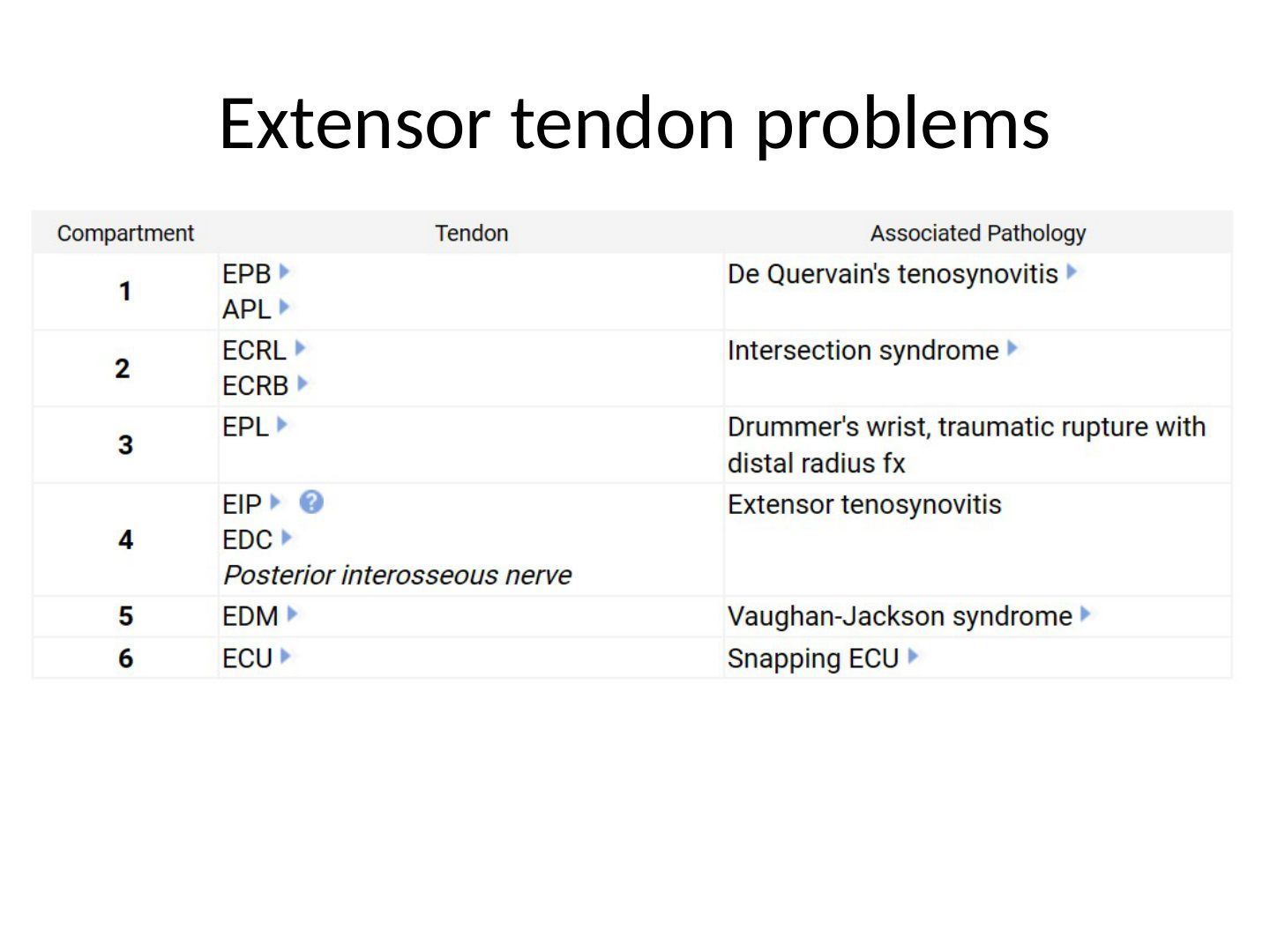
Q1-Q66 questions — tap to reveal all answerslist
- List the six extensor compartments and a condition associated with each.
- What is intersection syndrome and how is it managed?
- What is drummer wrist?
- What is snapping ECU syndrome and what is its mechanism?
- What is the differential for snapping ECU and how is it managed?
- What is Wartenburg syndrome?
Q1.List the six extensor compartments and a condition associated with each.
- 1: de Quervain (APL/EPB)
- 2: Intersection syndrome (ECRL/ECRB with APL/EPB)
- 3: EPL rupture / drummer wrist
- 4: EDC tenosynovitis
- 5: Vaughan-Jackson
- 6: Snapping ECU
Q2.What is intersection syndrome and how is it managed?
- Inflammatory condition at the crossing of the 1st (APL/EPB) and 2nd (ECRL/ECRB) dorsal compartments
- Conservative: rest, wrist splinting, steroid injection to the 2nd compartment
- Rarely surgery: release of the 2nd dorsal compartment ~6cm proximal to the radial styloid
Q3.What is drummer wrist?
- Tenosynovitis of the 3rd dorsal compartment containing EPL
- Classically in drummers who play for long periods without rest
Q4.What is snapping ECU syndrome and what is its mechanism?
- Secondary to attenuation or rupture of the ECU subsheath (6th compartment); attenuation gives subluxation, rupture gives dislocation
- Ulnar-sided tears: ECU subluxates on supination and reduces on pronation
- Radial-sided tears: ECU subluxates on supination and lies on top of the torn subsheath on pronation
- Flexion and supination elicit a painful snap; subluxation can cause ECU tendonitis
Q5.What is the differential for snapping ECU and how is it managed?
- DDx: TFCC tear, hook of hamate fracture, ulnar styloid fracture, ulnar styloid impaction syndrome, ulnar tunnel syndrome, pisotriquetral arthritis
- Non-operative: wrist splint or long arm cast immobilised in pronation and slight radial deviation
- Operative: ECU subsheath reconstruction +/- wrist arthroscopy; direct repair in acute cases, extensor retinaculum flap for chronic
- Wrist arthroscopy shows concurrent TFCC tears in 50%
Q6.What is Wartenburg syndrome?
- Compression of the superficial radial nerve (SPN) by the brachioradialis (BR) and ECRL
- Not related to the extensor compartment diseases above
▸ Slide 780 · Trigger fingerHand Soft Tissue · 6 questions expand

Q1-Q66 questions — tap to reveal all answerslist
- What is trigger finger and what is its pathology?
- What conditions are associated with trigger finger?
- What is the Green classification of trigger finger?
- What are the success rates of non-operative and operative treatment?
- When is FDS slip release performed?
- How is paediatric trigger finger managed?
Q1.What is trigger finger and what is its pathology?
- Stenosing tenosynovitis at the A1 pulley
- Fibrocartilaginous metaplasia of tendon and/or pulley: chondrocyte proliferation, increased type III collagen
- Chronic hyperglycaemia creates collagen cross-links and impairs collagen degradation
- Usually involves FDP and spares FDS
- Trigger thumb associated with a 4th (variable annular) pulley in up to 75%
Q2.What conditions are associated with trigger finger?
- Associated conditions: RA, calcific tendinitis, septic tenosynovitis, carpal tunnel syndrome, congenital trigger thumb
- Medical diseases: diabetes, amyloidosis, hypothyroidism, sarcoidosis, gout/pseudogout
Q3.What is the Green classification of trigger finger?
- Type 1: pain on A1 pulley
- Type 2: spontaneously corrects
- Type 3: passively corrects
- Type 4: rigid
Q4.What are the success rates of non-operative and operative treatment?
- Splinting, activity modification, NSAIDs: 40-97% effective
- Corticosteroid injection: 60-90% effective
- Percutaneous A1 release: success >90%, earlier return to work than open
- Open release: satisfactory in >90%; higher recurrence in diabetic patients
Q5.When is FDS slip release performed?
- Release of A1 pulley and 1 slip of FDS (usually ulnar slip) for RA or recurrent/persistent trigger finger
- In RA, FDS slip excision without A1 release may prevent exacerbation of ulnar drift at the MCPJ/volar subluxation
Q6.How is paediatric trigger finger managed?
- Notta's node (proximal to A1 pulley), flexion contracture and triggering
- Surgery at 2-4 years of age to prevent interphalangeal joint contracture
- May need release of remaining FDS slip and A3 pulley
- Success rate >90%
▸ Slide 781 · Glomus tumor: Tumors of glomus body which is a perivascular temperature regulatiHand Soft Tissue · 7 questions expand

Q1-Q77 questions — tap to reveal all answerslist
- What is a glomus tumour?
- What history is taken for a suspected glomus tumour?
- How does a glomus tumour present?
- What special tests are used for a glomus tumour?
- What investigations are used for a glomus tumour?
- What is the differential diagnosis of a glomus tumour?
- What is the management of a glomus tumour?
Q1.What is a glomus tumour?
- Tumour of the glomus body, a perivascular temperature-regulating structure
- Neuromyoarterial neoplasm of smooth muscle with eponychium metaplasia
- 75% in the hand, 50% subungual
Q2.What history is taken for a suspected glomus tumour?
- Age and premorbid status
- Ask about the triad of paroxysmal pain, exquisite tenderness and cold intolerance
Q3.How does a glomus tumour present?
- Subungual mass with bluish discolouration
- Nail ridging
- Triad of paroxysmal pain, exquisite tenderness and cold intolerance
- 50% have bone erosion
Q4.What special tests are used for a glomus tumour?
- Love's test: pressure to a pinhead causes exquisite pain (sens 100%, spec 78%)
- Hildreth test: inflating a tourniquet abolishes the pain with Love's test (sen 92%, spec 91%)
Q5.What investigations are used for a glomus tumour?
- XR: bone erosion from pressure effect
- MRI: T1 isodense, T2 hyperintense with strong gadolinium enhancement
- Histology: 3 subtypes - mucoid hyaline, solid and angiomatous; round cells with dark nuclei
Q6.What is the differential diagnosis of a glomus tumour?
- Dupuytren's exostosis
- Gout
- Inclusion cyst
- Osteomyelitis
- Amelanotic melanoma
Q7.What is the management of a glomus tumour?
- Marginal excision
- Low recurrence (20% quoted in the notes)
- Counsel for nail ridging and nailbed grafting
▸ Slide 782 · Giant cell tumor of tendon sheathHand Soft Tissue · 3 questions expand
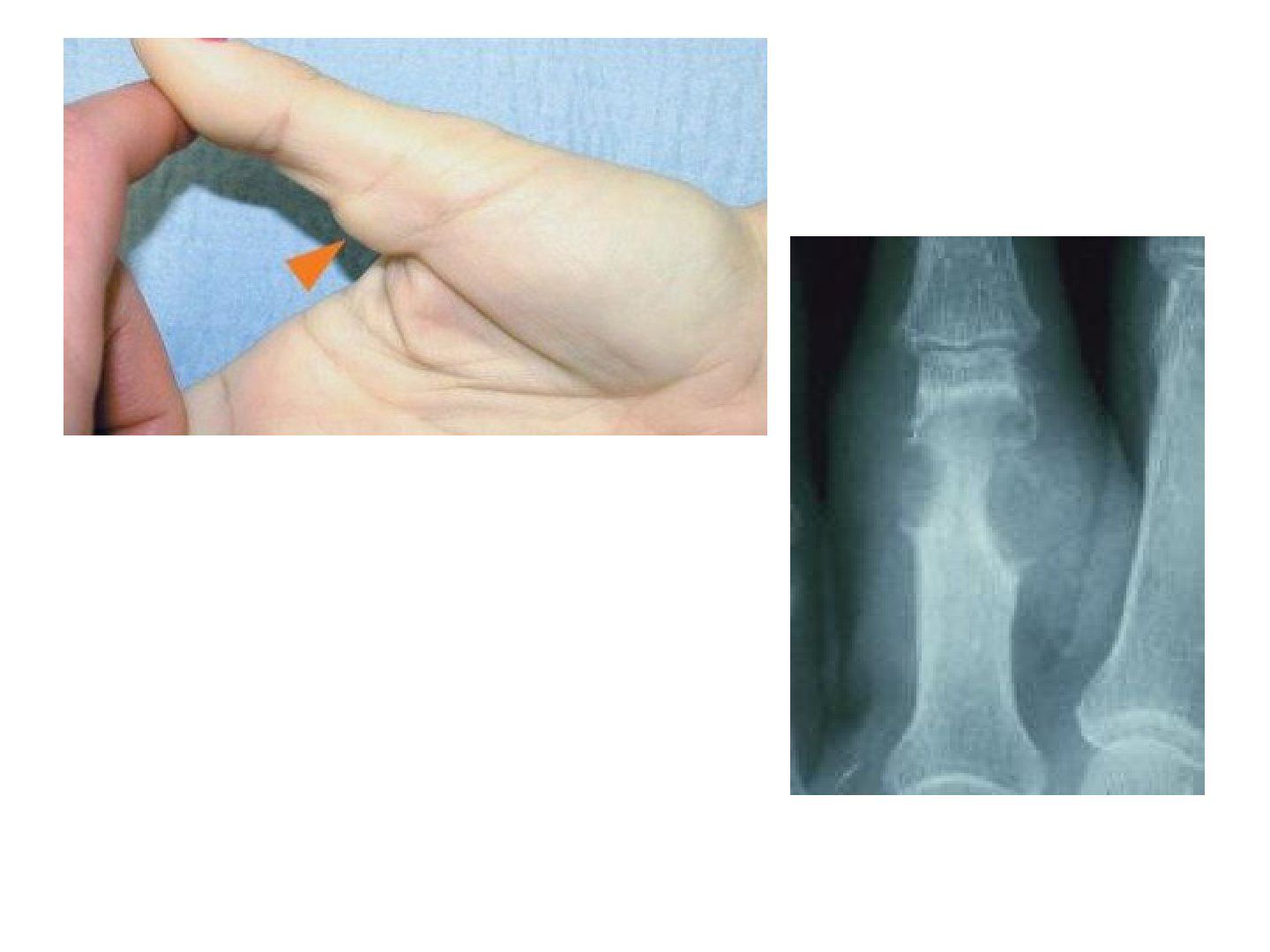
Q1-Q33 questions — tap to reveal all answerslist
- What is giant cell tumour of tendon sheath?
- What investigations and histology are seen?
- How is giant cell tumour of tendon sheath managed?
Q1.What is giant cell tumour of tendon sheath?
- 2nd most common hand soft tissue tumour
- Arises from the flexor tendon sheath, usually between A1 and A2
- PVNS of the tendon sheath
- Examine for NV and bone involvement
Q2.What investigations and histology are seen?
- XR: bone indentation up to 5%
- MRI: T1 and T2 hypodense
- Histology: multinucleated cells with haemosiderin staining, vascular villi with stromal cell infiltration, xanthoma
Q3.How is giant cell tumour of tendon sheath managed?
- Excision + H2O2 irrigation (to oxidise residual cells)
- High recurrence up to 50%, locally aggressive
▸ Slide 783 · MelanomaHand Soft Tissue · 7 questions 2 check expand
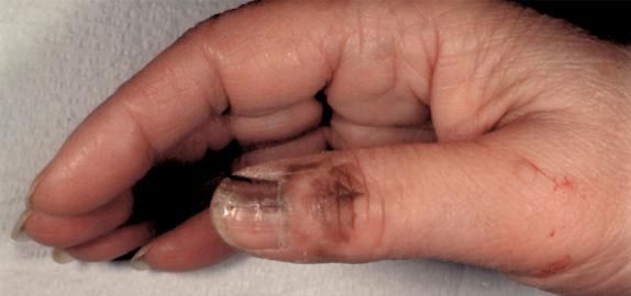
Q1-Q77 questions — tap to reveal all answerslist
- Describe the presentation of subungual melanoma.
- What history is important in subungual melanoma?
- What does the ABCDEF history cover in melanoma?
- What risk factors and symptoms of metastasis are asked about?
- What investigations are performed?
- What is the Breslow classification and how does it guide treatment?
- What are the poor prognostic factors?
Q1.Describe the presentation of subungual melanoma.
- Pigmented band from the germinal matrix
- Nail fold involvement = Hutchinson sign
- DDx: subungual haematoma, onychomycosis, benign longitudinal melonychia (straight border, single band)
Q2.What history is important in subungual melanoma?
- Age and premorbid status
- Current disability and complications, chronicity, history of trauma
- Then take the ABCDEF history and ask about metastatic symptoms
Q3.What does the ABCDEF history cover in melanoma?
- Age 50-70 years
- Band + breadth >3mm, irregular border
- Coloured + change (rapid increase in size, unhealing dystrophic nail)
- Digit (thumb > big toe > index finger)
- Extension (Hutchinson sign) and FHx
Q4.What risk factors and symptoms of metastasis are asked about?
- Risk factors: sun exposure, family history, fair skin, multiple naevi, immunosuppression
- Metastatic symptoms: LN, lung, liver, brain
Q5.What investigations are performed?
- LFT and LDH
- CXR for lung mets
- MRI
- PET-CT
Q6.What is the Breslow classification and how does it guide treatment?
- I <0.75mm (survival >90%); II 0.76-1.5mm; III 1.6-4mm; IV >4mm (survival 50%)
- Sentinel LN biopsy with planning for radical LND and adjuvant chemo
- WLE: <1mm -> 1cm margin; 1mm or more -> 2cm margin + SLN biopsy
Q7.What are the poor prognostic factors?
- Male
- Ulceration
- Metastases
- Subungual 5yr 40%, H&N lesion
- Basic science: melanocytes from neural crest, separate from dermis by basement membrane; Clark level 1-5; acral lentiginous subtype starts at nailfold/germinal matrix
Breslow classification: I <0.75mm, II 0.76-1.5mm, III 1.6-4mm, IV >4mm — outdated — This is Breslow's original prognostic classification; current AJCC 8th edition T categories use 1/2/4mm thresholds (T1 <1.0mm, T2 >1.0-2.0mm, T3 >2.0-4.0mm, T4 >4.0mm), with T1 subdivided at 0.8mm and thickness rounded to the nearest 0.1mm — source
Subungual melanoma 5-year survival 40% — contested — Survival varies widely by study and stage; 5-year overall survival ~40% in an MD Anderson cohort but 5-year melanoma-specific survival 56-82% in other series, so a single 40% figure is potentially misleading — medium confidence — source
▸ Slide 784 · Ridged nailHand Soft Tissue · 2 questions expand

Q1-Q22 questions — tap to reveal all answerslist
- What history is important in a ridged nail?
- How is a ridged nail managed?
Q1.What history is important in a ridged nail?
- Age and premorbid status
- History of infection or trauma (poor suture technique, missed germinal matrix injury)
- Current disability: cosmesis, hypersensitive scar, previous paronychia or osteomyelitis
Q2.How is a ridged nail managed?
- Observation
- <2mm: scar resection + primary closure
- 2mm or more: scar resection + 2nd toe nailbed graft
▸ Slide 785 · Hook nail volar curving of nail matrixHand Soft Tissue · 3 questions expand
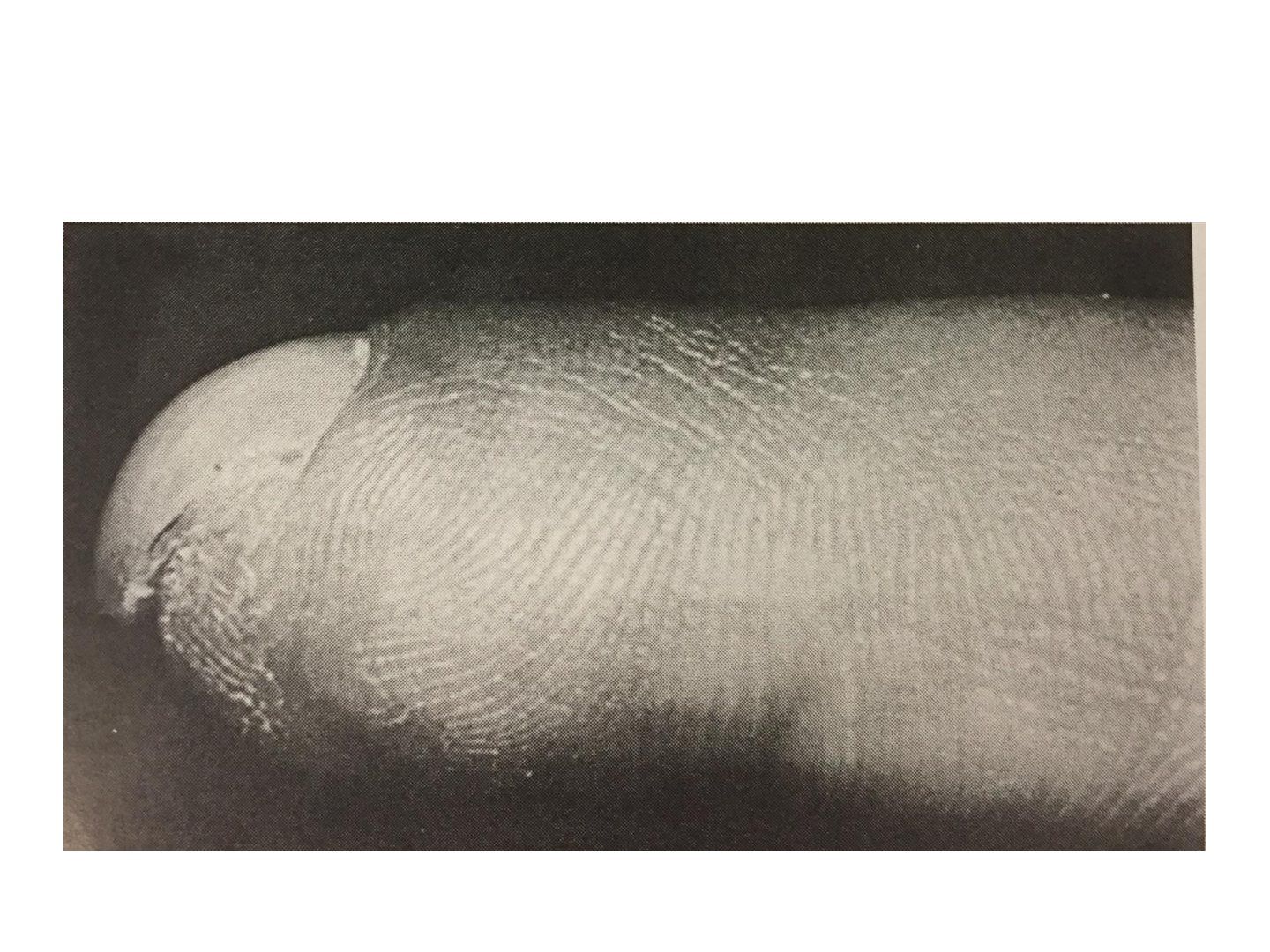
Q1-Q33 questions — tap to reveal all answerslist
- What is a hook nail and what causes it?
- What history and investigation are needed for a hook nail?
- What is the management of a hook nail?
Q1.What is a hook nail and what causes it?
- Volar curving of the nail matrix
- Cause: curved matrix upon suture
- Or a shortened bone
Q2.What history and investigation are needed for a hook nail?
- Age and premorbid status
- Current disability: cosmesis, pain from ingrown nail
- XR to check residual distal phalanx length
Q3.What is the management of a hook nail?
- Observation
- Antenna procedure
- If due to tight suture: soft tissue procedure to reconstruct the volar tip defect (VY or cross finger flap)
- If shortened bone: VBG from 2nd toe or K wire to support the nail bed
▸ Slide 786 · The three sites of injection in a tricompartmental wrist arthrogram:Hand Soft Tissue · 3 questions expand

Q1-Q33 questions — tap to reveal all answerslist
- What are the three sites of injection in a tricompartmental wrist arthrogram?
- Why are injections staged and how is sensitivity improved?
- What do you look for in the radiocarpal joint injection?
Q1.What are the three sites of injection in a tricompartmental wrist arthrogram?
- The midcarpal space
- The distal radioulnar joint (DRUJ)
- The radiocarpal joint (RCJ)
Q2.Why are injections staged and how is sensitivity improved?
- A ruptured ligament can act as a valve, so injecting at few points improves sensitivity
- Wait ~1 hour between injections for contrast to fade away
- Sequence RC, DRUJ, midcarpal several hours apart to eliminate the one-way valve phenomenon
- Dynamic views open up small tears
Q3.What do you look for in the radiocarpal joint injection?
- Contrast pooling and abnormal communication
- Communication with the pisotriquetral joint via the pisotriquetral orifice is normal
- There should be no communication with the midcarpal joint
▸ Slide 787 · CULAHand Soft Tissue · 2 questions expand

Q1-Q22 questions — tap to reveal all answerslist
- What is the Swanson classification of congenital limb anomalies?
- What is the Oberg-Manske-Tonkin classification?
Q1.What is the Swanson classification of congenital limb anomalies?
- I Formation
- II Differentiation
- III Duplication
- IV Overgrowth, V Undergrowth
- VI Constriction band, VII Syndrome
Q2.What is the Oberg-Manske-Tonkin classification?
- Malformation/deformation: constriction ring, trigger finger
- NOS/dysplasia: hypertrophy, tumourous
▸ Slide 788 · Clinical photo showing paed patient left hand with preaxial polydactylyHand Soft Tissue · 8 questions expand
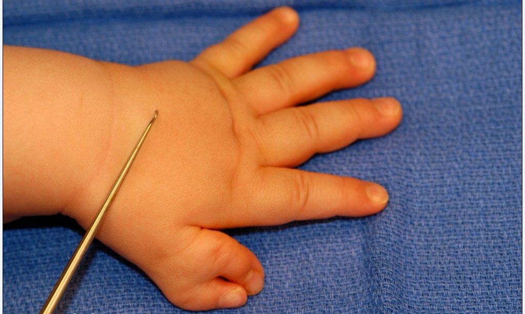
Q1-Q88 questions — tap to reveal all answerslist
- What is the likely diagnosis and Wassel type in this patient, and which findings support it?
- What history is taken in preaxial polydactyly?
- What are the local examination findings to document in preaxial polydactyly?
- What radiological classification systems are used for preaxial polydactyly and what does Hung's classification describe?
- Which systemic conditions are associated with type 7 preaxial polydactyly?
- What is the management and timing of surgery?
- What is the treatment by Wassel type?
- What are the six steps of ablation and reconstruction and the possible complications?
Q1.What is the likely diagnosis and Wassel type in this patient, and which findings support it?
- Paediatric left hand preaxial polydactyly
- Radial thumb is small but has a complete nail, distal phalanx and suspected IPJ
- First webspace contracted
- Likely type IV (commonest; IPJ seen in 47%); next most common type 2
Q2.What history is taken in preaxial polydactyly?
- Age, other abnormalities, hand function
- Family history (FHx)
Q3.What are the local examination findings to document in preaxial polydactyly?
- LSW APB
- Length, size, webspace, alignment, position, bifurcation
- MSSSV: mobility, sensibility, strength, stability, vascularity
- Check for other systemic diseases (usually sporadic)
Q4.What radiological classification systems are used for preaxial polydactyly and what does Hung's classification describe?
- Wassel classification on XR
- Hung's classification: 4 subgroups of type IV
- Hypoplastic 12%, ulnar deviated 64%, divergent 15%, convergent 9%
- Convergent is the most complex and most likely to have residual deformity
Q5.Which systemic conditions are associated with type 7 preaxial polydactyly?
- Holt-Oram syndrome, Fanconi anaemia, Blackfan-Diamond anaemia
- Imperforate anus, cleft palate, tibial defects
- Usually sporadic; type 7 is the type commonly associated with these syndromes
Q6.What is the management and timing of surgery?
- Parental counselling: incidence 1 in 1000
- Offer operation at 1 year old (less anaesthetic risk, before thumb function fully develops)
- Goal: single mobile stable thumb that is cosmetically acceptable
- Reconstruct according to Lister parameters: length, stability, position
Q7.What is the treatment by Wassel type?
- 1-2: modified Bilhaut-Cloquet (ablate radial thumb + RCL reattachment) - reduce nail ridging, less scar, less growth disturbance; disadvantage lig laxity (ligament laxity)
- 3-6: resection + reconstruction
- 7: small - excision + ligament reconstruction; large - fusion
- Excise the radial thumb if the remaining thumb is ~80% of the contralateral
Q8.What are the six steps of ablation and reconstruction and the possible complications?
- Isolate NV bundle and collateral ligament (osteoperiosteal sleeve from the deleted thumb)
- Reconstruct the joint surface (obliquity, enlargement, osteotomy, reduction, chondroplasty), then reattach collaterals
- APB preservation and reattachment; EPL, FPL realignment; 1st webspace reconstruction
- Complications: bone (late angular deformity), joint (instability, stiffness), soft tissue (scar, abnormal tendon excursion), cosmesis (ridged nail, size, narrowed 1st web)
▸ Slide 789 · Post-axial polydactylyHand Soft Tissue · 3 questions expand
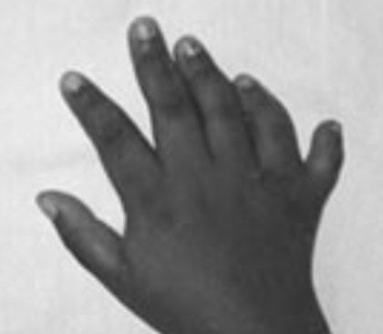
Q1-Q33 questions — tap to reveal all answerslist
- What is postaxial polydactyly and what genetic conditions are screened for?
- What is the aim and timing of surgery for postaxial polydactyly?
- How is postaxial polydactyly treated by type?
Q1.What is postaxial polydactyly and what genetic conditions are screened for?
- Too high SHH activity at the ulnar limb bud
- 10x more common in African Americans (AD)
- Thorough genetic workup (chondroectodermal dysplasia or Ellis-van Creveld syndrome EVC gene, AR, 'six-fingered dwarfism')
Q2.What is the aim and timing of surgery for postaxial polydactyly?
- Aim is mainly cosmesis
- Timing: 1 year old
Q3.How is postaxial polydactyly treated by type?
- Type A - well formed digit: reconstruction (preserve radial digit, reattach ulnar digit soft tissue and muscle)
- Type B - rudimentary skin tag (vestigial): tie off before 1 year
▸ Slide 790 · Ulnar dimelia (Mirror hand)Hand Soft Tissue · 3 questions expand
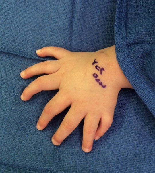
Q1-Q33 questions — tap to reveal all answerslist
- What is ulnar dimelia (mirror hand)?
- What neurovascular anomalies can accompany ulnar dimelia?
- What are the two types of ulnar dimelia?
Q1.What is ulnar dimelia (mirror hand)?
- Very rare
- Absence of the radial ray, duplication of the ulnar carpal, metacarpal and phalanx bones, and symmetric polydactyly
- Swanson classification type III
Q2.What neurovascular anomalies can accompany ulnar dimelia?
- Duplication of the ulnar nerve
- Duplication of the ulnar artery
- Abnormal arterial arches, shortening of the radial nerve, absence of the radial artery
Q3.What are the two types of ulnar dimelia?
- Type 1: one lunate and one trapezoid bone, one index finger
- Type 2: two lunate and two trapezoid bones, two index fingers
▸ Slide 791 · This is a clinical photo showing bilateral thumb angulation with the apex radialHand Soft Tissue · 3 questions expand
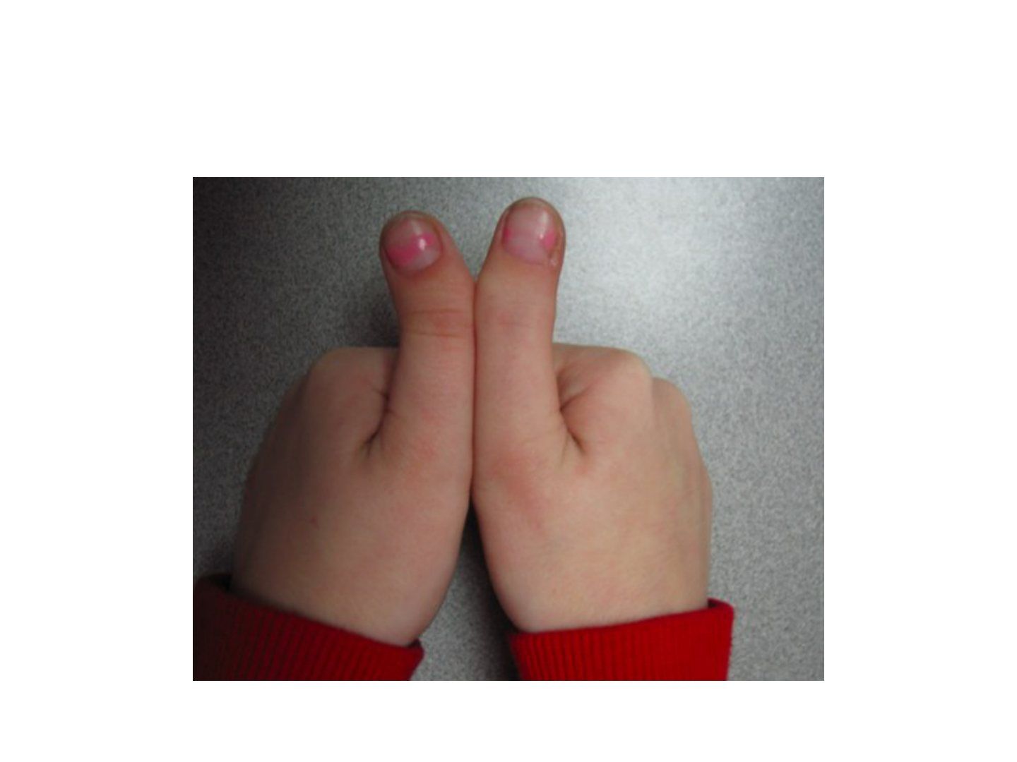
Q1-Q33 questions — tap to reveal all answerslist
- Describe the clinical photo and the likely diagnosis.
- What do you assess and what systemic associations are checked?
- What is the management of a triphalangeal thumb with delta phalanx?
Q1.Describe the clinical photo and the likely diagnosis.
- Bilateral thumb angulation with the apex radial
- Need an X-ray
- Likely triphalangeal thumb with a delta phalanx (small wedge-shaped ossicle) on the radial side causing angulation
Q2.What do you assess and what systemic associations are checked?
- Address the patient's complaint and exclude problems: length, instability
- Triphalangism is associated with Holt-Oram, Diamond-Blackfan, Fanconi anaemia, imperforate anus, tibial defects
Q3.What is the management of a triphalangeal thumb with delta phalanx?
- If small: excise + reconstruct the ligament
- If large: fuse with the proximal or distal phalanx
▸ Slide 792 · Types of curly fingersHand Soft Tissue · 8 questions 1 check expand
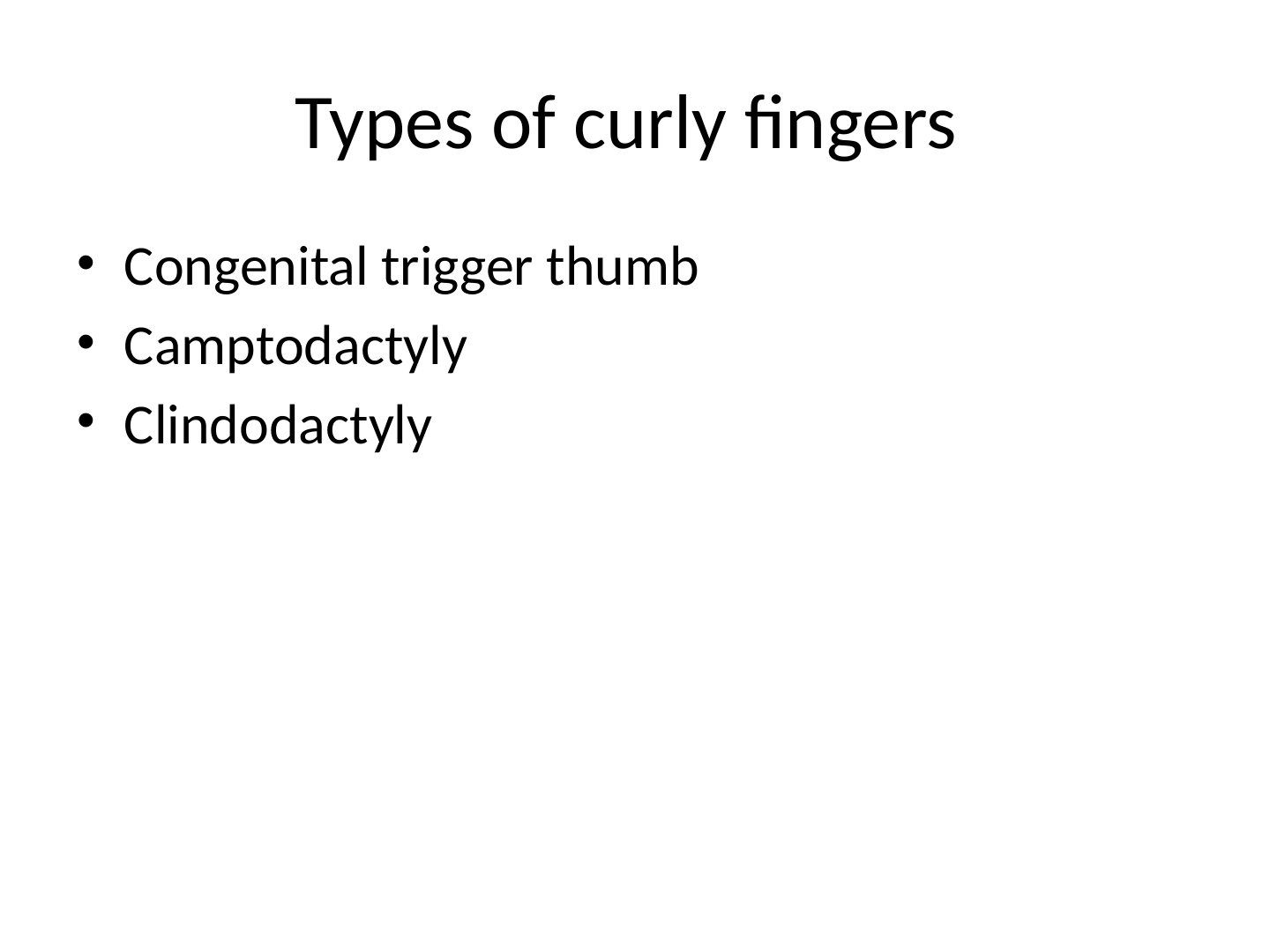
Q1-Q88 questions — tap to reveal all answerslist
- What is the pathophysiology and clinical presentation of congenital trigger thumb?
- What is the differential diagnosis of congenital trigger thumb?
- What is the management of congenital trigger thumb and when is surgery indicated?
- What is congenital clasp thumb and how is it classified (Tsuyuguchi)?
- What is camptodactyly and how is it classified (Benson)?
- How is Benson type 2 camptodactyly managed?
- What is clinodactyly and what are its types?
- Describe the classification of the Av pulley of the thumb.
Q1.What is the pathophysiology and clinical presentation of congenital trigger thumb?
- Nodular thickening of FPL causing size mismatch between flexor sheath and FPL, disrupting gliding
- IPJ flexion deformity with MCPJ compensatory hyperextension
- Notta's nodule at FPL; bilateral in 30%
- Associations: mucopolysaccharidosis, Gaucher's disease, arthrogryposis
Q2.What is the differential diagnosis of congenital trigger thumb?
- Clasp thumb: extensor agenesis/hypoplasia
- Hypoplastic thumb
Q3.What is the management of congenital trigger thumb and when is surgery indicated?
- 1st line stretching if flexible; 50% resolve by 2 years
- If unresolved >2 years, only <10% resolve --> OT
- Release before 3 years, otherwise increased IPJ FFC if done after 4 years
- Release A1 pulley and preserve oblique pulley; 90% successful, low risk of recurrence
Q4.What is congenital clasp thumb and how is it classified (Tsuyuguchi)?
- Extensor agenesis/hypoplasia
- Associations: arthrogryposis (congenital joint contractures affecting two or more areas of the body), digitotalar dysmorphism, Freeman-Sheldon syndrome, X-linked MASA syndrome
- Type I: supple with no other digital anomaly; Type II: contracture +/- other digital anomalies
- Type III: rigid associated with arthrogryposis and marked soft-tissue deficits
- Mx: serial splint/stretching; tendon transfer PI to EPL; reconstruction at 3-5 years old
Q5.What is camptodactyly and how is it classified (Benson)?
- Congenital FFC of PIPJ with webbed volar skin and no crease; DIPJ normal
- Cause: abnormal lumbrical/FDS/extensor; LF > RF
- Benson: 1 infant (most common, stretching); 2 adolescent from abnormal FDS; 3 multiple digits with syndrome (non-op)
- Benson 4: Kirner deformity = 2-plane deformity of LF, apex dorsal-ulnar
- Mx: 1st line stretching; OT if FFC >60 degrees; FDS transfer/tenotomy/fusion
Q6.How is Benson type 2 camptodactyly managed?
- Adolescent presentation due to abnormal FDS
- If flexible, perform FDS to radial LB transfer
- For comparison: type 1 (infant) is most common and treated with stretching; type 3 is non-operative and treated later with osteotomy/fusion
Q7.What is clinodactyly and what are its types?
- LF curved towards RF; autosomal dominant
- Type I: normal length (most common); Type II: short length; Type III: significant angulation with delta phalanx
- Delta phalanx = C-shaped epiphysis and longitudinal bracketed diaphysis
- Partial closure of PP head or MP base physis; USG screening for Down syndrome (look for simian crease as well=single palmar crease)
Q8.Describe the classification of the Av pulley of the thumb.
- Type I: transverse, parallel to A1, with a gap between Av and A1
- Type II: no gap between Av and A1
- Type III: triangular/oblique Av pulley with fibres converging to the radial side
50% of congenital trigger thumbs resolve by 2 years and only <10% resolve if unresolved beyond 2 years — imprecise/optimistic — Published resolution rates vary widely: about 30-50% over several years, with some studies reporting 32-76% by 5 years; resolution is uncommon after age 2 and surgery is generally not advised before age 3. — medium confidence — source
▸ Slide 793 · 0Clinical photo of this childs right hand showing a hypoplastic thumb. I am alsHand Soft Tissue · 9 questions expand
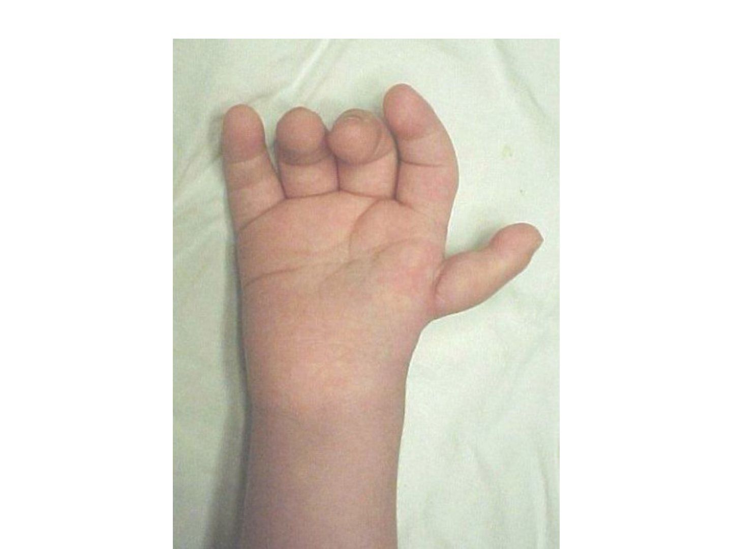
Q1-Q99 questions — tap to reveal all answerslist
- What is the aetiology of thumb hypoplasia and what examination is needed?
- Describe the LSWAP assessment of the thumb.
- Describe Blauth's classification of the hypoplastic thumb.
- What are the aims and principles of management of thumb hypoplasia?
- How is the first MCPJ stabilised in thumb hypoplasia?
- What are the four basic surgical principles of pollicization?
- Which tendon transfers realign the motor system in pollicization?
- What are the principles of pollicization (Buck-Gramcko)?
- What are the complications of pollicization?
Q1.What is the aetiology of thumb hypoplasia and what examination is needed?
- Undergrowth malformation (Swanson). This is due to a undergrowth (according to Swanson); according to OMT (Oberg-Manske-Tonkin), due to a defect in the AER in the limb bud
- Check the rest of the limb for associated longitudinal preaxial deficiency; thenar eminence may be absent
- 60% bilateral; 80% associated with other abnormalities e.g. TAR, Fanconi anemia, VACTERL syndrome
- X-ray to assess bony anatomy and presence of radial deficiency
Q2.Describe the LSWAP assessment of the thumb.
- Size: nail 70% opposite thumb, 133% index finger girth, nail width 105% of index finger
- Length (reaching IF PIPJ); Alignment sagittal/coronal/axial; Position (abduction, opposition)
- Webspace: I-II IMA 45-55 degrees
- Mobility CMCJ > IPJ > MCPJ; stability UCL > RCL; sensibility, strength, vascularity, growth
Q3.Describe Blauth's classification of the hypoplastic thumb.
- 1: deficient thumb, extrinsics and intrinsics normal
- 2: deficient intrinsics and collaterals, thumb in adducted position
- 3A: deficient extrinsics, CMCJ stable
- 3B: deficient extrinsics, CMCJ unstable
- 4: floating thumb (pouce flotant); 5: absent thumb/aplasia
Q4.What are the aims and principles of management of thumb hypoplasia?
- Goal: stable, mobile, sensate, opposable thumb; Lister: position, stability, strength
- 1-3a: deepen webspace +/- soft tissue reconstruction (thenar, extrinsic) +/- stabilise MCPJ (UCL)
- Webspace: Tajima flap, Z-plasty, four-flap plasty; aim 55-60 degrees 1st-2nd MC angle
- Opponensplasty with FDS IV or Huber; release pollex abductus (anomalous FPL-EPL connection 20%)
- 3b-5: ablation +/- reconstruction (toe hand / pollicization)
Q5.How is the first MCPJ stabilised in thumb hypoplasia?
- Use available local tissue
- Reinforce with FDS IV
- MCPJ chondrodesis (subphyseal arthrodesis) for global instability
Q6.What are the four basic surgical principles of pollicization?
- Shortening of the index finger ray to a 3-bone system
- Appropriate positioning of the new thumb
- Creation of a wide supple first webspace
- Realignment of the motor system to achieve a good soft tissue balance
Q7.Which tendon transfers realign the motor system in pollicization?
- EI --> EPL
- EDCII --> APL
- 1st DI --> APB
- 1st PI --> adductor pollicis
Q8.What are the principles of pollicization (Buck-Gramcko)?
- Severity of radial club hand and thumb hypoplasia does not always correlate; if concomitant problem, wrist first (wrist position needed to balance tendons), thumb later
- Pollicization 1/2-1 year after wrist centralization, before the hand matures at 3 years (Before hand mature @ 3 year)
- Shorten ~1 segment (excise MC shaft, preserve base x insertion of ECRL, preserve head as 'carpal bone')
- Fix in 45 degrees abduction, 100-120 degrees pronation, fix in hyperextend (so will not hyperextend in the end)
- Create webspace; interosseous to abduction (palmar)/adduction (dorsal); shorten extensor, no need to shorten flexor
Q9.What are the complications of pollicization?
- First webspace contracture
- Stiffness
- Excessive length
- Malrotation
- Lack of opposition
▸ Slide 794 · Radial club handHand Soft Tissue · 12 questions expand

Q1-Q1212 questions — tap to reveal all answerslist
- What are the features of radial club hand?
- Describe the modified Bayne and Klug classification.
- What history and local examination are required in radial club hand?
- What systemic associations and investigations are needed in radial club hand?
- What is the goal of management and when is surgery indicated in radial club hand?
- What are the components of treatment in radial club hand?
- Describe centralization versus radialization for wrist relocation.
- What are the roles of the multidisciplinary team in radial club hand?
- What does radial lengthening and ulnar osteotomy involve in radial club hand?
- When is pollicization performed in radial club hand and what are the bone steps?
- What is the length of the radius in radial club hand?
- What nerve, vessel and muscle anomalies occur in radial club hand?
Q1.What are the features of radial club hand?
- Preaxial longitudinal deficiency with radial deviation of the wrist and radial bowing
- Associated with hypoplastic thumb; comment on the elbow crease
- Failure of formation (Swanson); OMT classification; SHH pathway
- Example: modified Bayne type III
Q2.Describe the modified Bayne and Klug classification.
- I: deficient distal radial epiphysis
- II: deficient distal and proximal radial epiphysis
- III: partial aplasia
- IV: complete absence (most common)
Q3.What history and local examination are required in radial club hand?
- Hx: age, developmental Hx, other abnormalities
- Local: presence of radius and radial carpal bones; thumb (size, length, position, webspace, alignment, mobility, stability, sensation, strength, vascularity)
- Elbow range (extension contracture) and bicep function
- Contralateral limb - 50% bilateral
Q4.What systemic associations and investigations are needed in radial club hand?
- Associations: VACTERL, Holt-Oram syndrome, TAR, Fanconi's anaemia (AR, aplastic anaemia, fatal, needs BMT), trisomy 13/18
- Ix: CBC, renal USG, echocardiogram, spine X-ray
Q5.What is the goal of management and when is surgery indicated in radial club hand?
- Goal: maximise function; multidisciplinary (paediatrician, physio, ortho)
- Most are non-op: limited life expectancy, well-adapted adults, mild deformity, elbow contracture precludes recon
- Indication for surgery: insufficient carpus support; OT at 6-12 months old
- C/I: lack of elbow flexion or short life expectancy
Q6.What are the components of treatment in radial club hand?
- Elbow ROM physio/serial casting to preserve ROM; splint taut radial structures from infancy
- Wrist relocation: ulnarization > radialization > centralization (highest recurrence)
- Forearm: radius lengthening + ulnar osteotomy
- Finger pollicization
Q7.Describe centralization versus radialization for wrist relocation.
- Centralization (at 1 yo): 3rd MC with ulna; radial soft tissue release + tendon transfer; dorsal bilobed flap; single transcarpal K wire
- Release tight radial anlage; tighten ECU, imbricate ulnar wrist capsule, proximalise hypothenar
- Tendon transfer of radial flexor and extensor to ulnar carpus (radial, musculocutaneous nerve and radial artery may be absent)
- Correction limited by the median nerve (pre-op soft tissue distraction lengthening)
- Radialization: 2nd MC with ulna, overcorrect to avoid recurrence, but more pressure on the distal ulnar physis
Q8.What are the roles of the multidisciplinary team in radial club hand?
- Paediatrician: screen for other organ anomalies
- Physio: maintain wrist and finger motion
- Ortho: deformity correction and preserve forearm growth
- Goal of management is to enhance/maximise the function
Q9.What does radial lengthening and ulnar osteotomy involve in radial club hand?
- Osteotomy to correct bowing
- +/- lengthening of the radius and ulnar closing wedge osteotomy
- Pin x 3 months (need to be prolonged), then physiotherapy + splintage till adulthood
Q10.When is pollicization performed in radial club hand and what are the bone steps?
- Hypoplastic thumb addressed around 1 ½ - 2 y.o before hand develop full function
- New thumb tip at PIPJ region (MCPJ becomes CMCJ)
- Reduce 1 phalanx, rotate 140 degrees, cut at MC neck with MCPJ hyperextended
- Plus webspace reconstruction and tendon transfer (abduction/adduction by intrinsic muscles, shortening of extensor tendon)
Q11.What is the length of the radius in radial club hand?
- Radius is usually 60% of normal length
Q12.What nerve, vessel and muscle anomalies occur in radial club hand?
- Radial wrist extensors and extrinsic thumb motors usually absent/aberrant
- Radial nerve generally absent below elbow; median nerve and MCN absent
- Radial artery usually absent
- Muscles: absent FPL, FCR, APL, EPL, thenar muscles
▸ Slide 795 · Describe clinical photo:Hand Soft Tissue · 7 questions expand

Q1-Q77 questions — tap to reveal all answerslist
- A child presents with the hand shown. What findings and syndromic associations should be documented?
- How is syndactyly assessed and counselled?
- What are the specific risks of syndactyly release?
- Describe the surgical steps of syndactyly release.
- What is the rule for random flap design in syndactyly release?
- Classify syndactyly.
- Describe Poland syndrome.
Q1.A child presents with the hand shown. What findings and syndromic associations should be documented?
- Immature hand with complete syndactyly of R/F to L/F; nail well formed
- Ulnar deviation of R/F; no brachydactyly
- Screen for associated syndromes: Poland (absent pec major), Apert (craniofacial abnormalities)
- Also consider acrocephalosyndactyly (e.g. Apert) and acrocephalopolysyndactyly (e.g. Carpenter)
Q2.How is syndactyly assessed and counselled?
- Hx: family Hx, birth Hx; PE: movement, NV status, other limb, Allen test
- X-ray to rule out complex syndactyly
- Single syndactyly unlikely associated (1 in 2000 live births); 30% AD with incomplete penetrance
- Aim: as many independent functional digits as possible, improve hand span and dexterity
- Timing 1-1.5 years; border digits earlier <6 months to prevent angular growth; stage if multiple digits
Q3.What are the specific risks of syndactyly release?
- Recurrence 8-60% due to web creep
- Need for skin graft
- Injury to NV bundle with necrosis
- Residual angular deformity; nail deformity
Q4.Describe the surgical steps of syndactyly release.
- Separation with zigzag incision (apex not beyond midline)
- Recreation of commissure with dorsal pantaloon flap or butterfly flap
- Resurface: interdigitating flap radial side, skin graft ulnar side
- Separate bone if complex; nail reconstruction - may consider Buck gramcko flap to recreate the nail fold
- Vessel to one digit; nerve split as proximal as possible; expect 22% skin deficit, FTSG from groin
Q5.What is the rule for random flap design in syndactyly release?
- Random flap base : height should be 1:1
Q6.Classify syndactyly.
- Simple: only soft tissue; incomplete (DP spared) or complete (DP involved)
- Complex: side-to-side fusion +/- bone (nail sharing)
- Complicated (accessory digit/ abnormal bone)
Q7.Describe Poland syndrome.
- Hand + heart (dextrocardia) + chest deformity (also Sprengel)
- Absent pec major sternal head; hypoplasia of breast
- Hand: syndactyly, hypoplasia/absence of metacarpals or phalanges, absent extensors/flexors, carpal coalition, radioulnar synostosis, nail agenesis
- Patho: subclavian artery interruption proximal to internal thoracic artery at 2 weeks
- Surgery: central distraction lengthening; toe-hand transfer for thumb/little finger; release syndactyly
▸ Slide 796 · Apert syndrome (acrocephalosyndactyly type 1)Hand Soft Tissue · 5 questions expand
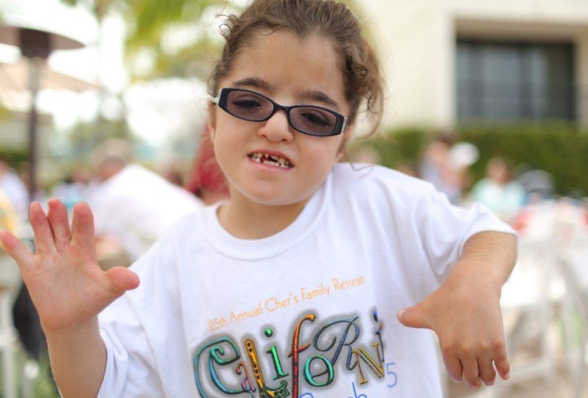
Q1-Q55 questions — tap to reveal all answerslist
- What is the genetic basis of Apert syndrome and how does it relate to the phenotype?
- What are the hand features of Apert syndrome?
- Describe the Upton classification of the Apert hand.
- List the craniofacial features of Apert syndrome.
- Describe the surgical plan in Apert syndrome.
Q1.What is the genetic basis of Apert syndrome and how does it relate to the phenotype?
- Acrocephalosyndactyly type 1; FGF family receptor mutation (FGFR2)
- Severe craniofacial + mild hand: gain of function in FGFR2c affinity for FGF2 (expressed in craniofacial area)
- Mild craniofacial + severe hand: loss of function in FGFR2c specificity, now binds FGF10 (more expressed in hands)
Q2.What are the hand features of Apert syndrome?
- Spoon shaped with tapered ends
- Border: simple +/- clinodactyly; middle 3 complex
- Triphalangeal thumb
Q3.Describe the Upton classification of the Apert hand.
- I: spade (IF to RF fused), thumb free
- II: mitten (thumb to RF fused)
- III: rosebud (all fused)
Q4.List the craniofacial features of Apert syndrome.
- Frontal bossing, flat occiput
- Wideset eyes, lower outer canthus
- Short maxilla, prominent jaw
- MR (mental retardation)
Q5.Describe the surgical plan in Apert syndrome.
- Staged surgery
- Border digit separation at 6 months - PP osteotomy, fix mid axis with K wire
- Central digits at 1 year
- Correct delta phalanx
▸ Slide 797 · Describe photo:Hand Soft Tissue · 7 questions expand
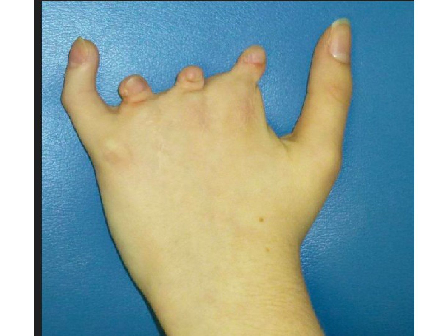
Q1-Q77 questions — tap to reveal all answerslist
- Describe the clinical photo and diagnosis.
- How does symbrachydactyly differ from constriction band and cleft hand?
- How is symbrachydactyly assessed?
- What are the associations and pathogenesis of symbrachydactyly?
- What is the management of symbrachydactyly?
- Describe the Blauth and Gekeler classification of symbrachydactyly.
- What is symphalangism and how is it managed?
Q1.Describe the clinical photo and diagnosis.
- Deformity of the left hand; grossly short I/F, M/F and R/F with nail plate still visible
- L/F angular deformity; thumb appears normal
- Symbrachydactyly: short central digits with thumb and little finger preserved; starts at MP
Q2.How does symbrachydactyly differ from constriction band and cleft hand?
- Different from constriction band: nail/DP present, no acrosyndactyly/fenestrations, no constriction bands
- Different from cleft hand: metacarpals present
Q3.How is symbrachydactyly assessed?
- Hx: birth history; look for other anomalies such as Poland syndrome
- PE local: assess if pinch is achieved; assess flexor and extensor tendon function
- Systemic assessment for associated anomalies
Q4.What are the associations and pathogenesis of symbrachydactyly?
- Association: Poland syndrome - hypoplasia of chest wall (absence of sternocostal head of pec major), hypoplasia of breast, syndactyly, brachysyndactyly
- Middle phalanx chondrification and differentiation of the 2 heads of pec major occur at a similar time (44-47 days)
- Proposed vascular mechanism: subclavian artery hypoplasia
Q5.What is the management of symbrachydactyly?
- Lengthen MC so thumb can touch the tip of other digits
- Transfer with a mobile joint (at least 1 digit for pinch)
- Vascularized/non-vascularized toe transfer; bilateral second toe for thumb + little finger pincer grip
- In-phasic tendon transfer; deep webspace; prosthesis
Q6.Describe the Blauth and Gekeler classification of symbrachydactyly.
- Type 1 = short fingers
- Type 2 = oligodactylic = atypical cleft hand (central portion aplastic, border digits less affected)
- Type 3 = monodactylic (all fingers gone except thumb aplasia)
- Type 4 = peromelic (resembles transverse amputation at the MCPJ level)
Q7.What is symphalangism and how is it managed?
- Fusion of PIPJ
- Hereditary or non-hereditary
- Non-hereditary: related to Poland's syndrome and Apert's syndrome; hereditary is related to hearing loss
- Mx: observation / bone / soft tissue procedure
▸ Slide 798 · Cleft handHand Soft Tissue · 7 questions expand
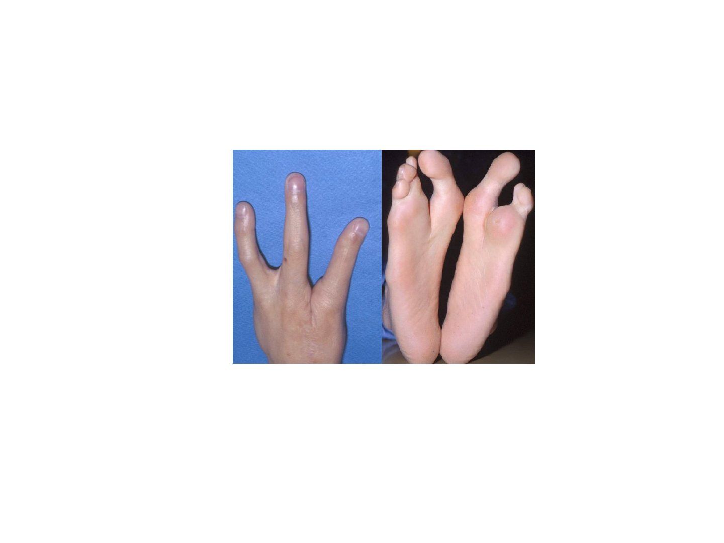
Q1-Q77 questions — tap to reveal all answerslist
- What are the features of a cleft hand?
- Which associations should be looked for in cleft hand?
- Describe the Manske classification of cleft hand.
- What are the genetic associations of cleft hand?
- What are the steps of cleft hand reconstruction?
- Which cleft hand types are observed and what is the surgical timing?
- What is the pathology of cleft hand?
Q1.What are the features of a cleft hand?
- Absent MF to radial digit (thumb); LF always present
- Absent MC as evident by cleft proximal to MCPJ
Q2.Which associations should be looked for in cleft hand?
- Cleft foot
- Bilaterality
Q3.Describe the Manske classification of cleft hand.
- 1-3: thumb and IF present
- 1: thumb web normal; 2A: mild narrowing; 2B: severe narrowing
- 3: syndactylised thumb + IF syndactyly
- 4: merged web (thumb and/or IF absent)
- 5: absent web (thumb absent)
Q4.What are the genetic associations of cleft hand?
- FHx AD
- Ectrodactyly-ectodermal dysplasia-cleft (EEC) syndrome
- Sensorineural hearing loss
- Syndactyly and polydactyly
Q5.What are the steps of cleft hand reconstruction?
- Recreate web commissure
- Excise intervening MC + reconstruct interMC ligament
- Transposition if >3 MC present
- Type 5 - pollicization
Q6.Which cleft hand types are observed and what is the surgical timing?
- Function okay in type 1 and 4 --> observe
- Type 2, 3, 5 --> surgery
- Type 3: separation at 6 months; others at 1 year
Q7.What is the pathology of cleft hand?
- Central deficiency with varied absence of the long ray
- Due to wedge-shaped degeneration at the centre (AER)
▸ Slide 799 · Describe photo:Hand Soft Tissue · 7 questions expand

Q1-Q77 questions — tap to reveal all answerslist
- Describe the findings in these clinical photos.
- Describe the Patterson classification of constriction band syndrome.
- What are the risk factors for constriction band syndrome?
- What are the associations and investigations in constriction band syndrome?
- What is the surgical technique for constriction band release?
- What is the timing of surgery in constriction band syndrome?
- What is constriction band syndrome (Streeter dysplasia)?
Q1.Describe the findings in these clinical photos.
- Left: band over distal leg, distal edema, no ischaemic change
- Right: 4 fingers with gross shortening, incomplete syndactyly with proximal region not fused and fenestrations visible --> acrosyndactyly
Q2.Describe the Patterson classification of constriction band syndrome.
- 1: simple
- 2: distal edema
- 3: acrosyndactyly (fusion between the more distal portions of the digits)
- 4: amputation
Q3.What are the risk factors for constriction band syndrome?
- Prematurity
- Low birth weight
- Maternal smoking
- Drug exposure (decongestants, NSAIDs, antihypertensives)
Q4.What are the associations and investigations in constriction band syndrome?
- Rare for only one ring to be present as an isolated malformation
- Associations: club foot, Cleft lip/ palate
- Local: NV status; associated hand anomalies - syndactyly, hypoplastic digits, brachydactyly
- Ix: X-ray for presence of complex syndactyly
Q5.What is the surgical technique for constriction band release?
- Excision of the ring and subcutaneous tissue combined with Z-plasty
- If circulation compromised (EOT): release the band by longitudinal incision first to improve circulation
- Deep band: single side first, wait 3 months, then complete, to prevent NV compromise
Q6.What is the timing of surgery in constriction band syndrome?
- Timing of OT depends on the Patterson type
- Simple --> can be elective OT
- Acrosyndactyly: border at 6 months, central at 1 to 1.5 yrs old
Q7.What is constriction band syndrome (Streeter dysplasia)?
- A malformation due to intrauterine rings or bands which constrict fetal tissue
- Sporadic with no hereditary predisposition
▸ Slide 800 · Clinical photo of patients both hands showing an obvious discrepancy in size ofHand Soft Tissue · 7 questions expand
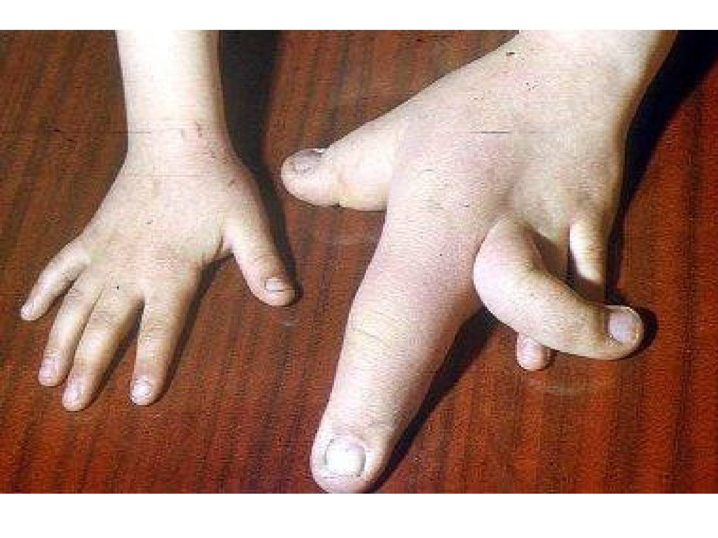
Q1-Q77 questions — tap to reveal all answerslist
- Describe the diagnosis and classification of this hand.
- What associations should be screened for in macrodactyly?
- Describe the natural history and classification of macrodactyly.
- What history, examination and investigation are needed in macrodactyly?
- What are the principles and bone procedures in macrodactyly management?
- What soft tissue and nerve procedures are used in macrodactyly?
- What are the complications of macrodactyly treatment?
Q1.Describe the diagnosis and classification of this hand.
- Discrepancy in size of the entire left hand, most marked in the index finger; angular deformity of the middle finger
- Macrodactyly
- Swanson classification: overgrowth; OMT classification: dysplasia (hypertrophy)
Q2.What associations should be screened for in macrodactyly?
- NF, Proteus syndrome, Ollier's disease, Maffucci syndrome
- Congenital lymphoedema (Milroy's disease)
- Klippel-Trenaunay-Weber syndrome, Beckwith-Wiedemann syndrome
Q3.Describe the natural history and classification of macrodactyly.
- No known cause; 90% unilateral; 70% more than one finger; I/F is the most common
- Associated with lipofibromatous hamartoma of nerve, especially the median nerve
- Natural history - it will grow!
- Static = large at birth, proportional growth to other digits; dynamic = progressive disproportionate (more associated with NF)
Q4.What history, examination and investigation are needed in macrodactyly?
- Hx: functional problems, birth history, family history
- Local PE: joint, tendon, collaterals, NV; digital Allen test
- Systemic: screen for associated syndromes (NF, Proteus, Ollier's, Maffucci, Milroy's, KTWS, Beckwith-Wiedemann)
- Ix: X-ray
Q5.What are the principles and bone procedures in macrodactyly management?
- Principle: preserve a functional digit if possible; prime parents for multiple operations and even amputation; refer paediatrician
- Longitudinal growth: epiphysiodesis when digit reaches adult size
- Circumferential overgrowth: serial reduction osteotomy or longitudinal narrowing osteotomy
Q6.What soft tissue and nerve procedures are used in macrodactyly?
- Debulking done half at a time, 3 months apart
- Nerve stripping
- Nerve: median nerve excision (lipohamartoma) / neurectomy
- Amputation
Q7.What are the complications of macrodactyly treatment?
- Digital stiffness
- Chronic digital pain or edema
▸ Slide 801 · AP Xray: radial shortening and increase inclination, V shaped carpus, carpal ulnHand Soft Tissue · 7 questions expand
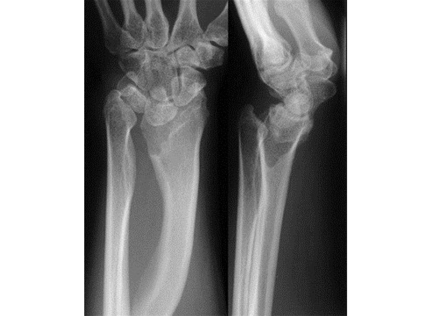
Q1-Q77 questions — tap to reveal all answerslist
- What is the diagnosis and what are the AP X-ray findings?
- What are the lateral X-ray findings in Madelung deformity?
- What is the differential diagnosis of Madelung deformity?
- What are the clinical features of Madelung deformity?
- Describe the Vander and Watson classification by aetiology.
- How is Madelung deformity managed according to skeletal maturity?
- Describe the pathophysiology and genetics of Madelung deformity.
Q1.What is the diagnosis and what are the AP X-ray findings?
- Diagnosis: Madelung deformity
- Radial shortening and increased radial inclination
- V-shaped carpus
- Carpal ulnar translation
Q2.What are the lateral X-ray findings in Madelung deformity?
- Volar tilting of the radius
- Dorsal subluxation/dislocation of the ulna
- Volar subluxation of the carpus
Q3.What is the differential diagnosis of Madelung deformity?
- HME type III
- Malunion
- MED (multiple epiphyseal dysplasia)
- JIA
Q4.What are the clinical features of Madelung deformity?
- Hx: gymnast? trauma; pain from ulnar impaction; median nerve symptoms (Medial nerve sx?)
- Stiffness: decreased wrist extension, radial deviation and supination
- PE: median nerve, ROM, ulnar impaction signs, tendon attrition
Q5.Describe the Vander and Watson classification by aetiology.
- 1. Post-traumatic
- 2. Dysplastic (dyschondrosteosis, diaphyseal aclasis aka HME)
- 3. Genetic (e.g. Turner's syndrome, Leri-Weill dyschondrosteosis)
- 4. Idiopathic
Q6.How is Madelung deformity managed according to skeletal maturity?
- Observe/conservative by avoiding trauma
- Skeletally immature: ulnar epiphysiodesis + radial physeal bar fat interposition (release Vickers ligament, need MRI)
- Skeletally mature: radial corrective osteotomy +/- distal ulnar shortening osteotomy
- DRUJ arthroplasty (very controversial)
Q7.Describe the pathophysiology and genetics of Madelung deformity.
- AD, female; congenital dyschondrosis of the distal radial physis --> partial growth deficiency --> excessive radial inclination and volar tilt --> ulnar carpal impaction
- Caused by disruption of the ulnar volar physis of the distal radius
- Repetitive trauma/dysplastic arrest - tethered Vickers ligament (thick fibrous band from DR to lunate and TFCC on volar side)
- Leri-Weill dyschondrosteosis: pseudoautosomal dominant, SHOX gene
▸ Slide 802 · Hand ulnar deviated at wrist + pronationHand Soft Tissue · 7 questions expand

Q1-Q77 questions — tap to reveal all answerslist
- What are the clinical features of ulnar club hand?
- Describe the Bayne classification of ulnar deficiency.
- What are the clinical assessment points in ulnar club hand?
- What are the aims and principles of management of ulnar club hand?
- How are types 1-3 ulnar club hand managed?
- How is type 4 ulnar club hand managed?
- What is the pathology of ulnar club hand?
Q1.What are the clinical features of ulnar club hand?
- Hand ulnar deviated at the wrist + pronation
- Absent crease at wrist and elbow --> suspect radiohumeral synostosis
- RF and LF syndactyly; +/- loss of ulnar digits
- Hand on flank posture
Q2.Describe the Bayne classification of ulnar deficiency.
- 0: deficiencies in carpal/hand only
- 1: undersized ulnar with both growth centres present
- 2: partial aplasia
- 3: absent ulna
- 4: radiohumeral synostosis
Q3.What are the clinical assessment points in ulnar club hand?
- Usually unilateral
- Check elbow ROM
- Assess compensatory wrist and shoulder motion
- Document border syndactyly if present
Q4.What are the aims and principles of management of ulnar club hand?
- Aim: restore mobility + length; joint stability
- Separate border syndactyly if any at 6 months
Q5.How are types 1-3 ulnar club hand managed?
- Forearm - ulnar: release ulnar soft tissue, ulnar lengthening; if absent ulna excise fibrous anlage
- Forearm - radius: realignment osteotomy
- Elbow - stable: radial head excision
- Elbow - unstable: radial head excision + one bone forearm
Q6.How is type 4 ulnar club hand managed?
- Mostly operative as patient is unable to reach the mouth
- Correct pronation and angulation at the radiohumeral synostosis
Q7.What is the pathology of ulnar club hand?
- Postaxial longitudinal deficiency
- Example: fibular hemimelia
▸ Slide 803 · Symphalangism , failure PIPJ formationHand Soft Tissue · 4 questions expand
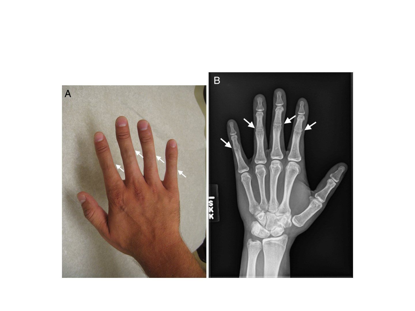
Q1-Q44 questions — tap to reveal all answerslist
- What is symphalangism?
- What are the clinical features and associations of symphalangism?
- What are the aetiological types of symphalangism?
- What is the management of symphalangism?
Q1.What is symphalangism?
- Failure of PIPJ formation
- Failure of segmentation
Q2.What are the clinical features and associations of symphalangism?
- Clinically absent skin crease and stiff joint
- Associated with syndactyly, Poland syndrome and Apert syndrome
Q3.What are the aetiological types of symphalangism?
- Hereditary or non-hereditary
- Non-hereditary: related to Poland's syndrome and Apert's syndrome
- Hereditary: related to hearing loss
Q4.What is the management of symphalangism?
- Observation
- Bone or soft tissue procedure if indicated
▸ Slide 804 · krukenbergHand Soft Tissue · 6 questions expand
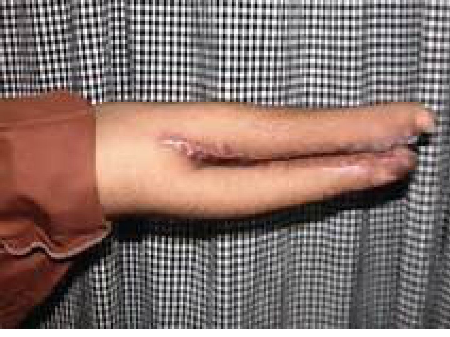
Q1-Q66 questions — tap to reveal all answerslist
- What is the pathology and differential diagnosis of transverse deficiency?
- Describe the prosthetic timeline for a child with transverse deficiency.
- What are the components of an upper limb prosthesis?
- How does an above-elbow prosthesis operate?
- How does a below-elbow prosthesis operate?
- What is the Krukenberg procedure and its indications/prerequisites?
Q1.What is the pathology and differential diagnosis of transverse deficiency?
- Ddx: constriction band, symbrachydactyly
- Patho: AER truncation
Q2.Describe the prosthetic timeline for a child with transverse deficiency.
- 1st line: prosthesis
- 6-9 months: passive device
- 15-24 months: body power prosthesis
- >3 years: myoelectric
Q3.What are the components of an upper limb prosthesis?
- Cable
- Figure 8 harness
- Socket
- Elbow hinge
Q4.How does an above-elbow prosthesis operate?
- Elbow movement is activated when the shoulder extends and depresses
- Terminal device can lock the elbow first, then be activated by shoulder abduction and flexion
Q5.How does a below-elbow prosthesis operate?
- Retraction of the shoulder opens the hand by cable
Q6.What is the Krukenberg procedure and its indications/prerequisites?
- Convert the forearm into a pincer apparatus (pronator teres; radius and ulna function independently)
- Indication: bilateral + significant deficiency + reliance on tactile feedback (vision loss); no access to prosthesis
- Prerequisites: length >8cm; pronator teres power normal