Hand Bone
Metacarpal fractures and MCP injuries
Metacarpal shaft, head and base fractures, MCP subluxation, CMC dislocation, classification.
21 questions
4 source pages
2 images
Images appear with the first question taken from each source page — tap a question to open it.
Q1What are the defining features of each Mason grade of radial head fracture?📷▸
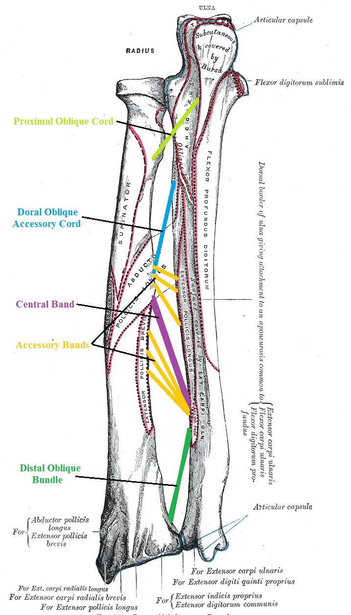
- 1: minimally displaced
- 2: displaced >2mm or >30% articular
- 3: comminuted fracture
- 4: fracture + elbow dislocation
Q2What are the normal radiographic relationships of the forearm?▸
- US to CP 180 degrees on XR
- RS to BT 180 degrees on XR
Q3How is radial bowing measured?▸
- Max radial bow at 60% of radial length; value <10% of radial length
- x/y x 100%, where y = bicep tuberosity to ulnar end of distal radius; x = bicep tuberosity to site of maximum radial bow
- Value = vertical distance between the radial bone and the line at maximum radial bow
Q4What are the clinical telltale signs of a complex MCPJ dislocation?▸
- Deformity less striking than expected due to bayonetting
- Palpable metacarpal head
- Dimple on the volar side
- Also rule out open wound and NV deficit, and document flexor function
- A sesamoid bone is present in 70% of index finger MCP joints
Q5Describe the closed reduction technique for an MCPJ dislocation.▸
- Adequate sedation and analgesia
- Direct pressure over the PP with wrist in flexion to relax intrinsic and extrinsic flexors
- Apply volar and distal directed force, followed by flexion of the MCPJ
- NO traction - may cause jamming of the volar plate into the MCPJ and convert to a complex dislocation
- Looks complex but still try CR as 50% still able to reduce
Q6What is a Kaplan lesion?▸
- Metacarpal head buttonholes into the palm
- Volar plate ruptured
- Radial: lumbricals; ulnar: FDP
- Distal/posterior: natatory ligament; proximal/anterior: superficial transverse metacarpal ligament
- Index finger: lumbrical radial, flexor tendon ulnar
Q7Why is the dorsal approach used for open reduction of an MCPJ dislocation?▸
- Less neurovascular injury
- Easier retrieval of the intraarticular seasamoid
- More direct for fixation of MC head fractures
- BUT need to incise the volar plate
- Volar approach tackles the pathology directly but puts the radial NV bundle at risk
Q8How is a simple MCPJ dislocation managed definitively?▸
- Reduction with flexion
- Immobilise in 60deg flexion
- Simple: immobilise for 2 weeks, then progressive ROM
- Complex (interposed volar plate +/- sesamoid): open reduction
Q9What is the difference in volar plate behaviour between MCPJ and PIPJ dislocations?▸
- MCPJ: volar plate attached to the PP -> will trap (same as thumb), because of the checkrein ligament
- PIPJ: volar plate attached to the PP -> will not trap
Q10What does the X-ray show in this MCPJ dislocation?▸
- Fracture of the 2nd MC head and subluxation of the 2nd MCPJ
- No sesamoid trapped inside the joint
- A sesamoid bone is present in 70% of index finger MCP joints
Q11What is the most common pattern and site of MCPJ dislocation?▸
- Most common pattern of injury is hyperextension
- More common in the index finger
Q12How is open reduction of an MCPJ dislocation performed through a dorsal approach?▸
- Midline incision, split the extensor
- The fibrocartilaginous volar plate is seen but is difficult to differentiate from articular cartilage (both white)
- Make a small incision to confirm the tissue, then complete a longitudinal incision to reduce the MC head
Q13How do Kaplan lesion tissue relations differ between the index and little finger?▸
- IF: lumbrical radial, flexor tendon ulnar
- LF: lumbrical radial, FDB and ADM ulnar
Q14What must be assessed in a metacarpal shaft or neck fracture?▸
- Site and pattern, stability, angulation/displacement/translation
- Associated injury: open wounds, CMCJ dislocation
- Rule out compartment syndrome
- Assess malrotation
Q15Describe the Jahss maneuver.▸
- Jahss maneuver (for neck #)
- MCPJ and PIPJ flexed 90deg
- Dorsal directed force through the proximal phalanx
Q16What angulation and shortening are acceptable in metacarpal fractures?▸
- No rotation is acceptable
- MC 2&3: 15deg; MC 4: 30deg; MC 5: 40deg
- Shortening 2-5mm acceptable
- Mnemonic: 20/30/40/50
Q17What are the operative indications for metacarpal fractures?▸
- Multiple or open fractures
- Unacceptable alignment
Q18What fixation options are used for metacarpal fractures?▸
- K wires parallel or crossed
- Plate if comminution
- External fixator if intra-articular comminution
Q19What do you assess in a 5th CMCJ fracture dislocation?📷▸
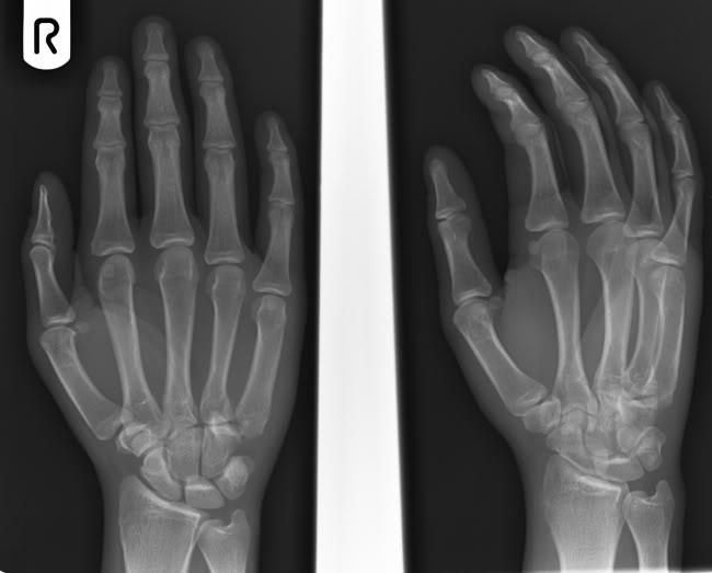
- Soft tissue cover
- NV: motor branch of ulnar nerve near hook of hamate
- Compartment status
- Rotation deformity
- Cascade of MCPJ (draw a curve line on X-ray)
Q20Why is closed reduction alone unlikely to be stable in a 5th CMCJ fracture dislocation?▸
- The dislocation is pulled by ECU
- Close reduction is not likely to be stable
Q21How is a 5th CMCJ fracture dislocation managed operatively?▸
- CR + KWF (K wire fixation) to carpal bone and other MC bones
- Risk: motor branch of ulnar nerve
- K wire must not perforate the volar side