38 slides
▸ Slide 702 · Hand - boneHand Bone · 5 questions expand
Slide render
Question list
Q1-Q55 questions — tap to reveal all answerslist
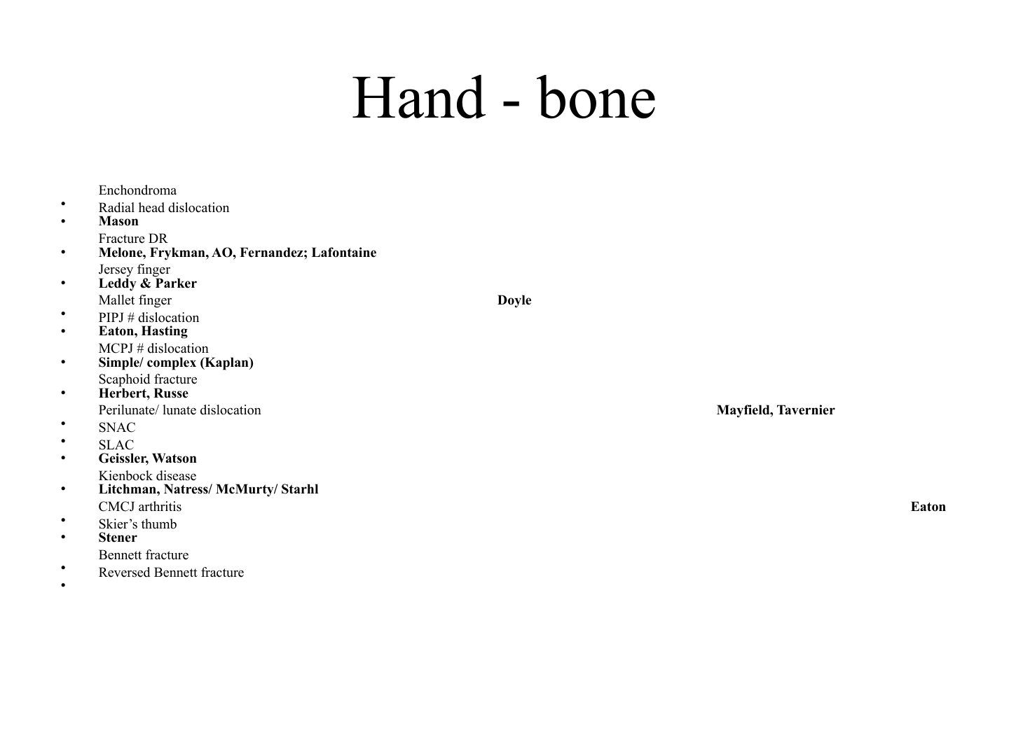
- Which classifications are used for radial head dislocation and distal radius fracture on this slide?
- Which eponymous classifications apply to tendon injuries of the hand?
- Which eponymous classifications apply to finger joint fracture-dislocations?
- Which eponymous classifications apply to carpal and wrist conditions?
- What classification is used for CMCJ arthritis and which other hand conditions are listed on this slide?
Answers · Q & A
Q1.Which classifications are used for radial head dislocation and distal radius fracture on this slide?
- Radial head dislocation — Mason classification
- Distal radius fracture — Melone, Frykman, AO and Fernandez classifications
- Lafontaine criteria applied to distal radius fracture instability
Q2.Which eponymous classifications apply to tendon injuries of the hand?
- Jersey finger — Leddy & Parker classification
- Mallet finger — Doyle classification
Q3.Which eponymous classifications apply to finger joint fracture-dislocations?
- PIPJ fracture dislocation — Eaton, Hasting
- MCPJ fracture dislocation — simple/complex (Kaplan)
Q4.Which eponymous classifications apply to carpal and wrist conditions?
- Scaphoid fracture — Herbert, Russe
- Perilunate/lunate dislocation — Mayfield, Tavernier
- SNAC/SLAC — Geissler, Watson
- Kienbock disease — Litchman, Natress/McMurty/Starhl
Q5.What classification is used for CMCJ arthritis and which other hand conditions are listed on this slide?
- CMCJ arthritis — Eaton classification
- Skier's thumb — Stener
- Bennett fracture and reversed Bennett fracture
- Enchondroma
▸ Slide 703 · Describe Xray:Hand Bone · 5 questions expand
Slide render
Question list
Q1-Q55 questions — tap to reveal all answerslist
- Describe the X-ray findings of this hand lesion.
- What is the differential diagnosis of a lytic bone lesion?
- How do you assess and manage a suspected enchondroma?
- What is the epidemiology and pathogenesis of enchondroma?
- How do you distinguish enchondroma from chondrosarcoma?
Answers · Q & A
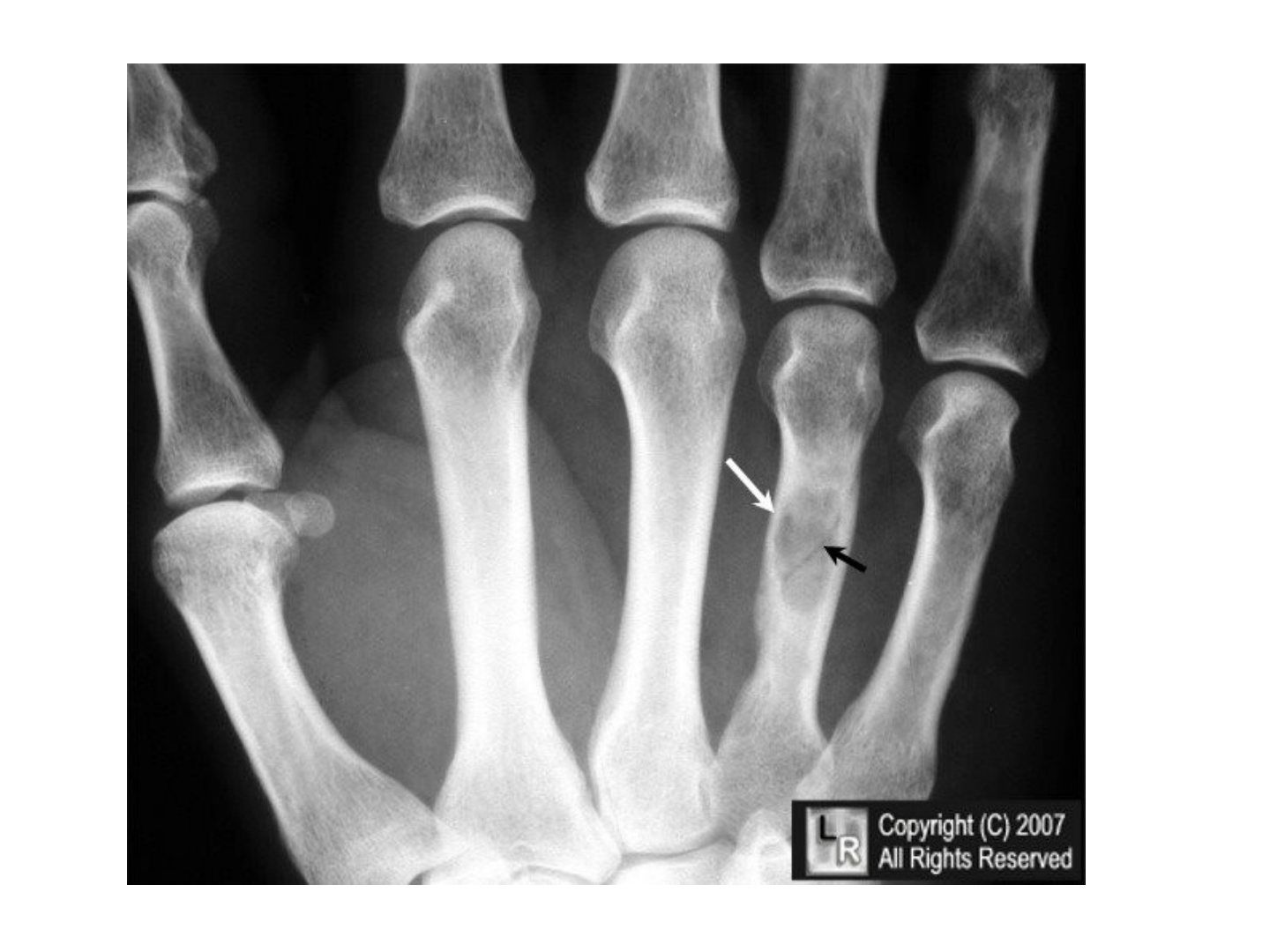
Q1.Describe the X-ray findings of this hand lesion.
- Solitary, central, diaphyseal, osteolytic lesion at the 4th metacarpal
- Matrix +/- rings and arcs suggestive of calcification
- Narrow transitional zone and sclerotic border; mild cortical expansion, no periosteal reaction, cortical breakage
- Overall suggestive of a benign tumour
Q2.What is the differential diagnosis of a lytic bone lesion?
- Fluid
- Fat
- Fibrous
- Chondral
Q3.How do you assess and manage a suspected enchondroma?
- Aim: establish diagnosis, rule out complications
- PMHx renal disease, trauma/infection, pain, overlying skin change, joint ROM
- X-ray very suggestive of enchondroma; may not need biopsy (+/- MRI)
- Ddx: enchondroma (if multiple -> Ollier/Maffucci), infection, brown’s tumour, chondrosarcoma
- No pathological fracture: observation or curettage with bone graft; pathological fracture: allow healing then curettage
Q4.What is the epidemiology and pathogenesis of enchondroma?
- M:F 1:1; most common 20-50 years; diaphysis/metaphysis
- Most common bone tumour in the hand
- Benign chondrogenic tumour of hyaline cartilage from abnormal chondroblast function in the physis
- Escaped chondrobloast from physis displaced to metaphyseal region and proliferate
- Rubin: physeal hyperplasia
Q5.How do you distinguish enchondroma from chondrosarcoma?
- Chondrosarcoma: cortical thickening and destruction, endosteal erosion/scalloping >50% of cortical width, larger >5cm
- Histology enchondroma: bland mature hyaline cartilage (blue balls) separated by normal marrow
- Histology chondrosarcoma: hypercellularity, plump nuclei, binucleate cells, giant cells with chromatin clumps
- Solitary malignant transformation 1%
▸ Slide 704 · Describe Xray:Hand Bone · 4 questions expand
Slide render
Question list
Q1-Q44 questions — tap to reveal all answerslist
- What injury is demonstrated and what associated injuries must be excluded?
- What is an Essex-Lopresti injury?
- What is the management of a radial head fracture with longitudinal radioulnar instability?
- What does the JBJS 2022 review (Sheth) advise?
Answers · Q & A
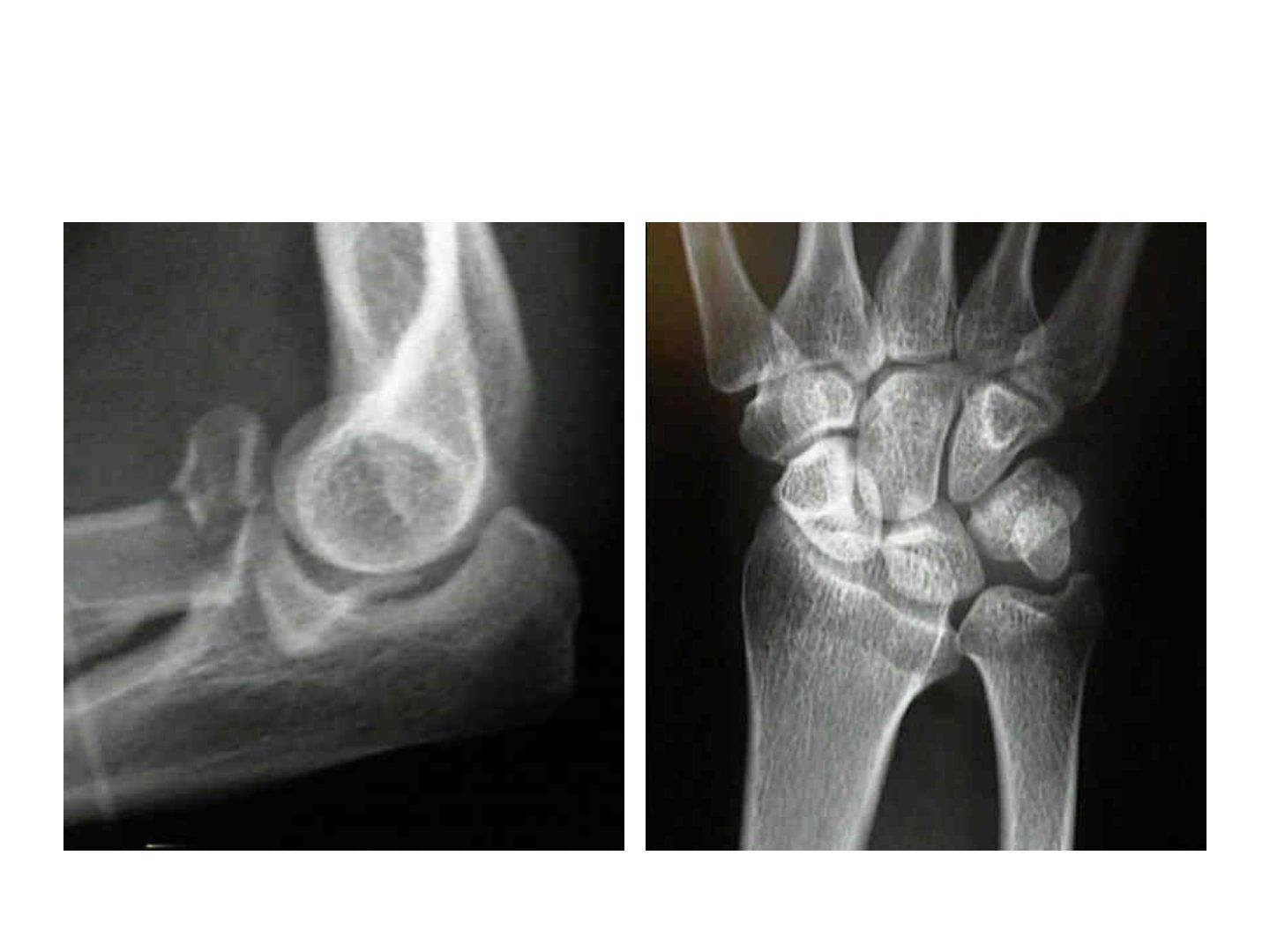
Q1.What injury is demonstrated and what associated injuries must be excluded?
- Comminuted radial head and neck fracture, Mason type 3
- No elbow dislocation or coronoid fracture
- Take X-ray one joint above and below, especially to rule out DRUJ instability (Essex-Lopresti injury)
Q2.What is an Essex-Lopresti injury?
- Radial head fracture + dislocation of the distal radioulnar joint + interosseous membrane disruption
- Incidence ~1-8%
Q3.What is the management of a radial head fracture with longitudinal radioulnar instability?
- Soft tissue: compartment syndrome, PIN palsy
- Bony: restore radial alignment to prevent proximal migration and limited rotational range
- Radial head + neck fixation or replacement depending on comminution
Q4.What does the JBJS 2022 review (Sheth) advise?
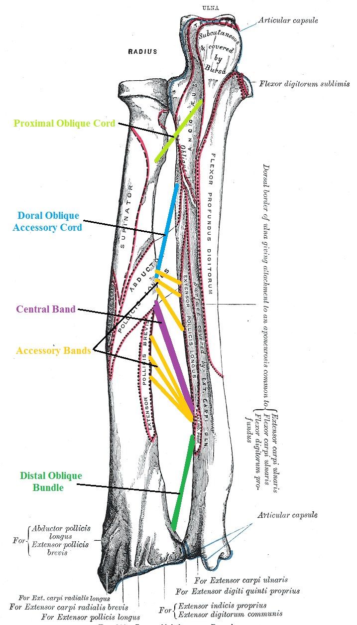
- Leads to longitudinal radioulnar instability
- Acute: restore radial length and temporary DRUJ immobilisation; indication for acute IOM repair/reconstruction unclear
- Chronic: restore radial length, wrist levelling +/- treatment of degenerative changes at RCJ or DRUJ
- Central band reconstruction debated but can restore forearm load transfer
▸ Slide 705 · Mason classificationHand Bone · 3 questions expand
Question list
Q1-Q33 questions — tap to reveal all answerslist
- What are the defining features of each Mason grade of radial head fracture?
- What are the normal radiographic relationships of the forearm?
- How is radial bowing measured?
Answers · Q & A
Q1.What are the defining features of each Mason grade of radial head fracture?
- 1: minimally displaced
- 2: displaced >2mm or >30% articular
- 3: comminuted fracture
- 4: fracture + elbow dislocation
Q2.What are the normal radiographic relationships of the forearm?
- US to CP 180 degrees on XR
- RS to BT 180 degrees on XR
Q3.How is radial bowing measured?
- Max radial bow at 60% of radial length; value <10% of radial length
- x/y x 100%, where y = bicep tuberosity to ulnar end of distal radius; x = bicep tuberosity to site of maximum radial bow
- Value = vertical distance between the radial bone and the line at maximum radial bow
▸ Slide 706 · RA patient with long term steroidHand Bone · 4 questions expand
Slide render
Question list
Q1-Q44 questions — tap to reveal all answerslist
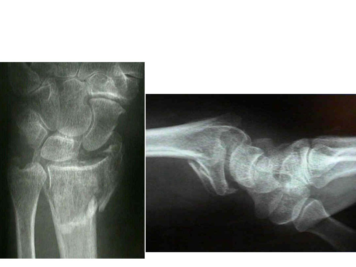
- Describe the X-ray in this RA patient on long-term steroids.
- Which soft tissue structures are at risk in this fracture, and what systemic factor affects management?
- What classifications are used for distal radius fractures?
- What is the management including instability criteria?
Answers · Q & A
Q1.Describe the X-ray in this RA patient on long-term steroids.
- Fracture distal radius with comminution at the volar aspect, intra-articular
- Joint volarly subluxed
- Ulnar styloid fracture
- Soft tissue at risk: SL ligament, TFCC
Q2.Which soft tissue structures are at risk in this fracture, and what systemic factor affects management?
- SL ligament and TFCC at risk (intra-articular fracture with volar subluxation)
- Ulnar styloid fracture present
- Systemic: manage osteoporosis (RA patient on long-term steroid)
Q3.What classifications are used for distal radius fractures?
- AO
- Fernandez: bending/shearing/compression/avulsion/combined or explosion
- Melone 4 components: undisplaced or die-punch/volar spike/separation or rotation/metaphyseal-diaphyseal comminution
- Frykman
Q4.What is the management including instability criteria?
- Systemic: osteoporosis
- Local: CR + slab
- Lafontaine criteria 3/5 = unstable: ulnar, intra-articular, dorsal comminution, dorsal angulation >20 degrees, age >60
- OT: volar plating, screen TFCC; if not stable splint in supination for 3 weeks
▸ Slide 707 · AP and lateral Xray of wrist with fracture of distal radius and distal ulnarHand Bone · 9 questions expand
Question list
Q1-Q99 questions — tap to reveal all answerslist
- What do you assess on the AP and lateral X-ray of a distal radius fracture?
- What are the LaFontaine criteria for instability and what alignment is acceptable?
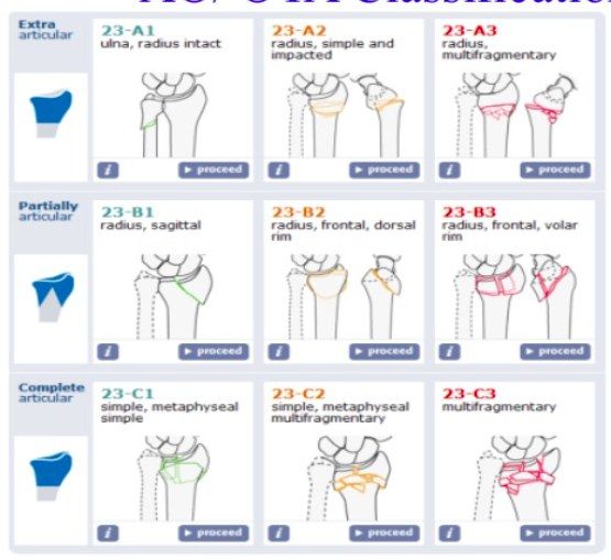
- Describe the AO classification of distal radius fractures.
- Describe the Frykman, Fernandez and Melone classifications of distal radius fractures.
- How is a distal radius fracture acutely managed?
- What is the definitive treatment of a 23-C2 distal radius fracture?
- Describe the modified volar Henry approach and plating.
- What is the Soong classification?
- What is the evidence for treating fragility distal radius fractures and why is a chauffeur fracture unstable?
Answers · Q & A
Q1.What do you assess on the AP and lateral X-ray of a distal radius fracture?
- AP: intra-articular (RCJ or DRUJ), comminution, radial height, radial inclination, ulnar styloid fracture
- Lateral: intra-articular, dorsal/palmar comminution, dorsal/volar tilt
- Beware soft tissue injury: TFCC, SL ligament (especially radial styloid fracture)
Q2.What are the LaFontaine criteria for instability and what alignment is acceptable?
- LaFontaine (1989): >3/5 predicts displacement; age >60, intra-articular, dorsal comminution, ulnar styloid base fracture, dorsal tilt >20 degrees
- AO modification adds osteoporosis, initial displacement >1cm, shortening >5mm, palmar comminution
- Acceptable: radial height <3mm loss (normal 12mm), radial inclination <5 degrees loss (normal 23 degrees)
- Articular step <2mm; no dorsal tilt (normal 11 degrees volar tilt)
Q3.Describe the AO classification of distal radius fractures.
- A: extra-articular (ulnar, radial simple, radial comminution)
- B: partial-articular (sagittal, dorsal, volar)
- C: complete articular (simple, metaphyseal comminuted, articular comminuted)
Q4.Describe the Frykman, Fernandez and Melone classifications of distal radius fractures.
- Frykman: odd numbers for extra-articular (1), RCJ (3), DRUJ (5), both (7); even numbers add ulnar styloid involvement
- Fernandez: bending, shearing, compression, avulsion, explosion
- Melone: lunate impaction (= Fernandez compression); 4 fragments: shaft, radial, medial volar, medial dorsal
Q5.How is a distal radius fracture acutely managed?
- CR under sedation (for pain relief and psychological relaxation) - not haematoma block - + slab
- History: need to ask age of menopause
- Treatment approach depends on patient and fracture factors
- Systemic: treat osteoporosis
Q6.What is the definitive treatment of a 23-C2 distal radius fracture?
- Volar locking plate for absolute stability and stable fixation
- Screen DRUJ: stable = free mobilisation; unstable = splint for 3 weeks
- 2.4mm LCP in buttressing mode; modified volar Henry approach between FCR and radial artery; plate proximal to watershed line
Q7.Describe the modified volar Henry approach and plating.
- Plane between FCR and radial artery; skin, subcut, FCR sheath and subsheath
- Split PQ; reduce fracture; +/- bone substitute
- Volar plate proximal to the watershed line
- Aim: anatomical reduction, stable fixation for early mobilisation
Q8.What is the Soong classification?
- Grade 0: dorsal to the critical line
- Grade 1: volar to the critical line but proximal to the volar rim
- Grade 2: volar to the critical line and on or distal to the volar rim
Q9.What is the evidence for treating fragility distal radius fractures and why is a chauffeur fracture unstable?
- Arora JBJS 2011: cast vs surgery >65, operation better grip strength but no overall clinical difference
- DRAFFT 1 (2019): K-wire vs volar locking plate no difference at 5 years; DRAFFT 2 (BMJ 2022): K-wire vs casting no difference at 12 months
- Chauffeur fracture: avulsion of the styloid by the RSC ligament, rupture of volar capsule and radiolunate ligament allows radiocarpal subluxation
- Brachioradialis insertion 17mm from the styloid tip, so no stabilising force
▸ Slide 708 · 22 yo ulnar sided wrist painHand Bone · 14 questions expand
Slide render

Question list
Q1-Q1414 questions — tap to reveal all answerslist
- What is the clinical presentation on this slide?
- How is ulnar variance measured on a wrist X-ray?
- What are the consequences of positive ulnar variance?
- How are ulnar styloid fractures classified (Fernandez and Jupiter)?
- What is the comprehensive classification with Q modification for distal ulna fractures?
- List the differential diagnosis of ulnar-sided wrist pain by tissue.
- Describe the traumatic types of the Palmer classification of TFCC injury.
- Describe the degenerative stages of the Palmer classification of TFCC injury.
- How is an acute TFCC tear initially managed?
- What are the three aims of treating a chronic TFCC injury?
- Chronic TFCC tear, no DRUJ arthritis, ulnar positive variance - what are the options?
- Why must a 2mm peripheral rim of TFCC be preserved during debridement?
- Chronic TFCC tear with ulnar negative variance - what are the options?
- What are the options when there is DRUJ arthritis or instability with a chronic TFCC tear?
Answers · Q & A
Q1.What is the clinical presentation on this slide?
- 22-year-old with ulnar-sided wrist pain
- Ulnar positive variance
- Type II ulnar styloid fracture (through the base)
Q2.How is ulnar variance measured on a wrist X-ray?
- One line tangential to the distal ulnar articular surface, perpendicular to the shaft
- Another line tangential to the lunate fossa, perpendicular to the shaft
- The interval between the two lines gives the ulnar variance
Q3.What are the consequences of positive ulnar variance?
- Ulnar impaction syndrome
- TFCC tear
- Ulnocarpal arthritis
- SL dissociation
Q4.How are ulnar styloid fractures classified (Fernandez and Jupiter)?
- Type 1: distal to base
- Type 2: through base
- Type 3: proximal to base
Q5.What is the comprehensive classification with Q modification for distal ulna fractures?
- Q1 styloid, Q2 simple neck, Q3 comminuted neck
- Q4 intra-articular, Q5 head + neck, Q6 meta-diaphyseal
Q6.List the differential diagnosis of ulnar-sided wrist pain by tissue.
- Bone: ulnar styloid fracture, hook of hamate fracture
- Cartilage: chondral lesions
- Joint: pisotriquetral arthritis, wrist ganglion
- Tendon: ECU subluxation or tendinitis
- Ligaments: LT ligament injury, TFCC
- Nerve: ulnar nerve entrapment; artery: ulnar artery thrombosis
Q7.Describe the traumatic types of the Palmer classification of TFCC injury.
- Traumatic tears are typed by location
- Central, ulnar, distal, radial
Q8.Describe the degenerative stages of the Palmer classification of TFCC injury.
- A: TFCC wear/thinning
- B: wear/thinning + lunate/ulnar chondromalacia
- C: TFCC perforation + lunate/ulnar chondromalacia
- D: LT ligament perforation
- E: + ulnocarpal arthritis
Q9.How is an acute TFCC tear initially managed?
- Conservative with splintage and NSAIDs
- Observe for 3 months
Q10.What are the three aims of treating a chronic TFCC injury?
- 1. Decrease ulnocarpal loading
- 2. Treat the TFCC tear
- 3. Treat instability
Q11.Chronic TFCC tear, no DRUJ arthritis, ulnar positive variance - what are the options?
- Wafer procedure
- Ulnar shortening
- TFCC debridement - leave a 2mm peripheral rim intact
Q12.Why must a 2mm peripheral rim of TFCC be preserved during debridement?
- To avoid iatrogenic instability
Q13.Chronic TFCC tear with ulnar negative variance - what are the options?
- Arthroscopic debridement
- Lunotriquetral ligament repair or debridement
Q14.What are the options when there is DRUJ arthritis or instability with a chronic TFCC tear?
- Darrach
- Sauve-Kapandji (SK)
- Bower - TFCC has to be repairable or reconstructable
- Ulnar head replacement
▸ Slide 709 · Left: Sauve Kapandji, right: DarrachHand Bone · 9 questions expand
Question list
Q1-Q99 questions — tap to reveal all answerslist
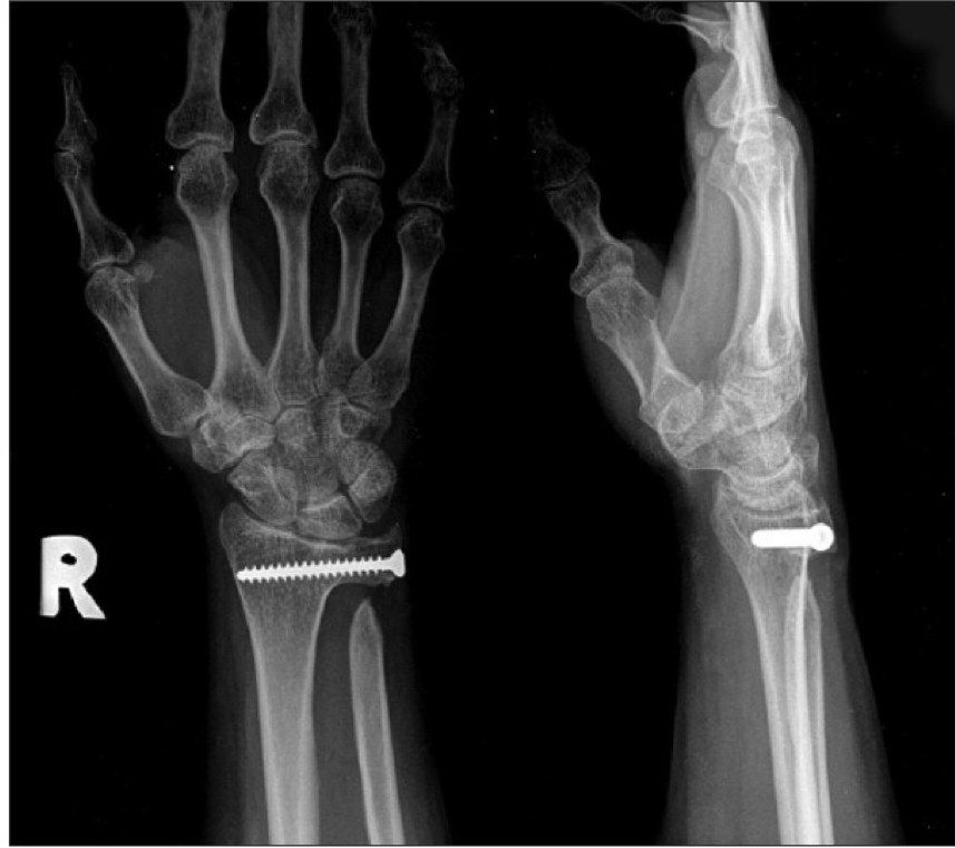
- What is the Sauve-Kapandji procedure?
- What are the problems of the Sauve-Kapandji procedure?
- What is the Darrach procedure?
- What are the complications of the Darrach procedure?
- How is the distal ulna stabilised to prevent convergence after loss of the DRUJ buttress?
- Why is Sauve-Kapandji preferred over Darrach in young patients?
- What is the general indication for the Sauve-Kapandji and Darrach procedures?
- What are the benefits of the Darrach procedure?
- Which muscles contract to cause ulnar convergence after loss of the DRUJ buttress?
Answers · Q & A
Q1.What is the Sauve-Kapandji procedure?
- DRUJ fusion with stump pseudoarthrosis 1cm proximal to the fusion
- Interposition of pronator quadratus
- Maintains ulnocarpal support (less ulnar translocation of carpus), mobile pronation/supination, good strength, better cosmesis than Darrach
- Indicated in the young, high demand patient, including RA
Q2.What are the problems of the Sauve-Kapandji procedure?
- Proximal ulnar impingement
- Failed fusion, or fusion of pseudoarthrosis
- Heterotopic ossification
- Ulnar sensory branch neuritis
- Screw impingement over the far cortex causing iatrogenic DQV
Q3.What is the Darrach procedure?
- Excisional arthroplasty from ulnar styloid to the level of the sigmoid notch
- Retain the ulnar styloid and its attached ligament
- Ulnar stump stabilised by interposed PQ, FCU/ ECU
Q4.What are the complications of the Darrach procedure?
- Loss of ulnar carpal support leading to ulnar translocation of the carpus
- Grip weakness
- Ulnar convergence impingement
Q5.How is the distal ulna stabilised to prevent convergence after loss of the DRUJ buttress?
- Pronator quadratus interposition
- Volar capsular flap
- ECU tenodesis - old distally based (tension depends on wrist position), new proximally based
- Alternative solution: ulnar head prosthesis
Q6.Why is Sauve-Kapandji preferred over Darrach in young patients?
- Maintains ulnocarpal support -> less ulnar translocation of the carpus
- Mobile in pronation and supination
- Good strength
- Better cosmesis
Q7.What is the general indication for the Sauve-Kapandji and Darrach procedures?
- Salvage procedures for DRUJ instability with arthritis
- Alternative is ulnar head replacement
Q8.What are the benefits of the Darrach procedure?
- Improves ROM
- Relieves pain
- Good cosmesis
- Suited to the old, low demand patient
Q9.Which muscles contract to cause ulnar convergence after loss of the DRUJ buttress?
- APL, EPB and PQ
- EPB arises from the radius, some APL from the ulna
- Convergence: 100% happen, 50% sx
▸ Slide 710 · Clinical photo:Hand Bone · 15 questions expand
Slide render
Question list
Q1-Q1515 questions — tap to reveal all answerslist
- Describe the Leddy and Packer classification of FDP avulsion.
- How is an acute FDP avulsion managed?
- What factors decide one- versus two-stage reconstruction in chronic FDP avulsion?
- Describe the two-stage reconstruction of FDP avulsion (Hunter and Salisbury).
- What are the options for pulley reconstruction?
- Why is the ring finger most commonly affected in FDP avulsion?
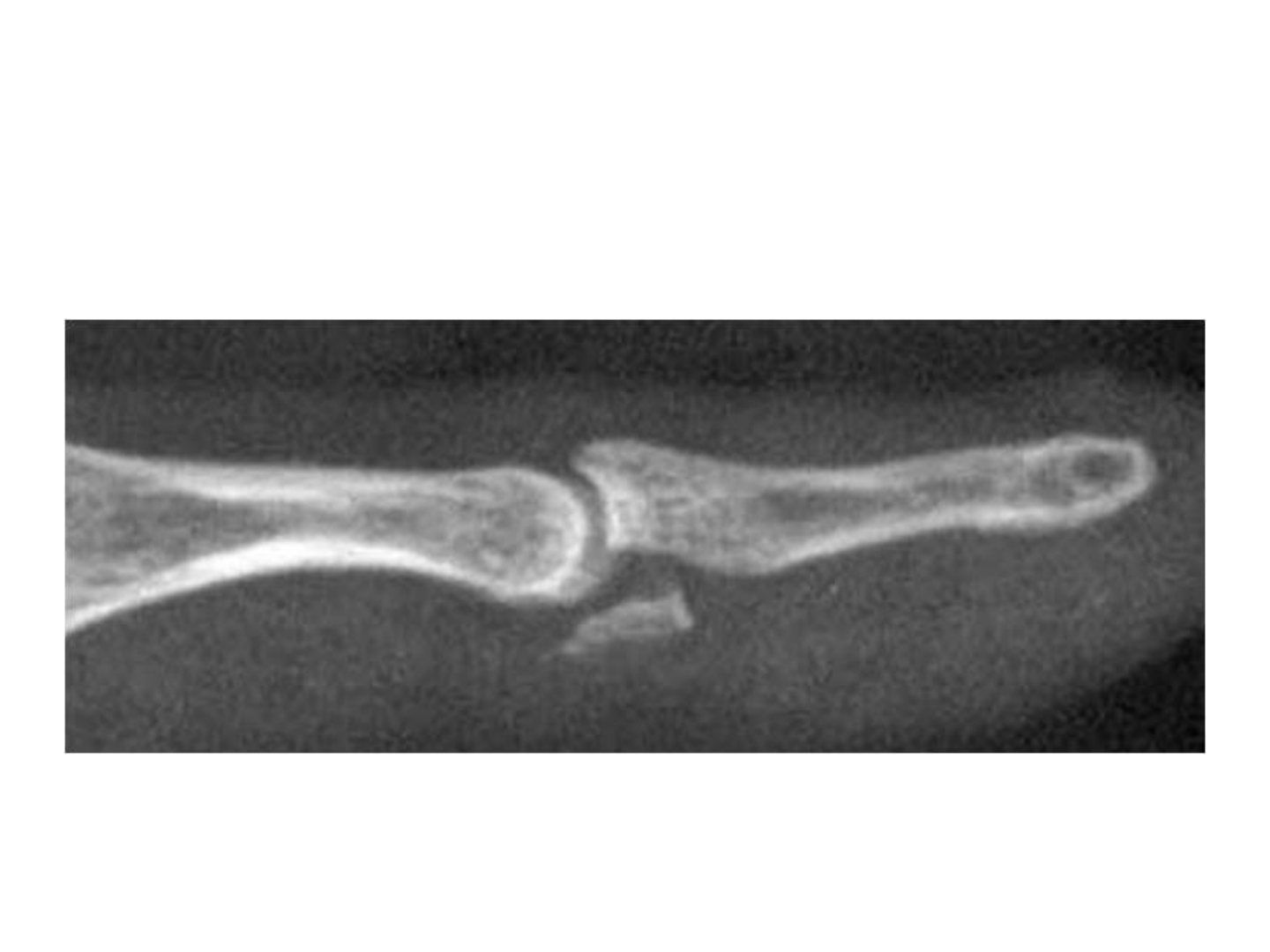
- What is the clinical presentation and X-ray finding in this FDP avulsion?
- What history is important in FDP avulsion?
- What examination findings are sought in FDP avulsion?
- What imaging is used in FDP avulsion?
- What are the complications of FDP avulsion surgery?
- What is the salvage procedure for a failed FDP avulsion reconstruction?
- When is a tension transfer performed in FDP avulsion?
- Describe the Paneva Holovich two-stage reconstruction.
- What are the advantages and disadvantages of the Paneva Holovich technique?
Answers · Q & A
Q1.Describe the Leddy and Packer classification of FDP avulsion.
- Type 1: retracts to the palm, both vinculae disrupted, will not retract beyond the wrist due to the lumbrical
- Type 2: retracts to the PIPJ, distal to A2 pulley, short vinculae disrupted
- Type 3: minimal retraction, distal to A4 pulley
- Type 4: double avulsion
- Type 5: DP base comminution
Q2.How is an acute FDP avulsion managed?
- Large fragment: screw fixation
- Small fragment: pull-out wire
Q3.What factors decide one- versus two-stage reconstruction in chronic FDP avulsion?
- Soft tissue envelope status
- Joint contracture
- Pulley status
- Also guided by chronicity and Leddy and Packer stage
Q4.Describe the two-stage reconstruction of FDP avulsion (Hunter and Salisbury).
- Stage 1: silicon rod placed to create a favourable tendon bed, rod sutured to FDS/FDP as donor; physio for ROM
- Stage 2 (3-4 months): rod retrieved, tendon graft passed through the mesothelium-lined psuedosheath
- Suture to distal first with pull-out wire technique (allows excursion adjustment)
- Suture to proximal at zone 5 with Pulvertaft technique (zone 3 if graft is soft)
Q5.What are the options for pulley reconstruction?
- Non-encircling: Kleinert ever-present rim, Karev belt-loop, Lister extensor retinaculum, Doyle and Blythe palmaris longus
- Encircling: Bunnell single-loop, Okutsu triple-loop
- Need at least 3 loops: one A2 loop and two A4 loops
- A2 loops pass deep to the extensor mechanism; A4 loops (around the bone) pass superficial to the extensors
Q6.Why is the ring finger most commonly affected in FDP avulsion?
- The RF tip is 5mm more prominent in grip in 90% of patients, exposing it to greater forces
- The ring finger cannot be fully extended when the middle and small finger MCPJ are flexed to 90deg, due to intertendinous connections
Q7.What is the clinical presentation and X-ray finding in this FDP avulsion?
- Clinical photo: DIPJ failed flexion, no open wound
- XR: volar DP base fracture
- Articular surface involvement, comminution, +/- DIPJ dislocation/subluxation
Q8.What history is important in FDP avulsion?
- Mechanism of injury
- Chronicity
- Function demands, occupation, smoking
Q9.What examination findings are sought in FDP avulsion?
- Scar
- ROM, active and passive
- Palpate the tendon along its sheath to the level of the palm
Q10.What imaging is used in FDP avulsion?
- USG
Q11.What are the complications of FDP avulsion surgery?
- Fracture
- Stiffness
- Persistent bowstring
Q12.What is the salvage procedure for a failed FDP avulsion reconstruction?
- DIPJ fusion
Q13.When is a tension transfer performed in FDP avulsion?
- If there is a non-functional motor unit
Q14.Describe the Paneva Holovich two-stage reconstruction.
- Stage 1: silicon rod in the flexor sheath, pulleys reconstructed; a loop between the proximal FDS and FDP stumps is created in the palm
- Stage 2: rod retrieved; FDS is cut proximally and reflexed distally through the pseudosheath
- FDS attached directly to FDP or secured with a button
Q15.What are the advantages and disadvantages of the Paneva Holovich technique?
- Advantage: FDS graft size known at stage 1, so the correct rod size is chosen
- Advantage: FDS is intrasynovial, so fewer adhesions
- Advantage: relies on one tenorrhaphy site
- Disadvantage: problems with tension at the distal tenorrhaphy site
▸ Slide 711 · Describe Xray:Hand Bone · 9 questions expand
Slide render

Question list
Q1-Q99 questions — tap to reveal all answerslist
- Describe the X-ray findings in this bony mallet finger.
- Classify mallet finger injuries using the Doyle classification.
- What are the indications and regime for conservative treatment with a mallet splint?
- What are the indications for operation in mallet finger and what techniques are used?
- What reconstruction options exist for chronic mallet finger?
- What are the complications of mallet finger and how is swan neck deformity treated?
- What history is important in mallet finger?
- What examination findings are important in mallet finger?
- What management considerations guide mallet finger treatment?
Answers · Q & A
Q1.Describe the X-ray findings in this bony mallet finger.
- Intra-articular fracture at the dorsal base of the distal phalanx with displacement
- Size ~50% of the articular surface, no comminution
- No subluxation of the DIPJ
Q2.Classify mallet finger injuries using the Doyle classification.
- Type 1: Close soft tissue mallet
- Type 2: laceration
- Type 3: deep laceration with tendon loss
- Type 4: fracture
- Type 4 subtypes: physeal injury, 20-50% art surface, >50% art surface of the articular surface
Q3.What are the indications and regime for conservative treatment with a mallet splint?
- Soft tissue mallet OR small dorsal fragment with no joint subluxation, injury <12 weeks
- Immobilise the DIPJ in extension for 6-8 weeks (2 weeks long mallet, 4 weeks short mallet splint)
- Warn of residual extension lag that will not affect function, and possible later swan neck deformity
Q4.What are the indications for operation in mallet finger and what techniques are used?
- Incongruent DIPJ, >25% fracture, >2mm displacement, open injury
- Screw fixation for a sizable fragment (>50%)
- Extension blocking K wire (Ishiguro technique): dorsal block wire plus a second volar wire holding the DIPJ in extension
- Pull out wire
Q5.What reconstruction options exist for chronic mallet finger?
- Terminal extensor tendon shortening
- Fowler central slip tenotomy
- Tenodermodesis (elliptical wound)
- Spiral oblique retinacular ligament reconstruction (PL graft from dorsal DP base, radial MP, volar PIPJ, ulnar PP)
- Salvage surgery: DIPJ fusion
Q6.What are the complications of mallet finger and how is swan neck deformity treated?
- Stiffness
- Swan neck deformity from secondary attenuation of the PIPJ volar plate and transverse retinacular ligament
- Mild: correct the mallet finger
- Severe: lateral band tenodesis / FDS tenodesis
Q7.What history is important in mallet finger?
- Mechanism of injury - hyperflexion, reducing by extension and vice versa
- Hand dominance, smoker, occupation
- Chronicity
Q8.What examination findings are important in mallet finger?
- Wounds
- Confirm no active DIPJ extension
- PROM in chronic cases
Q9.What management considerations guide mallet finger treatment?
- Open or closed
- Bony or soft tissue
- Size of the bony fragment
- Subluxation of the joint
- Timing of injury
▸ Slide 712 · PIPJ # subluxationHand Bone · 12 questions expand
Slide render

Question list
Q1-Q1212 questions — tap to reveal all answerslist
- Describe the Eaton classification of PIPJ fracture-dislocation.
- When does a PIPJ fracture-dislocation require surgery?
- How do you perform closed reduction of a PIPJ dislocation?
- How is stability assessed after reduction of a PIPJ fracture-dislocation?
- What is the definitive management of PIPJ injuries by direction?
- What are the surgical options for PIPJ fracture-dislocation?
- Describe the anatomy of the PIPJ.
- Describe the Eaton classification in terms of ligament disruption.
- What history is important in a PIPJ fracture-dislocation?
- What examination findings are important in a PIPJ fracture-dislocation?
- What are the components of the Suzuki frame for dynamic external fixation?
- What are the problems of a chronic PIPJ fracture-dislocation deformity?
Answers · Q & A
Q1.Describe the Eaton classification of PIPJ fracture-dislocation.
- Type 1: hyperextension
- Type 2: dislocation
- Type 3: fracture-dislocation, then graded by Hastings: I <30%, II 30-50%, III >50% articular surface
- Fracture fragment typically involves ~50% of the joint surface and is likely an avulsion from the volar plate
Q2.When does a PIPJ fracture-dislocation require surgery?
- Eaton III with Hastings II or III
- Irreducible dislocation
- Aim: stable joint for early mob & prevent stiffness
- Also request AP X-ray to check for a rotatory component
Q3.How do you perform closed reduction of a PIPJ dislocation?
- Dorsal: flex wrist and MCPJ, gentle hyperextension of the MP followed by palmar force
- Volar: reverse of dorsal
- Rotatory: traction with MCPJ and PIPJ at 90deg flexion to relax the lateral band
- Check NV status and document collateral and volar plate integrity
Q4.How is stability assessed after reduction of a PIPJ fracture-dislocation?
- Assess PIPJ stability range - unstable if >30deg flexion is needed to remain reduced
- Elson test for volar dislocation
- Post-reduction XR: articular fragment size, PIPJ Shenton line and V sign
Q5.What is the definitive management of PIPJ injuries by direction?
- Dorsal, stable, no fracture: dorsal block splint allowing active flexion
- Dorsal, unstable: operative treatment
- Volar: extension splint full time 3 weeks, then 3 weeks nocturnal splint
- Lateral: buddy taping
Q6.What are the surgical options for PIPJ fracture-dislocation?
- Dynamic external fixation (Suzuki frame) for comminuted/open - maintains length by ligamentotaxis
- ORIF with screw for a sizeable fragment
- Extension block pinning for a congruent joint
- Hemihamate graft for comminuted/chronic cases; volar plate arthroplasty in chronic cases
- Chronic <50% surface: volar plate arthroplasty (K wire 2-3 weeks + dorsal block splint, then dynamic splint 6-8 weeks)
- Chronic >50%: ORIF + hemihamate autograft (distal dorsal hamate at 4/5 CMCJ) with K wire fixation
Q7.Describe the anatomy of the PIPJ.
- Hinged joint, congruent with the intercondylar sulcus
- 3-sided box
- Stability from bone and soft tissue (volar plate + collaterals)
- 2/3 of soft tissue stabilisers must be injured for dislocation to occur
Q8.Describe the Eaton classification in terms of ligament disruption.
- 1: partial disruption between the proper and accessory collaterals
- 2: complete disruption of both collaterals + volar plate
- 3: fracture - loss of bony stability, collaterals stay with the proximal fragment, unstable++
Q9.What history is important in a PIPJ fracture-dislocation?
- Mechanism of injury
- Chronicity
- Premorbid status
Q10.What examination findings are important in a PIPJ fracture-dislocation?
- Skin: wounds or dimple
- Deformity: chronic dislocation, dorsal pseudoboutonniere
- NV status
Q11.What are the components of the Suzuki frame for dynamic external fixation?
- Traction wire, reduction wire and hook wire (1.2mm)
- Allows PIPJ movement and maintains length by ligamentotaxis
Q12.What are the problems of a chronic PIPJ fracture-dislocation deformity?
- Resorbed bone fragment
- Contracted soft tissue
- Scar over the fracture site
▸ Slide 713 · Volar plate:Hand Bone · 4 questions expand
Slide render
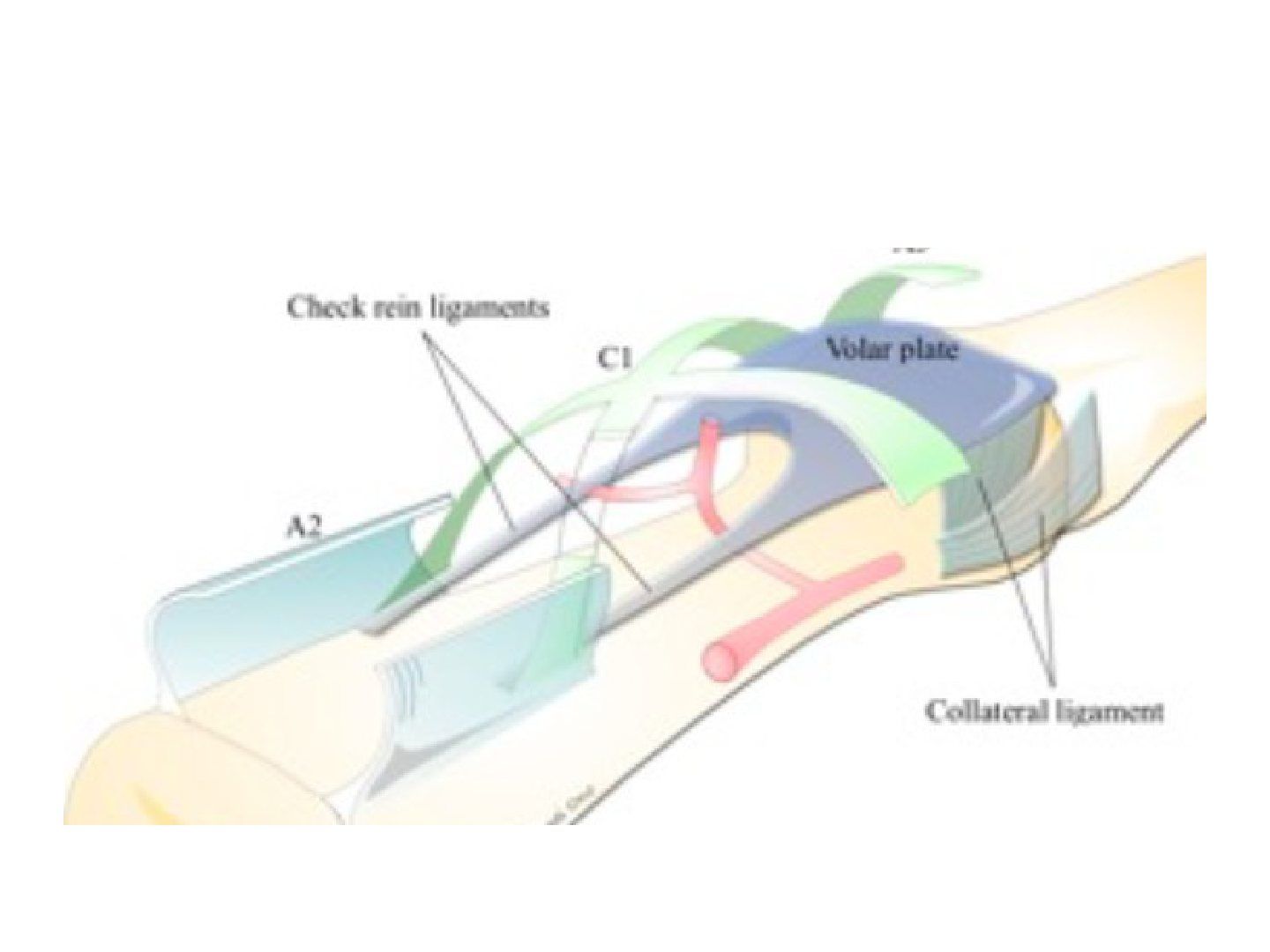
Question list
Q1-Q44 questions — tap to reveal all answerslist
- What is the function of the volar plate?
- Describe the anatomy of the MCP volar plate.
- Describe the collateral ligament anatomy.
- Why do complex dislocations occur at the MCPJ but not the PIPJ?
Answers · Q & A
Q1.What is the function of the volar plate?
- Reinforces the joint capsule
- Enhances joint stability
- Limits hyperextension
- Loose in flexion, tight in extension
Q2.Describe the anatomy of the MCP volar plate.
- Thickening of the joint capsule volar to the MP joint
- Origin: metacarpal head
- Insertion: periarticular surface of the proximal phalanx, via checkrein ligaments
Q3.Describe the collateral ligament anatomy.
- Origin from the collateral recess
- Proper collateral attached to the metacarpal head (dorsal one)
- Accessory collateral attached to the volar plate (volar one)
- Tighten in extension
Q4.Why do complex dislocations occur at the MCPJ but not the PIPJ?
- PIPJ: checkrein inserts into the distal PP, stronger, so no complex dorsal dislocation
- MCPJ: checkrein inserts into the proximal PP, stronger, so complex dislocation happens in dorsal dislocation
▸ Slide 714 · Xray showing fracture of 2nd MC head and subluxation of 2nd MCPJ, no sesamoid trHand Bone · 10 questions expand
Slide render
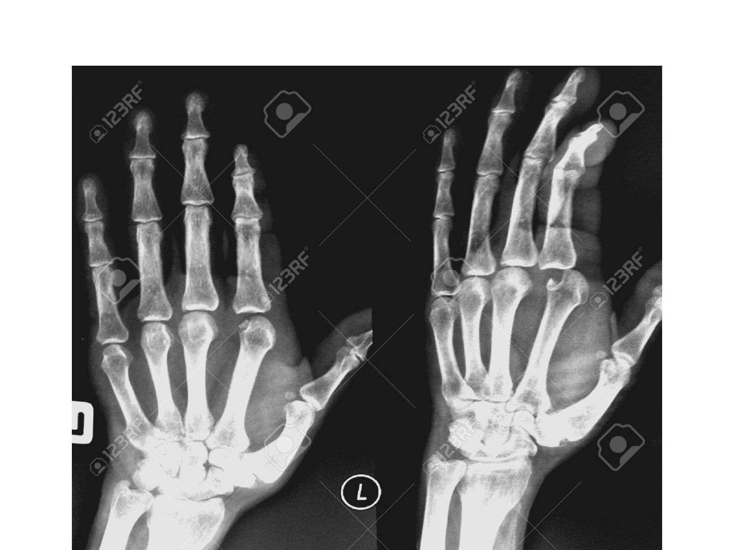
Question list
Q1-Q1010 questions — tap to reveal all answerslist
- What are the clinical telltale signs of a complex MCPJ dislocation?
- Describe the closed reduction technique for an MCPJ dislocation.
- What is a Kaplan lesion?
- Why is the dorsal approach used for open reduction of an MCPJ dislocation?
- How is a simple MCPJ dislocation managed definitively?
- What is the difference in volar plate behaviour between MCPJ and PIPJ dislocations?
- What does the X-ray show in this MCPJ dislocation?
- What is the most common pattern and site of MCPJ dislocation?
- How is open reduction of an MCPJ dislocation performed through a dorsal approach?
- How do Kaplan lesion tissue relations differ between the index and little finger?
Answers · Q & A
Q1.What are the clinical telltale signs of a complex MCPJ dislocation?
- Deformity less striking than expected due to bayonetting
- Palpable metacarpal head
- Dimple on the volar side
- Also rule out open wound and NV deficit, and document flexor function
- A sesamoid bone is present in 70% of index finger MCP joints
Q2.Describe the closed reduction technique for an MCPJ dislocation.
- Adequate sedation and analgesia
- Direct pressure over the PP with wrist in flexion to relax intrinsic and extrinsic flexors
- Apply volar and distal directed force, followed by flexion of the MCPJ
- NO traction - may cause jamming of the volar plate into the MCPJ and convert to a complex dislocation
- Looks complex but still try CR as 50% still able to reduce
Q3.What is a Kaplan lesion?
- Metacarpal head buttonholes into the palm
- Volar plate ruptured
- Radial: lumbricals; ulnar: FDP
- Distal/posterior: natatory ligament; proximal/anterior: superficial transverse metacarpal ligament
- Index finger: lumbrical radial, flexor tendon ulnar
Q4.Why is the dorsal approach used for open reduction of an MCPJ dislocation?
- Less neurovascular injury
- Easier retrieval of the intraarticular seasamoid
- More direct for fixation of MC head fractures
- BUT need to incise the volar plate
- Volar approach tackles the pathology directly but puts the radial NV bundle at risk
Q5.How is a simple MCPJ dislocation managed definitively?
- Reduction with flexion
- Immobilise in 60deg flexion
- Simple: immobilise for 2 weeks, then progressive ROM
- Complex (interposed volar plate +/- sesamoid): open reduction
Q6.What is the difference in volar plate behaviour between MCPJ and PIPJ dislocations?
- MCPJ: volar plate attached to the PP -> will trap (same as thumb), because of the checkrein ligament
- PIPJ: volar plate attached to the PP -> will not trap
Q7.What does the X-ray show in this MCPJ dislocation?
- Fracture of the 2nd MC head and subluxation of the 2nd MCPJ
- No sesamoid trapped inside the joint
- A sesamoid bone is present in 70% of index finger MCP joints
Q8.What is the most common pattern and site of MCPJ dislocation?
- Most common pattern of injury is hyperextension
- More common in the index finger
Q9.How is open reduction of an MCPJ dislocation performed through a dorsal approach?
- Midline incision, split the extensor
- The fibrocartilaginous volar plate is seen but is difficult to differentiate from articular cartilage (both white)
- Make a small incision to confirm the tissue, then complete a longitudinal incision to reduce the MC head
Q10.How do Kaplan lesion tissue relations differ between the index and little finger?
- IF: lumbrical radial, flexor tendon ulnar
- LF: lumbrical radial, FDB and ADM ulnar
▸ Slide 715Hand Bone · 2 questions expand
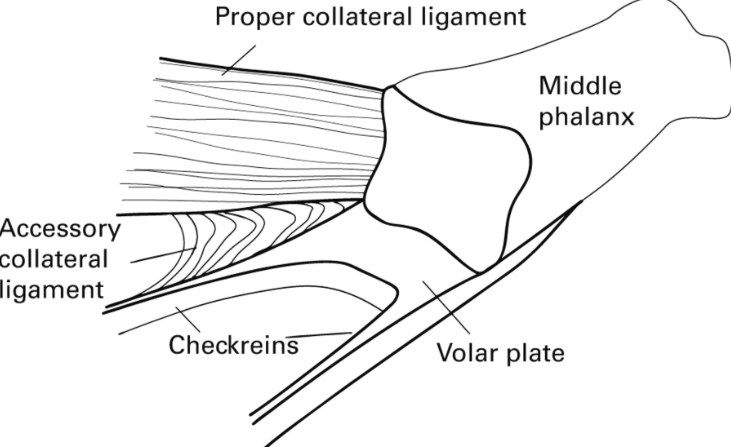
Question list
Q1-Q22 questions — tap to reveal all answerslist
- What hand bone condition is illustrated on this slide?
- What are the key teaching points from this slide?
Answers · Q & A
Q1.What hand bone condition is illustrated on this slide?
- Not covered in the speaker notes - the slide image is the only source
Q2.What are the key teaching points from this slide?
- Not covered in the speaker notes
▸ Slide 716 · XR showing a short oblique fracture of proximal 1/3 of index finger PP with no iHand Bone · 7 questions expand
Slide render
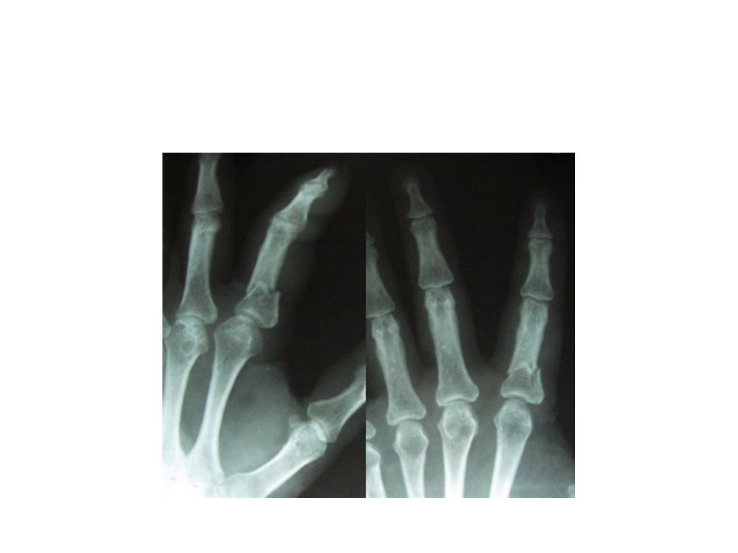
Question list
Q1-Q77 questions — tap to reveal all answerslist
- What are the deforming forces in a proximal phalanx fracture?
- How is malrotation assessed in a phalangeal fracture?
- What are the nonoperative indications for a proximal phalanx fracture?
- What is the nonoperative regime for a proximal phalanx fracture?
- What are the operative indications for a proximal phalanx fracture?
- What fixation is used for phalangeal shaft, base and head fractures?
- Describe the X-ray findings in this proximal phalanx fracture.
Answers · Q & A
Q1.What are the deforming forces in a proximal phalanx fracture?
- Central slip extends the distal fragment
- Interossei flex the proximal fragment
Q2.How is malrotation assessed in a phalangeal fracture?
- Tenodesis check
- Scissoring upon making a fist
Q3.What are the nonoperative indications for a proximal phalanx fracture?
- transverse/ short oblique pattern
- Stable at >50 degrees ROM
- Angulation <20deg
- Displacement <2mm
- No malrotation
Q4.What is the nonoperative regime for a proximal phalanx fracture?
- Dorsal splint with MCPJ at 90deg flexion
- Intrinsic forces act through the extensor mechanism -> tension band effect
Q5.What are the operative indications for a proximal phalanx fracture?
- Long spiral fracture
- Displacement, angulation or rotation
- Multiple digits
Q6.What fixation is used for phalangeal shaft, base and head fractures?
- Transverse shaft: K wire (antegrade or retrograde), Lister wiring, Bouquet pinning
- Spiral/oblique shaft: cross K wires, plate, lag screws
- Base: cross K wire or plate
- Head: type 1 undisplaced, type 2 unstable displaced unicondylar, type 3 unstable bicondylar/comminuted
Q7.Describe the X-ray findings in this proximal phalanx fracture.
- Short oblique fracture of the proximal 1/3 of the index finger PP
- No intra-articular extension
- Apex volar with minimal coronal malignment
- No rotational deformity
▸ Slide 717 · MC fractureHand Bone · 5 questions expand
Slide render

Question list
Q1-Q55 questions — tap to reveal all answerslist
- What must be assessed in a metacarpal shaft or neck fracture?
- Describe the Jahss maneuver.
- What angulation and shortening are acceptable in metacarpal fractures?
- What are the operative indications for metacarpal fractures?
- What fixation options are used for metacarpal fractures?
Answers · Q & A
Q1.What must be assessed in a metacarpal shaft or neck fracture?
- Site and pattern, stability, angulation/displacement/translation
- Associated injury: open wounds, CMCJ dislocation
- Rule out compartment syndrome
- Assess malrotation
Q2.Describe the Jahss maneuver.
- Jahss maneuver (for neck #)
- MCPJ and PIPJ flexed 90deg
- Dorsal directed force through the proximal phalanx
Q3.What angulation and shortening are acceptable in metacarpal fractures?
- No rotation is acceptable
- MC 2&3: 15deg; MC 4: 30deg; MC 5: 40deg
- Shortening 2-5mm acceptable
- Mnemonic: 20/30/40/50
Q4.What are the operative indications for metacarpal fractures?
- Multiple or open fractures
- Unacceptable alignment
Q5.What fixation options are used for metacarpal fractures?
- K wires parallel or crossed
- Plate if comminution
- External fixator if intra-articular comminution
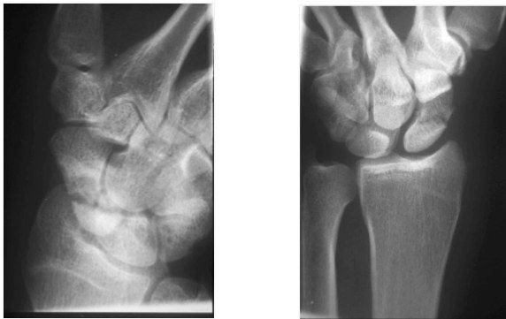
▸ Slide 718 · PA and scaphoid XR showing patients wrist with scaphoid waist fractureHand Bone · 20 questions expand
Question list
Q1-Q2020 questions — tap to reveal all answerslist
- Describe the X-ray findings of this scaphoid waist fracture.
- Why are scaphoid fractures prone to nonunion?
- Describe the Herbert classification of scaphoid fractures.
- What is the blood supply of the scaphoid?
- What is the conservative management and union rate for a scaphoid waist fracture?
- What is the evidence for cast versus surgery in scaphoid waist fractures?
- What is the summary diagnosis and which further views are requested?
- What history is taken in a scaphoid fracture?
- What examination findings are sought in scaphoid fracture?
- What is the acute first aid for a scaphoid fracture?
- Which patient and fracture factors guide definitive scaphoid management?
- What is the aim and technique of surgical fixation of a scaphoid waist fracture?
- How is a grossly displaced scaphoid fracture approached?
- Describe the percutaneous approaches to the scaphoid.
- Describe the dorsal open approach to the scaphoid.
- Describe the volar (Russe) approach to the scaphoid.
- What is a good lateral (SPC) view and how is displacement measured?
- On PA and AP wrist views, where is the ulnar styloid seen?
- If a scaphoid fracture is suspected but not seen on initial X-ray, what is the next step?
- Which approach is used for fixation of an undisplaced scaphoid fracture?
Answers · Q & A
Q1.Describe the X-ray findings of this scaphoid waist fracture.
- Complete, displaced >1mm, horizontal oblique scaphoid waist fracture
- Unstable according to the Herbert classification (Herbert B)
- Acute - no cyst, sclerosis or arthritis
- Associated SL widening; no carpal collapse or other fracture
Q2.Why are scaphoid fractures prone to nonunion?
- Biological: retrograde blood supply, no tendon attachment, 80% covered with cartilage, lacks osteogenic cambium layer, bathed in synovial fluid, small bone contact
- Mechanical: connects the two carpal rows, area of high stress; proximal fragment extended/radially deviated by SL and DIC, distal fragment flexed/ulnar deviated around the RSC
- Delayed presentation: 25% not seen on initial X-ray
Q3.Describe the Herbert classification of scaphoid fractures.
- A: stable - tubercle, horizontal waist, treated conservatively
- B: unstable
- C: delayed (6 weeks)
- D: nonunion
- Russe adds orientation: horizontal oblique, transverse, vertical oblique
Q4.What is the blood supply of the scaphoid?
- All supply is retrograde
- Proximal 80% via the dorsal carpal artery
- Distal 20% via the superficial palmar branch
- No anastomosis between them
Q5.What is the conservative management and union rate for a scaphoid waist fracture?
- Short arm cast with thumb left free, wrist slightly extended, functional cup position
- Cast for 8 weeks, union rate 90-95%
- Scaphoid cast management is reliable in undisplaced stable waist fracture with union rate up to 90%
- Duration by site: 8/10/12 weeks and union 90, 80, 60% for distal/waist/proximal fractures
- No consensus on long versus short arm cast or thumb immobilisation
- Netherlands meta-analysis (J Trauma 2011, 500 patients): no difference in union, pain, grip, time to union or osteonecrosis
Q6.What is the evidence for cast versus surgery in scaphoid waist fractures?
- SWIFT trial (Dias, Lancet 2020): fractures displaced 2mm or less should be treated with cast immobilisation, with early fixation of confirmed nonunion
- Surgery gives similar long-term outcomes but more complications
- McQueen (JBJS 2008): surgery shortens time to union (9 vs 14 weeks) and earlier return to work, but no difference in union rate, ROM or grip at 2 years
Q7.What is the summary diagnosis and which further views are requested?
- Herbert B scaphoid waist fracture, suspicious of SL widening
- Better lateral view for displacement and humpback deformity
- Stress view for SL dissociation
Q8.What history is taken in a scaphoid fracture?
- Age, hand dominance, occupation, functional demand
- Mechanism of injury, delayed presentation, symptoms
Q9.What examination findings are sought in scaphoid fracture?
- Localised tenderness over the anatomical snuffbox and SL interval
- Rule out other injuries
Q10.What is the acute first aid for a scaphoid fracture?
- Analgesics, rest, elevate
- Short arm thumb spica slab
Q11.Which patient and fracture factors guide definitive scaphoid management?
- Timing, orientation, associated injuries
- Displacement, site
- Patient counselling
Q12.What is the aim and technique of surgical fixation of a scaphoid waist fracture?
- Reduce with stable fixation and compression; union rate up to 97%
- Percutaneous approach with K wire as a joystick for reduction
- Headless compression screw with differential pitch targeting the central longitudinal axis
Q13.How is a grossly displaced scaphoid fracture approached?
- Volar approach for ORIF - direct visualisation of the whole scaphoid, correct humpback deformity, preserve dorsal blood supply
- Dorsal approach if the fracture is proximal
Q14.Describe the percutaneous approaches to the scaphoid.
- Volar: incision distal to STTJ, entry at scaphoid tubercle abutting STT; 12G hypodermic needle as guide; reduce by hyperextension and ulnar deviation; bean view - axis from middle third of scaphoid tubercle to distal half of proximal pole
- Dorsal: pronate and flex until the scaphoid is seen as a double ring; guidepin at centre; length 18-24mm (-4mm from measured)
Q15.Describe the dorsal open approach to the scaphoid.
- Longitudinal incision over Lister tubercle, between the 3rd and 4th compartments, between EPL and EDC
- Incise extensor retinaculum and dorsal capsule up to the DIC; flex the wrist over a bolster to reveal the proximal pole
- Danger: superficial branch of radial nerve, dorsal carpal branch of radial artery
- Pros: can address SL, also good for perilunate dislocation
Q16.Describe the volar (Russe) approach to the scaphoid.
- Incision along FCR then towards the scaphoid tubercle
- Incise RSC (preserve the RSC stump to prevent humpback) and long radiolunate ligament
- Pros: good exposure, correct humpback, avoids the dorsal carpal branch of the radial artery
- Cons: radial artery at risk
Q17.What is a good lateral (SPC) view and how is displacement measured?
- SPC view: volar cortex of pisiform should lie between the middle 1/3 of the interval between volar cortex of scaphoid and capitate
- Displacement >1mm = displaced
- Radiolunate angle >15deg dorsal = DISI; capitolunate >15deg volar = VISI
- Intra-scaphoid angle >35deg = humpback deformity
Q18.On PA and AP wrist views, where is the ulnar styloid seen?
- PA view: ulnar styloid is peripheral (ulnar)
- AP view: ulnar styloid is central
Q19.If a scaphoid fracture is suspected but not seen on initial X-ray, what is the next step?
- Scaphoid series: PA, lateral, ulnar deviated (scaphoid view), oblique PA with wrist extension 20deg + ulnar deviation 20deg, 45deg pronation and supination
- If still negative: thumb spica cast and repeat scan in 2/52
- Consider CT and MRI
Q20.Which approach is used for fixation of an undisplaced scaphoid fracture?
- Distal fracture: volar percutaneous
- Proximal fracture: dorsal percutaneous
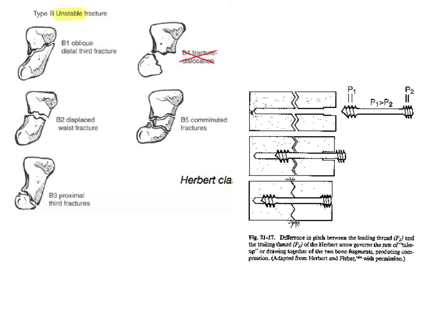
▸ Slide 719 · Herbert screwHand Bone · 2 questions expand
Slide render
Question list
Q1-Q22 questions — tap to reveal all answerslist
- What is a Herbert screw?
- How does the Herbert screw achieve interfragmentary compression?
Answers · Q & A
Q1.What is a Herbert screw?
- A headless compression screw used for scaphoid fixation
- It has a differential pitch
Q2.How does the Herbert screw achieve interfragmentary compression?
- The lead of the leading (distal) thread is larger than the trailing (proximal) thread
▸ Slide 720 · Scaphoid nonunionHand Bone · 8 questions 1 check expand

Question list
Q1-Q88 questions — tap to reveal all answerslist
- Describe the X-ray findings of scaphoid nonunion.
- What investigations are used in scaphoid nonunion and why?
- What is the natural history and pathoanatomy of scaphoid nonunion?
- What are the principles of scaphoid nonunion surgery?
- Which bone graft is chosen in scaphoid nonunion with AVN or deformity?
- Which surgical approach is preferred for scaphoid nonunion?
- What history is important in scaphoid nonunion?
- What is the surgical aim in scaphoid nonunion?
Answers · Q & A
Q1.Describe the X-ray findings of scaphoid nonunion.
- Scaphoid fracture with sclerosis and cystic change
- Relevant negatives for malalignment/SNAC: no obvious radial styloid arthritis, no carpal collapse, Gilula lines intact
Q2.What investigations are used in scaphoid nonunion and why?
- XR lateral and scaphoid series: humpback deformity, displacement, DISI
- CT 1mm cuts: cyst, bone stock, deformity
- MRI with contrast: proximal pole AVN
Q3.What is the natural history and pathoanatomy of scaphoid nonunion?
- Natural history is predictable: 97% arthrosis in 5 years, 90% symptomatic
- Distal scaphoid flexes, capitate is pulled proximally, compression on scapholunate -> SC arthritis
Q4.What are the principles of scaphoid nonunion surgery?
- Debridement of necrotic bone and exposure of a healthy vascularised bed
- Alignment: correction of scaphoid malalignment
- Biology: bone grafting
- Mechanical stability: stabilisation
Q5.Which bone graft is chosen in scaphoid nonunion with AVN or deformity?
- AVN -> vascularised bone graft: dorsal 1,2 ICSRA, volar radial carpal artery
- Deformity -> opening wedge interposition graft (Fisk)
- No deformity -> cancellous graft (arthroscopic) or modified Russe inlay cortical graft
- OA/SNAC at presentation -> non-salvageable
Q6.Which surgical approach is preferred for scaphoid nonunion?
- Mostly volar, as it causes less disruption to the dorsal carpal branch of the radial artery
Q7.What history is important in scaphoid nonunion?
- Age, hand dominance, functional demand, occupation, smoking
- Mode and time of injury, treatment received
- Symptoms
Q8.What is the surgical aim in scaphoid nonunion?
- Realign the fracture and promote union with bone graft + stable fixation
Fact check
Untreated scaphoid nonunion causes arthrosis in 97% within 5 years — imprecise — The 97% figure (Ruby 1985) applied to nonunions already at least 5 years old in a symptomatic series; a 102-case series found 22% arthritis at <5 years, 75% at 5-9 years and 100% at >=10 years. It is not '97% by 5 years'. — medium confidence — source
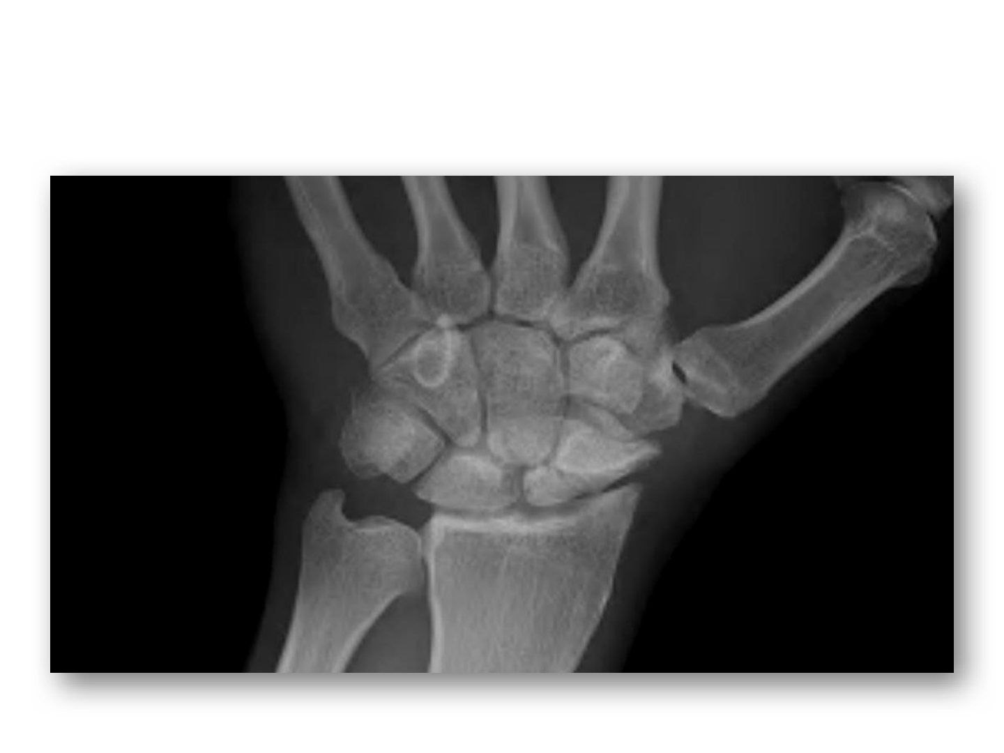
▸ Slide 721 · Scaphoid nonunion advanced collapseHand Bone · 6 questions expand
Slide render
Question list
Q1-Q66 questions — tap to reveal all answerslist
- Describe the X-ray findings of a stage III SNAC wrist.
- How is SNAC wrist managed by stage?
- What is the aim and technique of four corner fusion?
- When can PRC be used in SNAC and what are its drawbacks?
- What is the aim and what are the means of surgery in SNAC wrist?
- Why is the lunate relatively resistant to osteoarthritis?
Answers · Q & A
Q1.Describe the X-ray findings of a stage III SNAC wrist.
- Scaphoid nonunion with OA change at the radial styloid and distal scaphoid, scaphocapitate and capitolunate joints
- Collapsed carpal height as indicated by Nattrass and Youm indices
- Lateral: increased SL angle (may be due to humpback deformity) and increased capitolunate angle -> DISI deformity
Q2.How is SNAC wrist managed by stage?
- I: radial styloidectomy + excision of distal scaphoid
- II: excision of radial styloid + 4 corner fusion or PRC
- III: four corner fusion
- Pan arthritis: wrist fusion
Q3.What is the aim and technique of four corner fusion?
- Eliminate the arthritic midcarpal joint (usually capitolunate)
- Maintain carpal height
- Also fuse hamate and triquetrum to increase fusion rate
- Fusion rate also depends on smoking status
Q4.When can PRC be used in SNAC and what are its drawbacks?
- SNAC 2 with SC joint arthritis and intact midcarpal (LC) joint
- Decreased carpal height may cause decreased grip strength and risk of future OA
Q5.What is the aim and what are the means of surgery in SNAC wrist?
- Aim: removal of the pain generator +/- maintain carpal height in 4 corner fusion
- Means: excisional arthroplasty / limited fusion / wrist fusion
Q6.Why is the lunate relatively resistant to osteoarthritis?
- The lunate is spherical; even if orientation is disrupted it is unlikely to develop OA
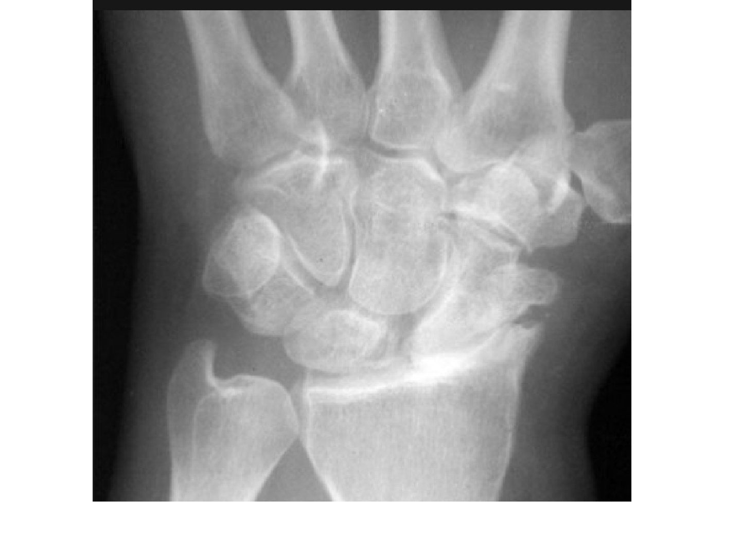
▸ Slide 722 · Widened SL interval (terry Thomas sign: >3mm)Hand Bone · 7 questions expand
Slide render
Question list
Q1-Q77 questions — tap to reveal all answerslist
- Describe the X-ray findings of scapholunate dissociation.
- Describe the Watson (SLAC) classification.
- Describe the Geissler classification.
- How is Watson's test performed?
- How is SL dissociation managed (Garcia-Elias)?
- Compare PRC with four corner fusion.
- What factors are taken into account when managing SL dissociation?
Answers · Q & A
Q1.Describe the X-ray findings of scapholunate dissociation.
- Widened SL interval >3mm (Terry Thomas sign)
- Gilula line broken - intercarpal alignment disrupted, DISI, scaphoid flexed
- Lateral view: SL angle >60deg (DISI)
- OA change between scaphoid and radius; midcarpal joint not involved
- Likely SLAC Watson stage 2; mention carpal height
Q2.Describe the Watson (SLAC) classification.
- I: radial styloid
- II: scaphoid fossa
- III: capitolunate
- IV: pan-arthritis
Q3.Describe the Geissler classification.
- Arthroscopic classification for acute SL dissociation
- Grades: hemorrhage, incongruent, probe through, scope through
- Stage: pre dynamic/dynamic/static/reducible DISI/irreducible DISI/SLAC
Q4.How is Watson's test performed?
- Elbow in 90deg flexion, neutral forearm rotation
- Scaphoid flexes on radial deviation and extends on ulnar deviation
- Examiner's thumb gives a dorsal directed force to the scaphoid tubercle; wrist deviated from ulnar to radial
- Positive if a clunk is felt, with dorsal subluxation of the scaphoid over the dorsal rim of the radius
Q5.How is SL dissociation managed (Garcia-Elias)?
- Predynamic/acute dynamic: dorsal repair + K wire fixation
- Chronic, no SLAC, reducible DISI: reconstruction - anatomical (BLB/BRB) or non-anatomical with capsulodesis (Blatt DIC, Linscheid ECRL, Brunelli FCR)
- Chronic, no SLAC, not reducible DISI: STT fusion + radial styloidectomy
- SLAC 1: radial styloidectomy + SL reconstruction/ PIN and AIN neurectomy; 2: scaphoidectomy + 4 corner/PRC; 3: 4 corner or total wrist fusion; 4: total wrist fusion
Q6.Compare PRC with four corner fusion.
- Systematic review 2015 J Hand surg Eur Saltzman: 4 corner fusion: better grip strength
- PRC: better ROM, fewer complications; no implant, no nonunion problems
- PRC cons: reduced grip strength, longer rehab, 100% OA at 10 years
- PRC prerequisites: RSC intact, no CL arthritis, stable midcarpal joint
Q7.What factors are taken into account when managing SL dissociation?
- Integrity and quality of the dorsal SLL
- Posture of the scaphoid (DISI)
- Carpal alignment and cartilage status
▸ Slide 723 · Describe Xray:Hand Bone · 10 questions expand
Slide render
Question list
Q1-Q1010 questions — tap to reveal all answerslist
- Describe the X-ray findings of a perilunate dislocation.
- Outline the acute management of a perilunate dislocation.
- Describe Tavernier's manoeuvre.
- Describe the Mayfield circle of perilunate instability.
- What is the definitive operative management of a perilunate dislocation?
- What is the prognosis after perilunate dislocation?
- What are the boundaries of the space of Poirier?
- Describe the pathoanatomy of perilunate instability.
- Describe the volar extrinsic wrist ligaments.
- Describe the intrinsic and dorsal wrist ligaments.
Answers · Q & A
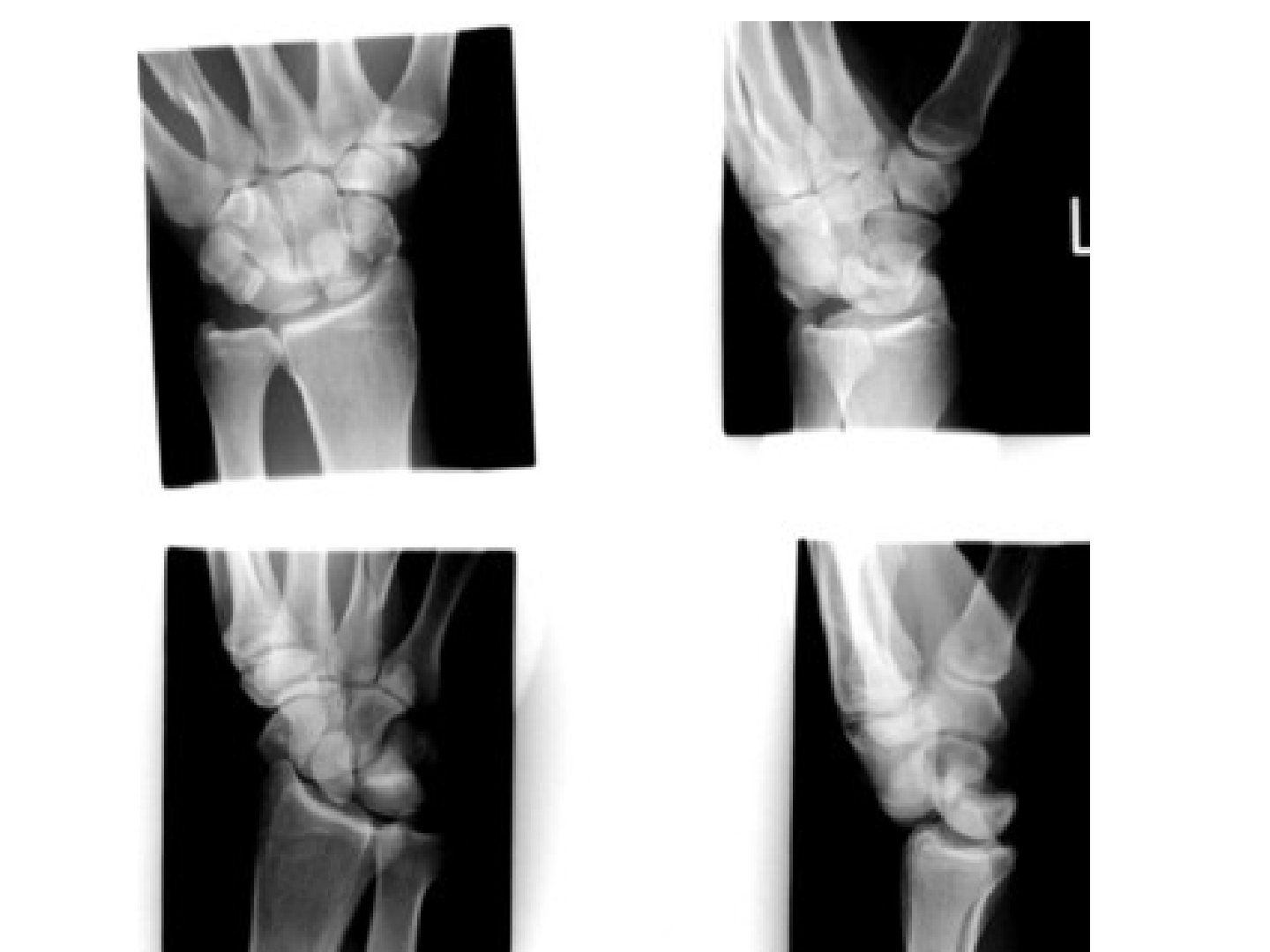
Q1.Describe the X-ray findings of a perilunate dislocation.
- AP: displaced scaphoid waist fracture, disrupted Gilula line at the proximal and distal curvatures
- Piece of pie sign - overlap of capitate and lunate
- +/- scaphoid cortical ring sign, +/- radial styloid fracture
- Lateral: loss of colinearity of radius, lunate and capitate = trans-scaphoid perilunate dislocation
Q2.Outline the acute management of a perilunate dislocation.
- ATLS, AMPLE history; assess median nerve, wounds, compartments
- Closed reduction under sedation; back slab; closely monitor neurovascular status
- CT scan
- If closed reduction fails: OR with volar approach
Q3.Describe Tavernier's manoeuvre.
- Prolonged traction with wrist extension
- Stabilise the lunate volarly
- Reduce the capitate onto the lunate with wrist flexion
Q4.Describe the Mayfield circle of perilunate instability.
- I: SLL / scaphoid fracture
- II: capitolunate
- III: LT
- IV: dorsal radiocarpal ligament, lunate dislocation (unopposed SRL pull)
- Intrinsic weakest point: space of Poirier, between lunate and capitate
Q5.What is the definitive operative management of a perilunate dislocation?
- Refer hand specialist
- Aim: anatomical restoration of carpal alignment and stable fixation for ligaments to heal
- Fix scaphoid/styloid, repair ligaments with suture anchors, transfix unstable carpal bones
- Pinning: 1. lunate to radius (prevent DISI), 2. transfix SL and LT, 3. CL if capitolunate unstable (CIND)
- Pin for 8 weeks, cast for 12 weeks; encourage early finger mobilisation
- Dorsal approach: Berger flap (repair intrinsic SL ligament and extrinsic radiotriquetral); volar approach: release carpal tunnel, repair LT and radiocarpal ligaments
Q6.What is the prognosis after perilunate dislocation?
- 36% posttraumatic arthritis
- Residual scapholunate instability
- Stiffness - 50% of movement lost
- CRPS
Q7.What are the boundaries of the space of Poirier?
- Intrinsic weakest point of the space of Poirier without ligament b/w lunate & capitate
- Bound by volar deltoid ligament (intrinsic) superiorly, RSC radially
- RLT (long radiolunate) inferiorly, UC ligament ulnarly
Q8.Describe the pathoanatomy of perilunate instability.
- Disruption follows Mayfield's classification (radial to ulnar)
- Greater versus lesser arc injuries
- Classical greater arc: radial styloid -> scaphoid fracture -> capitate fracture -> ulnar styloid fracture
Q9.Describe the volar extrinsic wrist ligaments.
- Long volar arch = RSC + UCL; short volar arch = RL + UL
- RSC is strongest to prevent ulnar translocation
- Long RL / short RL (ligament of Testut, located between SRL/LRL)
- UT / UL / UC blend with the TFCC
Q10.Describe the intrinsic and dorsal wrist ligaments.
- Intrinsic: LT, SL (volar and dorsal)
- Dorsal extrinsic: DIC (scaphoid to triquetrum), DRC (radius to triquetrum)
- Dorsal intrinsic: SL, LT
▸ Slide 724 · Moneim Classification - based on presence of intercarpal dissociationHand Bone · 4 questions expand
Slide render

Question list
Q1-Q44 questions — tap to reveal all answerslist
- Describe the Moneim classification.
- Describe the Dumontier classification.
- Which ligaments are involved in perilunate instability?
- Describe the three column fixation concept.
Answers · Q & A
Q1.Describe the Moneim classification.
- Based on the presence of intercarpal dissociation
- Type 1: without associated intercarpal dissociation
- Type 2: with associated intercarpal dissociation
Q2.Describe the Dumontier classification.
- Based on the presence of radial styloid fracture
- Type 1: purely ligamentous or only a small cortical avulsion
- Type 2: associated with a large radial styloid fracture
Q3.Which ligaments are involved in perilunate instability?
- RSC, LRL, SRL, UC, UT, UL
Q4.Describe the three column fixation concept.
- Radial column: radial styloid to scaphoid pinning
- Intermediate column (lunate facet): SRL and RSC ligament repairs + radiolunate K wire
- Ulnar column: DRUJ pinning
- Conservative if stable; otherwise ligament repair/reconstruction
▸ Slide 725 · AP and lateral Xray showing abnormalities over lunate with sclerosis and collapsHand Bone · 9 questions 1 check expand
Slide render
Question list
Q1-Q99 questions — tap to reveal all answerslist
- Describe the X-ray findings of this lunate pathology.
- How is carpal collapse quantified?
- What are the hypotheses and risk factors for Kienbock disease?
- Describe the Lichtman classification of Kienbock disease.
- What are the surgical aims in early versus late Kienbock disease?
- Why are symptoms rather than radiology the guide to treatment in Kienbock disease?
- What examination findings are important in Kienbock disease?
- What are the Zapico lunate morphology types and what is the vascular supply prevalence?
- Describe the Bain arthroscopic classification of Kienbock disease.
Answers · Q & A
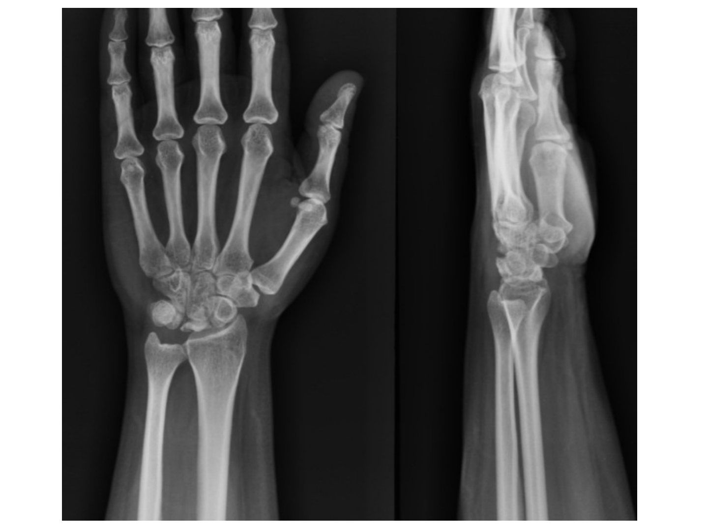
Q1.Describe the X-ray findings of this lunate pathology.
- Lunate sclerosis with collapsed height; suspected carpal height collapse
- No features of STT arthritis; signet ring sign on AP view
- DISI on lateral view with reduced SLA
- Ulna minus on PA X-ray, radial inclination within normal limits
- Consistent with Lichtman type IIIB Kienbock disease; Ddx infection, trauma
Q2.How is carpal collapse quantified?
- Nattrass index (carpal height / capitate) <1.57
- Youm index (carpal height / 3rd metacarpal) <0.54
- Stahl index (lunate height to width ratio) <0.53
- McMurtry (ulnar central axis to capitate / ulnar axis to 3rd MC) 0.27-0.33
Q3.What are the hypotheses and risk factors for Kienbock disease?
- Avascular necrosis of the lunate of unknown aetiology; biological, mechanical or mixed cause
- Biological: single intraosseous/extraosseous vessel variation; 7-20% have a single palmar vessel
- Mechanical: ulnar minus, increased radial inclination, lunate morphology (Zapico 1 trapezoidal worst)
- Ask about age, functional demands, occupation; risk factors such as alcohol and steroid intake
- CT: coronal split; MRI: vascularity (decrease T1 signal), articular congruency (decreased T1 signal suggests AVN)
Q4.Describe the Lichtman classification of Kienbock disease.
- 1: X-ray normal
- 2: sclerosis without collapse
- 3: collapse - 3A normal scaphoid alignment, 3B fixed scaphoid rotation with ring sign, 3C coronal split
- 4: pan arthritis
- Bain arthroscopic classification looks for articular surface involvement
Q5.What are the surgical aims in early versus late Kienbock disease?
- Early (I-IIIA): revascularize + redistribute load
- biologically revascularize: 4,5 ECA pedicle graft, vascularised pisiform transfer, 2nd dorsal intermetacarpal artery
- Mechanical: UV minus -> radial shortening; UV positive/neutral -> radial wedge osteotomy or capitate shortening
- Postop Exfix to protect joint level procedure
- Stage 3B: excise lunate + rolled PL graft and STT fusion, more motion preserved, or SC fusion (larger fusion area, better load transfer); or PRC
- Late stage: remove pain generator and transfer load to radial column and salvage remaining carpal height
- Late/arthritis: PRC or total wrist fusion/replacement
Q6.Why are symptoms rather than radiology the guide to treatment in Kienbock disease?
- Radiological findings do not correlate well with patient symptoms
- Treat if symptomatic: start conservative with activity modification, splint, NSAID for 3-6 months
Q7.What examination findings are important in Kienbock disease?
- ROM
- Swelling
- Tenderness just distal to Lister tubercle
Q8.What are the Zapico lunate morphology types and what is the vascular supply prevalence?
- Zapico: 1 trapezoidal (worst), 2 rectangular, 3 double facet
- Majority of lunates have both dorsal and palmar vessels with rich anastomoses
- 7-20% have a single palmar vessel supplying the whole lunate
- Intraosseous prevalence: Y > I > X
Q9.Describe the Bain arthroscopic classification of Kienbock disease.
- 0: 0 (no involvement)
- 1: proximal lunate
- 2a: proximal lunate + radius articular surface
- 2b: split of both distal and proximal lunate surfaces
- 3: both lunate + radius
- 4: 3 + proximal capitate
Fact check
Youm index <0.54 and Nattrass index <1.57 indicate carpal collapse — imprecise — These are the normal population means, not fixed cut-offs: Youm carpal height ratio is 0.54 +/- 0.03 (normal range about 0.51-0.57) and Nattrass index is 1.57 +/- 0.05 (range about 1.52-1.62). Compare with the contralateral wrist rather than a single threshold. — medium confidence — source
▸ Slide 726 · Wrist fusionHand Bone · 3 questions expand
Slide render
Question list
Q1-Q33 questions — tap to reveal all answerslist
- What position is the wrist fused in and why?
- What are the functional movements of the wrist?
- How much of wrist ROM comes from each joint?
Answers · Q & A
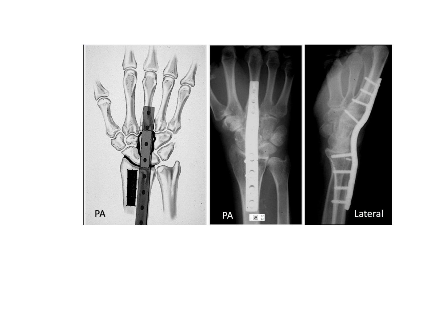
Q1.What position is the wrist fused in and why?
- Extension 20deg for grip strength
- Neutral or 10deg ulnar deviation
Q2.What are the functional movements of the wrist?
- 40deg flexion/extension
- 10deg radial deviation
- 30deg ulnar deviation
Q3.How much of wrist ROM comes from each joint?
- 60% radiocarpal joint
- 30% intercarpal
- 10% intracarpal
▸ Slide 727 · 1st CMCJ OA narrowing of joint space, subchondral sclerosis, dorsal and radialHand Bone · 12 questions expand
Slide render

Question list
Q1-Q1212 questions — tap to reveal all answerslist
- Describe the X-ray findings of first CMCJ osteoarthritis.
- Describe the Eaton and Littler classification.
- What are the key examination findings and tests in first CMCJ OA?
- What is the conservative management of first CMCJ OA?
- What are the operative options for first CMCJ OA by stage?
- Describe the pathoanatomy of first CMCJ osteoarthritis.
- What history is important in first CMCJ OA?
- What is the aim of treatment in first CMCJ OA?
- Describe the Wagner approach to trapeziectomy.
- What are the advantages and disadvantages of trapeziectomy?
- What is the functional position for first CMCJ fusion and when is it contraindicated?
- Which nerves are targeted in first CMCJ denervation?
Answers · Q & A
Q1.Describe the X-ray findings of first CMCJ osteoarthritis.
- Joint space narrowing, subchondral sclerosis
- Dorsal and radial subluxation >1/3, osteophytes
- Also arthritis in the STTJ
- MCPJ hyperextension deformity; narrowing of the 1st webspace
- Eaton stage IV; request a Robert's view (hyperpronation, thumb flat on cassette)
Q2.Describe the Eaton and Littler classification.
- I: increased joint space
- II: <1/3 subluxation, osteophyte <2mm
- III: >1/3 subluxation, osteophyte >2mm
- IV: pantrapezial arthritis
Q3.What are the key examination findings and tests in first CMCJ OA?
- Look: square off sign (dorsoradial prominence), MCPJ hyperextension, adducted and flexed 1st MC
- Feel: tenderness, grind, crank, torque and retropulsion tests
- Move: ROM, 1st webspace (45-55deg between 1st and 2nd MC), STT radiocarpal grinding test
- Rule out Finkelstein, Eichhoff and RCJ grind; associations include carpal tunnel (50%) and trigger finger
Q4.What is the conservative management of first CMCJ OA?
- Lifestyle modification, splint, NSAIDs
- Steroid injection: 80% pain relief in Eaton 1
- Physio: 1st webspace stretching and 1st dorsal interosseous strengthening
Q5.What are the operative options for first CMCJ OA by stage?
- Early (stage 1-3): arthroscopic debridement + thermal shrinkage; first MC basal osteotomy
- Later (stage III/IV): trapeziectomy +/- webspace contracture release, or Eaton's procedure (trapeziectomy + LRTI)
- Newer: suspensionplasty with suture button, APL (Weilby) or ECRL; less commonly 1st CMCJ arthroplasty or fusion
- Fusion contraindicated in stage IV (exacerbates STTJ arthritis); unable to put hand flat
- Davis 2012 RCT and Cochrane 2015: no long-term benefit of LRTI over trapeziectomy alone; LRTI had more adverse events
- MCPJ hyperextension: EPB transfer to PP base, volar capsulodesis, or MCPJ fusion
Q6.Describe the pathoanatomy of first CMCJ osteoarthritis.
- Biconcave saddle joint
- Beak ligament (trapezium volar tubercle to ulnar 1st MC base) prevents dorsal subluxation on key pinch
- Weakened beak ligament -> dorsal subluxation on pinch -> see-saw movement -> cartilage degeneration -> further instability
- Recent evidence: dorso-radial ligament more important than beak ligament, maintaining the relationship between 1st and 2nd MC
Q7.What history is important in first CMCJ OA?
- Risk factors: occupation
- Pain
- Functional limitations
Q8.What is the aim of treatment in first CMCJ OA?
- Stable, painless thumb
- Restore thumb function of opposition
Q9.Describe the Wagner approach to trapeziectomy.
- Incision between dorsal and volar glabrous skin
- Interval between APL and thenar muscles (or scope)
Q10.What are the advantages and disadvantages of trapeziectomy?
- Advantages: simple procedure, good pain relief, preserves ROM
- Disadvantages: splintage 4-6 weeks
- Loss of thumb length and pinch strength (50-60%)
- Metacarpal subsidence and instability
Q11.What is the functional position for first CMCJ fusion and when is it contraindicated?
- Radial and palmar abduction 30deg, 15deg pronation
- Cons: unable to put the hand flat on the table
- Contraindicated in stage IV - exacerbates STTJ arthritis
Q12.Which nerves are targeted in first CMCJ denervation?
- Thenar branch of the median nerve
- Superficial branch of the radial nerve entering the CMCJ
- Articular branches of the palmar cutaneous branch
- Nerve of Cruveilhier
- Dorsal articular nerve of the first dorsal interosseous space
▸ Slide 728 · Gamekeepers thumb (6 week) / Skiers thumb (acute)Hand Bone · 8 questions expand
Slide render
Question list
Q1-Q88 questions — tap to reveal all answerslist
- What is injured in gamekeeper's/skier's thumb and where do the avulsions occur?
- How do you differentiate partial from complete UCL injury?
- What is a Stener lesion and what is its mechanism?
- What investigations are used for UCL injury?
- How is incomplete versus complete UCL injury managed?
- What is the outcome after UCL injury treatment?
- What is the difference between gamekeeper's and skier's thumb?
- Where does the adductor pollicis attach?
Answers · Q & A
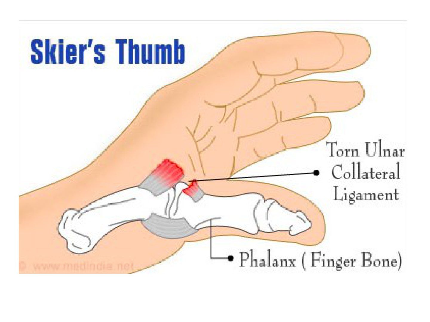
Q1.What is injured in gamekeeper's/skier's thumb and where do the avulsions occur?
- Injury to the ulnar collateral ligament, important for lateral pinch
- Avulsion distally 50%, proximally 40%, midsubstance 10%
Q2.How do you differentiate partial from complete UCL injury?
- Radial stress test at neutral and 30deg (neutral tests accessory + proper, 30deg tests proper)
- Complete: 15deg more laxity compared with normal; at 30deg, no endpoint
- Grade 1: pain, no laxity; grade 2: soft endpoint; grade 3: no endpoint
- Also rule out a Stener lesion
Q3.What is a Stener lesion and what is its mechanism?
- Complete UCL rupture trapped superficial to the adductor pollicis aponeurosis
- Prevents healing or causes unpredictable healing; fibrotic mass -> needs operative treatment
- Mechanism: forced hyperabduction of the thumb
Q4.What investigations are used for UCL injury?
- XR: avulsion fracture
- USG/MRI: Stener lesion
Q5.How is incomplete versus complete UCL injury managed?
- Incomplete: thumb spica cast 4 weeks, then splint 2 weeks, then gentle ROM; no strenuous exercise for 3 months
- Complete acute: repair UCL +/- suture anchor or pullout wire, then repair aponeurosis to extensor tendon sheath, K wire with MCPJ at 20deg flexion, thumb spica
- Chronic/Stener: static reconstruction with PL graft; dynamic reconstruction with EIP, EPB or adductor pollicis
Q6.What is the outcome after UCL injury treatment?
- Samora (CJSM 2013): >90% excellent outcome
- Acute and chronic injuries achieved comparable results (acute = chronic)
Q7.What is the difference between gamekeeper's and skier's thumb?
- Gamekeeper's thumb = chronic injury (6 weeks)
- Skier's thumb = acute injury
Q8.Where does the adductor pollicis attach?
- Ulnar base of the proximal phalanx of the thumb
▸ Slide 729 · Xray hand showing 1st MC base intra-articular fracture suluxation with CMCJ radiHand Bone · 13 questions expand
Slide render
Question list
Q1-Q1313 questions — tap to reveal all answerslist
- What are the key facts about 1st metacarpal fractures?
- Describe the X-ray findings in this hand injury.
- How does the 1st metacarpal displace in a Bennett fracture?
- What is a Bennett fracture and which X-ray view best shows displacement?
- Why is surgery usually needed for a Bennett fracture?
- What is the PAET reduction manoeuvre?
- Is it necessary to fix the ulno-volar fragment in a Bennett fracture?
- How is CRPP performed for a Bennett fracture?
- When is ORIF used for 1st MC base fractures and what implants are used?
- Describe the Wagner approach for ORIF of the 1st MC base.
- How is a Rolando fracture treated and what is its prognosis?
- What is opposition of the thumb?
- What is a reverse Bennett fracture and what must you look for?
Answers · Q & A
Q1.What are the key facts about 1st metacarpal fractures?
- 80% involve the MC base, most common is Bennett (partial intra-articular)
- Others: Rolando (complete intra-articular +/- comminuted) and extra-articular
- Mechanism is typically axial force to the thumb
- CMCJ allows large sagittal movement; up to 30deg angulation is accepted
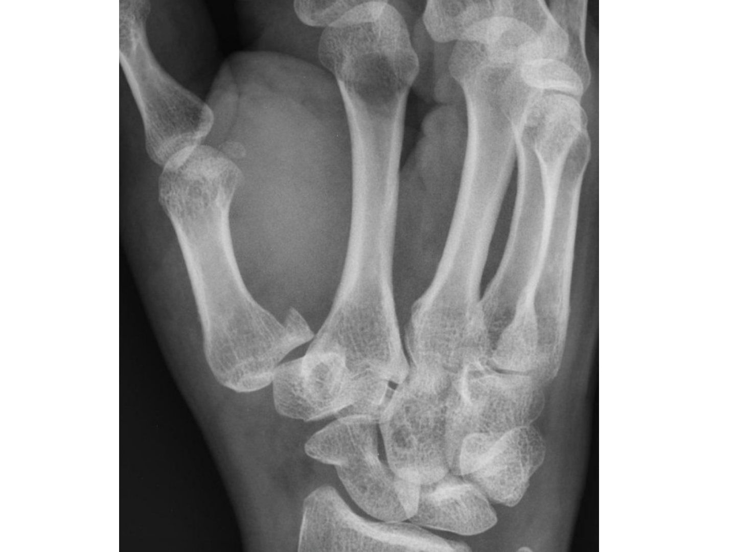
Q2.Describe the X-ray findings in this hand injury.
- 1st MC base intra-articular fracture-subluxation
- CMCJ radial and dorsal subluxation
- 1st MC is supinated, shortened, adducted and flexed
Q3.How does the 1st metacarpal displace in a Bennett fracture?
- Shortened with radial and dorsal subluxation by APL
- Supination by EPL and adductor pollicis
- Adduction by adductor pollicis
Q4.What is a Bennett fracture and which X-ray view best shows displacement?
- 2-part volar ulnar intra-articular fracture of the 1st MC base
- Known to be easy to reduce but difficult to hold
- Hyperpronated thumb (Robert's) view best assesses displacement
Q5.Why is surgery usually needed for a Bennett fracture?
- Fracture is intra-articular, displaced with joint subluxation
- Conservative treatment only for undisplaced fractures ie 1mm
- Distal fragment is subjected to multiple tendon pull and prone to displacement
Q6.What is the PAET reduction manoeuvre?
- (1) Traction
- (2) Extension with pressure over dorsal apex
- (3) Abduction
- (4) Pronation of the MC
Q7.Is it necessary to fix the ulno-volar fragment in a Bennett fracture?
- No - fixation is not necessary to catch the ulno-volar fragment
Q8.How is CRPP performed for a Bennett fracture?
- Indicated if small fragment and reducible
- 1st pin: distal fragment to trapezium
- 2nd pin: 1st to 2nd MC to maintain length
- Followed by thumb spica cast immobilisation
Q9.When is ORIF used for 1st MC base fractures and what implants are used?
- If 1. large fragment, 2. irreducible
- ORIF with K wire or screw
- Lag screw for Bennett, plate for Rolando
- Post op casting x 4/52
Q10.Describe the Wagner approach for ORIF of the 1st MC base.
- Skin incision between volar and dorsal radial skin, curving volar towards the radial border of FCR (hockey stick incision)
- Plane between APL and thenar muscles (in plane radial and medial)
- Protect sensory branches of radial nerve
- Subperiosteal reflection of thenar muscle
Q11.How is a Rolando fracture treated and what is its prognosis?
- Base split into volar and dorsal fragment (Y fracture)
- ORIF with plate (T/L plate) or external fixation with Thoren traction (abduction + extension vectors)
- Connected in a Bandi outrigger splint for 2 weeks, then cast
- Worse prognosis
Q12.What is opposition of the thumb?
- Palmar abduction, flexion and pronation
Q13.What is a reverse Bennett fracture and what must you look for?
- Reverse Bennett = fracture of the base of the 5th MC
- CT to look for concomitant hamate fracture
▸ Slide 730 · 5th and 4th CMCJ dislocationHand Bone · 3 questions expand

Question list
Q1-Q33 questions — tap to reveal all answerslist
- What do you assess in a 5th CMCJ fracture dislocation?
- Why is closed reduction alone unlikely to be stable in a 5th CMCJ fracture dislocation?
- How is a 5th CMCJ fracture dislocation managed operatively?
Answers · Q & A
Q1.What do you assess in a 5th CMCJ fracture dislocation?
- Soft tissue cover
- NV: motor branch of ulnar nerve near hook of hamate
- Compartment status
- Rotation deformity
- Cascade of MCPJ (draw a curve line on X-ray)
Q2.Why is closed reduction alone unlikely to be stable in a 5th CMCJ fracture dislocation?
- The dislocation is pulled by ECU
- Close reduction is not likely to be stable
Q3.How is a 5th CMCJ fracture dislocation managed operatively?
- CR + KWF (K wire fixation) to carpal bone and other MC bones
- Risk: motor branch of ulnar nerve
- K wire must not perforate the volar side
▸ Slide 731 · Pisotriquetral arthritisHand Bone · 2 questions expand

Question list
Q1-Q22 questions — tap to reveal all answerslist
- What is pisotriquetral arthritis?
- What are the clinical features and management of pisotriquetral arthritis?
Answers · Q & A
Q1.What is pisotriquetral arthritis?
- Not covered in the speaker notes beyond the slide title
Q2.What are the clinical features and management of pisotriquetral arthritis?
- Not covered in the speaker notes
▸ Slide 732 · Rheumatoid handHand Bone · 8 questions expand
Slide render
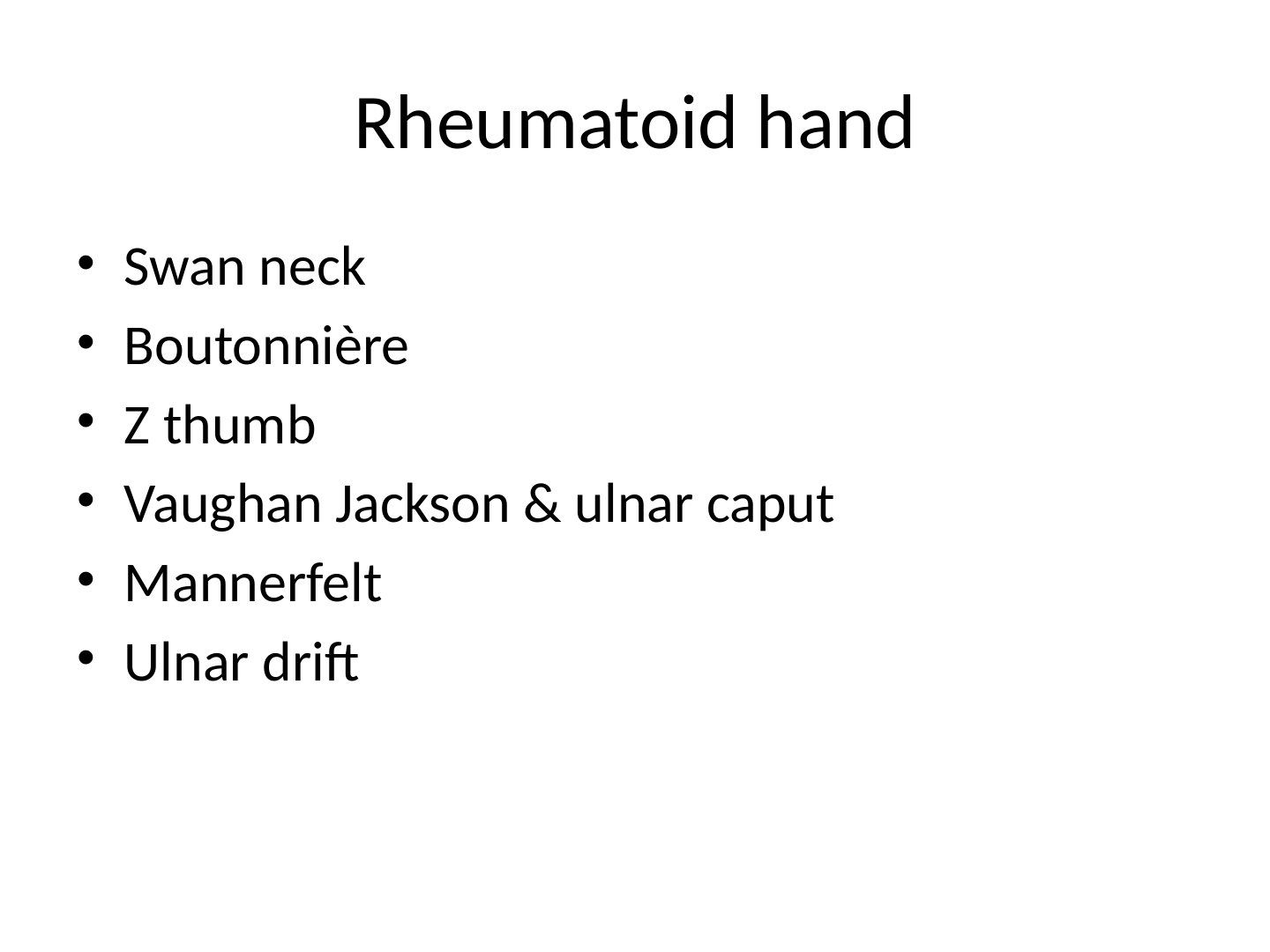
Question list
Q1-Q88 questions — tap to reveal all answerslist
- What is the pathophysiology of rheumatoid hand involvement?
- What happens in the hand in rheumatoid arthritis?
- What intrinsic factors in the hand cause MCPJ ulnar drift in RA?
- What are the classic deformities seen in the rheumatoid hand?
- What are the aims of treatment in the rheumatoid hand?
- What are the urgent indications in the rheumatoid hand?
- What is the priority in managing the rheumatoid hand?
- What are the ACR/EULAR 2010 diagnostic criteria for RA?
Answers · Q & A
Q1.What is the pathophysiology of rheumatoid hand involvement?
- RA is a progressive, systemic, cell-mediated (T-cell, MHC class II) autoimmune condition affecting the synovial lining of joints and tendon sheaths
- Rheumatoid factor = IgM autoantibodies directed against the Fc portion of IgG, forming immune complexes
- Pannus causes destruction of the articular surface through release of activated neutrophils and microvascular ischemia
- Stages: Stage 1: synovitis, Stage 2: destruction, Stage 3: deformity; synovial fluid becomes a Newtonian fluid (loss of shear thinning), altering tribology
- Genetics: associated with HLA DW4, DR4
Q2.What happens in the hand in rheumatoid arthritis?
- UC ligament synovitis causes dorsal subluxation of the ulnar head (caput ulnare)
- RC ligament synovitis + ECU subluxation causes ulnar translocation of the carpus
- Caput ulnare + ECU subluxation = Vaughan Jackson syndrome, with attrition rupture of extensor tendons (EDM, then EDC, then EI)
- MCs deviate radially; flexors and extensors sublux ulnar and volar
- Leads to MCPJ ulnar drift with ulnar intrinsic and collateral contracture
Q3.What intrinsic factors in the hand cause MCPJ ulnar drift in RA?
- Lateral pinch and power grip exert an ulnar directed force
- Small sloping ulnar condyle
- Gravity
- Attenuation of sagittal band and radial joint capsule
Q4.What are the classic deformities seen in the rheumatoid hand?
- Swan neck deformity
- Boutonniere deformity
- Z thumb
- Vaughan Jackson syndrome with ulnar caput
- Mannerfelt syndrome
- Ulnar drift
Q5.What are the aims of treatment in the rheumatoid hand?
- MDT and maximise medical treatment
- Treat according to function: painless, good hand function with good ROM, adequate power
- Prevent deformity and complication
Q6.What are the urgent indications in the rheumatoid hand?
- Acute carpal tunnel syndrome
- Locked trigger finger
- Impending tendon rupture
Q7.What is the priority in managing the rheumatoid hand?
- Aim: MDT, maximise medical treatment, treat according to function (painless, good ROM, adequate power); prevent deformity and complication
- Urgent: acute CTS, locked trigger finger, impending tendon rupture
- Reconstruction follows Souter's order of predictability: extensor tenosynovectomy + wrist stabilisation + ulnar head resection, then flexor tenosynovectomy, thumb, DIPJ, MCPJ
Q8.What are the ACR/EULAR 2010 diagnostic criteria for RA?
- Synovitis in >=1 joint plus no alternative diagnosis
- >=6/10 across 4 domains
- Joints involved 0-5; RF/anti-CCP 0-3; inflammatory marker 0-1; duration >6 weeks 0-1
▸ Slide 733 · Clinical photo showing patients both handsHand Bone · 7 questions expand
Slide render

Question list
Q1-Q77 questions — tap to reveal all answerslist
- What thumb and finger deformities accompany the MCPJ ulnar drift in this patient?
- What are the intrinsic causes of MCPJ dislocation in RA?
- What are the extrinsic causes of MCPJ dislocation in RA?
- How do you treat MCPJ dislocation in RA?
- What are the outcomes of MCPJ arthroplasty in RA?
- Why is arthroplasty advocated over fusion at the MCPJ in RA?
- What are the operative options for a supple versus a rigid MCPJ in RA?
Answers · Q & A
Q1.What thumb and finger deformities accompany the MCPJ ulnar drift in this patient?
- Boutonniere deformities of both thumbs
- Swan neck deformity in multiple other digits
- Prominent knuckles with radial deviation of the MCs; no scars and no active joint swelling
- Overall picture: symmetrical polyarthropathy with typical deformities of RA
Q2.What are the intrinsic causes of MCPJ dislocation in RA?
- Anatomical: ulnar condyle smaller and ulnar sloping condyles of IF and MF; radial collaterals weaker; EI and EDM on ulnar side
- Biomechanical: grip exerts ulnar directed force from the thumb
- Gravity causes ulnar subluxation
Q3.What are the extrinsic causes of MCPJ dislocation in RA?
- Carpal ulnar translocation causes radial deviation of MCs; extensor vector pulls MCPJ into ulnar drift
- MCPJ synovitis -> sagittal band erosion + volar plate stretching -> extensor tendon ulnar subluxation -> ulnar intrinsic contracture -> PP ulnar deviation
- Flexors also ulnar sublux, causing further ulnar drift
Q4.How do you treat MCPJ dislocation in RA?
- Splinting and activity modification (eg how to hold a mug)
- Supple: extensor tenosynovectomy, extensor realignment (radial sagittal band imbrication, RCL reefing, cross intrinsic transfer, volar plate release + collateral recon)
- Rigid: fusion or arthroplasty; advocate arthroplasty as MCPJ is >60% of hand function
- Arthroplasty: silicone/pyrocarbon; correct wrist radial deviation + collateral recon same OT
- Complications: infection, loosening, recurrence
Q5.What are the outcomes of MCPJ arthroplasty in RA?
- 70% satisfaction at 1 year, 69% survivorship at 20 years
- Silicone implant 63% fracture in 17 years
- Decreases ulnar deviation and extension lag
Q6.Why is arthroplasty advocated over fusion at the MCPJ in RA?
- MCPJ provides >60% of hand function
- Correct wrist radial deviation and collateral recon in the same OT
- Implant options: silicone or pyrocarbon
Q7.What are the operative options for a supple versus a rigid MCPJ in RA?
- Supple: extensor tenosynovectomy and extensor realignment (radial sagittal band imbrication, RCL reefing, cross intrinsic transfer, volar plate release + collateral recon)
- Rigid: fusion or arthroplasty
▸ Slide 734 · Priority: souter principleHand Bone · 7 questions expand
Slide render
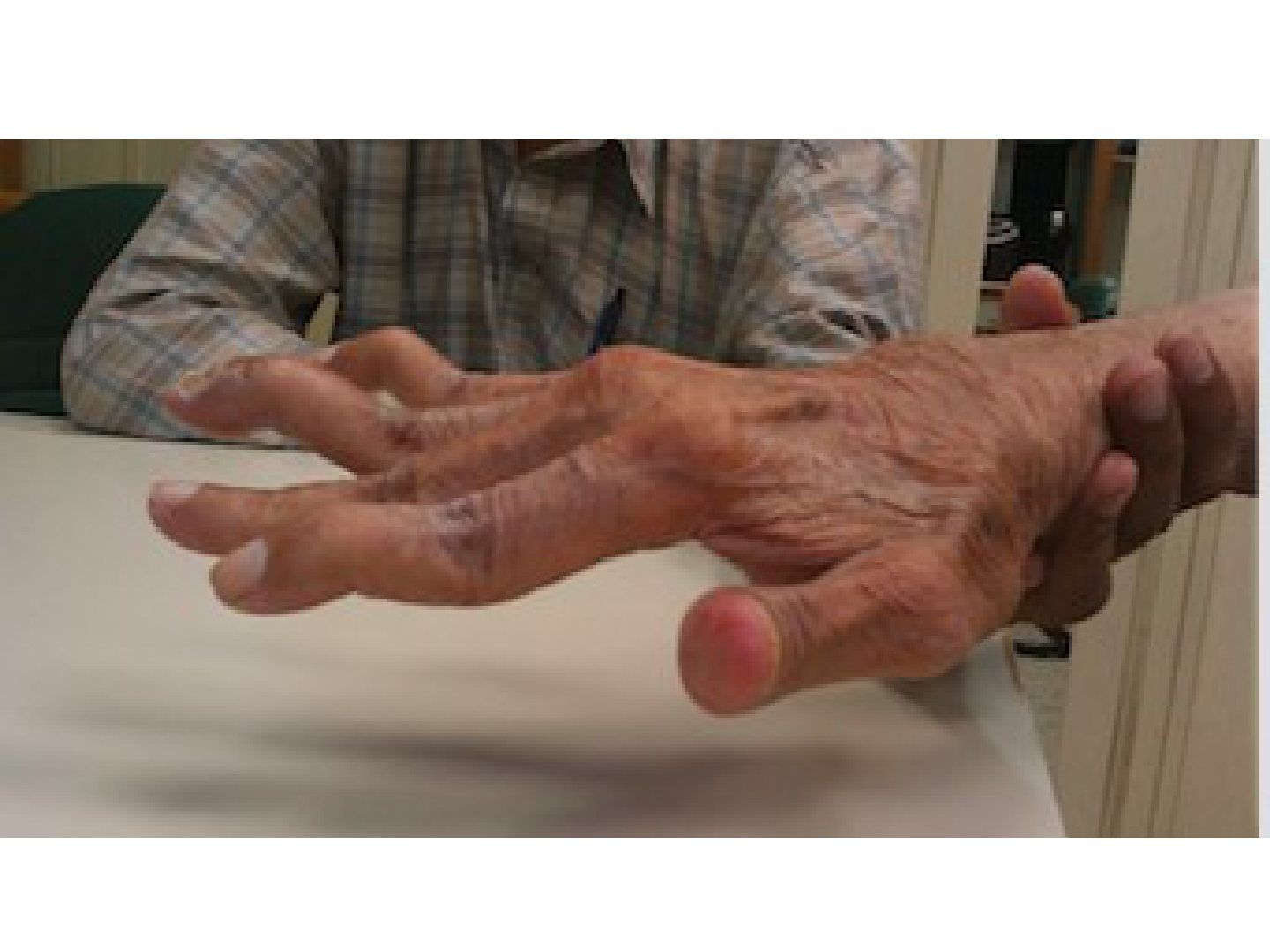
Question list
Q1-Q77 questions — tap to reveal all answerslist
- What is the Souter principle / priority in RA hand surgery?
- What are the 3 aetiologies of swan neck deformity?
- What perpetuates a swan neck deformity and what are the complications?
- What is the Nalebuff classification of swan neck deformity?
- How do you detect intrinsic tightness and why does it matter?
- How is swan neck deformity treated?
- How do you choose between FDS tenodesis and SORL reconstruction?
Answers · Q & A
Q1.What is the Souter principle / priority in RA hand surgery?
- Urgent: C spine, acute carpal tunnel, impending tendon rupture
- LL first: foot, hip, knee, ankle
- UL: proximal --> distal
Q2.What are the 3 aetiologies of swan neck deformity?
- MCPJ: joint volar subluxation, dorsal subluxation of lateral band
- PIPJ: volar plate attenuation / FDS rupture
- DIPJ: mallet finger (terminal extensor tendon rupture)
Q3.What perpetuates a swan neck deformity and what are the complications?
- Attenuation of transverse retinaculum; tightening of triangular ligament and intrinsics -> further dorsal dislocation of lateral band
- Hyperextension of PIPJ functionally lengthens extensor mechanism -> DIPJ in flexion
- Complication: intrinsic tightness (Bunnell test) + fixed deformity
Q4.What is the Nalebuff classification of swan neck deformity?
- 1: flexible and full ROM
- 2: intrinsic tightness
- 3: stiff PIPJ
- 4: arthritis
Q5.How do you detect intrinsic tightness and why does it matter?
- Use the Bunnell test
- It determines the choice between FDS tenodesis (no tightness) and SORL reconstruction + intrinsic release (intrinsic tightness)
Q6.How is swan neck deformity treated?
- Determine if there is intrinsic tightness
- Conservative if no complication: double ring splint at PIPJ (Murphy ring)
- No intrinsic tightness: FDS tenodesis (ulnar FDS slip cut, loop around A2 and suture back proximally with PIPJ in 40deg flexion)
- Intrinsic tightness: SORL reconstruction + intrinsic release
- DIPJ/PIPJ arthrosis: fusion
Q7.How do you choose between FDS tenodesis and SORL reconstruction?
- Check if DIPJ flexion deformity corrects upon PIPJ flexion; if not corrected, do SORL reconstruction
- PL vs lateral band as SORL graft: PL more bulky
- Lateral band may not be present when the underlying cause was terminal slip rupture
▸ Slide 735 · What is boutonniere?Hand Bone · 7 questions expand
Slide render

Question list
Q1-Q77 questions — tap to reveal all answerslist
- What is a boutonniere deformity?
- What is the pathophysiology of a boutonniere deformity?
- How is an acute boutonniere deformity managed?
- How is a chronic boutonniere deformity managed according to extension lag?
- Summarise the Matev reconstruction for chronic boutonniere deformity.
- What is the Burton classification of boutonniere deformity?
- What is the differential diagnosis of multiple boutonniere deformities?
Answers · Q & A
Q1.What is a boutonniere deformity?
- Button hole: the lateral band is the hole and the PIPJ passes through the hole
Q2.What is the pathophysiology of a boutonniere deformity?
- Central slip rupture (traumatic/inflammatory) -> failed extension at PIPJ; central slip transmits extension force to DIPJ via the intact lateral band connection
- With time bilateral lateral band volar subluxation with contracted transverse retinacular ligament and ORL
- Triangular ligament attenuated; subluxed lateral band becomes a flexor to PIPJ instead of extensor, exacerbating deformity
- Does not happen in acute central slip rupture as lateral bands are not yet subluxed
Q3.How is an acute boutonniere deformity managed?
- Conservative first +/- central slip repair
Q4.How is a chronic boutonniere deformity managed according to extension lag?
- Mostly conservative as less disabling than swan neck deformity
- <15 degrees (flexible/grade I): Capener splint to prevent ORL contracture; Fowler tenotomy (cut lateral band just proximal to DIPJ to let ORL flex the DIPJ)
- 30-40 degrees (rigid, grades II/III) with TRL/SORL contracture: TRL release + extensor recon (Matev)
- Arthritis: fusion
Q5.Summarise the Matev reconstruction for chronic boutonniere deformity.
- Cut ulnar slip of lateral band at DIPJ and radial slip at MP
- Connect ulnar proximal stump to radial distal to lengthen the terminal extensor
- Radial proximal loops around the central slip remnant as reconstruction for the central slip
- Put simply: ulnar LB to radial LB to lengthen extensor, radial LB recon CS
Q6.What is the Burton classification of boutonniere deformity?
- Stage 1: supple
- Stage 2: fixed contracture of lateral band
- Stage 3: joint involvement with fibrosis + volar plate
- Stage 4: arthritis
Q7.What is the differential diagnosis of multiple boutonniere deformities?
- RA
- Psoriasis
▸ Slide 736 · RA thumb deformityHand Bone · 6 questions expand
Slide render
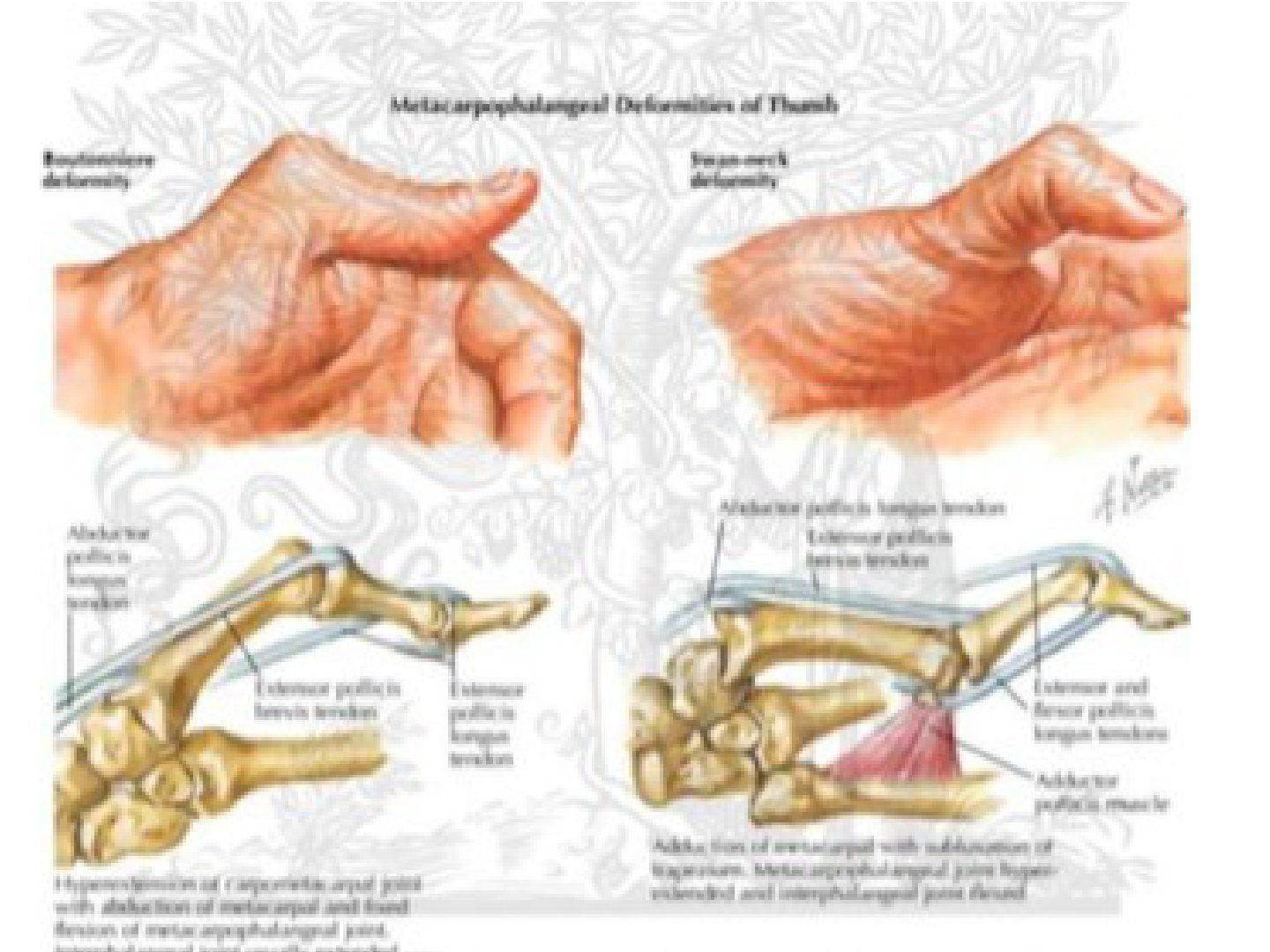
Question list
Q1-Q66 questions — tap to reveal all answerslist
- What is the Nalebuff classification of RA thumb deformity?
- Describe Nalebuff type I RA thumb deformity and its treatment.
- Describe Nalebuff type II RA thumb deformity and its treatment.
- Describe Nalebuff type III RA thumb deformity and its treatment.
- Describe Nalebuff type IV RA thumb deformity and its treatment.
- Describe Nalebuff types V and VI RA thumb deformity.
Answers · Q & A
Q1.What is the Nalebuff classification of RA thumb deformity?
- I: boutonniere of thumb (MCPJ disease)
- II: boutonniere + CMCJ involvement
- III: swan neck of thumb
- IV: gamekeeper thumb
- V: swan neck without IPJ involvement + MCPJ disease (MCPJ volar plate stretched)
- VI: arthritis mutilans
Q2.Describe Nalebuff type I RA thumb deformity and its treatment.
- Boutonniere of thumb: primary MCPJ disease with extensor hood synovitis; EPB attenuated, EPL subluxes ulnar and becomes a flexor of MCPJ
- Supple: synovectomy + EPL to EPB rerouting + IPJ fusion
- MCPJ rigid, IPJ supple: MCPJ fusion + EPL reroute
- MCPJ and IPJ rigid: IPJ fusion + MCPJ fusion (if CMCJ normal) or MCPJ arthroplasty (if CMCJ arthritis)
Q3.Describe Nalebuff type II RA thumb deformity and its treatment.
- Boutonniere + CMCJ involvement; primary pathology is CMCJ arthritis
- Tx: CMCJ fusion / trapeziectomy + soft tissue interposition
Q4.Describe Nalebuff type III RA thumb deformity and its treatment.
- Swan neck of thumb: CMCJ arthritis with subluxation, 1st webspace contracture, recruitment of EDB to hyperextend MCPJ to get the thumb out of the palm
- Supple: volar plate capsulodesis + FPL tenodesis
- Rigid: MCPJ arthrodesis +/- webspace release
Q5.Describe Nalebuff type IV RA thumb deformity and its treatment.
- Gamekeeper thumb: attenuation of UCL of MCPJ + contracture of 1st webspace
- Supple: UCL reconstruction + webspace release
- Rigid: fusion
Q6.Describe Nalebuff types V and VI RA thumb deformity.
- V: swan neck without IPJ involvement + MCPJ disease (MCPJ volar plate stretched)
- VI: arthritis mutilans - treatment is fusion
▸ Slide 737 · RA wrist deformityHand Bone · 7 questions expand
Slide render
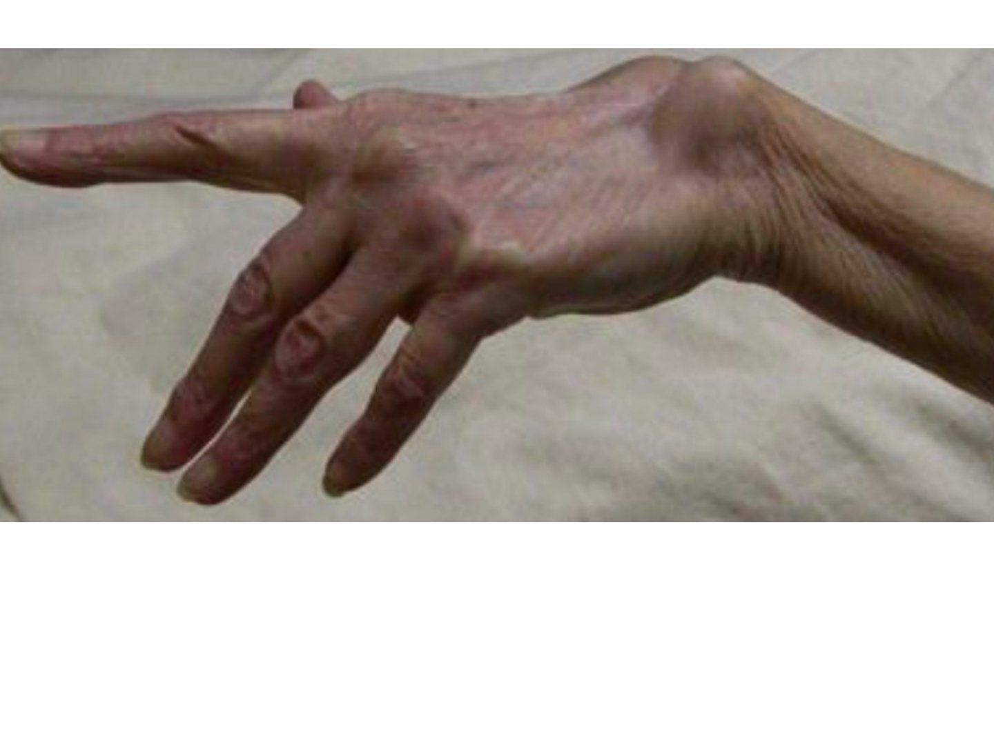
Question list
Q1-Q77 questions — tap to reveal all answerslist
- What features of RA wrist involvement should you look for in this patient?
- What is the differential diagnosis of finger drop in RA and how do you differentiate?
- What is the pathophysiology of caput ulnare and Vaughan-Jackson syndrome?
- What are the priorities in managing RA?
- How do you treat Vaughan-Jackson syndrome?
- When do you use a tendon graft versus a tendon transfer for ruptured extensor tendons in RA?
- How do you prevent further attritional extensor rupture in RA?
Answers · Q & A
Q1.What features of RA wrist involvement should you look for in this patient?
- Prominent ulnar styloid with finger drop of MF to LF
- +/- prominent knuckles +/- ulnar deviation of MCPJ
- Skin thin with loss of cutaneous fat; no scars or other joint swelling/deformity
- Assess the DRUJ for instability and check for radiocarpal joint involvement
Q2.What is the differential diagnosis of finger drop in RA and how do you differentiate?
- Tendon rupture - test tenodesis
- PIN palsy - test EPL and EI function; radial deviation of wrist (intact ECRL)
- MCPJ subluxation - try to reduce MCPJ - unable
- Sagittal band rupture - reduce MCPJ, able to maintain extension
- Intrinsic contracture - Bunnell test
Q3.What is the pathophysiology of caput ulnare and Vaughan-Jackson syndrome?
- RA involvement of DRUJ and UC ligament -> ulnar head dorsal subluxation
- ECU sheath synovitis -> ECU volar subluxation; with RC ligament erosion the carpus subluxes volarly, translocates ulnarly and supinates
- This places the carpal further away from the ulnar head and increase pressure over the extensor compartment -> progressive attrition rupture starting EDM, then EDC, then EI
Q4.What are the priorities in managing RA?
- Systemic: medical treatment; locally in the hand follow Souter's principle of predictable success
- Systemically: life threatening, patient preference, most symptomatic, sure win
- LL first, then UL; UL proximal first, then distal
Q5.How do you treat Vaughan-Jackson syndrome?
- First prevent further tendon rupture - give a splint
- Treat caput ulnare: Sauve-Kapandji (preferred in RA with ulnar translocation) or Darrach (not in unstable carpus; can do if wrist already fused)
- Darrach problems: ulnar impingement on radius (prevent by soft tissue interposition with PQ reattached dorsally), ulnar dorsal/volar instability (prevent by ECU tenodesis), decreased grip strength
- Bower not a good option as it requires intact TFCC
- Ruptured tendon: graft if good muscle quality with excursion >2cm (likely 2-stage, 2 anastomoses) vs transfer (side to side, EI or R/F FDS)
Q6.When do you use a tendon graft versus a tendon transfer for ruptured extensor tendons in RA?
- Tendon graft if good muscle quality with excursion >2cm, but likely needs 2 stages and has 2 anastomosis sites
- Tendon transfer (side to side, EI or R/F FDS) has one healing site but the donor unit downgrades one motor power grade
Q7.How do you prevent further attritional extensor rupture in RA?
- Extensor retinacular reconstruction
- Re-routing of the extensor retinaculum
- Tenosynovectomy
▸ Slide 738 · Failed thumb IPJ flexionHand Bone · 6 questions expand
Slide render
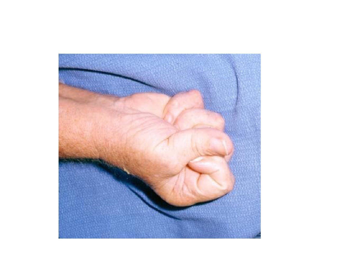
Question list
Q1-Q66 questions — tap to reveal all answerslist
- What are the causes of failed thumb IPJ flexion?
- What history do you take in a patient with failed thumb IPJ flexion?
- What is the differential diagnosis of failed thumb IPJ flexion in RA?
- How is FPL rupture in RA managed?
- How are ruptures of other digital flexor tendons in RA managed?
- How is trigger finger in RA managed?
Answers · Q & A
Q1.What are the causes of failed thumb IPJ flexion?
- FPL rupture (Mannerfelt syndrome)
- AIN palsy
- Trigger thumb
Q2.What history do you take in a patient with failed thumb IPJ flexion?
- Age + premorbid status
- Current disability: chronicity, decrease gripping power, median neuropathy with thenar muscle wasting and numbness
- Hand function
Q3.What is the differential diagnosis of failed thumb IPJ flexion in RA?
- Rupture (direct invasion, ischaemic, attrition - STT synovitis)
- Triggering (tenosynovitis, MCPJ subluxation, tendon nodule)
- AIN palsy (compression at lacertus fibrosus secondary to elbow synovitis)
- Advanced CTS (median nerve perineuritis, compression by synovitis)
Q4.How is FPL rupture in RA managed?
- Always tenosynovectomy; FPL rupture always needs synovectomy + spur excision
- IPJ fusion: stable thumb, good power, but loss of motion
- FDS4 transfer / free graft: motion preserved, but unpredictable motion gain and difficult tension adjustment
Q5.How are ruptures of other digital flexor tendons in RA managed?
- FDS only - observe
- FDP at wrist - suture to adjacent FDP
- FDP at finger - DIPJ fusion
- FDP + FDS - transfer intact FDS or tendon graft
Q6.How is trigger finger in RA managed?
- Tenosynovectomy
- 3 potential sites: carpal tunnel, A1, just distal to A2/FDS decussation
- Preserve A1 - otherwise increase ulnar drifting and MCPJ subluxation
- If severe ulnar drift, resect ulnar slip of FDS
▸ Slide 739 · Thoracic outlet syndromeHand Bone · 7 questions expand
Slide render
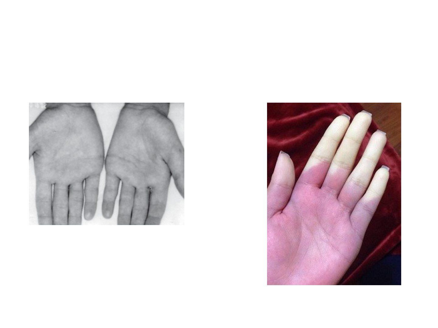
Question list
Q1-Q77 questions — tap to reveal all answerslist
- What is the presentation and epidemiology of thoracic outlet syndrome?
- What risk factors and history suggest thoracic outlet syndrome?
- What causes thoracic outlet syndrome?
- What clinical tests are used in thoracic outlet syndrome?
- What investigations are used in thoracic outlet syndrome?
- What is the management of thoracic outlet syndrome?
- What are the boundaries of the thoracic outlet and what is Paget-Schroetter syndrome?
Answers · Q & A
Q1.What is the presentation and epidemiology of thoracic outlet syndrome?
- Females > males (3:1); neurogenic 95%, venous 4%, arterial 1%
- Symptoms worse with overhead activities
- Neurogenic: pain at trapezius (96%), intrinsic minus hand, thenar and hypothenar wasting
- Vascular: Raynaud phenomenon (pale > red > cyanotic)
Q2.What risk factors and history suggest thoracic outlet syndrome?
- Symptoms worse with overhead activities
- Previous trauma, body builder, smoker / constitutional symptoms
- Look actively for the underlying cause (bone, soft tissue, vascular, neoplastic)
Q3.What causes thoracic outlet syndrome?
- Bone 30%: cervical rib, enlarged C7 transverse process, clavicle fracture, ACJ/SCJ dislocation
- Soft tissue 70%: muscular, scalene muscle anomaly, fibrous band / variant costoclavicular ligament insertion
- Vascular: aneurysm, thromboembolism
- Neoplastic: Pancoast tumour, bone met
Q4.What clinical tests are used in thoracic outlet syndrome?
- Tinel at Erb's point (upper trunk), 2-3cm above clavicle
- Subclavicle bruit
- Adson and costoclavicular test
- Wright manoeuvre and Roos test
Q5.What investigations are used in thoracic outlet syndrome?
- XR; CT or MRI
- NCT for MABCN (medial antebrachial cutaneous nerve)
- Doppler USG (venous)
- Angiogram (arterial)
Q6.What is the management of thoracic outlet syndrome?
- 1st line non-op / nerve block; OT if failed non-op for 6 months
- Address underlying pathology; decompression +/- revascularisation
- Decompression: 1st rib excision + anterior/middle scalenectomy +/- neurolysis of medial cord
- Complication: pneumothorax (PTx)
Q7.What are the boundaries of the thoracic outlet and what is Paget-Schroetter syndrome?
- Proximal interscalene space: anterior anterior scalene, posterior middle scalene, inferior 1st rib; contents trunk + vessel
- Middle costoclavicular space: anterior clavicle + subclavius, posterior 1st rib + scalenes, medial costoclavicular ligament, lateral upper scapular border; contents division + vessel
- Distal retropectoralis minor (thoraco-coraco-pectoral/subcoracoid) space: superior coracoid, anterior pectoralis minor, posterior ribs 2-4; contents cord + vessel
- Paget-Schroetter syndrome: venous TOS in well-developed young athletes; intermittent subclavian vein obstruction by abnormal costoclavicular ligament / scalene hypertrophy -> upper limb DVT