▸ Slide 636 · SportsSports · 2 questions expand

Q1-Q22 questions — tap to reveal all answerslist
- What is the topic of this section?
- Which sports injuries are covered in this section?
Q1.What is the topic of this section?
- Sports (section slide); no speaker notes available
Q2.Which sports injuries are covered in this section?
- Not covered in the speaker notes
▸ Slide 637 · Meniscal tearSports · 20 questions expand

Q1-Q2020 questions — tap to reveal all answerslist
- How does a meniscal tear present?
- What is the differential diagnosis of acute haemarthrosis (Frank Noyes JBJS 1980)?
- What MRI signal changes suggest a meniscal tear?
- How do you describe a meniscal tear?
- Which meniscal tears can heal by themselves?
- Which meniscal tears should undergo meniscectomy (classical teaching)?
- Which meniscal tears go for repair?
- What other factors affect meniscal healing?
- What disease and patient factors are considered when treating a meniscal tear?
- Describe the inside-out meniscal repair technique.
- What is the medial approach to the capsule for inside-out meniscal repair?
- What is the lateral approach to the capsule for inside-out meniscal repair?
- What are the advantages and disadvantages of outside-in meniscal repair?
- What are the advantages and disadvantages of all-inside meniscal repair?
- What are the generations of all-inside repair techniques?
- What must be preserved during meniscectomy and why?
- How can meniscal healing be improved?
- What are the options if the meniscus is not repairable?
- What is the definition of the red-red zone?
- What are the pros and cons of acute one-stage bucket handle repair with ACL reconstruction?
Q1.How does a meniscal tear present?
- Presents as delayed swelling
Q2.What is the differential diagnosis of acute haemarthrosis (Frank Noyes JBJS 1980)?
- ACL tear 72%
- Meniscal injury 62%
- Osteochondral fragment 20%
- Patella dislocation
Q3.What MRI signal changes suggest a meniscal tear?
- Globular signal is not specific
- Linear signal is more suggestive of a tear
Q4.How do you describe a meniscal tear?
- Timing
- Morphology: radial, longitudinal, transverse, bucket handle, complex
- Zone (RR 3-4mm from rim, RW, WW) from rabbit study
- Site (root/body), stability, length
- Partial thickness/ full thickness
- These affect whether the tear can heal
Q5.Which meniscal tears can heal by themselves?
- Stable longitudinal tear
- RR zone
- Partial thickness
- <5mm
Q6.Which meniscal tears should undergo meniscectomy (classical teaching)?
- Degenerative/complex/radial tear - radial tear breaks many longitudinal fibres
- Chronic tear
- WW zone
- Irreducible tear
- Leave a stable rim to maintain hoop stress (may not apply now)
Q7.Which meniscal tears go for repair?
- Acute, traumatic tear
- Vascular zone
- Reducible and stable
- Simple pattern (longitudinal/bucket handle)
- Root tear
Q8.What other factors affect meniscal healing?
- Age (young is <40 years old)
- Smoking
- Stability of the joint, malalignment
Q9.What disease and patient factors are considered when treating a meniscal tear?
- Disease factors: chronicity (<8/52 better healing), size, shape, location, displacement, associated injury
- Patient factors: age, functional demand, symptoms (pain/locking)
- Aim: relieve symptoms, restore the meniscus as much as possible
Q10.Describe the inside-out meniscal repair technique.
- Inside-out with nonabsorbable suture, vertical mattress = gold standard
- Higher biomechanical strength
- Problems: need an assistant, risk of needle stick injury and NV injury (saphenous, CPN)
Q11.What is the medial approach to the capsule for inside-out meniscal repair?
- Incision posterior to the MCL at the level of the joint line (one third above and two thirds below), knee in 90 degrees flexion
- Sartorial fascia incised, dissect anterior to semimembranosus, retract pes tendons anteriorly
- Saphenous nerve and vein lie posterior to the incision in this position
- Place a Henning retractor between the medial gastrocnemius and the capsule
Q12.What is the lateral approach to the capsule for inside-out meniscal repair?
- Interval between the ITB and biceps femoris, just posterior to the LCL at the level of the joint line
- Flex the knee to 90 degrees to allow the peroneal nerve to fall posteriorly
- Then use the interval between the lateral head of gastrocnemius and the capsule
Q13.What are the advantages and disadvantages of outside-in meniscal repair?
- For anterior 2/3 tears
- Pros: no assistant, no posterior incision, no needle stick, no NV injury
- Cons: Difficult reduction & oppose edge
Q14.What are the advantages and disadvantages of all-inside meniscal repair?
- Pros: quick, easy, all arthroscopic, single surgeon
- Cons: reduced strength, limited compression, variable resorption profile
Q15.What are the generations of all-inside repair techniques?
- 1st gen: described by Morgan - curved suture hooks through accessory posterior portals
- 2nd gen: T-fix - polyethylene bar with attached suture deployed through a sharp needle; inability to tension the knots
- 3rd gen: bioabsorbable meniscus repair device e.g. meniscus arrow
- 4th gen: Fastfix - 2 suture anchors connected by a nonabsorbable polyester suture with a pretied slip knot
Q16.What must be preserved during meniscectomy and why?
- Keep the rim and root intact, otherwise there is no hoop stress
- Meniscus functions = shock absorption, stability, load bearing, lubrication, proprioception - all affected
- Medial side degeneration is more common due to roll back
Q17.How can meniscal healing be improved?
- Trephination/shaving at the tear site to increase punctate bleeding; microfracture of the notch
- PRP: CORR 2015 no difference in reoperation rate; Orth J Sport Med systematic review found lower failure rates but no difference in PRO
- Exofibrin clot technique: chemotactic, mitogenic, acts as a scaffold to fill the defect -> inflammatory fibrovascular scar; 30-50ml blood stirred till clot, inserted under the repair site
Q18.What are the options if the meniscus is not repairable?
- Allograft with bone graft
- Artificial meniscus
Q19.What is the definition of the red-red zone?
- 3mm from the edge according to Warren and Arnoczky 1982 AJSM
Q20.What are the pros and cons of acute one-stage bucket handle repair with ACL reconstruction?
- Pros: one OT
- Biological: drilling of tunnels allows stem cells to seep into the joint -> good healing
- Mechanical: stable environment for meniscal healing
- Cons: contradictory rehab -> arthrofibrosis
▸ Slide 638 · Horizontal /cleavage tearSports · 2 questions expand

Q1-Q22 questions — tap to reveal all answerslist
- List the meniscal tear patterns shown.
- What are the MRI findings of a radial tear of the medial meniscus body?
Q1.List the meniscal tear patterns shown.
- Horizontal/cleavage tear
- Longitudinal/vertical tear
- Radial tear of the medial meniscus (body)
Q2.What are the MRI findings of a radial tear of the medial meniscus body?
- Sagittal: T2W signal change
- Coronal: meniscal extrusion defined as 3mm
- Axial: radial tear
▸ Slide 639 · T2W MRI right kneeSports · 5 questions expand
Q1-Q55 questions — tap to reveal all answerslist
- Describe the MRI findings of a meniscal root tear.
- What is the definition and incidence of a meniscal root tear?
- What is the Laprade classification of meniscal root tears?
- How is a meniscal root tear repaired?
- What are the consequences of meniscectomy?
Q1.Describe the MRI findings of a meniscal root tear.
- Coronal: extrusion of the medial meniscus (>3mm), vertical linear defect (truncation)
- Axial: fluid interposition at the meniscus root and posterior horn (high signal cleft)
- Sagittal: ghost sign - absence of the posterior horn of the meniscus
Q2.What is the definition and incidence of a meniscal root tear?
- Avulsion of the meniscus insertion or radial tears within 9mm of the insertion
- Incidence 7-13%
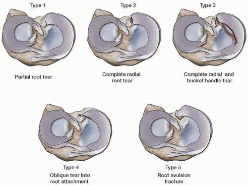
Q3.What is the Laprade classification of meniscal root tears?
- 1: partial stable root tear
- 2: complete root tear
- 3: bucket handle tear with complete root detachment
- 4: complex oblique or longitudinal tear with complete root detachment
- 5: bony avulsion of the root attachment
Q4.How is a meniscal root tear repaired?
- Restore the root in anatomical position by suture anchor or drill bone tunnel
Q5.What are the consequences of meniscectomy?
- Medial meniscectomy: contact area decreases 50-70%, contact stress increases 100%
- Lateral meniscectomy: contact area decreases 40-50%, contact stress increases 200-300% (convex lateral tibial plateau)
- So lateral meniscectomy has a much worse outcome
- Lateral meniscus occupies ~80% vs medial ~60% of the articular surface
- ~70% of load in the lateral and 50% in the medial compartment is transmitted through the menisci
▸ Slide 640 · Young boy with knee slappingSports · 10 questions expand
Q1-Q1010 questions — tap to reveal all answerslist
- What is a discoid meniscus and how does it develop?
- What is the common presentation of a discoid meniscus?
- What are the X-ray findings of a discoid meniscus?
- What are the MRI findings of a discoid meniscus?
- What is the Watanabe classification of a discoid meniscus?
- How is a discoid meniscus treated?
- What are the meniscofemoral ligaments?
- What are the dimensions and attachments of the meniscofemoral ligaments?
- What is the function of the meniscofemoral ligaments?
- What is a pseudotear and a Wrisberg rip?
Q1.What is a discoid meniscus and how does it develop?
- Abnormal development of the meniscus causing a thickened, discoid-shaped meniscus
- Embryology: failed regression of tissue
- 25% bilateral
Q2.What is the common presentation of a discoid meniscus?
- Often asymptomatic
- When symptomatic, usually secondary to a meniscal tear
- Knee clunk/click/lock after trivial trauma
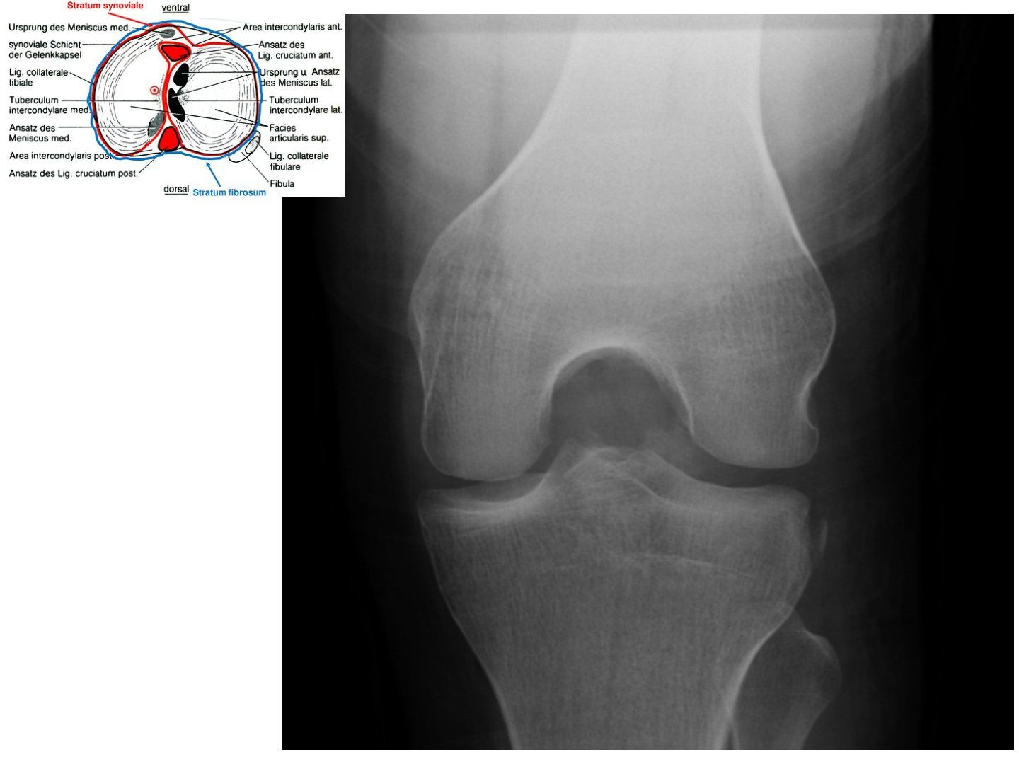
Q3.What are the X-ray findings of a discoid meniscus?
- Widened lateral joint space (11mm)
- Squaring of the lateral femoral condyle
- Hypoplastic lateral tibial spine
- Cupping/ flattening of lateral tibial plateau
Q4.What are the MRI findings of a discoid meniscus?
- Sagittal: consecutive 3 (5mm) cuts with continuous meniscus (bow tie sign)
- Transverse: transverse diameter in the mid body >15mm
- Coronal: medial-lateral height difference >2mm; meniscal width to maximal tibial width >20%
- Sagittal: ratio of the sum of both lateral horns to maximal meniscal diameter >75%
- R/O tear; +/- coronary ligament integrity
Q5.What is the Watanabe classification of a discoid meniscus?
- Incomplete (type 2)
- Complete (type 1)
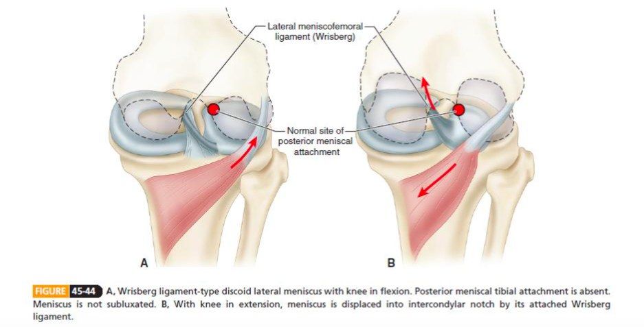
- Wrisberg type: absent meniscotibial ligament, only the meniscofemoral ligament remains; displaces into the intercondylar notch during extension
- May alternatively be classified as stable or unstable
Q6.How is a discoid meniscus treated?
- Surgery only for symptomatic patients
- Partial meniscectomy / meniscal repair for a tear
- Saucerization (5-8mm compared to the medial side) - reshapes to a semilunar shape, triangular cross-section, less click
- Repair of the meniscotibial ligament for peripheral attachment
- James Hui KSSTA 2021: meniscoplasty gives good mid- to long-term outcomes; concomitant repair/partial meniscectomy does not change outcomes
Q7.What are the meniscofemoral ligaments?
- Anterior MFL (ligament of Humphrey) - anterior to the PCL
- Posterior MFL (ligament of Wrisberg) - behind the PCL
- 70% of knees have either one, 6% have both
Q8.What are the dimensions and attachments of the meniscofemoral ligaments?
- Anterior MFL is thinner (<one third the diameter of the PCL), from the posterior horn of the LM to the distal edge of the femoral PCL attachment; can be mistaken for the PCL at arthroscopy
- Posterior MFL is usually larger (~half the diameter of the PCL), from the posterior horn of the LM to the medial femoral condyle, inserting near the posteromedial band of the PCL
Q9.What is the function of the meniscofemoral ligaments?
- Stabilisers and protectors of the posterolateral femorotibial compartment
- Increase congruity between the mobile lateral meniscus and lateral femoral condyle, protecting the posterior horn of the LM
- Anterior MFL supplements the anterior band of the PCL; posterior MFL supplements the posterior band; they may act as a splint in PCL injuries
- Anterior MFL is taut in flexion and lax in extension; posterior MFL is taut in extension and lax in flexion
Q10.What is a pseudotear and a Wrisberg rip?
- Pseudotear: the posterior MFL commonly causes a pseudotear of the posterior horn of the lateral meniscus on imaging - a normal vertical/oblique signal at the junction of the ligament of Wrisberg with the posterior horn
- Wrisberg rip: a longitudinal vertical tear of the posterior horn of the LM seen in association with ACL tears
- It is frequently missed and may be used as a secondary sign of ACL disruption
▸ Slide 641 · ACLSports · 39 questions expand
Q1-Q3939 questions — tap to reveal all answerslist
- What is the X-ray finding of an ACL tear?
- What are the sagittal MRI findings of an ACL tear?
- What are the coronal MRI findings of an ACL tear?
- What is the axial MRI finding of an ACL tear?
- What are the physical examination findings of ACL deficiency?
- Why examine the collateral ligaments at 30 degrees of flexion?
- What injuries are associated with an acute ACL tear?
- What is the anatomy and function of the anterolateral ligament (ALL)?
- What is the biomechanical difference between ALL reconstruction and LET?
- What are the results and downsides of the STABILITY trial (AJSM 2020)?
- What are the indications for ALL reconstruction or LET?
- What is the location of the ACL?
- What are the dimensions and composition of the ACL?
- What are the anteromedial and posterolateral bundles of the ACL?
- What is the blood and nerve supply of the ACL?
- What is the function of the ACL?
- What is the mechanism of an ACL injury and what causes locking?
- What is the natural history of the ACL-deficient knee (Frank Noyes JBJS 1983)?
- When should ACL reconstruction be performed?
- What factors determine whether to perform ACL reconstruction?
- What are the graft options and their loads to failure?
- What are the pros and cons of a BPTB graft?
- What are the pros and cons of a hamstring graft?
- What are the problems with allograft and artificial grafts?
- What are the advantages of autograft versus allograft?
- What do KT-1000 measurements show after ACL reconstruction?
- Should single or double bundle ACL reconstruction be used?
- What are the ideal properties of graft fixation?
- What are the types of suspensory and aperture fixation and their pros and cons?
- Which fixation is used in the femur, tibia and revision cases?
- What are the key steps of ACL reconstruction?
- Describe the operative details of ACL reconstruction.
- What complications can occur according to time sequence?
- Describe the phases of graft ligamentisation and healing.
- What are the 4 zones of tendon-to-bone healing?
- What are the phases of ACL rehabilitation?
- What are the principles of ACL rehabilitation?
- How is a dropped graft managed intra-operatively?
- What are the pros and cons of one-stage versus staged meniscus and ACL surgery?
Q1.What is the X-ray finding of an ACL tear?
- Segond fracture on X-ray = ALL avulsion
- Associated with ACL tear 75-100% of the time
Q2.What are the sagittal MRI findings of an ACL tear?
- Discontinuity/waviness of fibres on T2
- Abnormal orientation (too flat compared with Blumensaat's line)
- PCL buckled
- Anterior translation of the tibia
Q3.What are the coronal MRI findings of an ACL tear?
- Discontinuity of fibres
- Bone bruise in the LFC (middle 1/3) and lateral tibial plateau (posterior 1/3)
- Effusion
Q4.What is the axial MRI finding of an ACL tear?
- Fluid at the intercondylar notch = empty notch sign
Q5.What are the physical examination findings of ACL deficiency?
- Palpate the bony prominence; feel step off bilaterally
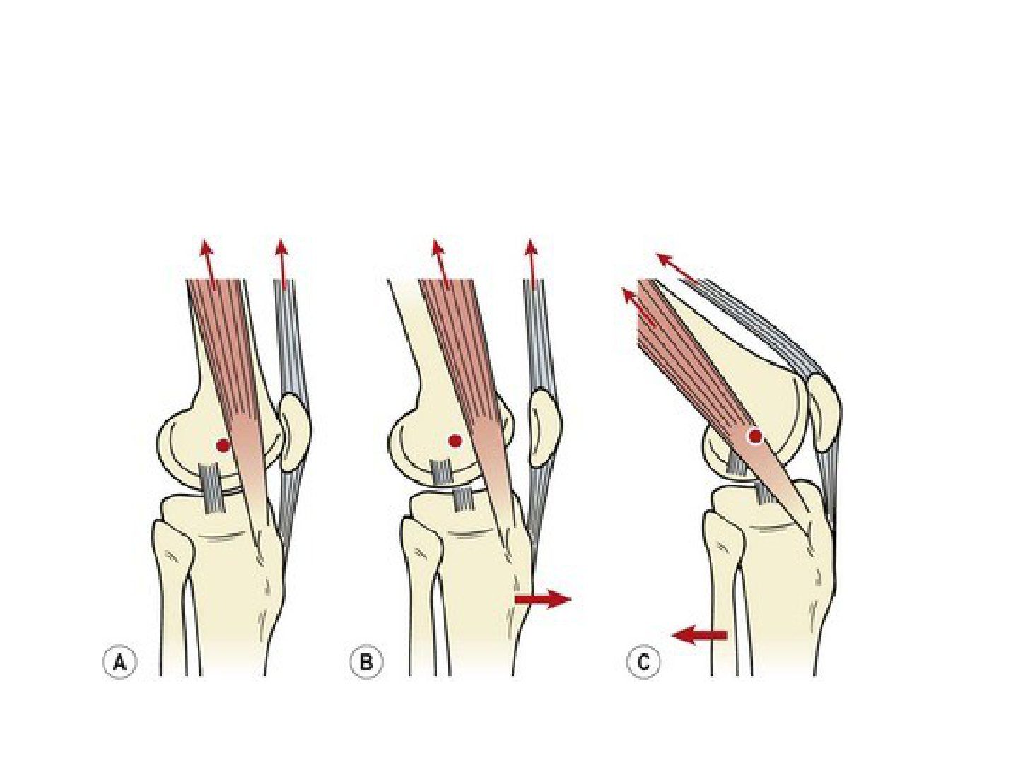
- Pivot shift (slide, jerk, clunk)
- IR + valgus anterolaterally subluxes the tibia
- Flexing the knee changes the ITB from extensor to flexor and reduces the tibia
Q6.Why examine the collateral ligaments at 30 degrees of flexion?
- At 30 degrees of flexion the capsule and cruciate ligaments relax
- This allows isolated assessment of the collateral ligaments
Q7.What injuries are associated with an acute ACL tear?
- Lateral meniscal tear in 54% of acute ACL injuries
- Unhappy triad = ACL + MCL + medial meniscus
Q8.What is the anatomy and function of the anterolateral ligament (ALL)?
- Origin: anterior and distal to the femoral attachment of the LCL
- Insertion: Gerdy tubercle on the tibia
- Contributes to tibial internal rotation stability
- Failure causes rotational instability; treated by ALL reconstruction or LET
Q9.What is the biomechanical difference between ALL reconstruction and LET?
- ALL - non-isometric (tight in extension, slack in flexion), allowing physiological internal rotation at 90 degrees flexion
- LET (modified Lemaire) - isometric, with a tendency to limit physiological internal rotation at 90 degrees flexion
Q10.What are the results and downsides of the STABILITY trial (AJSM 2020)?
- Adding LET to single bundle ACLR in young high-risk patients results in a statistically significant, clinically relevant reduction in graft rupture and persistent rotatory laxity at 2 years
- Downside: more pain and worse outcome scores in the first 3-6 months
- Slower return to sport
- Long-term studies suggest increased risk of lateral compartment OA
Q11.What are the indications for ALL reconstruction or LET?
- 2 of 3: Grade 2 pivot shift or greater
- Desire to return to high-risk/pivoting sport
- Generalised ligamentous laxity
Q12.What is the location of the ACL?
- Intra-articular, extrasynovial - no haematoma formation; synovial fluid has a surfactant effect
- From the medial surface of the LFC (bifurcate ridge) to the anterior tibia, just anterior and between the intercondylar eminences
Q13.What are the dimensions and composition of the ACL?
- ~33mm length, 11mm width
- 90% type 1 collagen, other type 3
Q14.What are the anteromedial and posterolateral bundles of the ACL?
- AM bundle: more isometric, tight with the knee in flexion, controls AP translation more
- PL bundle: tight in extension, controls rotation more
Q15.What is the blood and nerve supply of the ACL?
- Blood supply: middle geniculate artery
- Nerve supply: posterior articular branch of the tibial nerve
Q16.What is the function of the ACL?
- Provides 85% of the stability preventing anterior translation of the tibia
- Secondary restraint to valgus and varus stress and tibial rotation
- 2200N load to failure
Q17.What is the mechanism of an ACL injury and what causes locking?
- Non-contact pivoting injury
- Valgus + external rotation, partially flexed
- Landing with the knee in extension
- Cause of locking in an ACL tear: the stump
Q18.What is the natural history of the ACL-deficient knee (Frank Noyes JBJS 1983)?
- 1/3 have no/minimal symptoms in ADL or recreational activity
- 60% had repeated injury and meniscal/cartilage injury within 2 years
- 1/3 give up sport
- 1/3 deteriorate and require surgical stabilisation
Q19.When should ACL reconstruction be performed?
- Delay around 6/52
- Allow effusion and inflammation to subside
- Regain good ROM and good quadriceps control
- To avoid arthrofibrosis
Q20.What factors determine whether to perform ACL reconstruction?
- Return to sports, especially pivot type
- Prevent second injury - cartilage and meniscus
- Theoretically reduces DJD, yet no concrete evidence (ACL deficiency puts more stress on the posterior horn of the medial meniscus)
Q21.What are the graft options and their loads to failure?
- BPTB - 2600N, size 10mm
- Hamstring - 4000N, size 7-8mm
- Quadriceps tendon - 2500N, mainly for revision cases
- Return to sport/repeat injury and ROM/failure rate are the same for BPTB and hamstring
Q22.What are the pros and cons of a BPTB graft?
- Pros: early healing (animal study), bone-to-bone -> faster rehab
- Cons: patella fracture (stellate) or PT rupture 0.5%, graft-tunnel mismatch
- Anterior knee pain (15%)/PFJ OA - cannot kneel; patellar baja from retropatellar fibrosis; some flexion contracture
Q23.What are the pros and cons of a hamstring graft?
- Pros: high tensile strength, easier/smoother passage
- Cons: not used in generalised ligamentous laxity or complete MCL injury - do not jeopardise medial stability
- Saphenous nerve injury during harvest, premature harvest, knee flexor weakness, graft attritional rupture
Q24.What are the problems with allograft and artificial grafts?
- Allograft: fresh frozen risks infection; irradiation weakens the graft; quality not as good, not enough supply
- Artificial: no incorporation, a lot of synovitis and pain, cannot absorb energy (stress-strain curve has no toe phase and is steep)
Q25.What are the advantages of autograft versus allograft?
- Autograft: less immunogenicity, less disease transmission, faster incorporation, lower cost
- Allograft: saves harvesting time, bigger graft for revision, no donor site morbidity
- Grassi BJJ 2017: autograft has lower rates of postoperative laxity and fewer reoperations/complications; when irradiated grafts are excluded, outcomes are similar
Q26.What do KT-1000 measurements show after ACL reconstruction?
- <3mm compared with the normal side is considered stable, and BPTB is more stable
- If the difference is <5mm, BPTB and hamstring show no difference
- Ligament has more elastin, is more viscoelastic and elongates less compared with hamstring
Q27.Should single or double bundle ACL reconstruction be used?
- Meta-analysis: double bundle is better for rotational stability but clinical outcome is similar
- Prerequisite: footprint at least 11mm
- Freddie Fu CORR 2020: <14mm single, 14-18 controversial, >18 double
Q28.What are the ideal properties of graft fixation?
- Strong enough to avoid failure
- Stiff enough to restore load-displacement response
- Secure enough to resist slippage under cyclic loading
Q29.What are the types of suspensory and aperture fixation and their pros and cons?
- Suspensory: cortical (endobutton, staples, tie over post) or cancellous (transfix, rigidfix)
- Pros: more healing surface, more rigid (fixed at closed loop), adjustable length
- Cons: micromotion - bungee effect + windshield wiper effect -> tunnel widening
- Aperture fixation: interference screw (depends on friction, longer = more pull-out strength)
- Pros: fixation near the articular surface, less bungee effect; Cons: advancement of bone graft, graft/suture laceration, divergent -> less stability
Q30.Which fixation is used in the femur, tibia and revision cases?
- Femoral: usually suspensory +/- double fixation
- Tibial: usually aperture +/- double fixation
- Revision: poorer bone quality -> double fixation
- Reverse thread in the femoral tunnel of the right knee: the graft moves anteriorly and causes laxity (the isometric point is more posterior)
Q31.What are the key steps of ACL reconstruction?
- Low morbidity harvest of a biologically active graft
- Short arthroscopy time and prevent intra-articular injury
- Accurate identification of the tibial and femoral footprints
- Smooth passage of the graft through the tunnels
- Robust fixation and tensioning of the graft
- Appropriate rehabilitation
Q32.Describe the operative details of ACL reconstruction.
- GA/SA with tourniquet; graft harvesting and preparation (preconditioning reduces stress relaxation)
- Femoral tunnel: anatomical footprint, as posterior as possible but prevent blow out, coronally at 9-10 o'clock if done on the right knee
- Tibial tunnel: anatomical footprint (centre 10-11mm in front of the anterior border of the PCL), trajectory <75 degrees from horizontal (55 degrees guide used)
- Fixation: femoral endobutton, tibial interference screw
- Graft tensioning: 40N with the knee at 20-30 degrees flexion
Q33.What complications can occur according to time sequence?
- Intraoperative: graft harvest - BPTB patella fracture; hamstring infrapatellar branch of saphenous nerve injury, saphenous nerve injury, graft transection, contamination
- Intraoperative: tunnel malposition, graft fixation problems (interference screw laceration/divergence, endobutton blow out at lateral cortex), poor tensioning, injury to secondary stabilisers
- Postoperative: notch impingement (cyclops lesion around the tibial tunnel), stiffness/arthrofibrosis, infection, late patella fracture/anterior knee pain/patella tendon rupture (BPTB graft)
Q34.Describe the phases of graft ligamentisation and healing.
- Early healing 0-6 months: inflammatory cytokines (IL6, TNF alpha, TGF beta, metalloproteinase 1 and 13) digest collagen; graft undergoes central avascular necrosis; maximum increase in anterior laxity at 6 months
- Proliferation 6-12 months: Revascularization from the synovium, infrapatellar fat pad and pseudoligamentum mucosum, increased expression of the VEGF; Repopulation by progenitor cells from the autograft, bone tunnel and seeding fibroblasts from the residual stump lay down type III collagen
- Maturation/ligamentisation >12 months: osseous integration by enchondral ossification; collagen remodelled to type I collagen
Q35.What are the 4 zones of tendon-to-bone healing?
- Fibrous tissue surrounds the graft
- Collagen grows into the fibrous tissue
- Fibrocartilaginous tissue forms and then becomes bone
- This is fibrocartilaginous healing
Q36.What are the phases of ACL rehabilitation?
- Phase 1 (postop 1 week) [ROM + ADL]: cryotherapy for swelling and pain, obtain ROM 0-90 especially full extension, immediate weight bearing
- Phase 2 (postop 2-8 weeks) [strengthening]: full ROM, isometric and isotonic exercise, commence close and open chain exercise
- Phase 3 (postop 8-16 weeks) [functional]: isokinetic and endurance, increase resistance of OC/CC exercise from 9 weeks, plyometric exercise, normal running from 13 weeks
- Phase 4 (postop 16-22 weeks) [sport specific + injury prevention]: Neuromuscle ex optimization, jumping, agility; return to sport when HQ ratio >0.8
Q37.What are the principles of ACL rehabilitation?
- The cornerstone is ROM, strengthening, functional exercise and injury prevention, facilitating ligamentisation and graft healing
- Avoid excessive stress on the graft: initial closed chain (open chain: distal limb unsupported -> increases shear force by gravity)
- Eccentric quadriceps strengthening; isometric hamstring and quadriceps exercise
- Avoid isokinetic quadriceps or open chain exercise in the early post-op period
Q38.How is a dropped graft managed intra-operatively?
- Retrieve from the floor within 15 seconds
- Remove all sutures; soak in saline
- Soak in 2% chlorhexidine
- Soak in triple antibiotics (gentamicin, clindamycin, polymyxin)
- Minimise sutures in the graft, consider monofilament sutures; longer antibiotics and more frequent follow-up
Q39.What are the pros and cons of one-stage versus staged meniscus and ACL surgery?
- One stage pros: one GA
- Biological: drilling of tunnels provides MSC for meniscus healing
- Mechanical: restores a stable environment for meniscus healing
- Cons: contradictory rehab -> arthrofibrosis; for a locked knee go 2 stage (meniscus then early ACLR)
▸ Slide 642 · Principle: reduction of subluxed knee from IR + extension to valgus + flexionSports · 5 questions expand
Q1-Q55 questions — tap to reveal all answerslist
- Describe the technique and prerequisites of the pivot shift test.
- What is the principle of the pivot shift?
- What are the grades of pivot shift?
- What does a high-grade pivot shift indicate?
- What is a RAMP lesion and how is it classified (Thaunat)?
Q1.Describe the technique and prerequisites of the pivot shift test.
- IR tibia at knee extension
- Valgus force + gradual knee flexion
- Sudden glide/jump/clunk = reduction of the tibia relative to the femur
- + axial load to exaggerate
- Prerequisites: intact MCL and intact ITB
Q2.What is the principle of the pivot shift?
- IR of the tibia exaggerates ACL deficiency (especially the PL bundle)
- Valgus stress tightens the MCL -> decreases medial tibial plateau movement -> lateral tibial plateau translates forward
- Sudden reduction by the ITB on knee flexion (axis of pull posterior to knee COR/TEA -> ITB becomes a flexor)
Q3.What are the grades of pivot shift?
- 1: glide (may be physiological)
- 2: sublux with spontaneous reduction (jump)
- 3: subluxation +/- spontaneous reduction (clunk)
Q4.What does a high-grade pivot shift indicate?
- Isolated ACL tear is unable to produce a high-grade pivot shift
- Meniscus: LM root tear, ramp lesion, MM posterior horn tear
- Anterolateral complex: ITB (superior and deep), ALL, AL capsule - secondary restraint to anterior translation and IR (with LM)
- Generalised ligamentous laxity (Beighton score >=5/9)
Q5.What is a RAMP lesion and how is it classified (Thaunat)?
- 9-17% of ACL tears
- 1: capsulomeniscal junction lesions - very peripheral, low mobility at probing
- 2: partial superior lesions - stable, diagnosed only by the trans-notch approach (modified Gillquist manoeuvre)
- 3: partial inferior/hidden - not visible trans-notch, high mobility (meniscotibial ligament disruption)
- 4: complete tear in the red-red zone - very high mobility
- 5: double tear
▸ Slide 643 · 100 patients randomly assign OT/ conservative MxSports · 3 questions expand
Q1-Q33 questions — tap to reveal all answerslist
- What was the design of the study of operative versus conservative management?
- What were the key findings of the study?
- What happened to the conservatively managed patients?
Q1.What was the design of the study of operative versus conservative management?
- 100 patients randomly assigned to operative (ACL reconstruction) or conservative management
Q2.What were the key findings of the study?
- ACL reconstruction per se did not reduce risk of OA or improve outcome scores
- ACL reconstruction can reduce risk of 2nd meniscal injury
- Meniscal status is the most important predictor of OA
Q3.What happened to the conservatively managed patients?
- 1/3 of conservative patients later received ACL reconstruction due to instability
▸ Slide 644 · ACL complications and preventionSports · 6 questions expand

Q1-Q66 questions — tap to reveal all answerslist
- What are the intraoperative complications of ACL reconstruction related to graft harvest?
- What complications can occur during tunnel placement in ACL reconstruction?
- What complications can occur during graft passage in ACL reconstruction?
- What complications can occur during graft fixation in ACL reconstruction?
- What postoperative complications are related to ACL reconstruction?
- How are postoperative complications of ACL reconstruction classified?
Q1.What are the intraoperative complications of ACL reconstruction related to graft harvest?
- Hamstring: premature harvest, deep MCL injury, hematoma, saphenous nerve injury
- BPTB: fracture (patella or tibia)
- Both grafts: dropped graft
Q2.What complications can occur during tunnel placement in ACL reconstruction?
- MFC injury
- Lateral wall blowout
- Posterior wall blowout
Q3.What complications can occur during graft passage in ACL reconstruction?
- Under-sized graft
- Over-sized graft
Q4.What complications can occur during graft fixation in ACL reconstruction?
- Late button flip
- Screw breakage or slippage
- Screw divergence
- Graft laceration, advancement or rotation
- Screw protrusion into the joint
Q5.What postoperative complications are related to ACL reconstruction?
- Cyclops lesion
- Arthrofibrosis
- Septic arthritis
- Prominent tibial screw
Q6.How are postoperative complications of ACL reconstruction classified?
- Related to ACLR: cyclops, arthrofibrosis, septic arthritis, prominent tibial screw
- Not related to ACLR (listed as a separate category by the lecturer; no examples given in the notes)
▸ Slide 645 · 10mm widthSports · 7 questions expand
Q1-Q77 questions — tap to reveal all answerslist
- What are the dimensions and fixation of a BPTB graft?
- What is the classical placement of a BPTB graft and why?
- What is the problem if a BPTB graft is too long and how is the graft remedied?
- What are the tunnel remedies if a BPTB graft is too long?
- What is the problem if a BPTB graft is too short and how is it remedied?
- What happens if a BPTB graft is too wide or too narrow?
- How are patellar complications prevented during BPTB harvest?
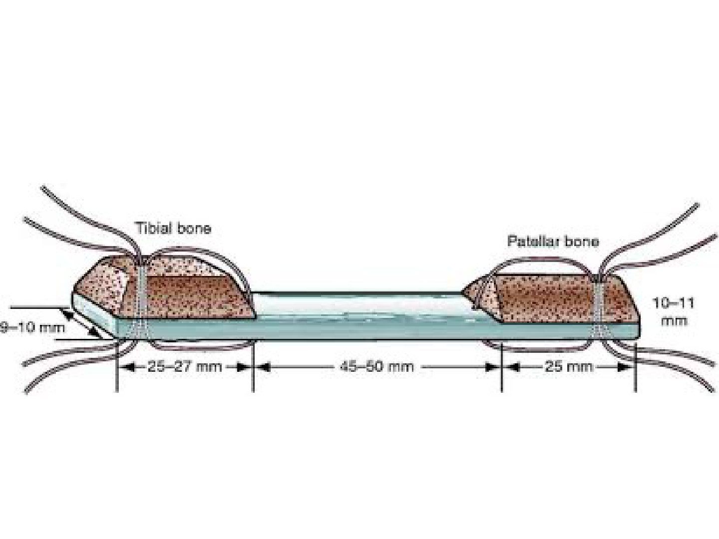
Q1.What are the dimensions and fixation of a BPTB graft?
- 10mm width, 90-100mm length
- Rigidfix/interference screw (proximal) + interference screw (distal)
Q2.What is the classical placement of a BPTB graft and why?
- Tibial bone --> femoral tunnel; patellar bone --> tibial tunnel
- Because the tibial metaphysis is weaker, and there is increased bone contact if the intraop tunnel is too wide
Q3.What is the problem if a BPTB graft is too long and how is the graft remedied?
- Too long: unable to use an interference screw
- Graft remedy: trim and flip 180 degrees back
- Graft remedy: self-twist 360 degrees to take up tension
Q4.What are the tunnel remedies if a BPTB graft is too long?
- Femur: recess graft and secure at aperture with bioscrew
- Tibia: secure bone plug in trough distal to tunnel
Q5.What is the problem if a BPTB graft is too short and how is it remedied?
- Too short: fixation not cortical
- Remedy: double fixation
Q6.What happens if a BPTB graft is too wide or too narrow?
- Too wide: bone plug not through tunnel; remedy graft trimming +/- notchplasty
- Too narrow: loosening; remedy alternative fixation
Q7.How are patellar complications prevented during BPTB harvest?
- Patella fracture: trapezoidal/bevel cut to increase the cross-sectional area of bone at the donor site
- Avoid transverse saw cut overruns
- Burr to round off corners
- Leave at least 1cm from the superior pole
▸ Slide 646 · Graft motion and fixationSports · 6 questions expand

Q1-Q66 questions — tap to reveal all answerslist
- What types of graft motion occur after ACL reconstruction?
- What are the consequences of graft motion greater than 3mm?
- Compare the healing time of BPTB and hamstring grafts.
- What are the types of aperture fixation and their pros and cons?
- What are the types of suspensory fixation and their pros and cons?
- How is the EndoButton loop length (EB-CL) calculated?
Q1.What types of graft motion occur after ACL reconstruction?
- Longitudinal motion: bungee effect
- Horizontal motion: windshield wiper effect
- Creep of graft: elongation
Q2.What are the consequences of graft motion greater than 3mm?
- Delayed incorporation
- Tunnel widening
Q3.Compare the healing time of BPTB and hamstring grafts.
- BPTB: healing <8-10 weeks
- Hamstring: tendon to bone >12 weeks
Q4.What are the types of aperture fixation and their pros and cons?
- Aperture fixation: anatomical, at joint level (femoral subchondral, tibial cancellous), direct linkage
- Pros: decrease graft motion, decrease widening of canal, decrease creep
- Cons: lower margin of error, graft attrition during insertion
Q5.What are the types of suspensory fixation and their pros and cons?
- Suspensory fixation: non-anatomical, distal level; mainly cortical (Endobutton, staple, tie on post) or cancellous (transfixation pin)
- Pros: more versatile
- Cons: increase graft motion
Q6.How is the EndoButton loop length (EB-CL) calculated?
- Socket length is subtracted from total channel length and 6 or 7mm added for turning radius
- If the number is a multiple of 5, that is the EB-CL; if not, select the next-largest multiple of 5
- Example: channel 34mm, socket 27mm --> 7 + 6 = 13 --> 15mm EB-CL, so 34 - 15 = 19mm of graft in the femoral tunnel
▸ Slide 647 · Interference screwSports · 5 questions expand

Q1-Q55 questions — tap to reveal all answerslist
- How does an interference screw achieve fixation and how is interference defined?
- What are the properties of bioabsorbable interference screws?
- What screw size and position give the best fixation?
- What is the purpose of reverse thread on an interference screw?
- What are the complications of interference screw use?
Q1.How does an interference screw achieve fixation and how is interference defined?
- Relies on friction between graft and bone tunnel
- Interference = amount by which the diameter of the screw exceeds the graft tunnel gap
Q2.What are the properties of bioabsorbable interference screws?
- Biphasic CaPO4 (mixture of HA and tricalcium phosphate) + polylactide (more hydrophobic than polyglycolic)
- Bioabsorbable over years; no need for removal; MRI compatible
- Graft strength and stiffness decrease at 6 weeks
- Titanium screws are also used; old material was stainless steel
Q3.What screw size and position give the best fixation?
- 1mm wider than tunnel
- Longer screw gives better fixation but only engage bone plug in BPTB (20-30mm best)
- Within 5mm proximal to joint; if too long may cause windswept effect
Q4.What is the purpose of reverse thread on an interference screw?
- Reverse thread for less insertion torque
Q5.What are the complications of interference screw use?
- Screw laceration of suture or graft
- Screw divergence = difference between tunnel angle and screw direction; >20 degrees = less stability
- BPTB only: bone plug advancement and plug cut off from graft
- If plug cut off: intact side to interference screw, cut side to Krakow and fix with post/button/screw in tunnel
▸ Slide 648 · Description:Sports · 10 questions expand
Q1-Q1010 questions — tap to reveal all answerslist
- What do you describe on an X-ray after ACL reconstruction?
- What are the three reasons for ACL reconstruction failure?
- What are the factors of recurrent or persistent instability (Pittsburgh classification)?
- How does the lecturer classify ACLR failure according to time frame?
- What do you look for on X-ray in a failed ACL reconstruction?
- What history and risk factors are relevant in a failed ACL reconstruction?
- What examination findings are relevant in a failed ACL reconstruction?
- What is the preop workup before revision ACL reconstruction?
- What are the intraoperative considerations in revision ACL reconstruction?
- What are the expectations and postop plan for revision ACL reconstruction?
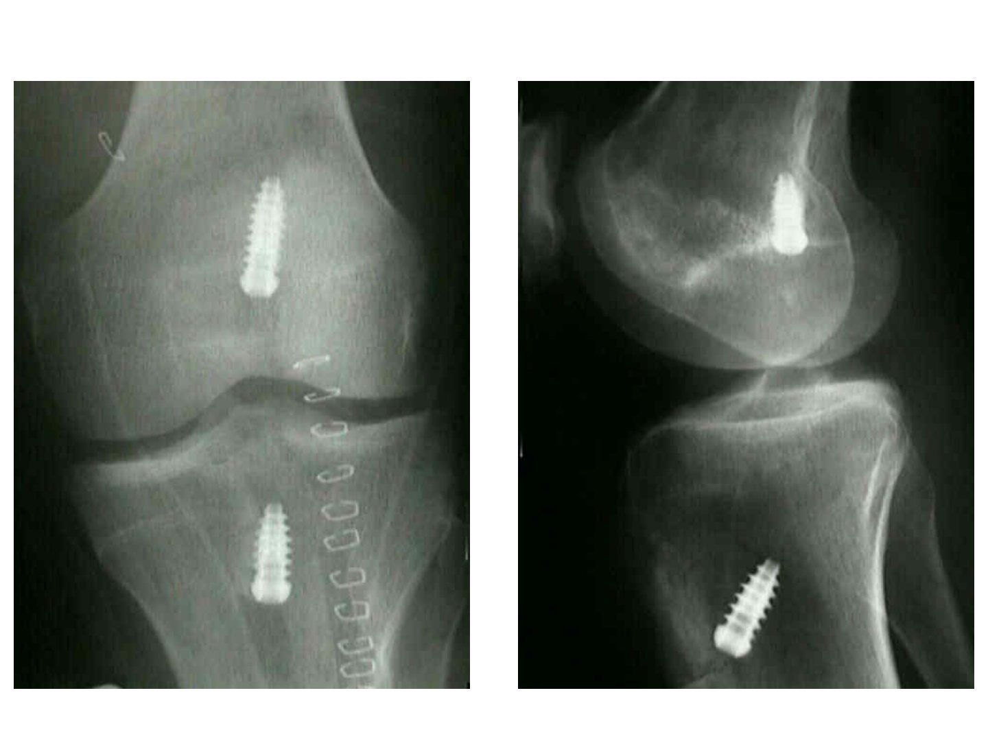
Q1.What do you describe on an X-ray after ACL reconstruction?
- Femoral and tibial tunnel position
- Single or double bundle
- Soft tissue or BPTB graft
- Bone quality and mode of fixation
Q2.What are the three reasons for ACL reconstruction failure?
- Recurrent instability
- Recurrent pain
- Loss of motion
Q3.What are the factors of recurrent or persistent instability (Pittsburgh classification)?
- Mechanical/technical (majority): wrong patient choice (knee abuser, malalignment), graft choice, graft size, tunnel position, tension (3tension in the notes), graft fixation, graft impingement, secondary stabilisers (meniscus, peripheral ligament)
- Failed graft incorporation: infection, immunological, avascularity, stress shielding
- Traumatic: reinjury, aggressive rehab
Q4.How does the lecturer classify ACLR failure according to time frame?
- Early (<3 months): failure of mechanical fixation or early biological problem (infection)
- Midterm (3-12 months): technical problems - tunnel malposition, impingement, graft elongation, unrecognised ligamentous injury
- Later (>1 year): trauma, underlying patient factor
Q5.What do you look for on X-ray in a failed ACL reconstruction?
- Details of surgery (single/double bundle, graft and fixation type, graft size)
- Tunnel position; tunnel widening >15mm may need 2-stage surgery; tibial slope; notch view for notch architecture
- Complications: patella fracture, posterior blowout, endobutton position, interference screw divergence
Q6.What history and risk factors are relevant in a failed ACL reconstruction?
- Age and premorbid status
- Activity level (knee abuser)
- Risk factors: ligamentous laxity, female, pivoting sports
- Current symptoms: pain, instability, mechanical symptoms, reinjury
Q7.What examination findings are relevant in a failed ACL reconstruction?
- Gait: varus thrusting
- Generalised ligamentous laxity
- Genu valgum
- ROM and evidence of loss of secondary restraints
- Position of previous scar
Q8.What is the preop workup before revision ACL reconstruction?
- XR (AP/lat/tunnel/skyline/notch)
- Scannogram for varus
- CT and MRI: tunnel, graft, incorporation, bone stock; look for other concomitant injuries
- Prerequisite: knee flexion >90 degrees, fixed flexion contracture <5
Q9.What are the intraoperative considerations in revision ACL reconstruction?
- Choice of graft (BPTB, HS for widened tunnel, graft size suboptimal, quad, allograft)
- Prepare screw/staple removal (revision ACL set)
- Tunnel planning: use old tunnel if position okay, new tunnel if severe malposition, two stage if old and new are close with not enough bone stock (dilatation >15mm)
- Revision notchplasty
- Fixation: endobutton, interference screw, tie over post, over the top, double fixation
- Send intraop culture if doubtful
Q10.What are the expectations and postop plan for revision ACL reconstruction?
- Expectation mx: results inferior to primary ACLR (increase rerupture rate and rehab time)
- One- vs two-stage operation
- Postop: individualised, less aggressive rehab plan
▸ Slide 649 · ACL post reconstruction XraySports · 5 questions expand

Q1-Q55 questions — tap to reveal all answerslist
- What should be assessed on an X-ray after ACL reconstruction?
- What is the optimal sagittal tunnel position after ACL reconstruction?
- What is the coronal clock-face position of the femoral tunnel?
- How can femoral tunnel position be predicted in transtibial versus transportal techniques?
- What should be checked for fixation quality on a post-ACL reconstruction X-ray?
Q1.What should be assessed on an X-ray after ACL reconstruction?
- Details of surgery: number of tunnels, graft type (bone plug/harvesting) and fixation method
- Complications: patella fracture, patella alta, posterior femoral blow-out, tunnel widening
- Quality of reconstruction and tunnel position
Q2.What is the optimal sagittal tunnel position after ACL reconstruction?
- Femur more posterior, tibia more anterior (to gain maximal AP length)
- Femoral: Blumensaat line divided into 4 parts - tunnel at the posterior 4th quadrant, must not breach the posterior cortex
- Tibia: plateau divided into 4 parts - tunnel at the anterior 2nd; too anterior causes notch impingement (double bundle can be more anterior)
Q3.What is the coronal clock-face position of the femoral tunnel?
- 10:30 and 1:30
Q4.How can femoral tunnel position be predicted in transtibial versus transportal techniques?
- Transtibial: can use the tibial tunnel to predict the femoral tunnel
- Transportal: by anteromedial portal
Q5.What should be checked for fixation quality on a post-ACL reconstruction X-ray?
- Interference screw: divergence <15 degrees
- Button: on cortex (may trap ITB / within bone)
- Tunnel widening
▸ Slide 650 · Paeditric Acl tearSports · 6 questions 1 check expand
Q1-Q66 questions — tap to reveal all answerslist
- When is surgery indicated in a paediatric ACL tear and what is the evidence on delay?
- What are the surgical options for a paediatric ACL tear?
- What is primary repair of a paediatric ACL tear and what is the evidence?
- What type of intra-articular reconstruction is used according to growth remaining?
- Why is a hybrid reconstruction used and how is growth considered?
- What technical principles minimise physeal damage in paediatric ACL reconstruction?
Q1.When is surgery indicated in a paediatric ACL tear and what is the evidence on delay?
- Quoted evidence (Clin sport med 2021 metanalysis, PLUTO study group): delayed ACL reconstruction for more than 12 weeks significantly increases risk of meniscal injuries and irreparable meniscal tears
- Take into account skeletal age, growth remaining, instability symptoms, aspiration for sport and associated injury
Q2.What are the surgical options for a paediatric ACL tear?
- Primary repair (ongoing BEAR trial)
- Extra-articular tenodesis
- Intra-articular reconstruction: transphyseal, hybrid or physeal sparing
Q3.What is primary repair of a paediatric ACL tear and what is the evidence?
- Primary repair is one option (ongoing BEAR trial)
- Preliminary 2-year follow-up results promising
Q4.What type of intra-articular reconstruction is used according to growth remaining?
- Transphyseal: <2 years of growth remaining
- Hybrid: 2-5 years of growth remaining (femur spared)
- Physeal sparing (over-the-top fixation): >5 years of growth remaining
Q5.Why is a hybrid reconstruction used and how is growth considered?
- Femur is spared in hybrid as it contributes 40% of lower limb growth
- Hybrid used when 2-5 years of growth remaining
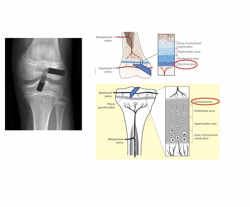
Q6.What technical principles minimise physeal damage in paediatric ACL reconstruction?
- Drilling damages the geminal layer ( 7-9% physis cross section); 7-9% of physis cross-section is sufficient to cause growth disturbance (germinal layer)
- Aim tunnel size 3-4% of physis cross-sectional area
- Soft tissue graft; centrally located vertical tunnels
- Avoid transphyseal screws; avoid overtensioning the graft
Clin Sport Med 2021 meta-analysis by the PLUTO study group shows delayed ACL reconstruction >12 weeks increases risk of meniscal injuries and irreparable meniscal tears — misattributed — PLUTO is a prospective multicentre descriptive epidemiology cohort of skeletally immature ACL tears, not a 2021 meta-analysis; evidence for irreparable medial meniscal tears shows risk rising by about 8 weeks (Everhart, AJSM 2019), and timing thresholds vary by study — medium confidence — source
▸ Slide 651 · BPTB graftSports · 4 questions expand

Q1-Q44 questions — tap to reveal all answerslist
- How do you recognise that a BPTB graft was used on a post-ACL reconstruction X-ray?
- How do you describe tunnel position on the coronal view?
- How do you describe tunnel position on the sagittal view?
- What other findings should be commented on in this post-ACLR X-ray?
Q1.How do you recognise that a BPTB graft was used on a post-ACL reconstruction X-ray?
- Defect in the patella and tibial tuberosity
- Bone inside the tunnel
- Fixation by bioabsorbable screws on both sides
Q2.How do you describe tunnel position on the coronal view?
- Graft position satisfactory; both tunnels obliquely aligned
- Femoral tunnel w.r.t. notch is at 10 o'clock position
- Tibial tunnel at 60 degrees w.r.t. knee joint (cf sagittally aligned)
Q3.How do you describe tunnel position on the sagittal view?
- Tibial tunnel 40% from anterior (2nd quarter) with entry point posterior to Blumensaat line (ideally comment with knee in full flexion)
- Femoral tunnel adequately posteriorly placed without posterior blow-out
Q4.What other findings should be commented on in this post-ACLR X-ray?
- There is patella Alta with Insall salvati ratio > 1.2, but I will compare it with preop XR
- No patellar fracture
- Screw position is not divergent
▸ Slide 652 · Femoral tunnel positionSports · 5 questions expand
Q1-Q55 questions — tap to reveal all answerslist
- What does the position of graft fixation represent, and when is it usually tensioned?
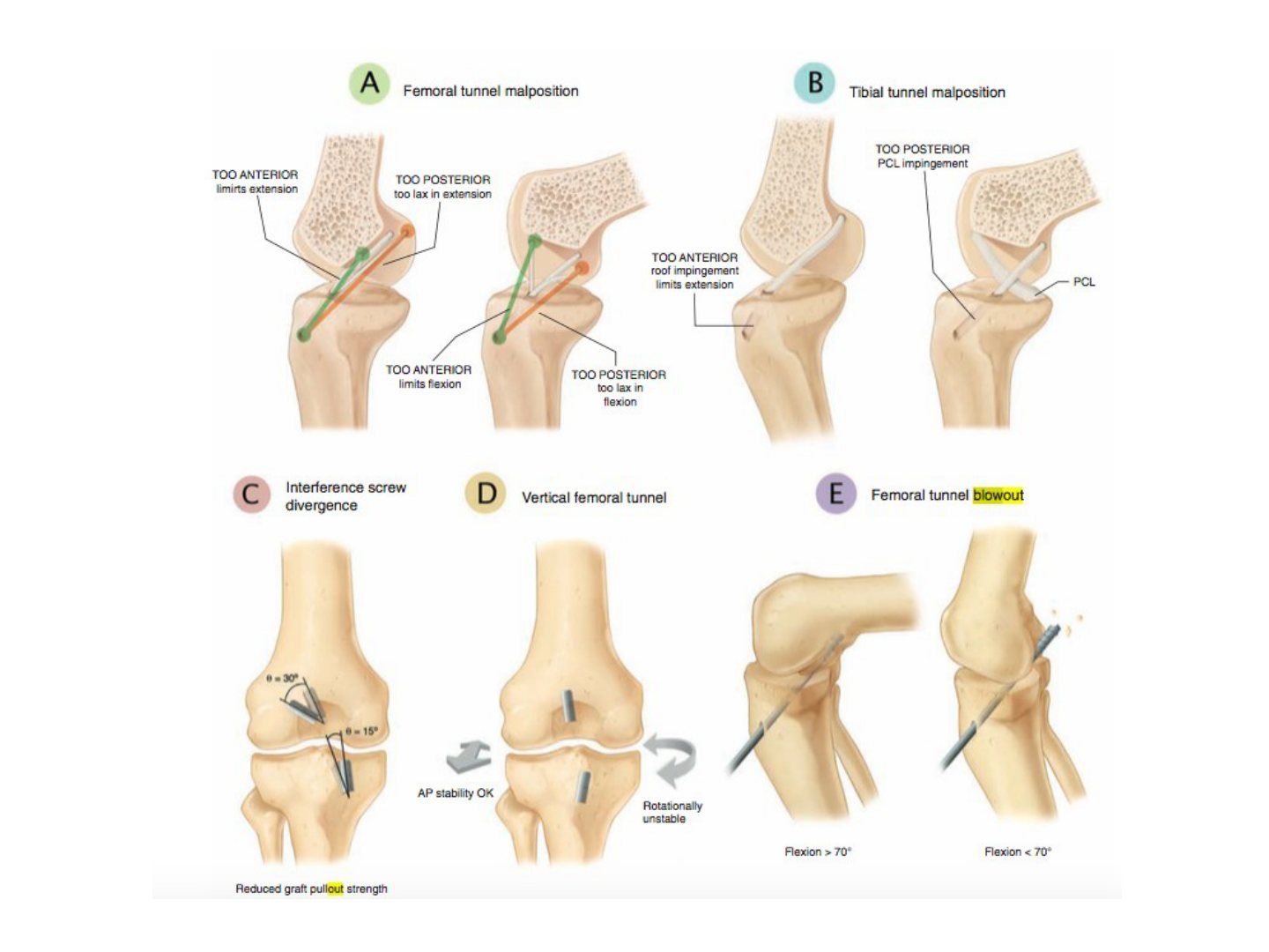
- What happens if the graft is fixed in extension with the femoral tunnel too anterior or too posterior?
- What are the consequences of a femoral tunnel placed too anterior when fixed in flexion?
- What are the consequences of a femoral tunnel placed too posterior when fixed in flexion?
- What are the effects of tibial tunnel malposition?
Q1.What does the position of graft fixation represent, and when is it usually tensioned?
- Position of fixing the graft represents the affected motion
- Usually tensioned in 20-30 degrees flexion and posterior drawer (maximum tension) --> most stable
- If fixed in extension, ensure full extension postop
Q2.What happens if the graft is fixed in extension with the femoral tunnel too anterior or too posterior?
- Too anterior: limit flexion
- Too posterior: lax in extension
Q3.What are the consequences of a femoral tunnel placed too anterior when fixed in flexion?
- Tight in flexion, lax in extension
- Fails to clear the resident ridge
- Physiologically not adequate flexion; graft gradually stretches out and becomes lax (biological problem)
Q4.What are the consequences of a femoral tunnel placed too posterior when fixed in flexion?
- Lax in flexion, tight in extension
- Rare
Q5.What are the effects of tibial tunnel malposition?
- Anterior: notch impingement, limits extension
- Posterior: impinges PCL, tight in extension
- Medial: impingement on medial femoral condyle
- Lateral: impingement on lateral femoral condyle
▸ Slide 653 · Tunnel malpositionSports · 6 questions expand
Q1-Q66 questions — tap to reveal all answerslist
- What is the resident ridge and what does it mark?
- What is the lateral bifurcate ridge?
- How does the AM bundle position over the femur change with knee movement?
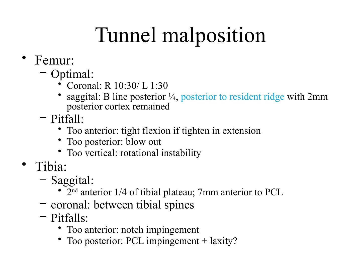
- What is the optimal femoral tunnel position?
- What are the pitfalls of femoral tunnel malposition?
- What is the optimal tibial tunnel position and what are its pitfalls?
Q1.What is the resident ridge and what does it mark?
- Also called the lateral intercondylar ridge
- Marks the anterior and superior border of the ACL insertion
Q2.What is the lateral bifurcate ridge?
- Ridge located between the AM and PL bundles
Q3.How does the AM bundle position over the femur change with knee movement?
- More posterior on flexion
- More superior on extension
Q4.What is the optimal femoral tunnel position?
- Coronal: right 10:30 / left 1:30
- Sagittal: B line posterior 1/4, posterior to the resident ridge with 2 mm posterior cortex remained
Q5.What are the pitfalls of femoral tunnel malposition?
- Too anterior: tight flexion if tightened in extension
- Too posterior: blow out
- Too vertical: rotational instability
Q6.What is the optimal tibial tunnel position and what are its pitfalls?
- Sagittal: 2nd anterior 1/4 of the tibial plateau, 7 mm anterior to PCL
- Coronal: between the tibial spines
- Too anterior: notch impingement
- Too posterior: PCL impingement + laxity?
▸ Slide 654 · Post ACLR stiffnessSports · 3 questions expand

Q1-Q33 questions — tap to reveal all answerslist
- What are the global causes of stiffness after ACL reconstruction?
- What causes loss of knee flexion after ACL reconstruction?
- What causes loss of knee extension after ACL reconstruction?
Q1.What are the global causes of stiffness after ACL reconstruction?
- Arthrofibrosis
- Infrapatellar contracture syndrome (fibrous hyperplasia of anterior knee - loss of flexion + extension + patellar entrapment)
- Infection
Q2.What causes loss of knee flexion after ACL reconstruction?
- Femoral tunnel too anterior, tightened and fixed in knee extension
- Excessively tensioned graft
- Suprapatellar pouch scarring
Q3.What causes loss of knee extension after ACL reconstruction?
- Tibial tunnel too anterior
- Notch scarring
- Cyclops lesion
▸ Slide 655 · ACL avulsionSports · 5 questions expand
Q1-Q55 questions — tap to reveal all answerslist
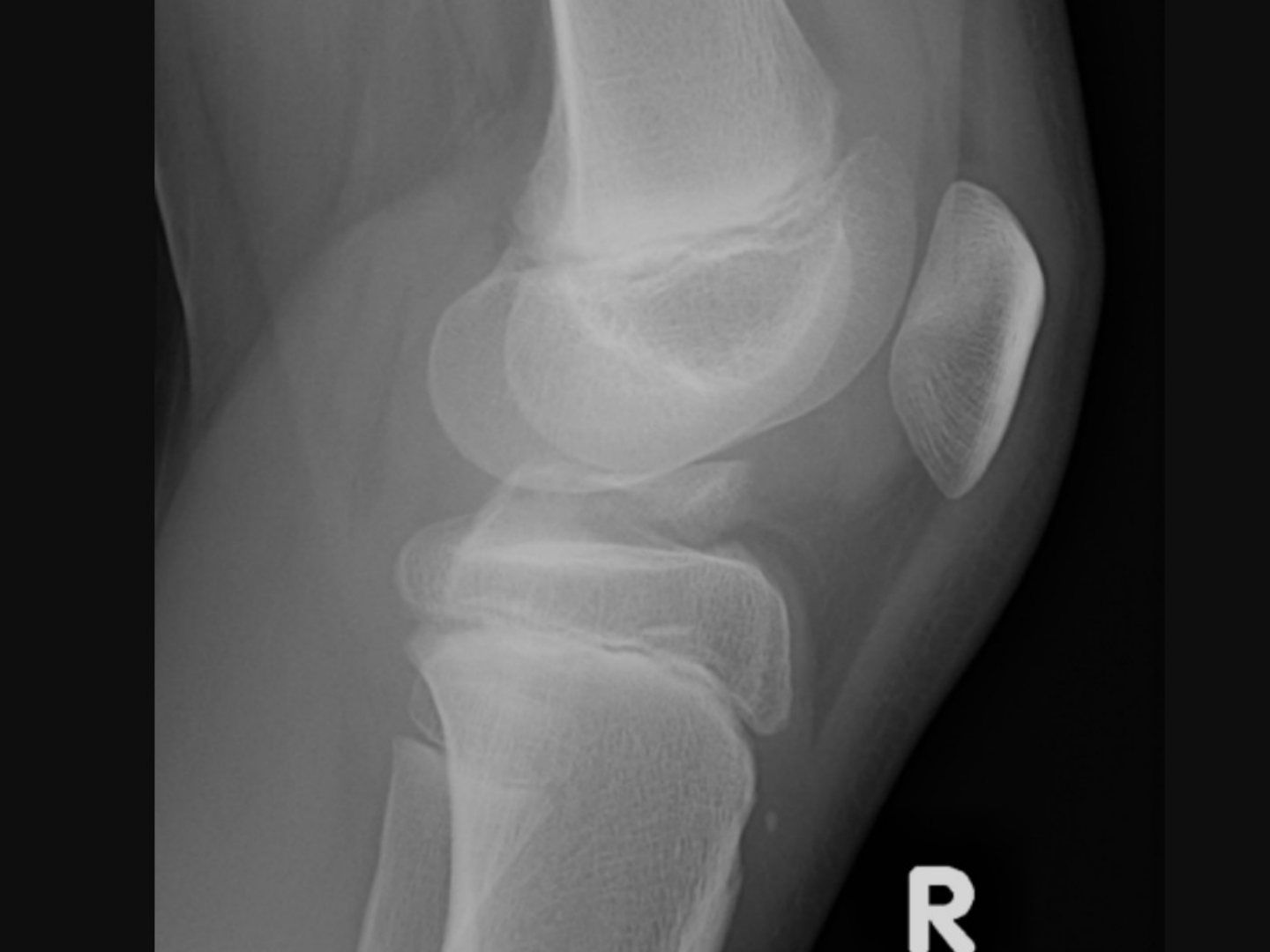
- Why does an ACL avulsion fracture occur rather than a midsubstance tear?
- What is the Meyers and McKeever classification of ACL avulsion fracture?
- How are ACL avulsion fractures treated?
- What are the difficulties and complications of ACL avulsion treatment?
- How is the knee immobilised and what fracture is associated with ACL avulsion?
Q1.Why does an ACL avulsion fracture occur rather than a midsubstance tear?
- Ligament stronger than physeal cartilage or cancellous bone
- ACL fibres in continuity with perichondrium of tibial epiphysis/cartilage
- Mechanism: hyperflexion or direct blow to flexed knee (e.g. fall from bicycle)
Q2.What is the Meyers and McKeever classification of ACL avulsion fracture?
- Non-displaced
- Minimally displaced with intact posterior hinge
- Completely displaced
Q3.How are ACL avulsion fractures treated?
- Long leg cast for types I and II
- Type II: CR before casting by hyperextension to reduce
- Treat early; arthroscopic or mini-open option
- Fixation: screw (better x2), suture anchor (usually not enough strength) or pull-out suture through bone tunnel
- Rehab similar to ACL reconstruction
Q4.What are the difficulties and complications of ACL avulsion treatment?
- Reduction difficulty: interposition of intermeniscal ligament and meniscus
- Recurvatum in children (fixation tethers anterior growth plate)
- Remove metallic screw at 3-4 mths (3-4 months) to avoid articular cartilage damage (difficult)
Q5.How is the knee immobilised and what fracture is associated with ACL avulsion?
- Cast in 20 degrees flexion to minimise distraction force; hyperextension increases tensile force and distracts the fragment
- Position is controversial; some put in 0 degrees but usually better reduced
- 80% are type 1 or 2
- Associated Segond fracture: disruption of meniscotibial portion of lateral capsule (direct evidence of lateral capsule, indirect for ACL)
▸ Slide 656 · PCL injurySports · 16 questions expand
Q1-Q1616 questions — tap to reveal all answerslist
- What are the aims of assessing a PCL injury?
- What are the usual injury mechanisms of PCL injury?
- What positive physical signs should be elicited in PCL injury?
- What tests indicate an associated PLC injury?
- How is the reverse pivot shift performed and how does it work?
- What imaging is used in PCL injury and how is stress view graded?
- What associated injuries must be assessed in PCL injury?
- Describe the anatomy of the PCL.
- Why does the PCL heal better than the ACL?
- What is the natural history of isolated PCL injury?
- What is the rehab protocol for non-operative management of PCL injury?
- What is the indication for surgery in PCL injury?
- What factors are considered in PCL reconstruction?
- Describe the all-arthroscopic transtibial PCL reconstruction technique.
- What are the pros and cons of all-arthroscopic versus inlay PCL reconstruction?
- What is the post-op rehabilitation after PCL reconstruction?
Q1.What are the aims of assessing a PCL injury?
- Assess severity
- Rule out associated injury
- Formulate management plan (also depends on patient's functional demand)
Q2.What are the usual injury mechanisms of PCL injury?
- Hyperextension/hyperflexion
- RTA dashboard injury (look for femoral neck fracture)
- Varus force on a flexed knee
- Fall onto the ground with a plantar-flexed foot
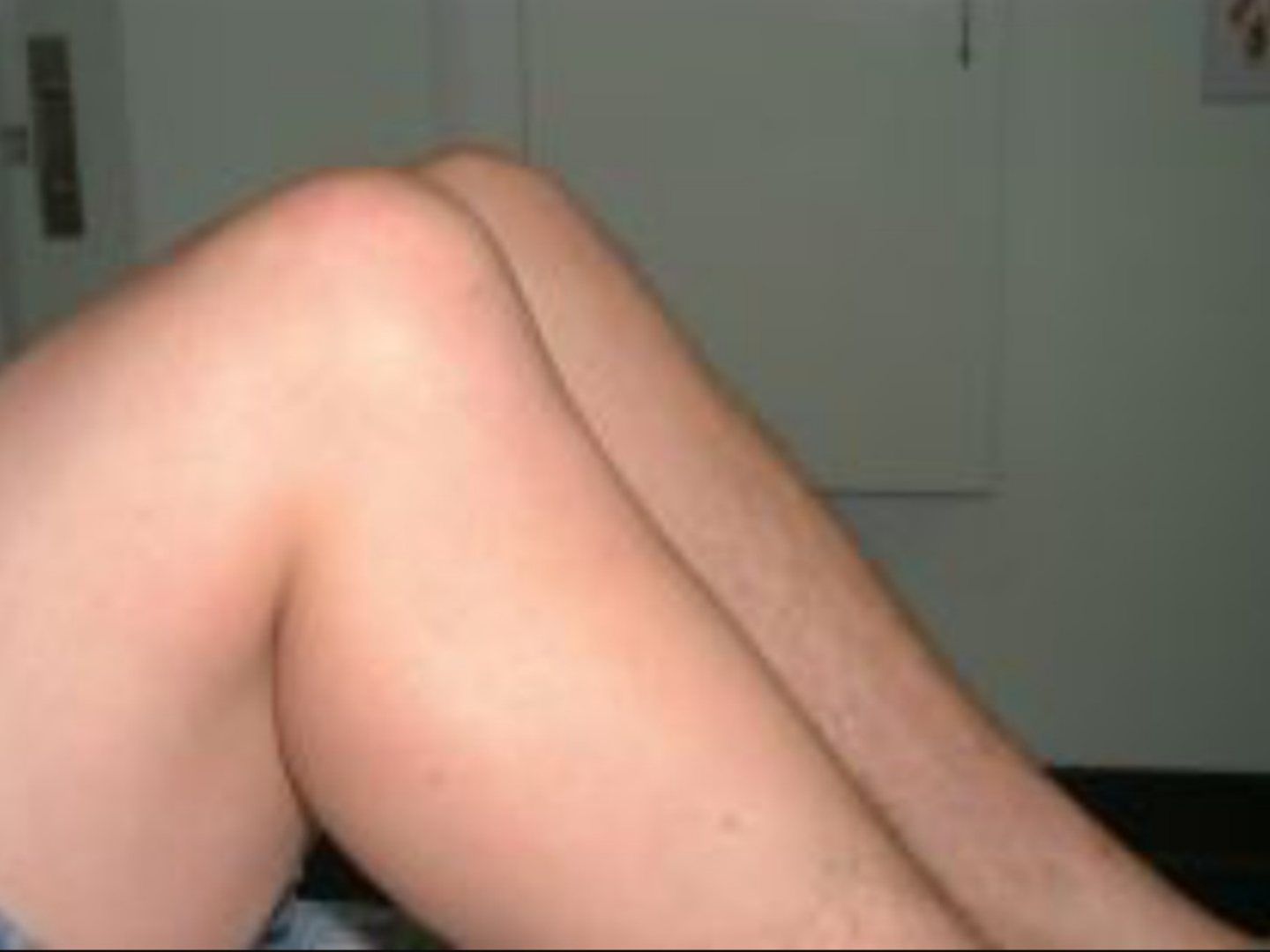
Q3.What positive physical signs should be elicited in PCL injury?
- Loss of medial tibial step-off with knee 90 degrees flexion
- Posterior sagging grading relative to medial femoral condyle: anterior, flush, posterior
- Posterior drawer grading: <5mm, 5-10mm, >10mm with no endpoint; rule out PLC in high grade
- Quadriceps active test: flex knee 60 degrees, stabilise foot and active extend knee
Q4.What tests indicate an associated PLC injury?
- Dial test at 30/90 degrees (positive at 90 = combined injury, 30 only = PLC)
- Recurvatum ER test: varus = LCL injury, ER = fabellofibular/popliteus tendon, recurvatum = PCL
- Posterolateral drawer test
- Reverse pivot shift - 26% sensitivity, 95% specificity
- Gait: lateral thrust
Q5.How is the reverse pivot shift performed and how does it work?
- Begins in flexion with ER and valgus stress applied, so tibia subluxes posteriorly
- Knee is then brought to full extension (reverse)
- ITB changes from flexor to extensor at ~30 degrees flexion, causing relocation (same principle)
- Sensitivity 26%, specificity 95%
Q6.What imaging is used in PCL injury and how is stress view graded?
- XR: avulsion fracture
- Stress view (posterior tibial cortex to posterior end of Blumensaat line at knee flexion 70 degrees) with kneeling or Telos
- 3-8mm = partial, 8-12mm = complete, >12mm = PLC injury
Q7.What associated injuries must be assessed in PCL injury?
- PLC injury
- Multiple ligament injury
- Knee dislocation
- Posterior hip dislocation
Q8.Describe the anatomy of the PCL.
- 38x13mm, 30% larger than ACL; intracapsular, extrasynovial ligament
- From MFC anterolateral surface (broad crescent-shaped footprint) to posterior tibial sulcus (1cm below joint line)
- Bundles: AL (easier to rupture, tight at flexion) / PM; AL anterior to PM at femoral insertion
- Meniscofemoral ligaments: Humphrey (anterior) and Wrisberg (posterior), contribute 20-30% stability
- Type I collagen; blood supply middle geniculate artery; nerve supply posterior articular branch of tibial nerve
- Primary restraint to posterior translation; secondary restraint to ER and varus; strength 2500-3000N
Q9.Why does the PCL heal better than the ACL?
- Mechanical: increased stability from femoromeniscal ligaments
- Biological: thick synovium sheath --> usually still extrasynovial after rupture (hematoma formation, no cytokine)
- ACL is also an intracapsular, extrasynovial ligament
Q10.What is the natural history of isolated PCL injury?
- Shelbourne 1999: 50% same sport, 30% lower level, 20% unable to play sport
- Complication: anterior knee pain (PFJ arthritis)
- Newer evidence: Agolley (prof Fares Haddad group) BJJ 2017 91.3% return to same level or higher
Q11.What is the rehab protocol for non-operative management of PCL injury?
- Grade 1-2: PWB + quad strengthening
- Grade 3: extension brace (Donjoy/posterior support) for 4-6 weeks, no flexion >50 degrees (PCL starts to take up tension around 50 degrees flexion)
- PWB walking + quads isometric exercises
Q12.What is the indication for surgery in PCL injury?
- Avulsion or peel-off lesion
- Multiple ligament injury / concomitant PLC injury
- Persistent symptoms (difficulty decelerating e.g. downstairs, PFJ pain) - observe 6-12 months
Q13.What factors are considered in PCL reconstruction?
- Type of graft (hamstring / BPTB / quadriceps + bone)
- Single/ double bundle: single bundle less demanding (reconstruct AL, tension at 90 degrees knee flexion)
- Chahla Arthroscopy 2017 systematic review - double bundle better IKDC score and posterior tibial translation/knee stability
- Inlay vs all-arthroscopic
Q14.Describe the all-arthroscopic transtibial PCL reconstruction technique.
- Supine on tourniquet; harvest graft
- Conventional portal + PM portal (injury to saphenous nerve)
- PM portal dissection of posterior capsule (protect popliteal artery)
- Drill tibial tunnel A to P, last few cm use hand drill, knee in 90 degrees flexion
- Tibial tunnel 15mm distal to articular surface --> less killer turn
- Femoral tunnel 7mm from articular surface at 1pm/11pm (shorter than ACL tunnel; consider aperture fixation if too short); tension at 90deg flexion with anterior drawer test
Q15.What are the pros and cons of all-arthroscopic versus inlay PCL reconstruction?
- All-arthroscopic pros: single position, can use hamstring graft; cons: difficult to pass graft, killer turn
- Inlay pros: avoid killer turn (less attrition), direct visualisation of NV bundle, better healing with bone union, biomechanically better
- Inlay cons: floppy lateral position, less graft choice (BPTB or quadriceps tendon + patella bone with bone peg)
Q16.What is the post-op rehabilitation after PCL reconstruction?
- Hinged knee brace locked in extension for ambulation and sleep for 6 weeks
- Protect against gravity
- Early motion in prone position
- Quad strengthening; no open chain hamstring or isolated hamstring exercise for 3 months
▸ Slide 657 · PLC:Sports · 7 questions expand

Q1-Q77 questions — tap to reveal all answerslist
- What are the static and dynamic structures of the PLC and their roles?
- What is the layer arrangement of the PLC?
- What is the anatomy of the fibular head insertion and structures to the fabella?
- What are the common mechanisms, symptoms and physical signs of PLC injury?
- What are the X-ray findings and varus stress grading in PLC injury?
- What is the Fanelli classification and conservative management of PLC injury?
- What are the indications for surgery, reconstruction options and evidence for repair vs reconstruction?
Q1.What are the static and dynamic structures of the PLC and their roles?
- Static (6): LCL, popliteus, popliteofibular ligament, fabellofibular ligament, arcuate ligament, capsule
- Dynamic: ITB, biceps femoris, lateral gastrocnemius
- Primary restraint to ER and varus; Secondary restrain to posterior translation
Q2.What is the layer arrangement of the PLC?
- Layer 1: ITB/biceps femoris (CPN in between)
- Layer 2: retinaculum
- Layer 3 superficial: LCL/FFL/ALL (LGA in between)
- Deep: arcuate/popliteal/PFL/capsule
Q3.What is the anatomy of the fibular head insertion and structures to the fabella?
- Fibular head, anterior to posterior: LCL, biceps femoris (long then short), arcuate ligament, popliteofibular, FFL
- Structures to fabella: capsular arm of short head of biceps, FFL, OPL, lateral gastrocnemius head, posterior lateral capsule
Q4.What are the common mechanisms, symptoms and physical signs of PLC injury?
- Mechanism: blow to anteromedial knee, varus blow to flexed knee, hyperextension injury
- Symptoms: instability in knee extension and upon pivoting
- PE: dial test positive if >15 degrees difference, ER recurvatum, posterolateral drawer with foot 15 degrees ER, reverse pivot shift
- (Peroneal nerve injury in 25% of patient with PLC injury)
Q5.What are the X-ray findings and varus stress grading in PLC injury?
- Avulsion fracture (arcuate fracture) of fibula head
- Varus stress (Laprade): <4mm = isolated PCL, 4-7mm = LCL + PCL, >7mm = LCL + PLC + XL
- Long leg standing XR: malalignment, determine mechanical axis +/- need for HTO
Q6.What is the Fanelli classification and conservative management of PLC injury?
- Fanelli A: increase ER only (PFL + popliteus)
- Fanelli B: increase ER + varus laxity in knee flexion (above + LCL)
- Fanelli C: increase ER + varus laxity in flexion and extension (above + ACL/PCL)
- Conservative: isolated grade 1-2 injury - extension knee brace + protected weight bearing x2 weeks, then quad strengthening
Q7.What are the indications for surgery, reconstruction options and evidence for repair vs reconstruction?
- Indications: bony avulsion, multiple ligament laxity, persistent symptoms, grade 3 injury (assess in MUA)
- Unstable (varus stress >7mm) + within 2 weeks of injury --> early repair then later staged recon; stable --> brace 6 weeks then one-stage recon (Laprade favours single stage early recon of PCL and PLC)
- Reconstruct LCL and popliteofibular ligament; graft: semitendinosus/achilles
- Larson: non-anatomical fibular-based figure of 8; LaPrade anatomical recon with 2 soft tissue grafts (LCL+PFL and popliteus); transtibial double bundle fixation
- CUHK arthroscopy paper: LaPrade technique gives better rotational stability
- Recon better than repair: Stannard failure 37% vs 9%; Levy 40% vs 6%; Geeslin AJSM 2016 metanalaysis – recon better outcomes than repairs in acute PLC injuries (meta-analysis)
▸ Slide 658 · Stieda Pellegrini sign (few weeks after injury): ossified lesion signifying avulSports · 7 questions expand
Q1-Q77 questions — tap to reveal all answerslist
- What is the Stieda Pellegrini sign?
- What is the function of the MCL and the usual mechanism of injury?
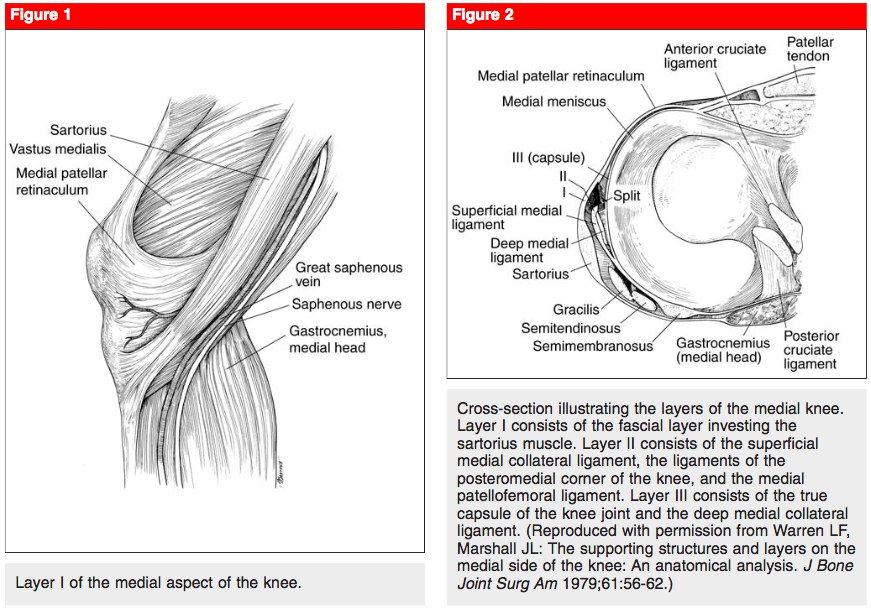
- What are the layers of the medial side of the knee?
- How is MCL instability graded and where do tears most commonly occur?
- When is operative management indicated in MCL injury?
- How is a grade 3 MCL tear with ACL tear managed?
- What are the classifications and reconstruction options for MCL tear?
Q1.What is the Stieda Pellegrini sign?
- Ossified lesion seen a few weeks after injury
- Signifies avulsion injury of MCL at the medial femoral condyle
Q2.What is the function of the MCL and the usual mechanism of injury?
- Primary restraint to valgus stress
- Secondary restraint to ER, ant/ post translation (anterior/posterior translation)
- Usual mechanism: valgus + ER
Q3.What are the layers of the medial side of the knee?
- Layer 1: sartorius and fascia, patella retinaculum (gracilis, semitendinosus, saphenous nerve)
- Layer 2: superficial MCL, POL, OPL, MPFL, semimembranosus
- Layer 3: deep MCL, capsule
Q4.How is MCL instability graded and where do tears most commonly occur?
- Medial gapping compared with opposite knee: <5mm, <10mm, >=10mm
- Grade 1: stretch injury (no loss of ligamentous integrity)
- Grade 2: incomplete tear (fibres opposed, endpoint at 30 degrees flexion valgus test)
- Grade 3: complete tear (no endpoint)
- Most common: femur side tear
Q5.When is operative management indicated in MCL injury?
- All try conservative management (hinged brace) first
- Grade III with: acute multiligamentous injury; acute displaced distal avulsion with Stener-like lesion (trapped torn end); subacute/chronic with persistent instability
- Femoral origin tears have better healing potential
Q6.How is a grade 3 MCL tear with ACL tear managed?
- Early MCL repair (within 2 weeks) + delay ACLR
- Or conservative Mx for MCL + delay ACL reconstruction (MCL needs protection, which may cause stiffness)
Q7.What are the classifications and reconstruction options for MCL tear?
- Taketomi (tibial side tear): under pes, Stener-like lesion, trapped inside joint
- Nakamura: type 1 femoral, type 2 tibial (none in his study), type 3 femoral with extension below the joint line
- Reconstruction: Bosworth (semitendinosus, distal intact, attached to femoral tunnel), Lind (gracilis looped over MCL), LaPrade
- Acute reconstruction is controversial
▸ Slide 659 · Posteromedial cornerSports · 4 questions expand

Q1-Q44 questions — tap to reveal all answerslist
- What is the definition and what are the components of the posteromedial corner?
- What is the role of the posteromedial corner?
- What is anteromedial rotatory instability (AMRI)?
- When is early repair of the posteromedial corner indicated?
Q1.What is the definition and what are the components of the posteromedial corner?
- From posterior margin of superficial MCL to PCL (does not include MCL)
- Components (2nd layer): semimembranosus expansion (5 arms: capsular, anterior, inferior, distal, direct)
- POL (ligament of Winslow), OPL, deep capsule, MM PH
Q2.What is the role of the posteromedial corner?
- Dynamic
- Secondary restraint to ER
- Restraint to valgus stress in extension
Q3.What is anteromedial rotatory instability (AMRI)?
- Medial tibial plateau anterior subluxation upon ER
- Signs: valgus stress + at 30 degrees ER
- Anterior drawer positive at 30 degrees ER
Q4.When is early repair of the posteromedial corner indicated?
- In multiligament injury
▸ Slide 660 · Skeletally matureSports · 14 questions expand
Q1-Q1414 questions — tap to reveal all answerslist
- What are the differentials and how do you assess a suspected osteochondral lesion of the medial femoral condyle?
- What imaging is used and what are the MRI criteria for instability?
- What is the Clanton and Delee classification of OCD?
- What is the Guhl arthroscopic classification and corresponding management?
- What is the definition, epidemiology and prognosis of OCD?
- What are the causes of OCD and the aim of management?
- What is the Outerbridge classification of chondral lesions?
- How is a sizable OCD flap repaired when the physis is open?
- What are the three broad categories of resurfacing/reconstruction options for OCD?
- What is the microfracture technique and what tissue does it produce?
- What are the OAT (osteochondral autograft transfer) plug requirements?
- How do ACI, MACI and AMIC differ?
- What prognostic factors suggest a good outcome in OCD?
- How is a high tibial osteotomy (HTO) performed?
Q1.What are the differentials and how do you assess a suspected osteochondral lesion of the medial femoral condyle?
- Differentials: osteonecrosis (SONK, post scope, secondary), osteoid osteoma, subchondral cyst, transient osteoporosis
- History to rule out differentials: mechanical symptoms suggest displacement; trauma history
- PE: Wilson's test (knee 30-90 degrees, pain with IR, relieved by ER); overall alignment; assess for instability. Gait: ER LL
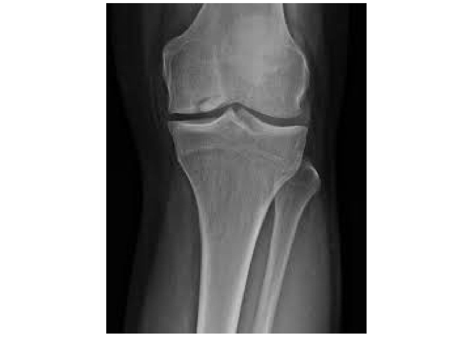
Q2.What imaging is used and what are the MRI criteria for instability?
- X-ray: weight bearing AP, lateral and notch view (flexed 30deg) to look for detached loose bodies; scannogram for overall alignment
- MRI: position, size, attachment, displacement, loose body
- Instability criteria: increased T2 signal >=5mm deep to lesion; focal defect >=5mm2 in articular surface
Q3.What is the Clanton and Delee classification of OCD?
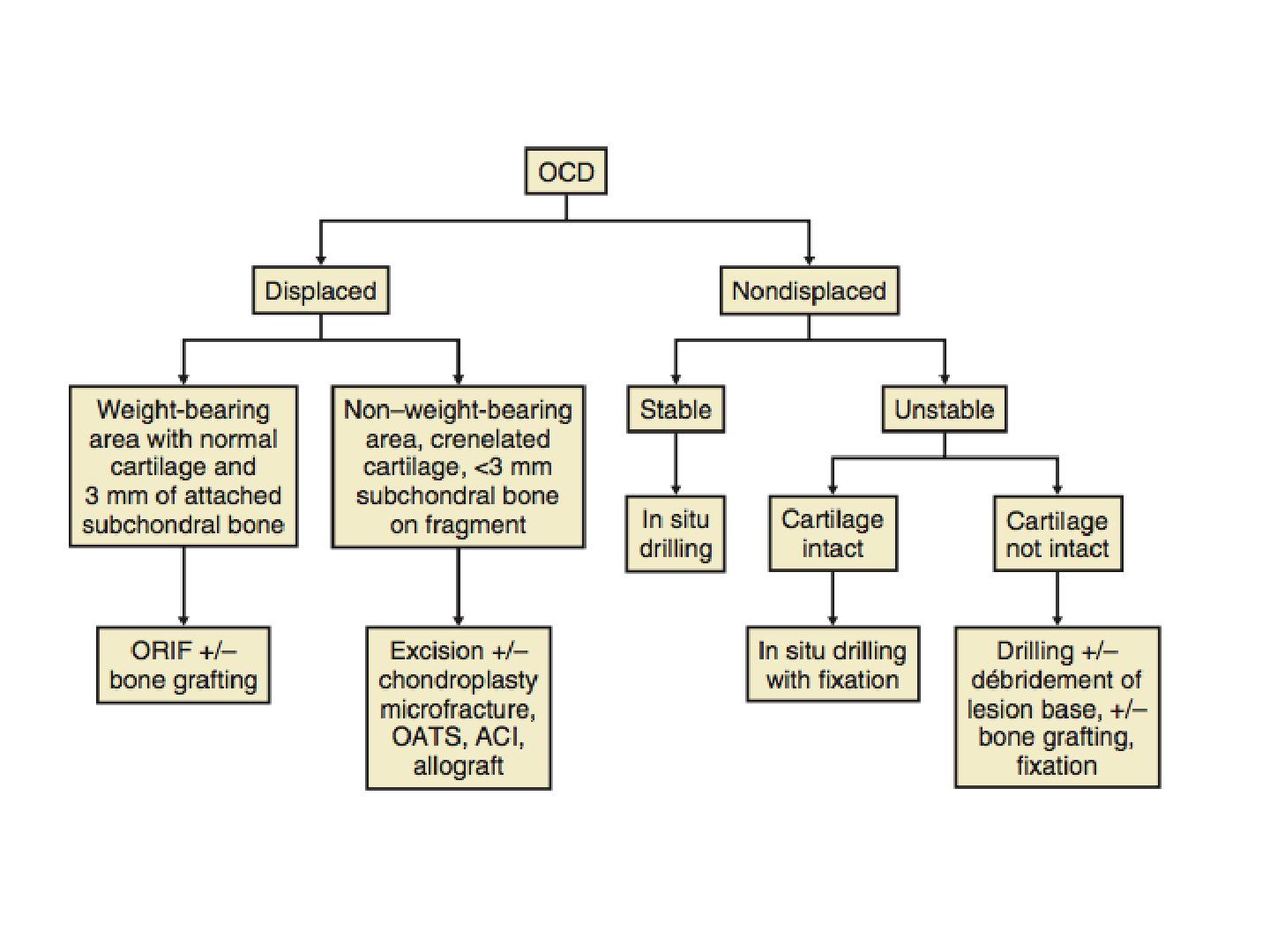
- 1. Depressed
- 2. Connected by osseous bridge
- 3. Detached, undisplaced
- 4. Displaced
Q4.What is the Guhl arthroscopic classification and corresponding management?
- 1. Intact, 1-3cm (activity modification/retrograde drilling)
- 2. Stable flap (headless screw)
- 3. Partially displaceable flap (remove fibrous tissue, chondroplasty, fix)
- 4. Complete detachment (remove, chondroplasty +/- OC allograft, ACI, MACI)
- Small (<2cm x 2cm) --> microfracture; large (>2cm x 2cm) --> chondroplasty
Q5.What is the definition, epidemiology and prognosis of OCD?
- Definition: subchondral bone lesion with delamination + sequestration +/- articular involvement
- Most common in knee (80% medial, 40% F, 20% bilateral, 10% patella); also talus and capitellum
- 70% resolve with conservative management
- PAPPAS: I <12y, II 12-20y, III >20y (III usually OA)
Q6.What are the causes of OCD and the aim of management?
- Constitutional: epiphyseal dysplasia with accessory nuclei separating from the epiphysis
- Vascular: watershed region from attachment of the PCL
- Traumatic: repetitive impingement of the tibial spine
- Aim: relieve symptoms and improve function; always start conservative with protected weight bearing and NSAIDs
- Surgery aims: restore joint congruity, increase local blood supply, address predisposing instability or malalignment
Q7.What is the Outerbridge classification of chondral lesions?
- 0 intact
- 1 softening
- 2 fissure not to subchondral bone
- 3 fissure to subchondral bone + diameter >1.5cm
- 4 subchondral bone exposed
Q8.How is a sizable OCD flap repaired when the physis is open?
- Repair/fixation for sizable flap with open physis
- Method of fixation: screw, bone peg, sutures
- Rehab: PWB 6-8 weeks
Q9.What are the three broad categories of resurfacing/reconstruction options for OCD?
- Resurfacing/ reconstruction options fall into three broad categories: MSC stimulation, substitution, cell-based biological replacement
- MSC stimulation: microfracture, drilling, abrasion chondroplasty
- Substitution options: OATS, osteochondral allograft
- Cell based, biological replacement options: ACI, MACI, AMIC
Q10.What is the microfracture technique and what tissue does it produce?
- OT x microfracture for fibrocartilage, others for hyaline
- Microfracture 3-4mm apart, break surface till just across the tidemark with a chondral pick with low energy
Q11.What are the OAT (osteochondral autograft transfer) plug requirements?
- Plugs should be cylindrical
- At least 8mm long
- 4-12mm diameter
Q12.How do ACI, MACI and AMIC differ?
- ACI: 2 procedures, expensive, can produce hyaline cartilage, not limited by size
- MACI: similar to ACI but with a matrix
- AMIC: one stage procedure
Q13.What prognostic factors suggest a good outcome in OCD?
- Open distal femoral physis
- Size <2cm
- No sclerosis on XR
- Location at medial femoral condyle posterolateral aspect
- No synovial fluid behind lesion on MRI
- High uptake on bone scan = good healing potential (grade II-IV, uptake at lesion, femoral, tibia)
Q14.How is a high tibial osteotomy (HTO) performed?
- Approach: direct anteromedial
- Identify the pes and release hamstrings
- Release the distal MCL fibres subperiosteally in one layer until the posterior border of the tibia
- Periosteal elevator to clear posterior structures off the tibia and place a radiolucent Homman retractor to protect the NV bundle
- Mark out biplanar osteotomy either ascending or descending; trajectory of the saw cut guided by K wires
- Apply Tomofix plate for FWB walking postop
▸ Slide 661Sports · 2 questions expand
Q1-Q22 questions — tap to reveal all answerslist
- What topic is covered in slide 661?
- What are the key learning points from slide 661?
Q1.What topic is covered in slide 661?
- Not covered in the speaker notes
Q2.What are the key learning points from slide 661?
- Not covered in the speaker notes
▸ Slide 662Sports · 5 questions expand
Q1-Q55 questions — tap to reveal all answerslist
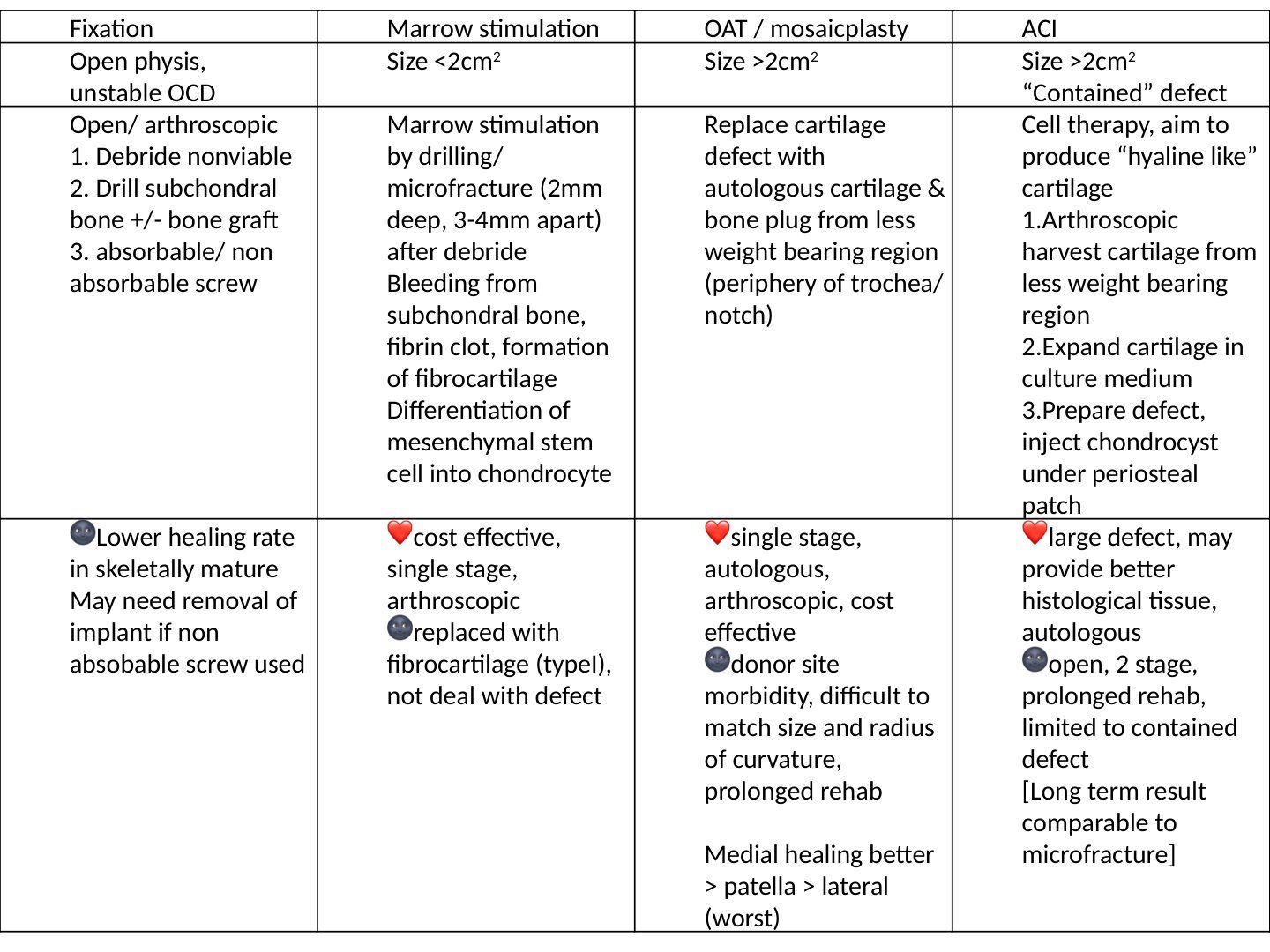
- What are the four surgical options compared on the slide and their size indications?
- How is fixation of an OCD performed and what are its limitations?
- How is marrow stimulation performed and what are its advantages and disadvantages?
- How is OAT/mosaicplasty carried out and what are its pros and cons?
- What is the technique and the pros and cons of ACI on the slide?
Q1.What are the four surgical options compared on the slide and their size indications?
- Fixation: open physis, unstable OCD
- Marrow stimulation: size <2cm2
- OAT/mosaicplasty: size >2cm2
- ACI: size >2cm2, contained defect
Q2.How is fixation of an OCD performed and what are its limitations?
- Open or arthroscopic: 1. debride nonviable, 2. drill subchondral bone +/- bone graft, 3. absorbable/non-absorbable screw
- Downside: lower healing rate in skeletally mature
- May need removal of implant if a non-absorbable screw is used
Q3.How is marrow stimulation performed and what are its advantages and disadvantages?
- Marrow stimulation by drilling/microfracture (2mm deep, 3-4mm apart) after debridement
- Bleeding from subchondral bone, fibrin clot, formation of fibrocartilage; differentiation of mesenchymal stem cells into chondrocytes
- Advantages: cost effective, single stage, arthroscopic
- Disadvantages: replaced with fibrocartilage (type I), does not deal with the defect
Q4.How is OAT/mosaicplasty carried out and what are its pros and cons?
- Replace cartilage defect with autologous cartilage and bone plug from a less weight bearing region (periphery of trochlea/notch)
- Advantages: single stage, autologous, arthroscopic, cost effective
- Disadvantages: donor site morbidity, difficult to match size and radius of curvature, prolonged rehab
- Healing: medial > patella > lateral (worst)
Q5.What is the technique and the pros and cons of ACI on the slide?
- Cell therapy aiming to produce hyaline-like cartilage
- Arthroscopic harvest from a less weight bearing region, expand in culture, prepare defect, inject chondrocyst under periosteal patch
- Advantages: large defect, may provide better histological tissue, autologous
- Disadvantages: open, 2 stage, prolonged rehab, limited to contained defect; long term result comparable to microfracture
▸ Slide 663 · AP Xray of ankle, lesion over medial talar dome, well defined radiolucent rim anSports · 6 questions expand

Q1-Q66 questions — tap to reveal all answerslist
- What are the X-ray findings and additional view needed for osteochondritis dissecans of the talus?
- What are the differential diagnoses for a talar dome lesion?
- What are the classifications of talar OCD?
- What history, examination and imaging are needed for talar OCD?
- What is the conservative treatment and aims of surgery in talar OCD?
- What determines the choice of surgery in talar OCD?
Q1.What are the X-ray findings and additional view needed for osteochondritis dissecans of the talus?
- Lesion over medial talar dome with well defined radiolucent rim
- Fragmented bony lesion superficial to the rim, detached from talus but undisplaced
- No subchondral cyst, no loose body, no degenerative change, no other signs of instability including talar tilt or avulsion fractures from fibula
- Need Saltzman view (hindfoot alignment view, standing weight bearing, XR from behind 20 degrees caudally) to comment on hindfoot alignment
- AL lesions: more associated with trauma, lower spontaneous recovery, usually displaced + symptomatic; PM: more common, larger and deeper
Q2.What are the differential diagnoses for a talar dome lesion?
- Ddx: GCT (giant cell tumour)
- Subchondral cyst
- Osteoid osteoma (most common site for OO in the foot)
Q3.What are the classifications of talar OCD?
- Berndt and Harty (XR): I small subchondral compression, II partial fragment detachment, III complete detachment undisplaced, IV complete detachment displaced
- Frankel (CT): roof intact cyst, communication cyst, undisplaced lesion, displaced lesion
- Hepple (MRI): cartilage edema, cartilage injury +/- bone edema, undisplaced lesion, displaced lesion, subchondral cyst
Q4.What history, examination and imaging are needed for talar OCD?
- History: trauma, premorbid and sports, pain, functional limitations
- PE: ankle ROM, tenderness, effusion, ligamentous stability (drawer test, syndesmosis), overall alignment
- CT (Ferkel) + MRI (Hepple) to delineate size, bony component and overlying cartilage integrity
- LL scannogram
Q5.What is the conservative treatment and aims of surgery in talar OCD?
- Conservative: NSAID, rest, cast immobilisation and NWB 6 weeks (success rate 20-50%)
- Aims: relieve symptoms and improve function
- Aims of operation: restore joint congruity, increase local blood supply, Address predisposing factors such as malalignment and ligament instability
- Technique: Diagnostic scope then decide whether to repair, resurface, reconstruct
Q6.What determines the choice of surgery in talar OCD?
- Site and size, displacement, overlying cartilage integrity
- Intact cartilage BH I or II --> retrograde drilling to stimulate growth
- Large and fixable --> fix with absorbable pin/headless screws
- Not fixable, cartilage not intact: contained --> chondroplasty/microfracture (cut off 2cm) +/- cartilage augmentation (AMIC/MACI/BMAC); uncontained --> OATS or mosaicplasty
- Wener Cartilage 2021: small stable conservative; healthy fragment fixation; necrotic excision + BMS; medium/deep autograft, BMAC, AMIC, ACI; large bone graft/allograft; failed biological - mini metal implants
- Overall quality of evidence poor: >90% of articles level III or IV
▸ Slide 664 · Xray showing laterally dislocated patella, no fracture seen, no loose body seenSports · 11 questions 2 check expand
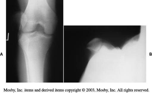
Q1-Q1111 questions — tap to reveal all answerslist
- What is the immediate management of a first-time lateral patellar dislocation?
- What history and examination findings are relevant to patellar instability?
- What X-ray views and measurements are used in patellar instability?
- How are CT and MRI used and what predicts recurrence risk?
- What are the types and anatomical causes of patellar instability?
- What are the surgical options for recurrent patellar dislocation and their complications?
- What is the normal function of the patella?
- What is normal patella movement during knee flexion and extension?
- What are the details of MPFL reconstruction?
- What are the types of trochleoplasty and their complications?
- How do congenital/habitual patellar dislocation differ from traumatic instability?
Q1.What is the immediate management of a first-time lateral patellar dislocation?
- CR under sedation (knee extended with medially directed force); X-ray shows laterally dislocated patella, no fracture or loose body
- First time: operate only if chondral lesion positive (with concomitant MPFL recon, Pedowitz AJSM 2019)
- Otherwise conservative: donut bracing, physiotherapy (VMO strengthening), ITB stretching, closed chain exercises
Q2.What history and examination findings are relevant to patellar instability?
- History: age at first dislocation, traumatic or atraumatic, associated bone or connective tissue dysplasias, mechanical symptoms
- Owls eye patella, overall LL alignment, Q angle (M 8-10, F 15-20) vs carry angle of elbow (M 5-10, F 10-15)
- Effusion, quad wasting, ROM
- J sign (patella alta + maltracking), apprehension (push lateral at 20 degrees flexion), patella shift (>2 quadrants abnormal), patella tilt (should pass neutral), grinding test
- Rotational profile and ligamentous laxity
Q3.What X-ray views and measurements are used in patellar instability?
- Standing scannogram; lateral with knee flexed 30 degrees: Blumensaat line, Caton-Deschamps ratio 1.2, trochlear dysplasia signs (flat trochlea = crossing sign, convex = double contour + spur sign)
- Merchant view (knee flexed 45 degrees, caudal directed beam 60deg from horizontal): sulcus angle (N=138), congruence angle (N=-6-16)
- Laurin view (knee flexed 20 degrees, cephalad beam): lateral PFA (N = lateral opening 10)
Q4.How are CT and MRI used and what predicts recurrence risk?
- CT: torsional malalignment, TTTG distance, trochlear dysplasia
- MRI: OC fracture in 40%
- Risk factors for recurrence: trochlear dysplasia, patellar alta, increased TTTG distance, patellar tilt
- PIS score (patellar instability severity score) >4 = high risk (age, bilateral instability, patellar tilt, patellar alta, TTTG distance, trochlear dysplasia)
Q5.What are the types and anatomical causes of patellar instability?
- Congenital
- Acquired: acute, chronic, voluntary (party trick), habitual (dislocates every time with knee flexion within normal ROM, usually painless), recurrent (trivial sprain leads to dislocation with knee extension, painful)
- Alignment (miserable malalignment): coronal Q angle, sagittal knee recurvatum/patella alta, axial femoral anteversion/tibial torsion
- Bone: trochlear dysplasia, patella hypoplasia (Wiberg classification)
- Soft tissue: weak VMO, tight lateral retinaculum, MPFL deficiency, weak hip abductors; general ligamentous laxity
- TTTG 15mm in Asian, 20mm in Caucasian
Q6.What are the surgical options for recurrent patellar dislocation and their complications?
- Principle: minimise destabilising forces while reconstructing bony and soft tissue restraints; recurrent --> MPFL reconstruction + correct risk factors
- Proximal: MPFL reconstruction, medial plication +/- lateral release/lengthening, VMO advancement; trochlear dysplasia --> trochleoplasty
- Distal: patella alta --> distal transfer of TT; increased TTTG --> medial transfer of TT; abnormal limb rotation --> derotational osteotomy
- Roux-Goldthwait (transfer lateral half of patellar tendon medially), Elmslie-Trillat (TT medialization), Maquet (anteriorization), Fulkerson (anteromedial + distal transfer)
- Complications: medial dislocation, medial PFJ arthritis, recurrence 5%, over-constraint leading to PFJ arthritis
Q7.What is the normal function of the patella?
- Sesamoid bone
- Increases moment arm of quadriceps
- Aids cartilage nourishment
- Protects knee joint
Q8.What is normal patella movement during knee flexion and extension?
- Extended: not engaged, most unstable as knee is locked in screw home mechanism with tibia ER (increases Q angle)
- Flexed 20 degrees: starts engagement, contact area moves proximal (increasing Q angle and stability)
Q9.What are the details of MPFL reconstruction?
- The only anatomical option
- Drill through patella or suture anchor
- Interference screw to femur at the Schottle point (confluence of the posterior cortex extension line, blumensaat lines and the posterior curving line of the posterior femoral condyle)
- Graft type: gracilis (size good) or semitendinosus (longer); latest study: semiT has better outcomes
Q10.What are the types of trochleoplasty and their complications?
- Dejour trochleoplasty (thick flap, need split): creates a gap beneath the trochlea surface, trochlea split longitudinally, two flaps collapsed into the gap and held with staples/screws/sutures
- Bereiter trochleoplasty (thin flap, no need split): elevates an osteochondral flap, thinned and moulded into a groove in the underlying bone created with osteotomes
- Resection trochleoplasty
- For Dejour B/D only; seldom done alone, technically demanding; complication: chondrolysis
Q11.How do congenital/habitual patellar dislocation differ from traumatic instability?
- Pathology is lateral tightness and quadriceps contracture rather than a defective medial constraint
- Treatment: early relocation to allow trochlear development, TT osteotomy, MPFL imbrication/recon
- Extensive release of thick fibrous band tethering patella to lateral intermuscular septum; quadriceps VY plasty
- Congenital: look for syndromes - arthrogryposis, nail-patella syndrome
Merchant's view is taken with the knee flexed 45 degrees and the beam directed caudally 60 degrees from horizontal — error — Merchant's original technique inclines the beam 30 degrees from the horizontal (equivalent to 60 degrees from vertical), knee flexed 45 degrees — source
Normal congruence angle on the Merchant view is -6 to 16 — misleading — Normal mean congruence angle is -6 degrees; values greater than +16 degrees are abnormal (lateral subluxation), so -6 and 16 are not the ends of a normal range — source
▸ Slide 665 · TRUE LATERAL XRAYSports · 5 questions expand

Q1-Q55 questions — tap to reveal all answerslist
- How do you assess patella alta on a lateral X-ray?
- What are the Dejour signs of trochlear dysplasia (based on CT)?
- How is the Merchant's view taken and what do you measure?
- How is the Laurin's view taken and what do you measure?
- What do you look for on CT and MRI in patellar instability?
Q1.How do you assess patella alta on a lateral X-ray?
- Blackburne-Peel index: ratio of perpendicular distance of patella articular surface to tibial plateau and patella articular surface (a/b); normal 0.8, alta >1.0, baja <0.5
- Caton-Deschamps: 0.6-1.3; >1.3 = alta, <0.6 = baja
- Insall-Salvati: patella tendon length / patella length (LT/LP); normal 0.8-1.2; alta >1.2, baja <0.8 (affected by patella and tibial tuberosity morphology)
- Blumensaat line: inferior pole of patella should sit on line drawn along superior aspect of intercondylar notch (rough guideline)
Q2.What are the Dejour signs of trochlear dysplasia (based on CT)?
- A: crossing sign - sulcus too shallow and ended prematurely anterior to groove
- B: supracondylar spur - flat trochlea with lateral condyle higher than medial
- C: double contour due to lateral convex condyle
- D: spur + double contour cliff
Q3.How is the Merchant's view taken and what do you measure?
- Knee flexed 45 degrees, beam aimed caudal
- Congruence angle: line connecting lowest pole of patella and lowest point of trochlear groove vs line bisecting the groove; normal should be negative 5 degrees (medial)
- Sulcus angle: lines along medial and lateral trochlear ridges; normal 138 degrees
Q4.How is the Laurin's view taken and what do you measure?
- Knee flexed 20 degrees, beam aimed cephalad
- Lateral patellofemoral angle: line along top of femoral condyle and lateral facet of patella
- Normal 10 degrees (tilt)
Q5.What do you look for on CT and MRI in patellar instability?
- CT: femoral version, trochlear dysplasia, lateral patella tilt, TTTG (lateral offset of tibial tuberosity from trochlear groove)
- MRI: MPFL injury (medial femoral epicondyle to superior medial patella)
- MRI: chondral lesion (usual medial patella and lateral femoral condyle, injured during relocation)
▸ Slide 666 · Middle age, M = F Recurrent atraumatic haemarthrosisSports · 9 questions 1 check expand
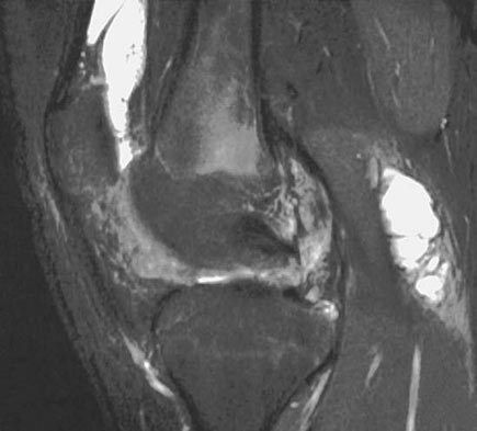
Q1-Q99 questions — tap to reveal all answerslist
- What are the lateral knee X-ray findings in this patient with recurrent atraumatic haemarthrosis?
- What is the differential diagnosis for recurrent atraumatic haemarthrosis with these X-ray findings?
- What is pigmented villonodular synovitis (PVNS)?
- How does the diffuse form of PVNS differ from the localized form?
- What are the MRI and histological features of PVNS?
- What is the management of PVNS?
- How would you clinically assess a patient with recurrent atraumatic haemarthrosis of the knee?
- What is the extra-articular equivalent of PVNS?
- Describe the arthroscopic portals used for PVNS of the knee.
Q1.What are the lateral knee X-ray findings in this patient with recurrent atraumatic haemarthrosis?
- Increased radiopacity over the suprapatellar region and posterior to the femoral condyles
- Well circumscribed, with no disruption of cortex, +/- nodular depression
- Cystic lesion over the femoral condyle with a sclerotic margin
- Mainly a soft tissue lesion
Q2.What is the differential diagnosis for recurrent atraumatic haemarthrosis with these X-ray findings?
- PVNS (pigmented villonodular synovitis)
- Haemophilia
- Synovial chondromatosis
- Inflammatory synovitis
Q3.What is pigmented villonodular synovitis (PVNS)?
- Idiopathic monoarticular neoplastic synovial disease
- Abundant proliferation of synovial villi and nodules
- CSF1 gene, genetic locus at chromosome 5q33; pexidartinib
- Can occur at any age; 50% have a history of trauma
- Presents with swelling and limited ROM; intra-articular
Q4.How does the diffuse form of PVNS differ from the localized form?
- Both forms are most common in the knee
- Diffuse: more extra-articular involvement
- Diffuse: more likely to be painful
- Diffuse: less well circumscribed on MRI
- Diffuse: higher recurrence (30% vs 8% after synovectomy)
Q5.What are the MRI and histological features of PVNS?
- MRI: low signal on both T1 and T2
- MRI: blooming artifact on gradient-echo sequences due to iron in hemosiderin (haemosiderin deposition)
- Histology: haemosiderin-stained multinucleated giant cells
- Mononuclear stromal cells; highly vascular villi with hyperplastic synovial cells
- Pigmented foam cells (lipid-laden histiocytes)
Q6.What is the management of PVNS?
- Nearly no role for conservative management
- Total synovectomy (marginal excision): open, arthroscopic or combined
- Arthroscopy: brownish or reddish inflamed synovium with frond-like papillary projections
- + Irradiation reduces recurrence to 10-20% (30-35Gy in 15 fractions, or 50Gy in 25 fractions)
- + TKI (e.g. imatinib)
- Hip PVNS: joint damage occurs early, so perform synovectomy early
Q7.How would you clinically assess a patient with recurrent atraumatic haemarthrosis of the knee?
- PE: confirm swelling, exclude a mass, document ROM
- Examine the contralateral limb for involvement
- Further imaging: MRI with gradient echo sequence
- MRI shows blooming effect over the area of concern, also involvement posterior to the knee
Q8.What is the extra-articular equivalent of PVNS?
- Giant cell tumour of the tendon sheath
Q9.Describe the arthroscopic portals used for PVNS of the knee.
- Posteromedial portal: 1cm above the joint line behind the MCL
- Posterolateral portal: 1cm above the joint line between the LCL and biceps tendon
PVNS is linked to the CSF1 gene on chromosome 5q33 — wrong chromosome/locus — CSF1 is on chromosome 1p13; PVNS/TGCT is driven by t(1;2)(p13;q35) fusing CSF1 to the COL6A3 promoter on 2q35. Loci at 5q22-31 are only uncommon alternative translocation partners. — source
▸ Slide 667 · Clinical photo and Xray showing knee subluxationSports · 14 questions expand
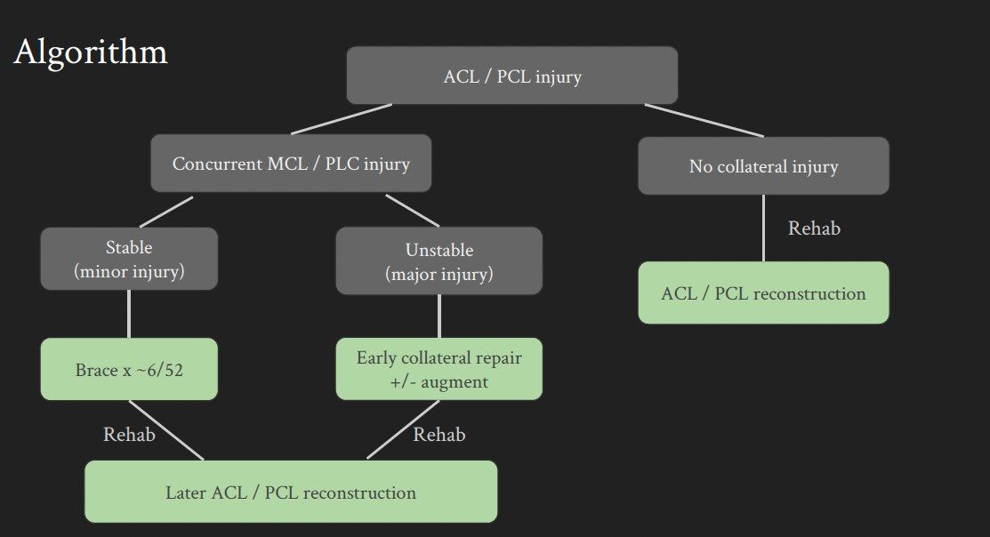
Q1-Q1414 questions — tap to reveal all answerslist
- What does the clinical photo show and what is the dimple sign?
- How do you assess a knee dislocation acutely?
- What associated injuries occur with knee dislocation and when is an ex-fix indicated?
- Scenario 2: dislocated knee with an absent pulse - what is your management?
- What signs suggest a knee dislocation that has spontaneously reduced?
- Describe the definitive (staged) management of a multiligamentous knee injury.
- Scenario 1: posterolateral dislocation with intact NV status - what is your management?
- Scenario 3: dislocated knee with a palpable pulse - what is your management?
- How do you classify a knee dislocation?
- How do you reduce a knee dislocation other than posterolateral?
- Why is a staged approach used in multiligamentous knee injury?
- Compare the graft choices for multiligamentous knee reconstruction.
- What are the controversies in operative treatment of knee dislocation?
- Describe the posterior approach to the knee.
Q1.What does the clinical photo show and what is the dimple sign?
- Posterior displacement of the tibia with lateral translation
- Dimple sign: puckering of anteromedial skin from the medial femoral condyle buttonholing through the medial retinaculum/capsule
- Indicates a posterolateral dislocation
- Contraindication to closed reduction (would cause skin necrosis)
Q2.How do you assess a knee dislocation acutely?
- ATLS protocol, AMPLE history, primary survey
- Local: rule out hip dislocation and open wound
- Distally document pulse, CR, neurology and compartment syndrome
- Systemic: secondary survey to rule out other injuries
- Recheck pulse and document nerve status after any reduction
- If stable, arrange CTA to look for an intimal tear
Q3.What associated injuries occur with knee dislocation and when is an ex-fix indicated?
- Direct vascular injury in 10-30%
- CPN injury in 10-40%, with complete recovery in only 20%
- Ex-fix if fracture dislocation, significant soft tissue compromise/open injury, vascular compromise or compartment syndrome
Q4.Scenario 2: dislocated knee with an absent pulse - what is your management?
- Analgesia, immediate closed reduction under sedation, splint with knee flexed 20-30 degrees, then recheck pulse
- Pulse returns: CT angiogram or serial ABI monitoring
- No return of pulse: vascular bypass, ex-fix knee, on-table angiogram, resect diseased segment and re-anastomose with reversed saphenous vein graft
- Delay >8 hours gives >80% amputation rate
- Prophylactic fasciotomy (EFFORT review 2020); Postop monitor CK for rhabdomyolysis and reperfusion syndrome
Q5.What signs suggest a knee dislocation that has spontaneously reduced?
- 20-50% reduce spontaneously before presentation
- Hyperextension compared with the normal side
- Popliteal ecchymosis
- NV injury at presentation
- Diffuse tenderness but NO effusion
Q6.Describe the definitive (staged) management of a multiligamentous knee injury.
- Aim: stable painless knee, full ROM, return to pre-injury activity
- Stage 1 at 2 weeks: PLC, PMC, MCL and LCL, meniscus +/- avulsion fracture fixation; hinged knee brace + NWB for 6 weeks
- Stage 2: delayed simultaneous ACL and PCL reconstruction (PCL before ACL), after good ROM and muscle bulk restored
- If vascular injury: no tourniquet, delayed surgery, inlay PCL
Q7.Scenario 1: posterolateral dislocation with intact NV status - what is your management?
- Acute: analgesics, immobilise with a slab then proceed to open reduction
- Medial parapatellar approach; reduce then check stability + NV condition
- Then slab or external fixator
- Avoid closed reduction in posterolateral dislocation (dimple sign - risk of skin necrosis)
Q8.Scenario 3: dislocated knee with a palpable pulse - what is your management?
- Analgesia, immediate closed reduction under sedation + splint with knee flexed 20-30 degrees, then recheck pulse
- Check symmetry of pulse and ABI
- ABI >0.9: serial monitoring; ABI <0.9 and asymmetric: CTA
- If CTA shows an intimal tear: anticoagulant, monitor, delayed OT for ligamentous reconstruction, no tourniquet, no tibial tunnel
- +/- MRI to document ligamentous laxity; classify with Schenck
Q9.How do you classify a knee dislocation?
- Kennedy: by direction - anterior/posterior/medial/lateral/rotational
- Schenck: by number of ligaments involved + fracture
- I = single cruciate; II = bicruciate; III = bicruciate + one collateral tear; IV = all 4; V = fracture
Q10.How do you reduce a knee dislocation other than posterolateral?
- CR under sedation, stabilise the femur and pull the tibia opposite to the direction of displacement
- Document NV status afterwards
- Apply a slab in 20-30 degrees knee flexion or an ex-fix
Q11.Why is a staged approach used in multiligamentous knee injury?
- Shorter OT time and each stage is simplified
- Reduces the risk of arthrofibrosis
- Delay definitive OT if vascular injury is present
- Stage 1 addresses the capsular tear (risk of compartment syndrome) and soft tissue status
Q12.Compare the graft choices for multiligamentous knee reconstruction.
- Autograft: no disease transmission, no added cost, documented healing/vascularisation; cons - donor site morbidity, longer OT
- Allograft: no donor site morbidity, shorter OT, does not further destabilise the knee; cons - availability, disease transmission, cost, biomechanics
- Synthetic: no donor site morbidity, shorter OT, readily available; cons - reactive synovitis, graft failure
Q13.What are the controversies in operative treatment of knee dislocation?
- Timing: Hohmann Knee 2017 metanalysis - early surgery <3 weeks improved Lysholm scores compared with delayed surgery
- Sheth 2019 ISAKOS J - early surgery may provide better functional outcomes without compromising ROM when using early postop mobilisation protocols
- Mook 2009 systematic review JBJS - early <3 weeks vs staged: similar need for additional treatment for arthrofibrosis, both significantly higher than delayed; delayed >3 weeks had less residual instability and ROM deficit; staged had highest excellent/good outcomes
- Jiang 2015 KSSTA - staged operation yields the best clinical results for KDIII; Marder 2021 - neither approach superior
- Repair vs reconstruction: Stannard 2005 PLC repair failure 37% vs 9% with reconstruction; Levy 2010 40% vs 6%
- Laprade AJSM 2019 –suggest single stage anatomic based recon of all ligament (single surgeon series); bicruciate reconstruction - simultaneous preferred at PWH
Q14.Describe the posterior approach to the knee.
- Incision: lazy S, proximal limb lateral, distal limb medial
- Open the popliteal fascia at opening of SSV and MSCN (small saphenous vein and medial sural cutaneous nerve), staying lateral to the MSCN
- Trace the MSCN proximally to protect the popliteal AV bundle
- Protect the common peroneal nerve along the posterior edge of biceps femoris
- Retract popliteal vessels laterally; ligate superior medial and middle geniculate arteries; elevate popliteus +/- soleus
▸ Slide 668 · Knee dislocationSports · 4 questions expand
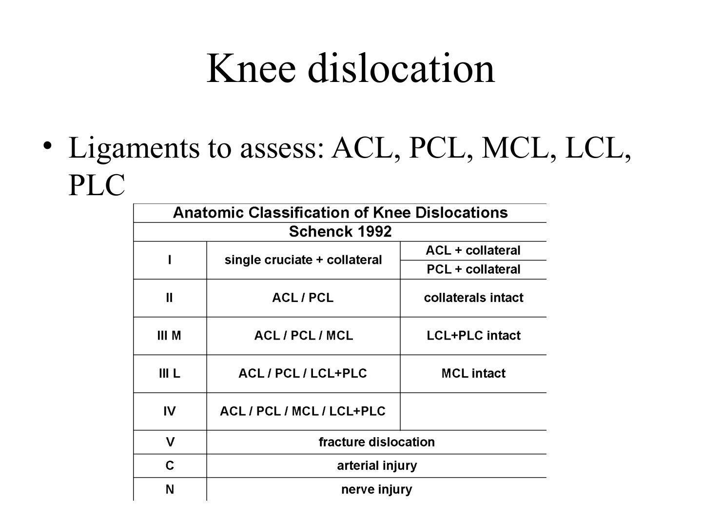
Q1-Q44 questions — tap to reveal all answerslist
- Define knee dislocation and explain why it is an orthopaedic emergency.
- How is knee dislocation classified (Kennedy and Schenck)?
- Which ligaments must be assessed in knee dislocation?
- What are the key complications of knee dislocation?
Q1.Define knee dislocation and explain why it is an orthopaedic emergency.
- Not covered in the speaker notes - the slide image is the only source
Q2.How is knee dislocation classified (Kennedy and Schenck)?
- Not covered in the speaker notes
Q3.Which ligaments must be assessed in knee dislocation?
- Ligaments to assess: ACL, PCL, MCL, LCL, PLC
Q4.What are the key complications of knee dislocation?
- Not covered in the speaker notes
▸ Slide 669 · When would you suspect knee dislocation?Sports · 8 questions 2 check expand

Q1-Q88 questions — tap to reveal all answerslist
- When would you suspect a knee dislocation?
- What are the common mechanisms of injury causing knee dislocation?
- How do you classify knee dislocation?
- Why is the popliteal artery prone to injury in knee dislocation?
- How do you investigate for vascular injury in knee dislocation?
- What is the timing of surgery for vascular injury and how is an intimal tear treated?
- Describe the approach and key steps of the posterior approach to the knee.
- What are the borders and contents of the popliteal fossa?
Q1.When would you suspect a knee dislocation?
- Multi-ligamentous injury (defined as two or more of ACL, PCL, PMC, PLC)
- High energy trauma
- Distal NV compromise
- 20-50% have spontaneously reduced by presentation
Q2.What are the common mechanisms of injury causing knee dislocation?
- Usually high energy e.g. RTA; can occur with relatively low energy injury in athletes
- Anterior (most common): LCP, intimal tear by traction
- Posterior (2nd commonest): axial loading to flexed knee, complete tear of popliteal artery
- Lateral: varus/valgus force, bicruciate tear + peroneal nerve
- Medial: varus/valgus force, bicruciate tear
- Rotational: irreducible
Q3.How do you classify knee dislocation?
- Kennedy: by direction (anterior/posterior/medial/lateral/rotational)
- Schenck: by number of ligaments involved + fracture
- I = single cruciate; II = bicruciate; III = bicruciate + one collateral tear; IV = all 4; V = fracture
Q4.Why is the popliteal artery prone to injury in knee dislocation?
- Incidence quoted as 20-60%
- Tethered proximally by the adductor hiatus (trifurcation) and inferiorly by the soleus arch
- Tied down medially and laterally by the geniculate arteries
- Also tethered by the 5 geniculate arteries
Q5.How do you investigate for vascular injury in knee dislocation?
- Document pulses; AAOS: ABI >0.9 admit + observe 24 hours, <0.9 angiogram, ischaemia explore
- Pulse -ve -> reduce -> pulse returns -> CTA
- Pulse -ve -> reduce -> pulse remains -ve -> vascular surgeon + OT
- Pulse +ve -> ABI <0.9 -> CTA +/- explore
- Pulse +ve -> ABI >0.9 -> serial monitoring for at least 72 hours
- Notes state ABI 100% sensitive, NPV 100%
Q6.What is the timing of surgery for vascular injury and how is an intimal tear treated?
- Emergency operation
- Ischaemic time >8hr = 90% amputation rate; <6hr = 6%
- Intimal tear: anticoagulation
- Subsequent operation: pre-op no tourniquet, delayed surgery, no tibial tunnel
- Nerve injury: ~30% peroneal nerve by traction, fair prognosis; explore if open wound, otherwise conservative first
Q7.Describe the approach and key steps of the posterior approach to the knee.
- GA, prone with bony prominences padded
- Lazy S incision with proximal limb lateral and distal limb medial to avoid the peroneal nerve
- Distally find the small saphenous vein and medial sural cutaneous nerve
- Trace proximally to reach the popliteal vein and tibial nerve in the popliteal fossa
- Incise the popliteal fossa longitudinally lateral to the vein
Q8.What are the borders and contents of the popliteal fossa?
- Diamond-shaped space over the posterior knee
- Bounded by semitendinosus/semimembranosus, biceps femoris and the 2 heads of gastrocnemius
- Floor is the posterior capsule of the knee; roof is the popliteal fascia
- Most superficial structure is the tibial nerve
ABI is 100% sensitive with a 100% negative predictive value for vascular injury after knee dislocation — overstated/contested — Mills et al (2004) reported 100%, but later cohort data show no single physical examination manoeuvre is 100% sensitive; a normal ABI can still miss intimal tears. Combine palpable pulses plus ABI >=0.9 and continue serial observation (48-72h). — medium confidence — source
Popliteal artery injury occurs in 20-60% of knee dislocations — imprecise range — Modern systematic reviews report weighted mean popliteal artery injury rates of about 8-18%; rates at the upper end (up to 60%+) are seen mainly in high-energy trauma and fracture-dislocations. — medium confidence — source
▸ Slide 670 · XR of the right proximal femur and right knee showing multiple well defined ossiSports · 4 questions expand

Q1-Q44 questions — tap to reveal all answerslist
- Describe the X-ray findings shown in this slide.
- What is the differential diagnosis for periarticular ossification/calcification?
- What is synovial chondromatosis?
- How is synovial chondromatosis managed?
Q1.Describe the X-ray findings shown in this slide.
- Multiple well defined ossifications surrounding the right hip and knee joints
- Calcifications are less well defined in the hip
- No associated bone erosions
Q2.What is the differential diagnosis for periarticular ossification/calcification?
- Dystrophic (vascular, tumour, infection, haematoma): synovial chondromatosis, synovial sarcoma, myositis ossificans
- Metastatic: renal osteodystrophy
- Calcinosis / tumoral calcinosis
Q3.What is synovial chondromatosis?
- Proliferative disease of the synovium associated with cartilage metaplasia
- Results in multiple intra-articular loose bodies
- MRI: low T1, high T2
Q4.How is synovial chondromatosis managed?
- Surgery as soon as possible
- Removal of loose bodies
- Synovectomy reduces recurrence rate
- Approach: posterior knee portal or open
▸ Slide 671Sports · 2 questions expand
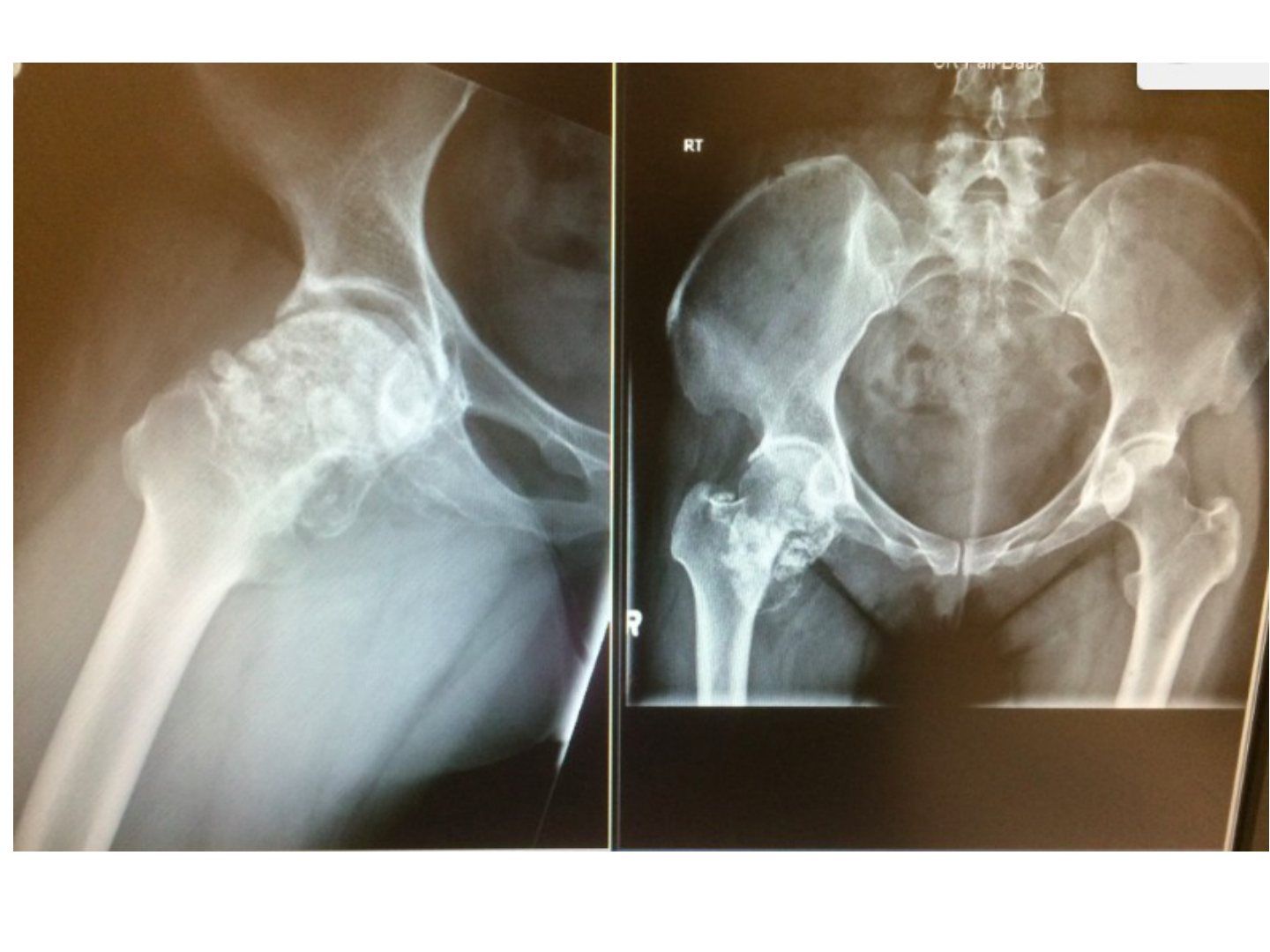
Q1-Q22 questions — tap to reveal all answerslist
- What is the clinical topic of this slide?
- What are the key learning points from this slide?
Q1.What is the clinical topic of this slide?
- Not covered in the speaker notes - the slide image is the only source
Q2.What are the key learning points from this slide?
- Not covered in the speaker notes
▸ Slide 672 · Shoulder stabilizerSports · 3 questions expand

Q1-Q33 questions — tap to reveal all answerslist
- What are the dynamic stabilizers of the shoulder?
- What are the static stabilizers of the shoulder?
- What are the roles of the glenohumeral ligaments?
Q1.What are the dynamic stabilizers of the shoulder?
- Rotator cuff
- Biceps
- Periscapular muscles
Q2.What are the static stabilizers of the shoulder?
- Bony: osteotomy, version
- Soft tissue: ligaments, labrum
- Negative intra-articular pressure
Q3.What are the roles of the glenohumeral ligaments?
- SGHL: inferior restraint at adduction
- MGHL: AP restraint at 0-45 degrees abduction + ER
- IGHL: AP + inferior restraint at 45-90 degrees abduction + ER
- CHL: primary ER restraint
▸ Slide 673 · 50/F shoulder pain + weaknessSports · 21 questions expand
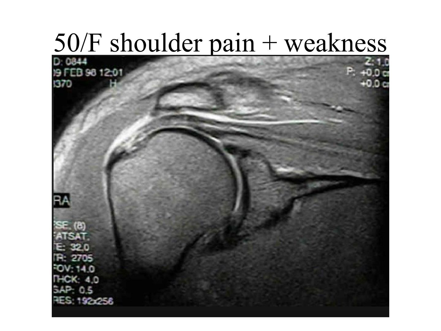
Q1-Q2121 questions — tap to reveal all answerslist
- What are the functions of the rotator cuff?
- What are the causes of rotator cuff tear?
- What are the causes of impingement and what is Neer's staging?
- What are the X-ray findings and special views in rotator cuff pathology?
- What do you look for on MRI in a rotator cuff tear?
- What are the indications for operative management of a cuff tear?
- How would you assess a patient with a suspected rotator cuff tear?
- How would you describe a rotator cuff tear on MRI and what associated findings do you comment on?
- What is the natural history of symptomatic cuff tears (Kim KSSTA 2017)?
- What conservative management is offered for a rotator cuff tear?
- What is the principle of cuff repair and how is the tendon mobilised to the footprint?
- How is a PASTA (partial articular supraspinatus tendon avulsion) tear repaired?
- When is acromioplasty performed and how is it done safely?
- What does double-row repair for massive cuff tears involve?
- What are the partial repair options for a massive cuff tear?
- What are the surgical choices for an irreparable cuff tear?
- How is shoulder arthroscopy set up and performed?
- What is the standard rehab protocol after cuff repair (<3cm tear/single cuff)?
- How does rehabilitation differ after massive or multiple cuff repair?
- Describe the footprint of the rotator cuff.
- What is the definition of a massive cuff tear and what are the clinical clues?
Q1.What are the functions of the rotator cuff?
- Dynamic stabilizer of the shoulder
- Force coupling to maintain a stable fulcrum for concentric rotation of the humeral head by neutralizing deltoid shear
- Vertical: inferior cuff (IS, TM, subscap) counteracts the superior pull of deltoid so the GHJ rotates instead of migrating superiorly
- Horizontal: IS + TM balanced by subscap
Q2.What are the causes of rotator cuff tear?
- Impingement: subacromial -> SS, infraspinatus, teres minor (SIT); subcoracoid -> subscap
- Trauma: middle-aged after shoulder dislocation -> SIT
- Young patient after fall with hyperabduction/ER -> subscap
Q3.What are the causes of impingement and what is Neer's staging?
- External: hooked acromion, ACJ arthritis/osteophytes
- Internal: throwers with tight posterior and lax anterior capsule; anterior subluxation on abd + ER; GT with SS impinging on posterosuperior glenoid
- Neer's staging: inflammation, fibrosis, tear with weakness
- Type 1 collagen replaced by type 2 (better compression strength, less resistant to tensile strength)
Q4.What are the X-ray findings and special views in rotator cuff pathology?
- AP: calcific tendonitis, cuff arthropathy, acromial sclerosis, GT cystic changes, Maloney's line
- Critical shoulder angle (normal 30-35): increased in RCT, reduced in OA
- Supraspinatus outlet view (Y view with 10 degrees caudal tilt) for Bigliani acromial morphology (flat, curved, hooked)
- Also look for os acromionale (os acromiale) and ACJ osteophyte
Q5.What do you look for on MRI in a rotator cuff tear?
- Number, size, retraction of tear; fatty infiltration/atrophy; bicep tendon pathology
- Coronal: SS tear, Patte retraction, superior migration of humeral head
- Sagittal: Goutallier fat atrophy 0-4 (0 normal, 1 fatty streak, 2 <50%, 3 50%, 4 >50%)
- Axial: biceps tendon subluxation (medial), subscapularis and IF tendon
- Ellman partial: I 3mm, II 3-6mm, III >6mm; A articular, B bursal, C intratendinous
- Size: small <1cm, medium 1-3cm, large 3-5cm, massive = 2 or more cuffs involved or >5cm
Q6.What are the indications for operative management of a cuff tear?
- Symptomatic failure of 6-12 months conservative management
- Partial thickness >50% PASTA (Ellman 3A), or bursal side >3mm (Ellman 2B)
- Full thickness + repairable
- Young <60 with acute tear: repair within 6 weeks
- Contraindications: irreparable tear, frozen shoulder
- Irreparable: Goutallier IV, GHJ arthritis, advanced age; SS retraction beyond glenoid; cannot pull cuff beyond articular surface even at 60 degrees abduction
Q7.How would you assess a patient with a suspected rotator cuff tear?
- Hx: history of injury, functional demand, symptoms (overhead activity, night pain) and functional limitation
- PE: identify pathology (rule out other causes of weakness e.g. nerve palsy, identify which cuff), severity of weakness
- Delineate cause (impingement) and complication (frozen shoulder)
- First-line investigation: X-ray and MRI
Q8.How would you describe a rotator cuff tear on MRI and what associated findings do you comment on?
- Morphology: crescent, U, L, massive
- Delineate number, size and retraction of the tear; which cuff is involved
- Comment on articular cartilage, associated ACJ arthritis, superior migration and the tangent line
- Associated biceps tendon pathology
Q9.What is the natural history of symptomatic cuff tears (Kim KSSTA 2017)?
- Symptomatic partial thickness: 26% progress at 2 years
- Symptomatic full thickness: 82% progress at 2 years
- Counsel the patient regarding the natural history
Q10.What conservative management is offered for a rotator cuff tear?
- NSAID
- Physio: strengthen periscapular muscles + deltoid recruitment exercises
- Subacromial injection if impingement symptoms are severe
- PRP: controversial; 2022 Bhandari meta-analysis showed better long-term pain relief than steroid
Q11.What is the principle of cuff repair and how is the tendon mobilised to the footprint?
- Principle: anatomical, secure, tension-free repair to lower retear rate
- Dissection between glenoid and cuff (not too medial 2cm or 1cm posterior - suprascapular nerve at risk)
- Anterior interval slide (to coracoid, more anatomical) and posterior interval slide (to glenoid, less anatomical)
- Release of the coracohumeral ligament
Q12.How is a PASTA (partial articular supraspinatus tendon avulsion) tear repaired?
- If 25% SST remains - convert to full thickness
- Small perforation - transtendinous repair
- Young PASTA - transtendinous in situ repair
- Old PASTA - complete the tear then repair
Q13.When is acromioplasty performed and how is it done safely?
- Not to be done routinely
- Indications: CA ligament fraying, bursal-side tear, anterolateral osteophyte, impingement on dynamic assessment
- Avoid the anteromedial corner (thoracoacromial artery bleeding)
- Together with bursectomy and biceps resection/tenodesis
Q14.What does double-row repair for massive cuff tears involve?
- Chen 2013 meta-analysis: higher rate of intact tendon healing, especially for large tears, but no better clinical outcome
- May not be possible with severe retraction
- Conventional: tendon strangulation, worse footprint restoration, synovial fluid leakage
- Transosseous: medial tendon strangulation, failure at musculotendinous junction, highest ultimate failure load
- Knotless: lower structural strength, suture slippage
Q15.What are the partial repair options for a massive cuff tear?
- Medialisation of footprint - no more than 10-12mm
- Patch repair (fascia lata) to share tension
- Augmentation with biceps
- Marginal convergence no longer recommended (non-anatomical)
Q16.What are the surgical choices for an irreparable cuff tear?
- Superior capsular reconstruction (>=6mm thick graft), medial anchor to superior glenoid
- Muscle transfer: LD for posterosuperior SST/IST tear +/- simultaneous RSA to restore ER; TM for better strength; pec major for subscapular/anterosuperior defect
- Old frail patient: debridement +/- balloon spacer (poly-L-lactide); START:REACTS (Lancet 2022) does not recommend the spacer
- Reverse shoulder arthroplasty
Q17.How is shoulder arthroscopy set up and performed?
- GA, lateral decubitus leaning 30 degrees posterior, parallel to the table; beanbag + correct head position
- Arm draped hanging out, abducted with slight flexion to relax the brachial plexus
- Landmarks: posterolateral corner of acromion and coracoid
- Posterior portal 2cm medial and inferior to the posterolateral corner, directed at the coracoid; danger - too inferior = axillary nerve, too medial = suprascapular nerve
- Rotator interval contents: coracohumeral ligament, SGHL, biceps; systematic view from anterior biceps, GHL, subscap, labrum, SS/IS
- Cuff repair needs lateral portals for anchors; Bankart repair uses anterosuperior and anteroinferior portals
Q18.What is the standard rehab protocol after cuff repair (<3cm tear/single cuff)?
- Phase 1 (first 6 weeks): muscle protection, swelling reduction, controlled mobilisation; abduction pillow, pendulum, scapular retraction exercises
- PROM not beyond 90 degrees in the first 2 weeks, increase to 120 degrees by 4 weeks, then AAROM to 90 degrees
- Phase 2 (up to 12 weeks): full AROM/PROM as tolerated, light isometric/isotonic exercises
- Phase 3: intensive strengthening (theraband, pulleys, press-ups); Phase 4: endurance training (multi-push and lift machines)
Q19.How does rehabilitation differ after massive or multiple cuff repair?
- Delay AROM and isotonic/isometric exercise until 10 weeks
- Can start AAROM from 4 weeks onwards
Q20.Describe the footprint of the rotator cuff.
- Overall dimension 20 x 25mm
- SST at the anterosuperior greater tuberosity
- Medial-lateral widths (SITS): SST 12mm, IST 13mm, TM 14mm, subscapularis 18mm
- 1.5-2mm from the articular cartilage
- Anteroposterior 20mm (SST + anterior IST)
Q21.What is the definition of a massive cuff tear and what are the clinical clues?
- >5cm (Cofield 4)
- >1 tendon involved (Europe)
- Retraction to the glenoid (Patte 3)
- >50% fatty infiltration (Goutallier 4); AHI <6mm
- Clinical clues: severe muscle atrophy, pseudoparalysis (active abduction <90 with full passive ROM and drop arm +), LHB rupture, ER lag or hornblower +
▸ Slide 674 · Suspension bridge theory (Burkhart)Sports · 3 questions expand

Q1-Q33 questions — tap to reveal all answerslist
- Describe the cable-crescent complex in the suspension bridge theory (Burkhart).
- What is the clinical relevance of the suspension bridge theory?
- What are the effects of rotator cuff deficiency?
Q1.Describe the cable-crescent complex in the suspension bridge theory (Burkhart).
- Cable- crescent complex
- Cable: thickened part at the GT insertion of SST and IST, at the CHL avascular zone, running perpendicular to SST fibres and spanning SST and IST insertions
- Crescent: thinner avascular zone of SST and IST distal to the cable (GT attachment)
- Anterior post: LHB
- Posterior post: behind IST
Q2.What is the clinical relevance of the suspension bridge theory?
- Forces can be transmitted as long as the anterior and posterior posts are maintained
- The cable can bypass the crescent and transfer load between the posts, shielding the crescent
- Explains why partial repair is feasible
Q3.What are the effects of rotator cuff deficiency?
- Loss of compressive centring forces
- Superior migration of the humeral head
- GHJ cartilage destruction; superior glenoid erosion
- Synovial fluid leakage; osteoporosis
- Humeral head destruction; articulation with acromion
- CA ligament erosion; ACJ destruction
▸ Slide 675 · Nonoperative mx for cuff tearSports · 3 questions expand
Q1-Q33 questions — tap to reveal all answerslist
- What are the phases of physiotherapy for a rotator cuff tear?
- What is the natural history of atraumatic rotator cuff tears (Moon group, JBJS 2014)?
- What is the role of injections in cuff disease?
Q1.What are the phases of physiotherapy for a rotator cuff tear?
- Phase 1: pain relief + ROM maintenance (pendular, postural exercises, active-assisted ROM, flexibility)
- Phase 2: strengthening
- Phase 3: functional training +/- work hardening
Q2.What is the natural history of atraumatic rotator cuff tears (Moon group, JBJS 2014)?
- 75% do not require OT
- Improvement occurs in 6-12 weeks
- Beyond 3 months, likely to need OT
Q3.What is the role of injections in cuff disease?
- Latest meta-analysis 2022 (Bhandari): steroid has short-term effects, PRP longer-term effects for the painful shoulder
- Neither alters the disease course nor facilitates tendon healing
▸ Slide 676 · Subcoracoid impingementSports · 5 questions expand
Q1-Q55 questions — tap to reveal all answerslist
- Define subcoracoid impingement.
- What are the clinical features of subcoracoid impingement and how is the impingement test performed?
- Describe the Lafosse classification of subscapularis tears.
- How is subcoracoid impingement managed?
- What anatomy predisposes to subcoracoid impingement?
Q1.Define subcoracoid impingement.
- Impingement of the lesser tuberosity with attached subscapularis on the coracoid during adduction, IR and forward flexion
- Similar pathology to subacromial impingement (type 1 replaced with type 2 collagen)
- Coracohumeral distance <6mm
- Related to subscapularis tear
Q2.What are the clinical features of subcoracoid impingement and how is the impingement test performed?
- Posture: 90 degrees FF, adduction + IR
- Impingement test: 90 degrees FF with forceful IR
- Pain = SST/SSC
- LA relief: subacromial -> SST; GHJ -> SSC
- Associated with biceps tendinitis
Q3.Describe the Lafosse classification of subscapularis tears.
- I: upper 1/3 partial
- II: upper 1/3 complete
- III: upper 2/3
- IV: whole subscapularis, head centred, fatty infiltration 1-3
- V: whole subscapularis, anterior subluxation, fatty infiltration 4-5
Q4.How is subcoracoid impingement managed?
- Mostly conservative: stretching +/- steroid injection
- OT indications: failure of conservative management; large subscapularis tear
- Procedure: coracoplasty (7mm clearance between coracoid and subscap) + subscap repair
- Open procedure requires reattachment of the conjoint tendon
- Type I/II: posterior portal, 1 anchor, single row; III/IV: AL portal, 2-3 anchors, double row; V: salvage
Q5.What anatomy predisposes to subcoracoid impingement?
- long/ too lateral coracoid
- Post latarjet (lateralized coracoid)
- Cuff tear with humeral head superior escape
▸ Slide 677 · Internal impingementSports · 5 questions expand
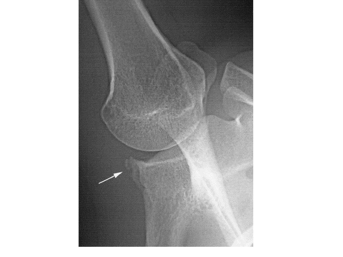
Q1-Q55 questions — tap to reveal all answerslist
- Define internal impingement of the shoulder.
- What is the pathogenesis of internal impingement?
- What are the associated lesions, clinical features and X-ray findings?
- How is internal impingement managed?
- What is GIRD and what are the throwing phases?
Q1.Define internal impingement of the shoulder.
- Impingement of the GT with attached SST on the posterosuperior glenoid during abduction, ER and extension
Q2.What is the pathogenesis of internal impingement?
- Tight posterior capsule causes microinstability
- GIRD (glenohumeral internal rotation deficit)
- Humeral head translates posteriorly
- Contact point shifts to the posterior and superior glenoid
- Posterior articular-sided cuff impinges on abduction + ER (GT and posterosuperior labrum in contact)
Q3.What are the associated lesions, clinical features and X-ray findings?
- Associated lesions: SLAP, GIRD
- Clinical: same total arc but ER > IR
- Posterior pain on apprehension test, relieved with relocated test
- XR: Bennett lesion on axillary view; glenoid retroversion from posterosuperior bone loss
Q4.How is internal impingement managed?
- Conservative: swimmer's stretch
- OT: arthroscopic debridement
- Posterior capsulotomy
- Anterior plication
Q5.What is GIRD and what are the throwing phases?
- GIRD: IR reduced by >25 degrees compared with the contralateral side (IR >25 reduction)
- Throwing phases: wind up, early cocking, late cocking, acceleration, deceleration, follow through
- In late cocking/acceleration, GIRD changes GHJ kinematics: tight posterior capsule, posterosuperior head shift, stretched anterior capsule, GT abuts posterosuperior labrum
▸ Slide 678 · Rotator cuff arthropathy loss of force couple and fixed fulcrumSports · 6 questions expand
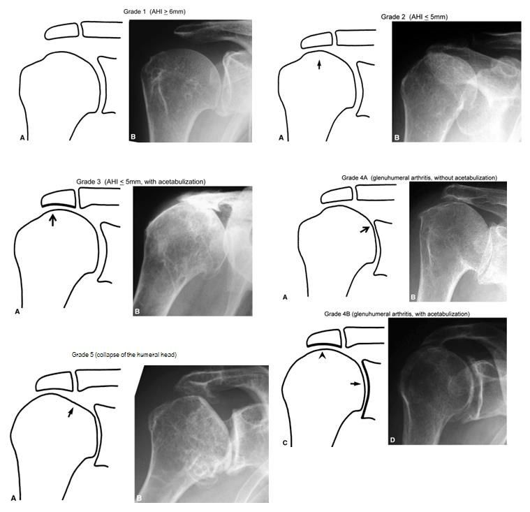
Q1-Q66 questions — tap to reveal all answerslist
- Describe the X-ray findings in rotator cuff arthropathy.
- Describe the Hamada classification of rotator cuff arthropathy.
- What imaging is needed to work up rotator cuff arthropathy?
- What history is important in rotator cuff arthropathy?
- What are the treatment options for rotator cuff arthropathy?
- What did Gill et al (CORR 2021) conclude about aTSA vs rTSA for primary glenohumeral OA?
Q1.Describe the X-ray findings in rotator cuff arthropathy.
- Superior migration of the humeral head
- Acetabularization / femoralization
- Signs of loss of force couple in the shoulder
Q2.Describe the Hamada classification of rotator cuff arthropathy.
- I: AHI >=6mm
- II: AHI <=5mm
- III: acetabulisation
- IV A: GHJ narrow, no acetabulisation
- IV B: GHJ narrow with acetabulisation
- V: head collapse
Q3.What imaging is needed to work up rotator cuff arthropathy?
- Ask for axillary view XR to better appreciate GHJ arthritis
- MRI: massive rotator cuff tear with retraction of SS and fat atrophy
- CT for glenoid bone stock and version
Q4.What history is important in rotator cuff arthropathy?
- Pain
- Stiffness
- Patient expectations
Q5.What are the treatment options for rotator cuff arthropathy?
- Conservative: NSAID, periscapular muscle strengthening, deltoid training
- Arthroscopic debridement
- Reverse arthroplasty (C/I: young, poor glenoid bone stock, non-functional deltoid)
- Other: resection arthroplasty/shoulder fusion (salvage); hemiarthroplasty for pain, not function
Q6.What did Gill et al (CORR 2021) conclude about aTSA vs rTSA for primary glenohumeral OA?
- No survivorship difference at 4 years between rTSA and aTSA
- Differences exist in revision risk between men and women
- Surgeons might select aTSA with an all-polyethylene glenoid to treat OA, despite the current popularity of rTSA
▸ Slide 679 · Classical indicationSports · 4 questions expand
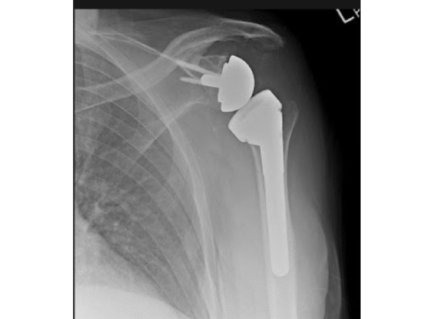
Q1-Q44 questions — tap to reveal all answerslist
- What are the classical indications and prerequisites for reverse shoulder arthroplasty (RSA)?
- What are Grammont's principles of RSA?
- What are the keys to success and the key steps of RSA?
- What are the complications of RSA and what is the Sirveaux classification?
Q1.What are the classical indications and prerequisites for reverse shoulder arthroplasty (RSA)?
- Elderly osteoporotic displaced 4-part fracture with comminuted tuberosities
- Cuff deficient (even 2 or 3 part fracture)
- Prerequisites: good glenoid bone stock, good deltoid power
Q2.What are Grammont's principles of RSA?
- Medialised centre of rotation increases deltoid moment arm (effective lever arm from the start of movement) and recruits more deltoid fibres for FF/abduction; inferiorises to pretension deltoid
- Fixed centre of rotation decreases shearing force to the glenoid
- Reverse polarity has inherent stability: centripedal forces pass into the glenosphere; deltoid shear converted to compression
- Maintenance of external rotation (LD transfer); subscapularis + pectoralis major preserve IR
Q3.What are the keys to success and the key steps of RSA?
- Keys: good exposure, correct sizing and version, stable fixation, satisfactory soft tissue tension
- Deltopectoral approach, biceps tenotomy, tag GT and LT fragments
- Retrieve humeral head, expose glenoid, clear labrum, guide pin, ream and screw baseplate
- Glenosphere with slight inferior overhang to prevent notching; trial
- Humeral retroversion 10 degrees; 3rd generation cementation; suture cuff to implant
Q4.What are the complications of RSA and what is the Sirveaux classification?
- General: bleeding, NV injury, infection, haematoma
- Loosening (old lateral COR designs had high glenoid torque)
- Inferior scapular notching - Sirveaux classification: pillar, reach lower screw, cross inferior screw, central peg
- Dislocation; acromial fracture
- Cosmesis - more squaring; lengthening of upper limb; cemented - more infection
▸ Slide 680 · Favard classifcation of glenoid bone erosionSports · 2 questions expand

Q1-Q22 questions — tap to reveal all answerslist
- Which classification is used for glenoid bone erosion in reverse shoulder arthroplasty?
- What patient and surgical factors increase notching in RSA?
Q1.Which classification is used for glenoid bone erosion in reverse shoulder arthroplasty?
- The Favard classification
Q2.What patient and surgical factors increase notching in RSA?
- Patient: rotator cuff arthropathy with a narrowed AHI (76% vs 38% in post-traumatic cases)
- Glenoids with superior erosion (type E2 glenoid wear)
- MRI grade 3 or 4 fatty infiltration of the infraspinatus
- Surgical: anterosuperior approach higher than deltopectoral (86% vs 56%)
- Neutral or superiorly tilted baseplates
▸ Slide 681 · Anterior shoulder dislocationSports · 14 questions expand
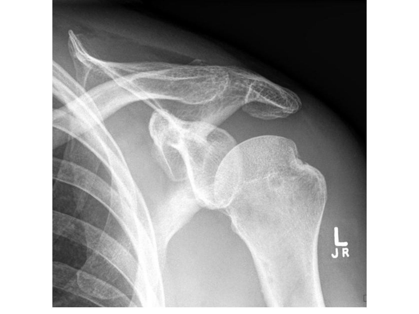
Q1-Q1414 questions — tap to reveal all answerslist
- What history and examination findings are important in anterior shoulder dislocation?
- Which X-rays should you take and how are the special views performed?
- What soft tissue injuries are seen on MRI after anterior dislocation?
- How do you reduce and immobilise an anterior shoulder dislocation?
- Explain the glenoid track / on-off track concept and how it guides surgery.
- Compare the Latarjet and Bristow procedures and describe screw fixation.
- What are the principles of definitive management in anterior shoulder instability?
- How is an arthroscopic Bankart repair performed and when is Latarjet indicated?
- What is the remplissage procedure and what are its complications?
- What other soft tissue and bony procedures are used for anterior instability?
- What are the outcomes and indications after anterior instability surgery?
- What is the Stanmore triangle classification of instability?
- What are the quoted association rates in anterior dislocation?
- What is the instability severity index score (Boileau JBJS 2007)?
Q1.What history and examination findings are important in anterior shoulder dislocation?
- Age - redislocation rate by Rowe: <20 years >90%, <30 60%, >40 20%
- Cofield: 84% athlete vs 30% non-athlete
- Times of dislocation, mechanism, occupation and sport, comorbid NM conditions
- PE: direction of instability, generalized ligamentous laxity, associated injury (cuff tear, SLAP, axillary nerve palsy 5%), infraclavicular plexus injuries
Q2.Which X-rays should you take and how are the special views performed?
- AP: fracture GT, Hill-Sachs lesion
- Axillary view / Velpeau axillary view
- Stryker notch (Hill-Sachs): supine, cassette under shoulder, palm on head, beam 10 degrees cephalic
- West point (Bankart): prone, shoulder on 7.5cm pad, arm 90 abducted, beam centred 25deg down and 25deg medial
- CT: glenoid inverted pear appearance, size of Hill-Sachs (70%), whether on or off track
Q3.What soft tissue injuries are seen on MRI after anterior dislocation?
- Bankart (90%)
- Labroligamentous complex + periosteal tear: Perthes, ALPSA (medial reattachment), GLAD
- HAGL
- Cuff tear (30-80% depending on age)
- SLAP (7%)
Q4.How do you reduce and immobilise an anterior shoulder dislocation?
- Reduction: traction-countertraction, Hippocratic, Stimson, Kocher (ER manoeuvre)
- Immobilise in IR for 2 weeks
- Itoi showed ER immobilisation decreased dislocation rate from 50% to 30%, but it could not be reproduced (compliance)
- AJSM 2015 meta-analysis: ER immobilisation no improvement, duration does not matter
- Cochrane review 2019: no difference
Q5.Explain the glenoid track / on-off track concept and how it guides surgery.
- Glenoid track = contact point of glenoid and humeral head in functional abd + ER = 83% of glenoid width (0.83D - d)
- Hill-Sachs interval (HSI) = distance from infraspinatus attachment to medial rim of the Hill-Sachs lesion
- HSI larger than glenoid track = off-track (engaging) lesion
- Bipolar bone loss concept predicts the risk of dislocation from pathological engagement of the hillsach lesion with the glenoid bone loss
- Glenoid bone loss <25%: on track = Bankart; off track = Bankart + remplissage
- Glenoid bone loss >25%: on track = Latarjet; off track = Latarjet +/- remplissage
Q6.Compare the Latarjet and Bristow procedures and describe screw fixation.
- Size: Bristow = coracoid tip; Latarjet = entire coracoid (anterior to CC)
- Position: Bristow <5mm from glenoid rim; Latarjet flush with rim
- Orientation: Bristow not rotated; Latarjet rotated 90 degrees (concavity along glenoid)
- Fixation: Bristow 1 screw; Latarjet 2 screws (4.0 cannulated or 4.5 cortical, below equator, <15 degrees divergent, within 5mm of rim)
- Effect: Bristow = checkrein; Latarjet = triple block (bone, conjoint tendon sling, capsule repair)
- Complications: nonunion, migration, cartilage injury, weak IR, stiff ER, lateral bone block -> OA
Q7.What are the principles of definitive management in anterior shoulder instability?
- Definitive management depends on: patient's age and activity level, number of dislocations, and associated injury
- Aim: restore the bony and soft tissue stabilizers of the shoulder, solid post-op rehab for early return to sport
- C/I to surgery: multidirectional instability, uncontrolled epilepsy, voluntary dislocation
Q8.How is an arthroscopic Bankart repair performed and when is Latarjet indicated?
- Arthroscopic Bankart repair restores the bumper + hammock effect of the labrum
- Latarjet if bony Bankart >25%, as soft tissue Bankart repair alone has up to 70% failure with recurrent dislocation
- Transfer coracoid with conjoint tendon (not CC ligament) through a vertical slit in subscap to the glenoid defect, fixed with screw anteroinferior
Q9.What is the remplissage procedure and what are its complications?
- Indication: large (>25%) and engaging Hill-Sachs lesion
- Suture infraspinatus and posterior capsule to the Hill-Sachs lesion, making the lesion extra-capsular
- If >30%, may need bone graft or even osteotomy
- Complications: loss of ER (especially with Latarjet), stiff IR (posterior tightness), dehiscence (partial filling)
Q10.What other soft tissue and bony procedures are used for anterior instability?
- Capsular shift if ligamentous laxity: tighten the anterior capsule, less ER, reduces the chance to engage (indirect)
- External osteotomy of the proximal humerus or fill with bone graft for a large Hill-Sachs lesion
- Subscapularis operations: Putti-Platt (double-breast subscap and anterior capsule), Magnuson-Stack (lateral subscap shift)
Q11.What are the outcomes and indications after anterior instability surgery?
- Young + male more recurrence, but symptom-free in between
- If operated, recurrence 5%, may lose ER
- No difference in outcome if operated early (after 1st time) or later (after 3-4th time)
- Thus the common indications are apprehension and recurrent dislocation
Q12.What is the Stanmore triangle classification of instability?
- 1. Structural, traumatic
- 2. Structural, atraumatic
- 3. Non-structural, muscle patterning
Q13.What are the quoted association rates in anterior dislocation?
- Bankart: acute 94%, chronic 49%
- Hill-Sachs: 74%
- Cuff tear: 30% (40s), 80% (60s)
- Axillary nerve: 5%
- Glenoid track (Yamamoto JBJS 2007); on/off track (Di Giacomo JARS 2014)
Q14.What is the instability severity index score (Boileau JBJS 2007)?
- Patient <20 years: 0/2 points
- Competitive sport, contact sport or forced overhead activity: 0/1 or 0/2
- Shoulder hyperlaxity: 0/1
- Hill-Sachs: 0/2, or loss of the sclerotic inferior glenoid contour: 0/2
- >6/10 = recurrence risk of 70%, so Latarjet is needed
▸ Slide 682 · Bankart avulsion of IGHL anterior band + torn periosteumSports · 4 questions expand

Q1-Q44 questions — tap to reveal all answerslist
- What is a Bankart lesion?
- What is a GLAD lesion?
- What is an ALPSA lesion?
- What is a Perthes lesion?
Q1.What is a Bankart lesion?
- Avulsion of the IGHL anterior band + torn periosteum
Q2.What is a GLAD lesion?
- Glenoid labral articular defect
- Shearing off of a portion of articular cartilage together with the labrum
Q3.What is an ALPSA lesion?
- Anterior labral periosteal sleeve avulsion
- Labrum-ligament complex detaches then reattaches medially and inferiorly
Q4.What is a Perthes lesion?
- Labrum-ligament + periosteum stripped off in continuity and not reattached
▸ Slide 683 · AP Xray of shoulder:Sports · 8 questions expand

Q1-Q88 questions — tap to reveal all answerslist
- Describe the AP X-ray findings in posterior shoulder dislocation.
- What are the indications for open surgery and how is reduction performed?
- What special tests and MRI findings relate to posterior instability?
- What are the surgical options for posterior instability?
- How is a chronic (delayed more than 2 weeks) posterior dislocation managed?
- Describe the McLaughlin procedure and its modification.
- What are the primary stabilizers against posterior dislocation?
- How are posterior dislocations classified?
Q1.Describe the AP X-ray findings in posterior shoulder dislocation.
- Incongruent GHJ; disruption of Maloney's line (normal parabola)
- Light bulb sign: internal rotation with loss of humeral head profile
- Trough sign: impaction fracture over the anteromedial humeral head (reverse Hill-Sachs)
- Vacant glenoid/rim sign: medial humerus to medial glenoid clear space >6mm
- Reduced elliptical overlap; associated fractures; need scapular Y or axillary view to confirm
- Causes: trauma 50% (fall on adducted IR arm), convulsion, electrical shock
Q2.What are the indications for open surgery and how is reduction performed?
- Initial management: ATLS/associated injury; less likely axillary nerve or cuff injury; Plain CT to look for indications for open surgery
- Major displaced LT fracture; large posterior glenoid fragment
- Reverse Hill-Sachs (anteromedial) engaged with posterior glenoid or >20% humeral head involvement
- Irreducible/impaction fracture
- CR under GA (usually muscular man) by Delpalma method (in-line traction, adduct + IR, lateral lift + ER)
- Intra-op check congruent reduction, stability and range; immobilise in ER and abduction
- Convert to open via deltopectoral approach; may address bone defect at the same time
Q3.What special tests and MRI findings relate to posterior instability?
- MRI: cuff, reverse Bankart, reverse HAGL, POLPSA
- Kim test: seated, 90 abd + firm axial load, further 45 elevation with down/backward force; pain +/- clunk = posteroinferior labral tear
- Jerk test: 90 abd + 90 IR, axial humeral force then adduction to FF; clunk = posterior labral or posterior subluxation
- Posterior load and shift (under GA): 1 not to rim, 2 to rim, 3 beyond rim, 4 dislocation
Q4.What are the surgical options for posterior instability?
- Indications: large bone loss, irreducible, unstable/recurrent instability
- Soft tissue (reduce intracapsular volume): reverse Bankart repair, closure of rotator interval (reverse remplissage)/inferior capsule
- Glenoid: bone graft if large reverse Bankart; posterior opening wedge osteotomy if excessive glenoid retroversion
- Humerus <30%: disimpaction + bone grafting, modified McLaughlin, McLaughlin, rotational osteotomy
- Humerus >30%: allograft, hemiarthroplasty
Q5.How is a chronic (delayed more than 2 weeks) posterior dislocation managed?
- Pathology - bone: cartilage damage, bone loss, osteoporosis; soft tissue: capsulolabral injury, cuff tear, contracture, adherence to NV bundle
- <4 weeks: try gentle CR; >4 weeks: OR
- Humeral bone loss <40%: McLaughlin, cortical window to disimpact + bone graft, rotational osteotomy
- Humeral bone loss >40%: young = bone graft; old = hemi or RSA
- Glenoid loss >25%: reverse Bristow; redundant capsule: inferior/lateral capsular shift
- During open reduction do not do subscap tenotomy - reserve for McLaughlin
Q6.Describe the McLaughlin procedure and its modification.
- Classically via deltopectoral approach
- A reverse remplissage: capsule + rotator interval + subscapularis filled into the defect
- Modified McLaughlin adds LT transfer: better bone healing, more secure fixation
Q7.What are the primary stabilizers against posterior dislocation?
- Posterior band of the IGHL
- Subscapularis
- Coracohumeral ligament (CHL)
Q8.How are posterior dislocations classified?
- By timing: acute vs chronic
- By underlying pathology: volitional, dysplastic, acquired
▸ Slide 684 · MechanismSports · 6 questions expand

Q1-Q66 questions — tap to reveal all answerslist
- What is the mechanism of inferior shoulder dislocation (luxatio erecta)?
- What are the clinical features of inferior shoulder dislocation?
- What associated injuries must be assessed for in inferior shoulder dislocation?
- How is closed reduction performed in inferior shoulder dislocation?
- What is the management of inferior shoulder dislocation?
- What is the outcome after inferior shoulder dislocation?
Q1.What is the mechanism of inferior shoulder dislocation (luxatio erecta)?
- Forceful abduction
- Levering the humeral head against the acromion out inferiorly
Q2.What are the clinical features of inferior shoulder dislocation?
- Salute position: locked humeral head at 110-160 degrees abduction
- Palpable humeral head at lateral chest wall and axilla
- May buttonhole through the inferior capsule, necessitating open reduction
Q3.What associated injuries must be assessed for in inferior shoulder dislocation?
- Cuff / labrum / IGHL injury
- Nerve: brachial plexus and axillary nerve injury
- Vascular: axillary artery and DCT injury
Q4.How is closed reduction performed in inferior shoulder dislocation?
- Traction and countertraction
- Traction along the humeral shaft
- Countertraction: bedsheet across the superior shoulder/neck
- Gradual reduction with adduction
Q5.What is the management of inferior shoulder dislocation?
- Mostly conservative
- Surgery for subcutaneous humeral head, NV injury, or recurrent dislocation
Q6.What is the outcome after inferior shoulder dislocation?
- Groh JSES 2010 / Malon 1990: 83% excellent/good for range and pain
- NV injury NOT associated with outcome
▸ Slide 685 · Identify the structureSports · 7 questions expand

Q1-Q77 questions — tap to reveal all answerslist
- Which structures should you identify on this arthroscopic view?
- What is the anatomy and contents of the rotator interval?
- What is frozen shoulder?
- What are the associations of frozen shoulder?
- What are the Reeves stages and MRI findings in frozen shoulder?
- How is frozen shoulder managed and what did the FROST trial show?
- Describe the Naviaser arthroscopic classification of frozen shoulder.
Q1.Which structures should you identify on this arthroscopic view?
- Long head of biceps and the biceps labral complex
- Glenoid and anterior labrum
- Subscapularis and the rotator interval
- Humeral head
Q2.What is the anatomy and contents of the rotator interval?
- Space between the supraspinatus and subscapularis
- Contents: SGHL, coracohumeral ligament, long head of biceps, capsule
- Diseases related to the rotator interval: frozen shoulder (tightening), LHB dislocation
Q3.What is frozen shoulder?
- Adhesive capsulitis: global loss of active and passive ROM
- Scarring and contracture of soft tissue (myofibroblast proliferation -> collagen type III), mainly involving rotator interval and coracohumeral ligament
- Presents with pain and global stiffness, especially limited ER
Q4.What are the associations of frozen shoulder?
- Immobilisation of the shoulder
- Previous shoulder pathology (cuff tear, dislocation) or lung/breast/shoulder surgery
- DM (poor prognosis)
- Thyroid disease
- Dupuytren contracture
- Distal clavicle OT
Q5.What are the Reeves stages and MRI findings in frozen shoulder?
- Painful phase: 3-9 months
- Frozen phase: 4-9 months
- Thawing phase: 5-26 months
- MRI: thickened IGHL
- Loss of joint volume (loss of axillary recess on arthrogram)
Q6.How is frozen shoulder managed and what did the FROST trial show?
- Physio + injection (only in painful phase)
- MUA (FEAR: flexion-extension-abduction-rotation); cons: labral injury, fracture
- Arthroscopic capsular release after frozen phase: rotator interval release to gain ER, posterior capsule release to gain IR (270 degrees)
- FROST trial (Lancet 2020): early structured PT, MUA and arthroscopic release - none superior for pain and function at 12 months
- MUA is most cost effective; arthroscopic release carries increased risk
Q7.Describe the Naviaser arthroscopic classification of frozen shoulder.
- Stage 1: erythematous/fibrinous synovium
- Stage 2: red, angry, thick synovium; thick contracted interval; tight joint space; adhesions in the inferior fold
- Stage 3: pink synovium
- Stage 4: no evidence of synovitis but persistent tight inferior fold and joint
▸ Slide 686 · Histological layers of the cuffSports · 7 questions expand
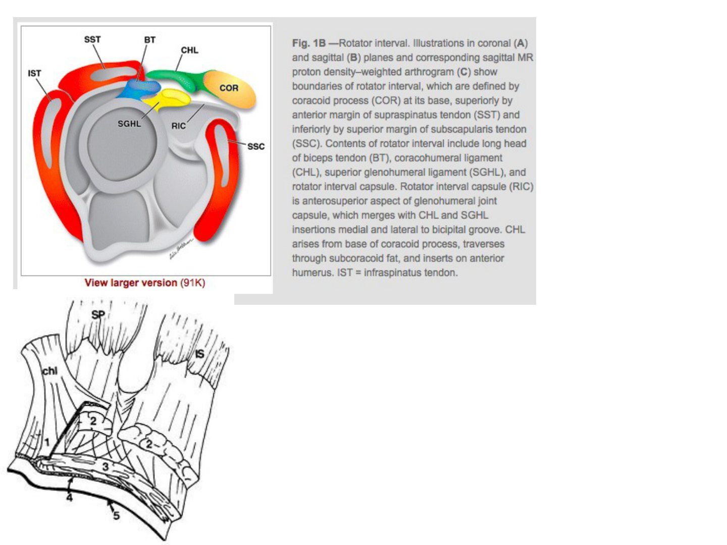
Q1-Q77 questions — tap to reveal all answerslist
- How many histological layers make up the rotator cuff and what is each layer?
- Which layer of the cuff forms the rotator cable and what is it made of?
- Which arteries supply the rotator cuff?
- Between which layers of the cuff does the blood supply run?
- What is the critical zone of Codman?
- Which side of the rotator cuff is more vascular and which side is stronger?
- On which side do most rotator cuff tears occur?
Q1.How many histological layers make up the rotator cuff and what is each layer?
- Layer 1: 1mm, coracohumeral ligament (CHL) fibres
- Layer 2: 3-5mm, dense fibres parallel to the tendon
- Layer 3: 3mm, loose fibres at 45 degrees to the tendon
- Layer 4: thick collagen bands merging with CHL (rotator cable)
- Layer 5: 2mm, shoulder capsule
Q2.Which layer of the cuff forms the rotator cable and what is it made of?
- Layer 4: thick collagen bands that merge with the CHL
- This structure is the rotator cable
Q3.Which arteries supply the rotator cuff?
- Subscapular artery (Subscap A)
- Suprascapular artery (Suprascap A)
- AHCA and PHCA
Q4.Between which layers of the cuff does the blood supply run?
- Between layers 2 and 3
Q5.What is the critical zone of Codman?
- 1cm from the humeral tuberosity insertion
Q6.Which side of the rotator cuff is more vascular and which side is stronger?
- The bursal side is more vascular
- The articular side has only half the strength of the bursal side
Q7.On which side do most rotator cuff tears occur?
- Most tears are articular sided
▸ Slide 687 · Describe MRI:Sports · 12 questions expand

Q1-Q1212 questions — tap to reveal all answerslist
- What did the MRI arthrogram show?
- What does SLAP stand for and what is it associated with?
- Describe the Snyder classification of SLAP lesions.
- What is the history and what examination tests are used for a SLAP lesion?
- Describe how the crank test is performed and what counts as positive.
- What X-ray finding suggests internal impingement?
- What is the function of the SLAP/biceps complex?
- What are the normal labral variants and their frequencies?
- What is the nerve supply of each rotator cuff muscle?
- Where can the suprascapular nerve be compressed and what is the effect?
- What will you look for on physical exam if there is a cyst at the spinoglenoid notch?
- What is the treatment and rehabilitation after a SLAP lesion?
Q1.What did the MRI arthrogram show?
- Signal change/linear signal at the superior labrum, likely SLAP lesion
- Cystic lesion at the spinoglenoid notch
Q2.What does SLAP stand for and what is it associated with?
- Superior Labrum from Anterior to Posterior tear
- Associations: cuff tear, shoulder dislocation
- Internal impingement (frequent thrower), GIRD
Q3.Describe the Snyder classification of SLAP lesions.
- I: fraying of superior labrum
- II: biceps anchor detachment + labral fraying
- III: superior labrum bucket-handle tear
- IV: bucket-handle tear with extension into biceps
- V: SLAP + Bankart; VI: SLAP + labral flap; VII: SLAP + extension into MGHL origin
- Types 5-7 are the Maffet modification (total 7 types)
Q4.What is the history and what examination tests are used for a SLAP lesion?
- Hx: deep shoulder pain, clicking; acute traction injury or recurrent throwing action
- PE: O'Brien's test, crank test, anterior shoulder instability
- SLAP lesion increases strain on the anterior band of IGHL
- Scapular dyskinesia
Q5.Describe how the crank test is performed and what counts as positive.
- Sitting, arm flexed to 90 degrees; examiner adjacent to affected shoulder holds the flexed elbow/forearm
- Joint load applied along the axis of the humerus with one hand; the other hand performs humeral rotation while the shoulder is elevated in the scapular plane
- Can be repeated supine
- Positive if symptoms reproduced (usually pain), with or without a click, usually during external rotation
Q6.What X-ray finding suggests internal impingement?
- Bennett lesion
Q7.What is the function of the SLAP/biceps complex?
- Restraint to external rotation, anterior and inferior translation
- Depresses the humeral head in abduction and external rotation
Q8.What are the normal labral variants and their frequencies?
- Normal 85%
- Sublabral foramen 12%: 1-3 o'clock, medial extension of contrast rather than lateral, smooth margin, <3mm, at biceps origin
- Buford complex (absent labrum + cord-like MGHL) 1.5%
- Sublabral foramen + cord-like MGHL 1%; meniscoid 1%
Q9.What is the nerve supply of each rotator cuff muscle?
- Supraspinatus/infraspinatus: suprascapular nerve
- Teres minor: axillary nerve
- Subscapularis: upper and lower subscapular nerves
Q10.Where can the suprascapular nerve be compressed and what is the effect?
- Suprascapular notch (suprascapular ligament): affects both SS and IS; suprascapular artery runs above the ligament; ganglion cyst from labral injury, fracture callus
- Spinoglenoid notch (spinoglenoid ligament): affects IS only; paralabral ganglion cyst from SLAP/posterior labral tear, traction injury in volleyball players
- Cyst at spinoglenoid notch: infraspinatus weakness (ER with arm at side) with no scar or winging
- Ddx: C5 palsy, brachial plexus injury
- Treatment: conservative; surgical excision of SOL/release of spinoglenoid ligament
Q11.What will you look for on physical exam if there is a cyst at the spinoglenoid notch?
- Infraspinatus weakness (external rotation with the arm on the side) with no scar or winging
- Ddx: C5 palsy, brachial plexus injury
Q12.What is the treatment and rehabilitation after a SLAP lesion?
- Conservative or operation: type 1/3 debridement, type 2 repair
- Type 4: debridement +/- biceps tenodesis if large biceps tendon involvement
- Rehab: 1-4 weeks active + passive assisted flexion in scapular plane; 4-12 weeks active ROM and isometrics; 12 weeks onwards strengthening
- Return to sports at 6 months
▸ Slide 688 · Humeral Avulsion of the Glenohumeral Ligament (HAGL)Sports · 5 questions expand

Q1-Q55 questions — tap to reveal all answerslist
- What is a HAGL lesion and why does it matter?
- Which part of the IGHL is involved and what are the patterns of failure?
- What is the association of HAGL with instability and failed stabilization?
- What MRI sign is seen in HAGL and what is the West Point classification?
- What is the management and rehabilitation of HAGL?
Q1.What is a HAGL lesion and why does it matter?
- Humeral Avulsion of the Glenohumeral Ligament
- Injury to the inferior glenohumeral ligament causing instability/pain
- Commonly a missed cause of recurrent shoulder instability
Q2.Which part of the IGHL is involved and what are the patterns of failure?
- Anterior band most common (93%)
- Medial (glenoid) versus lateral (humerus)
- Failure at labral complex 40% > intrasubstance tear 35% > humeral insertion 25%
Q3.What is the association of HAGL with instability and failed stabilization?
- 10% of recurrent anterior shoulder dislocators
- 20% of failed anterior stabilization
- 30% of shoulder instability patients without a Bankart lesion
Q4.What MRI sign is seen in HAGL and what is the West Point classification?
- MRI: J sign at the inferior pouch
- West Point classification: posterior/anterior; bony/soft tissue; with or without Bankart
Q5.What is the management and rehabilitation of HAGL?
- Conservative: 90% success
- Open or arthroscopic repair: similar outcome
- Rehab: anterior lesion limit ER; posterior lesion limit IR
- Good prognosis
▸ Slide 689 · Which photo shows popeye sign?Sports · 14 questions expand

Q1-Q1414 questions — tap to reveal all answerslist
- What does the popeye sign indicate?
- What do you consider when managing a long head of biceps rupture?
- What are the physical examination findings in a long head of biceps rupture?
- What are the physical examination findings of a distal biceps rupture?
- What factors are considered in managing a distal biceps tendon rupture?
- What are the risk factors for distal biceps tendon rupture?
- What investigations are performed for a distal biceps tendon rupture?
- When is a distal biceps rupture treated conservatively versus operatively?
- What are the treatment options for long head of biceps rupture?
- How is distal biceps repair performed and what are the complications?
- What fixation options are used for distal biceps repair?
- What is the anatomy and blood supply of the biceps tendon relevant to rupture?
- What are false positives for the hook test?
- What are the biomechanical tolerances of the distal biceps tendon?
Q1.What does the popeye sign indicate?
- Left: long head of biceps rupture with Popeye sign
- Right: reverse popeye sign = distal biceps tendon rupture
Q2.What do you consider when managing a long head of biceps rupture?
- Age
- Power (supination)
- Pain due to biceps tendinitis
- Cosmesis
- Associated injury (rotator cuff tear)
Q3.What are the physical examination findings in a long head of biceps rupture?
- Tenderness over the biceps groove
- Assess cuff power
- Elbow flexion and forearm supination power
Q4.What are the physical examination findings of a distal biceps rupture?
- Hook test from lateral to medial (from medial blocked by lacertus fibrosis)
- Ruland biceps squeeze test
- Loss of biceps tracking with passive forearm rotation
- Supination loss 50%, flexion loss 30%, endurance loss 80-90%
Q5.What factors are considered in managing a distal biceps tendon rupture?
- Age and function
- Power
- Cosmesis
- Chronicity of tear
- Degree of tear
Q6.What are the risk factors for distal biceps tendon rupture?
- Steroid use and smoking
- Male, dominant hand
- Prexisting pain (radiobicipital bursitis)
Q7.What investigations are performed for a distal biceps tendon rupture?
- XR to rule out avulsion fractures
- MRI: retraction and partial vs complete tear
Q8.When is a distal biceps rupture treated conservatively versus operatively?
- Conservative in low functional demand (accepts 50% supination, 30% flexion loss, 80-90% endurance loss)
- Operative in young and fit patients to regain supination power
- Perform within weeks
Q9.What are the treatment options for long head of biceps rupture?
- Tenodesis to proximal humerus (suture anchor/interference screw/keyhole procedure)
- Tenodesis: improve ~10% flexion power only, improve cosmesis
- Tenotomy for biceps tendinosis/subluxation with pain in a low-demand patient
- Approach: arthroscopic or mini-open
Q10.How is distal biceps repair performed and what are the complications?
- Single incision: cubital fossa transverse incision (BR/PT); protect LABCN (exits between biceps and brachialis) and PIN by lateral retraction + supination; ligate recurrent radial artery lying superficial to biceps tendon; locate radial tuberosity; incise radiobicipital bursa
- Single incision: decreased risk of synostosis/HO but more LABCN injury
- Two incision: posterior approach at PL elbow (ECU/EDC, between Kocher and Kaplan); less dissection to antebrachial fossa, less radial nerve/PIN injury but more synostosis (disrupts IOM + ulnar periosteum)
- Complications: LABCN injury, SRN injury, PIN injury, synostosis, HO, proximal radius fracture
Q11.What fixation options are used for distal biceps repair?
- Single incision: suture anchor / cortical button / bioabsorbable screw
- JBJS 2014 systematic review: lower complication rate with cortical button
- Double incision: pull-through suture
Q12.What is the anatomy and blood supply of the biceps tendon relevant to rupture?
- LH biceps: further from the forearm rotational axis, supinator, inserts at proximal aspect of radial tuberosity
- SH biceps: flexor, inserts at distal aspect of radial tuberosity
- Blood supply: proximal brachial artery, distal posterior interosseous artery; watershed area at zone 2
Q13.What are false positives for the hook test?
- False positive for positive hook test:
- Bicipital aponeurosis
- Partial tear
- Underlying brachialis tendon
Q14.What are the biomechanical tolerances of the distal biceps tendon?
- Elbow at 90 degrees, no load: distal biceps sustains 50N
- Elbow at 90 degrees with 1kg load: 112N
- Force to rupture 200N
- Repair needs to withstand 50N
▸ Slide 690 · Shoulder arthrodesisSports · 4 questions expand
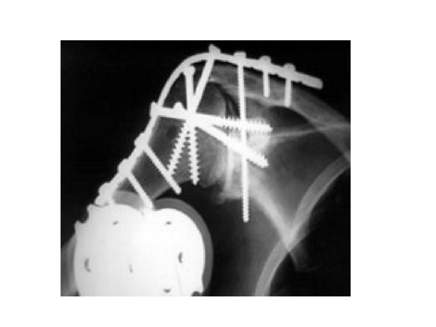
Q1-Q44 questions — tap to reveal all answerslist
- What is the goal of shoulder arthrodesis?
- What are the indications and contraindications for shoulder arthrodesis?
- What position is the shoulder arthrodesed in?
- Describe the technique of shoulder arthrodesis.
Q1.What is the goal of shoulder arthrodesis?
- Stable base for the upper limb optimizing hand and elbow function
- Rest at side without excess scapular winging
- Reach head for combing, midline for dressing, back pocket
Q2.What are the indications and contraindications for shoulder arthrodesis?
- Indications: stabilize paralytic disease (brachial plexus injury); recurrence of instability with multiple failed operations; irreparable deltoid and cuff arthropathy
- C/I: advanced bilateral shoulder disease, contralateral arthrodesis, ipsilateral elbow arthrodesis
- C/I: no scapulothoracic movement (need residual 60 degrees scapulothoracic movement)
Q3.What position is the shoulder arthrodesed in?
- 30 degrees abduction
- 30 degrees forward flexion
- 30 degrees internal rotation
Q4.Describe the technique of shoulder arthrodesis.
- Cuff resected; LH biceps tenodesis; decortication
- Extra-articular: 4.5mm recon plate from scapular spine/acromion to humeral shaft
- Intra-articular: humeral head to glenoid neck; acromion to humeral head; clavicle to glenoid
- Check intraoperatively able to reach mouth and adduction close to body
▸ Slide 691 · Calcified tendinitisSports · 4 questions expand

Q1-Q44 questions — tap to reveal all answerslist
- What is the pathophysiology of calcific tendinitis?
- Describe the Uhthoff classification of calcific tendinitis.
- What is calcific tendinitis associated with and how is it investigated?
- What is the treatment of calcific tendinitis?
Q1.What is the pathophysiology of calcific tendinitis?
- Local ischemia at codman zone -> fibrocartilaginous metaplasia -> CaHA deposition -> inflammation
- Resorption phase: CaHA dissolution and engulfment by macrophages
- HA - Alizarin red staining +
Q2.Describe the Uhthoff classification of calcific tendinitis.
- Pre-calcific phase: fibrocartilaginous metaplasia leading to crystal deposition
- Calcific phase: formative, resting, resorptive (phagocytic action, most painful)
- Post-calcific phase: restoration of fibroblasts
Q3.What is calcific tendinitis associated with and how is it investigated?
- Subacromial impingement, DM, hypothyroidism
- MRI to look for a cuff tear
Q4.What is the treatment of calcific tendinitis?
- Conservative: NSAIDs, physiotherapy, steroid
- USG-guided aspiration and lavage (barbotage): 2 needles, one in one out
- Surgical decompression
▸ Slide 692 · What does these photo shows and what are the underlying pathology?Sports · 3 questions expand

Q1-Q33 questions — tap to reveal all answerslist
- What do the photos show and what is the underlying pathology?
- What are the causes and management of medial scapular winging?
- What are the causes and management of lateral scapular winging?
Q1.What do the photos show and what is the underlying pathology?
- Left: lateral winging of scapula -> trapezius weakness due to accessory nerve palsy
- Other cause of lateral winging: rhomboid weakness due to dorsal scapular nerve
- Right: medial winging -> serratus anterior weakness due to long thoracic nerve palsy
Q2.What are the causes and management of medial scapular winging?
- Causes: overhead activity with head tilted to the other side; iatrogenic during anaesthesia
- Mx: observe for 6 months, then pect major transfer (pectoralis major transfer)
Q3.What are the causes and management of lateral scapular winging?
- Causes: iatrogenic during lymph node dissection
- Mx: observe; if failed, Eden-Lange levator scapulae and rhomboid transfer (medial border to lateral border)
▸ Slide 693 · Xray showing ACJ dislocation with marked superior displacement of the distal endSports · 7 questions expand
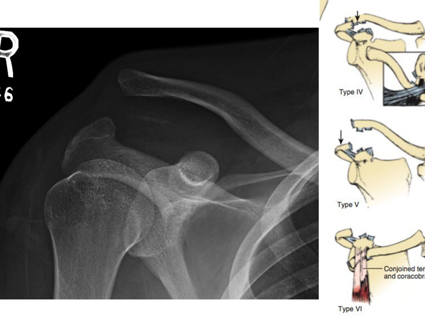
Q1-Q77 questions — tap to reveal all answerslist
- What does the X-ray show and what must be considered?
- How is an ACJ injury assessed?
- How does the Rockwood classification guide management?
- How does the surgical treatment differ between acute and chronic ACJ dislocation?
- What is the modified Weaver-Dunn procedure?
- What is done for isolated ACJ arthritis without instability, and what is the ACJ anatomy?
- What happens if an ACJ dislocation is left untreated?
Q1.What does the X-ray show and what must be considered?
- ACJ dislocation with marked superior displacement of the distal clavicle
- Associated fracture, especially floating shoulder
- Will need further X-ray to classify; start with history and examination
Q2.How is an ACJ injury assessed?
- Acute versus chronic (>3 weeks)
- Compare contralateral side; axillary view (AP translation)
- Zanca view: 10 degrees cephalic tilt (vertical translation); +/- stress view
Q3.How does the Rockwood classification guide management?
- 1-3: static stabilizers disrupted (AC sprain, AC torn, CC torn) -> conservative
- 4-6: dynamic (posterior through trapezius, deltoid + trapezius torn, inferior below coracoid) -> surgical
- 3 +/- operation
Q4.How does the surgical treatment differ between acute and chronic ACJ dislocation?
- Acute – repair; CC fixation with Bosworth screw, CC sling, fibre tape or tightrope
- ACJ fixation: hook plate
- If associated arthritis: modified Weaver-Dunn
- Chronic – reconstruct; with ACJ arthrosis, resect the distal clavicle
Q5.What is the modified Weaver-Dunn procedure?
- Excision of distal clavicle
- Transfer of the acromial end of the CA ligament to the distal clavicle
- CC fixation
- Repair of the deltotrapezial fascia
Q6.What is done for isolated ACJ arthritis without instability, and what is the ACJ anatomy?
- Excision of 7-8mm (<1.5cm) of distal clavicle (too much jeopardizes capsule); can be done arthroscopically
- ACJ is a diarthrodial joint with an articular disc connecting axial to appendicular skeleton
- Stabilizers: superior (most important)/anterior/posterior/inferior; ~8 degrees rotation; Kenny Howard brace
Q7.What happens if an ACJ dislocation is left untreated?
- The shoulder becomes protracted
▸ Slide 694 · GHJ arthritisSports · 7 questions expand

Q1-Q77 questions — tap to reveal all answerslist
- What are the causes of glenohumeral arthritis?
- What is the primary OA triad and X-ray finding?
- What are the nonoperative and operative options for GHJ arthritis?
- What are the contraindications, survival and complications of total shoulder arthroplasty?
- When are hemiarthroplasty and reverse shoulder arthroplasty indicated?
- When is arthroscopic debridement or a CAM procedure performed?
- When is shoulder arthrodesis indicated in glenohumeral arthritis?
Q1.What are the causes of glenohumeral arthritis?
- More common in females
- Primary osteoarthritis
- Secondary: post-traumatic, arthritis of dislocation, inflammatory/crystalline arthritis, osteonecrosis, neuropathic (Charcot arthropathy)
- Rotator cuff arthropathy
Q2.What is the primary OA triad and X-ray finding?
- Anterior capsular contracture, posterior glenoid wear, posterior humeral subluxation
- XR: osteophytes circumferentially at the humeral head, "goat's beard"
Q3.What are the nonoperative and operative options for GHJ arthritis?
- Nonoperative: physical therapy, NSAIDs, intra-articular injections
- Operative: total shoulder arthroplasty, hemiarthroplasty, reverse shoulder arthroplasty
- Also arthroscopic debridement, CAM procedure, arthrodesis
Q4.What are the contraindications, survival and complications of total shoulder arthroplasty?
- C/I: lack of deltoid or rotator cuff function, active infection, Charcot arthropathy
- 10-year survival 92-95%
- Complications: glenoid/humeral component loosening, infection, fracture, nerve injury, cuff tear
- Higher prosthetic joint infection risk within 3 months of prior arthroscopy
Q5.When are hemiarthroplasty and reverse shoulder arthroplasty indicated?
- Hemi: younger patient, irreparable RC tear + insufficient bone stock, osteonecrosis without glenoid involvement
- Ream-and-run over glenoid has early failure, not recommended, poorer outcomes
- RSA: irreparable/large RC tear with intact deltoid; 10-year survival ~90-95%
- RSA complications: scapular notching, infection, dislocation/instability, nerve injuries (higher than TSA)
Q6.When is arthroscopic debridement or a CAM procedure performed?
- Debridement: mild-moderate OA without structural alteration; mechanical symptoms from loose bodies or small humeral head lesions due to AVN
- CAM (comprehensive arthroscopic management): younger patient
- Debridement, chondroplasty, synovectomy, loose body removal, humeral osteoplasty (goat's beard), capsular release, decompressions, axillary nerve decompression, biceps tenodesis
Q7.When is shoulder arthrodesis indicated in glenohumeral arthritis?
- Paralysis, recurrent infection, severe soft tissue deficiency, failed replacement surgery
▸ Slide 695 · AVN of humeral headSports · 3 questions expand
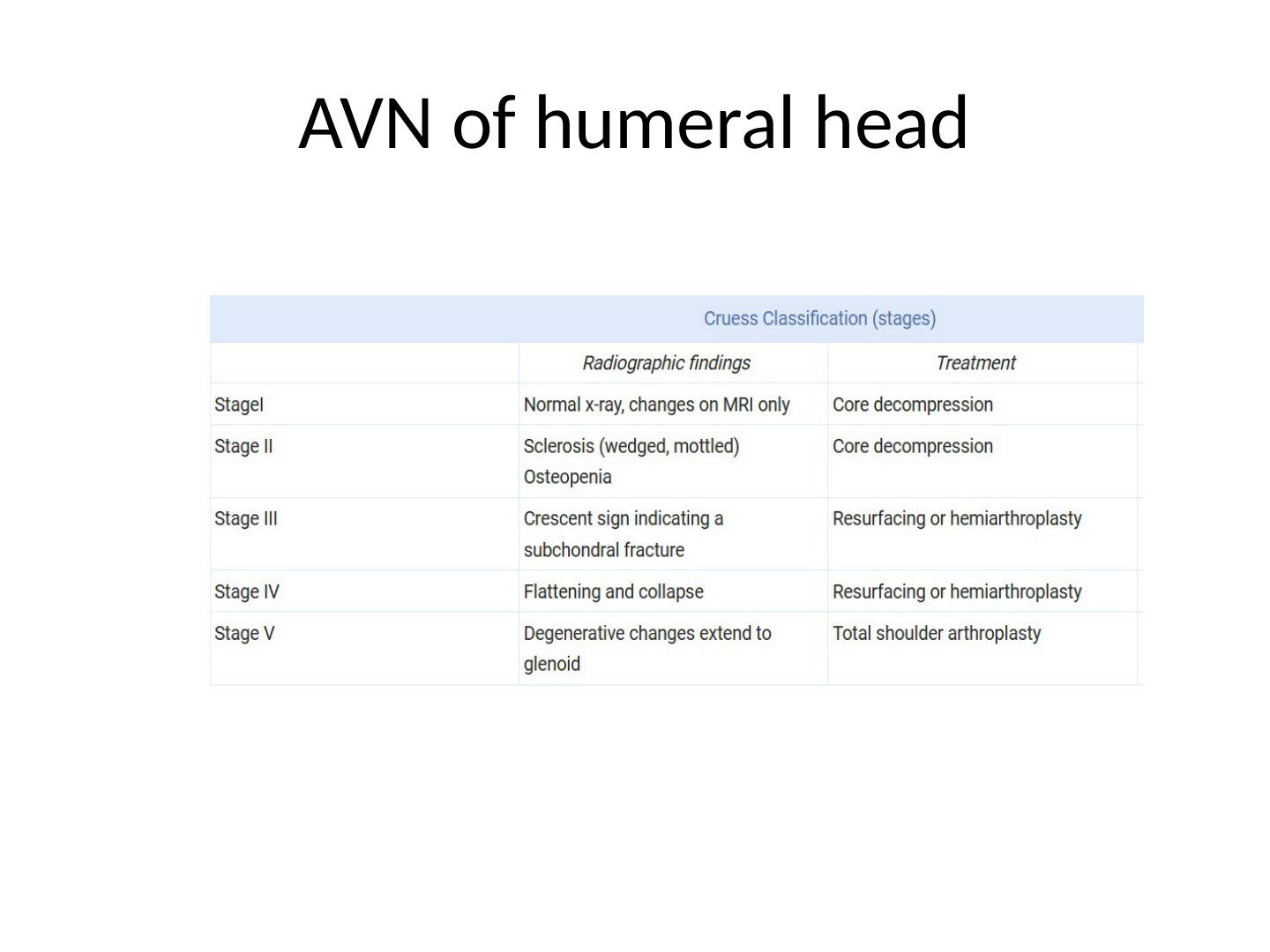
Q1-Q33 questions — tap to reveal all answerslist
- What is the most common initial site of avascular necrosis of the humeral head?
- What is the nonoperative management of humeral head AVN?
- What operations are used for humeral head AVN and when?
Q1.What is the most common initial site of avascular necrosis of the humeral head?
- Superior middle portion of the humeral head
- Aetiology is similar to avascular necrosis of the hip
Q2.What is the nonoperative management of humeral head AVN?
- Pain medications, activity modification, physical therapy
- Operative treatment depends on the Cruess stage of disease
Q3.What operations are used for humeral head AVN and when?
- Core decompression + arthroscopy (confirm cartilage integrity) for early disease (Cruess I-II)
- Humeral head resurfacing for stage III with focal chondral defects and sufficient epiphyseal bone stock
- Hemiarthroplasty for moderate disease (Cruess III-IV)
- Total shoulder arthroplasty for advanced stage (Cruess V)
▸ Slide 696 · Femoral acetabular impingementSports · 9 questions 1 check expand

Q1-Q99 questions — tap to reveal all answerslist
- What are the X-ray findings in femoroacetabular impingement?
- What are the symptoms and signs of FAI?
- Describe the alpha angle and its normal and abnormal values.
- What other radiographic measurements are used in FAI?
- What is the role of CT and MRI/MR arthrogram in FAI?
- What are the treatment options and what does hip arthroscopy involve?
- What are the indications for hip arthroscopy?
- What are the compartments examined in hip arthroscopy and which require traction?
- What are the portals used in hip arthroscopy?
Q1.What are the X-ray findings in femoroacetabular impingement?
- Cam lesion at the femoral neck: pistol grip appearance, decreased neck offset, aspherical head (SCFE, Perthes, fracture malunion)
- Pincer lesion: anterosuperior acetabular rim overhanging
- Retroversion: cross cover sign, posterior wall medial to femoral head centre, prominent ischial spine
- Coxa profunda vs protrusio (head medial to ilioischial line); excessive CEA
Q2.What are the symptoms and signs of FAI?
- Previous injury or childhood hip problems
- Mechanical hip pain, difficulty sitting, pain in flexion and internal rotation
- PE: limited flexion and IR; positive anterior impingement test (FADDIR)
Q3.Describe the alpha angle and its normal and abnormal values.
- Angle between the central axis of the femoral neck and the line from femoral head centre to where the head loses sphericity
- Normal <42 degrees; >42 degrees suggestive of head-neck offset deformity
- >50-55 degrees indicates cam deformity
Q4.What other radiographic measurements are used in FAI?
- Head-neck offset ratio: draw 2 lines parallel to neck central axis, one touching anterior head and one touching anterior neck, difference of the 2 lines divided by head diameter; >0.17 normal
- CEA >40; anterior CEA >20; herniation pit
- False profile view (pelvis 65 degrees from film) for anterior CEA
- Dunn view (hip flexion 90, abduction 20, neutral rotation) for CAM
Q5.What is the role of CT and MRI/MR arthrogram in FAI?
- CT: bony architecture, measure alpha angle
- MRI/MR arthrogram: labral injury (more in pincer lesion), cartilage injury (more in cam lesion)
- Countercoup lesion (unfavorable as difficult to attach posterior pathology)
Q6.What are the treatment options and what does hip arthroscopy involve?
- Conservative: NSAIDs, physiotherapy
- Operative: arthroscopic cam trim +/- labral debridement/repair; Ganz surgical dislocation; periacetabular osteotomy; THR
- Hip arthroscopy: GA/SA on traction table, ~50 lb traction (pudendal nerve risk), X-ray guidance, 70 degree scope
- Risks: direct cartilage injury, HO, instability from iatrogenic capsular deficiency (prevented by meticulous capsulotomy and closure/plication)
- Nerve risks: pudendal (traction), superior gluteal (anterolateral portal), lateral cutaneous nerve of thigh/femoral vessels (anterior portal), sciatic (posterolateral portal), LFCA ascending branch
Q7.What are the indications for hip arthroscopy?
- Intra-articular: FAI/labral lesion, chondral lesion, loose bodies, synovial disease
- Extra-articular: AIIS impingement, capsular tear, iliopsoas pathology
Q8.What are the compartments examined in hip arthroscopy and which require traction?
- Central (intra-articular): acetabular rim and fossa, femoral head, AIIS, capsule - requires traction
- Peripheral (lateral to labrum): HN junction, iliopsoas, zona orbicularis, medial synovial fold - no traction needed
- Lateral (peritrochanteric): ITB, trochanteric bursa - no traction
- Deep gluteal: piriformis, ischial tuberosity (hamstring origin) - no traction
Q9.What are the portals used in hip arthroscopy?
- Anterolateral portal first (primary viewing portal): 2cm anterior and superior to anterosuperior corner of GT
- Anterior portal (central viewing): intersection of vertical line from ASIS and superior ridge of GT
- Distal anterolateral (peripheral viewing): 3-5cm distal to anterolateral portal
- Posterolateral (posterior viewing): 1cm posterior and superior to GT
Alpha angle normal <42 degrees; >42 degrees is suggestive of a head-neck offset deformity — Misleading threshold: 42 degrees is the mean in normal controls, not a diagnostic cutoff — The usual diagnostic cutoff for cam morphology is >55 degrees (50 widely used); recent syntheses suggest 57-60 degrees and many normal hips exceed 42 degrees — source
▸ Slide 697 · False profile viewSports · 2 questions expand

Q1-Q22 questions — tap to reveal all answerslist
- What is a false profile view used for?
- What radiographic parameter is measured on the false profile view?
Q1.What is a false profile view used for?
- Not covered in the speaker notes
- Slide title: False profile view
Q2.What radiographic parameter is measured on the false profile view?
- Not covered in the speaker notes
- Slide title: False profile view
▸ Slide 698 · Patient complaining of snapping sound upon hip flexionSports · 5 questions expand

Q1-Q55 questions — tap to reveal all answerslist
- What is coxa saltans and who is affected?
- What are the causes of external, internal and intracapsular snapping hip?
- How do you examine a snapping hip?
- What investigations are used for snapping hip?
- What is the management of snapping hip?
Q1.What is coxa saltans and who is affected?
- Snapping sound on hip flexion
- Common in athletes and dancers in their teens/20s
- Usually the patient can reproduce the symptoms; +/- pain
Q2.What are the causes of external, internal and intracapsular snapping hip?
- External: posterior IT band sliding over greater trochanter; glut max (gluteus maximus; repeated gluteus injections -> contracture/fibrosis); fibrotic bursa; exostosis
- Internal: iliopsoas (prominent iliopectineal ridge, slide over femoral head, iliopsoas bursa, lesser trochanter exostosis)
- Intracapsular: loose body, synovial chondromatosis, labral tear
Q3.How do you examine a snapping hip?
- External: visible snap; pressing on the greater trochanter abolishes it; Ober’s +ve (limited adduction when hip placed in extension)
- Internal: audible snap when hip moves from flexed + ER to extended + IR
Q4.What investigations are used for snapping hip?
- X-ray (looking for chondromatosis)
- USG (dynamic assessment)
- MR arthrogram (rule out loose body/labral tear)
- Iliopsoas bursography
Q5.What is the management of snapping hip?
- Nonoperative: activity modification, physiotherapy, steroid injection
- USG-guided injection (GT bursa, iliopsoas sheath, intra-articular)
- Operative: excision of GT bursa with Z-plasty of ITB; release of iliopsoas tendon; resect lesser trochanter (distal part only); removal of loose body or labral repair
▸ Slide 699 · Stress fractureSports · 7 questions expand
Q1-Q77 questions — tap to reveal all answerslist
- Define a stress fracture.
- What is the difference between a fatigue and an insufficiency fracture?
- What is the pathogenesis of a stress fracture?
- What do X-ray, bone scan and MRI show in a stress fracture?
- What are the high-risk sites for stress fractures?
- What are the sport-specific stress fractures?
- What is the Arendt grading and when is operative treatment indicated?
Q1.Define a stress fracture.
- A fracture occurring at a stress below the ultimate tensile strength
Q2.What is the difference between a fatigue and an insufficiency fracture?
- Fatigue: repetitive stress under ultimate tensile strength and above endurance limit; normal bone; overuse history, insidious onset, prodromal pain, local tenderness/swelling
- Insufficiency: normal stress, abnormal bone; sudden pain with no prodromal symptoms
Q3.What is the pathogenesis of a stress fracture?
- 1. Bone formation < bone resorption
- 2. Decrease in ultimate tensile strength with time
- 3. Microfracture, crack initiation and crack propagation
Q4.What do X-ray, bone scan and MRI show in a stress fracture?
- X-ray normal in the initial 2-3 weeks; later lucent line with sclerotic edges and periosteal reaction
- Bone scan sensitive but not specific
- Acute: linear increase in uptake in all 3 phases; healing: normalises sequentially phases 1-3; soft tissue: only phases 1 and 2
- MRI: STIR sequence
Q5.What are the high-risk sites for stress fractures?
- Tibial anterior cortex
- NOF (superolateral), medial malleolus, talus
- 2nd and 5th metatarsals, patella
- Hallux sesamoids
Q6.What are the sport-specific stress fractures?
- Runner: tibial shaft, NOF, 2nd metatarsal
- Basketball: 5th metatarsal, navicular
- Football: metatarsals, hallux sesamoids
Q7.What is the Arendt grading and when is operative treatment indicated?
- Grading by Arendt (X-ray, bone scan, MRI)
- High grade, high risk -> operative treatment
▸ Slide 700 · Tennis elbowSports · 6 questions expand

Q1-Q66 questions — tap to reveal all answerslist
- What is lateral epicondylitis and what is the pathoanatomy?
- What is the differential diagnosis of tennis elbow?
- What are the physical examination findings in tennis elbow?
- What conservative and operative treatment is used for tennis elbow?
- How does the tennis elbow brace work?
- What is the evidence for PRP versus corticosteroid in tennis elbow?
Q1.What is lateral epicondylitis and what is the pathoanatomy?
- Overuse injury from eccentric overload at the common extensor origin
- Microtear begins at the ECRB origin
- Histology: angiofibroblastic hyperplasia, disorganized collagen (degenerative process)
Q2.What is the differential diagnosis of tennis elbow?
- Cervical radiculopathy
- Radiocapitellar OA
- Radial tunnel syndrome
- PLRI of the elbow
Q3.What are the physical examination findings in tennis elbow?
- Tenderness at the ECRB origin
- Decreased grip strength (in entrapment syndromes)
- Resisted wrist extension with elbow extended; resisted extension of long fingers
- Maximum wrist flexion -> pain; passive wrist flexion in pronation -> pain
Q4.What conservative and operative treatment is used for tennis elbow?
- Conservative: NSAIDs, physiotherapy, shockwave, brace worn 3-4cm distal to the common extensor origin, steroid/PRP injection
- Operative: release and debridement of ECRB origin
- Lift ERCL off ECRB (located deep and posterior to ECRL); excise degenerative tissue; decorticate epicondyle; repair capsule if breached; Side to side closure of the tendon
Q5.How does the tennis elbow brace work?
- Inhibits the traction force by extensors on the common extensor origin
- Inhibits full expansion of the muscle belly
- Worn 3-4cm distal to the common extensor origin
Q6.What is the evidence for PRP versus corticosteroid in tennis elbow?
- PM R 2020 Tang et al meta-analysis: PRP better long-term pain and function; corticosteroid most improvement short term
- Latest Cochrane review 2021 does not support PRP: no evidence of benefit
▸ Slide 701 · Platelet rich plasmaSports · 6 questions 1 check expand
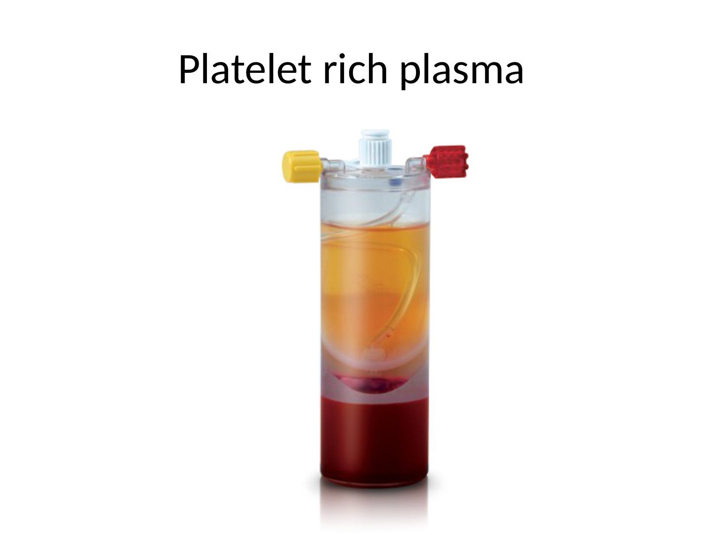
Q1-Q66 questions — tap to reveal all answerslist
- What is PRP and how is it prepared?
- What is the optimal platelet concentration in PRP?
- What are the indications for PRP in orthopaedics?
- What growth factors do platelets release and what is their proposed role?
- What is the clinical evidence for PRP in soft tissue healing, OA and fracture healing?
- What is the evidence for PRP in ACL reconstruction, meniscal repair, cuff repair and tendinopathy?
Q1.What is PRP and how is it prepared?
- Plasma from one's own blood enriched with autologous platelets
- Centrifugation separates the platelet-rich layer from whole blood; calcium chloride used to initiate platelet activation
- Spin twice: 3200RPM for 15minutes then 2 minutes at 2000RPM with calcium chloride
- 3 layers: lowest blood cells, middle PRP, top platelet-poor plasma
Q2.What is the optimal platelet concentration in PRP?
- 3-5x of whole blood
- >5x will inhibit healing
Q3.What are the indications for PRP in orthopaedics?
- Controversial use for possible stimulation of bone and soft tissue healing
Q4.What growth factors do platelets release and what is their proposed role?
- PDGF, TGF-beta, VEGF, IGF-1, EGF, CTGF, FGF-2
- Important role in the inflammatory cascade response after injury
- Proposed: increase ECM deposition, Reduced pro-apoptotic signals, minimise joint inflammation
Q5.What is the clinical evidence for PRP in soft tissue healing, OA and fracture healing?
- Soft tissue healing: no consensus for acute ligament, tendon or muscle injuries or chronic tendinopathies
- OA: RESTORE trial (JAMA 2021) and PEAK trial (BJJ 2022) showed no difference vs saline
- Fracture healing/fusion: limited evidence for bone formation
Q6.What is the evidence for PRP in ACL reconstruction, meniscal repair, cuff repair and tendinopathy?
- ACL: does not support ligamentisation/graft maturation; may improve donor site outcomes and decrease patellar tendon gap
- Meniscal repair: no clear evidence; rotator cuff repair: no benefit
- Lateral epicondylitis: Cochrane review 2021 - no benefit; midsubstance Achilles: not supported
- Patellar tendinopathy: AJSM 2016 Laprade group PRP vs saline, no difference
Optimal PRP concentration is 3-5x whole blood and >5x will inhibit healing — Oversimplified: the exact inhibitory threshold is not established — Reviews commonly cite 3-4x (600,000-900,000/µL) as optimal and >1.2 million/µL unfavourable; in vitro studies show a plateau with reduced collagen synthesis at high concentrations — medium confidence — source