Short Stature
Paediatric elbow - condyle and radial head
Radial neck, lateral condyle and medial epicondyle fractures with acute and chronic sequelae.
29 questions
5 source pages
4 images
Images appear with the first question taken from each source page — tap a question to open it.
Q1Describe the O'Brien classification of radial neck fractures.📷▸
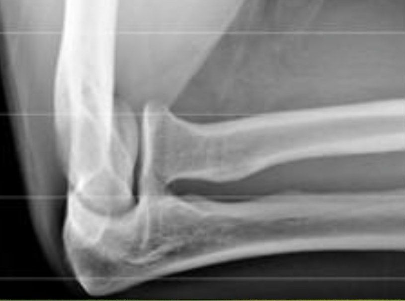
- I: <30 degrees angulation
- II: 30-60 degrees
- III: >60 degrees
- Median age 9-10 years; usually a valgus loading injury of the elbow
Q2What associated injuries and X-ray views are relevant to radial neck fractures?▸
- Associated conditions: elbow dislocation, medial epicondyle fracture
- Greenspan radiocapitellar view: elbow 90 degrees flexion, thumb up, beam 45 degrees from table
Q3How is a radial neck fracture treated according to angulation?▸
- <30deg: immoblization in situ (<30 degrees: immobilisation in situ)
- >30 degrees: attempt closed reduction
- Techniques: Patterson (traction, varus stress, thumb pressure), Israeli (supination + flexion, finger pressure, pronate to force radial head through), Chambers (Esmarch circumferential pressure)
- If still >30 degrees: percutaneous K wire reduction (joystick technique); also try Metazeau technique
Q4What are the complications of radial neck fracture?▸
- Overgrowth (more common)
- Radial head AVN
- Radioulnar synostosis
- Myositis ossificans
- Open reduction: greater loss of motion, increased osteonecrosis and synostosis vs closed reduction
Q5What is normal radial neck angulation?▸
- 0-15 degrees lateral
- 5 degrees posterior to 10 degrees anterior
Q6Describe the X-ray findings and suspected diagnosis.▸
- Flake of bone over lateral metaphyseal region, no gross malalignment
- Suspected lateral condyle fracture
- Minimally displaced on X-ray; need to see articular surface displacement before planning management
- Get internal oblique XR; urgent MRI to look for articular step; if MRI unavailable, arthrogram (inject from posterior to avoid injury to articular surface and major NV bundle)
Q7What is the mechanism of a lateral condyle fracture?▸
- Push off: axial loading by the radial head
- Pull off: common extensor origin
Q8What is the Milch classification?▸
- Type 1: fracture lateral to trochleocapitellar groove, SH type 4, more stable
- Type 2: fracture medial to trochleocapitellar groove, SH type 2 (does not cross lateral condyle epiphysis), less stable, worse prognosis
Q9What are the Jakob and Song classifications?▸
- Jacob: Jakob 1: <2mm displacement (conservative if articular cartilage confirmed smooth)
- Jakob 2: 2-4mm (OR KWF); Jakob 3: severe displacement +/- rotated fragment (OR KWF)
- Song classification (1-5); 3 or above unstable, needs surgery
- Song: 1 limited metaphyseal fracture line, 2 lateral gap, 3 medial and lateral gap, 4 >2mm displacement no rotation, 5 rotation
Q10What is the definitive management of a lateral condyle fracture?▸
- >2mm displacement in any of the 3 views -> OR + KWF
- CR: extension + supination to relax common extensor origin, then varus + anteromedial force
- OR: oblique lateral wound centred at lateral epicondyle (between BR and triceps); avoid posterior dissection to preserve blood supply
- Check anterior and lateral cortex smoothness; fix with 2 divergent pins (joystick reduction)
Q11Why are lateral condyle fractures prone to non-union and what are the late complications?▸
- Poor vascular supply, intra-articular, large pull from extensor origin
- Late complications: nonunion, AVN
- Deformity (cubital valgus from lateral condyle AVN, varus from overgrowth or fishtail); PL subluxation of proximal radius and ulna
Q12How do you manage a delayed presentation with non-union and deformity?▸
- Yes to OT due to future valgus, instability and tardy ulnar nerve palsy (Masada JBJS 1990)
- Aim: achieve union; debride + bone graft + screw horizontally (K wire gives no compression)
- Only debride anterior, leave posterior intact; avoid aggressive reduction (risk AVN and fishtail); Make sure to have preop imaging to rule out AVN
- Alternative: watchful waiting with later osteotomy; ORIF associated with stiffness and AVN (Jakob), but recent studies suggest surgery is a safe option
Q13What are the main arteries supplying the distal humerus?📷▸
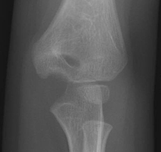
- Radial recurrent artery (comes from the radial collateral artery)
- Interosseous recurrent artery (comes from the middle collateral artery)
Q14Which structures of the distal humerus are supplied by end arteries, and why does this predispose to AVN?▸
- Lateral condyle epiphysis: entry posterolateral to the capsule origin and proximal to the articular cartilage
- Lateral condyle has an end-artery supply; the lateral crista of the trochlea is supplied from it
- Lateral part of the medial crista: crosses the posterior distal humerus metaphysis as an end artery (medial part has multiple vessels)
- Disruption causes lateral condyle AVN (valgus, fishtail) and AVN of the lateral part of the medial crista
Q15What is a fishtail deformity and what are its types?▸
- Area between medial ossification centre and lateral condyle ossification centre resorbs/fails to develop
- Type 1 (more common): sharp angled wedge; persistent gap between lateral condyle physis and medial trochlear due to underdevelopment of the lateral crista
- Type 2: smooth edge; AVN of the lateral part of the medial crista of the trochlea
Q16What causes cubital varus and cubital valgus after lateral condyle fracture?▸
- Cubital varus: overgrowth (50%)
- Cubital varus from fishtail: malunion of lateral part of medial crista (type 1) or medial crista AVN (type 2)
- Cubital valgus: nonunion leading to lateral condyle AVN
- Cubital valgus: physeal arrest
Q17How is non-union defined and managed?▸
- Definition: 3 months
- Displaced >2mm + no K wire fixation -> lateral condyle AVN
- Before physeal closure: osteotomy + BG (displaced <1cm, large fragment)
- After physeal closure: freshen edges + bone graft + compression; severe valgus -> osteotomy; ulnar nerve palsy -> transposition
Q18What is the Bado classification of Monteggia fractures?📷▸
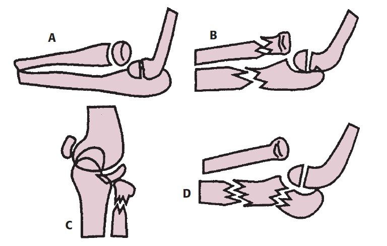
- I: anterior dislocation + mid-proximal 1/3 ulna fracture, apex anterior (hyperpronation or hyperextension; reduce with flexion)
- II: posterior dislocation, apex posterior (axial load with flexed elbow; reduce with extension)
- III: lateral dislocation + ulnar metaphyseal fracture (extension + varus stress; reduce with flexion)
- IV: fracture of both radius and ulna + anterior dislocation (type I plus radial shaft failure; reduce with flexion); children I, adults II
Q19What is the Monteggia variant (type 1 equivalent) classification?▸
- Type I: isolated radial head dislocation
- Type II: radial neck fracture + ulna fracture
- Type III: isolated radial neck fracture
- Type IV: elbow dislocation
Q20What is the acute management of a Monteggia fracture?▸
- Assess other injury, neurovascular status, open wound
- Reduce ulna fracture: flexion, traction, supination + direct pressure on radial head
- Radius reduces after ulna is reduced and length restored
- Immobilise in supination (tighten IOM) and flexion for type I; extension (<60) for type II
Q21What is the definitive management and what if the radial head does not reduce?▸
- Aim: concentric RCJ, anatomical ulnar fixation (length, angulation, rotation)
- Plate or K wire + cast (supination in I/III) for <10 years; ORIF if >10 years, comminuted or length unstable
- If radial head not reduced: confirm anatomical ulnar fixation
- Rule out annular ligament or radial nerve interposition; plastic deformity of ulna
Q22What nerve injuries and radial head instability complications occur?▸
- Most common in Bado type II and III; radial or median nerve; PIN (10%), AIN
- Observe 3 months; if no recovery -> explore
- <6 weeks + non-anatomical ulna reduction -> revise fixation + radial head OR
- >6 weeks -> osteotomy
Q23What are the sequelae and management of a late-presenting Monteggia fracture?▸
- Sequelae: cubital valgus, valgus/PLRI instability, ulnar nerve and PIN palsy, heterotopic ossification, OA
- Indications: All <12yo; normal concave radial head + convex capitellum
- Ulna osteotomy + open reduction of the radial head +/- annular ligament reconstruction
- Kocher approach for radial head (protect PIN); check stability; Bell-Tawse annular ligament reconstruction (lateral triceps fascia strip); cast 6 weeks
- Complications: residual radial head dislocation, ulnar nerve palsy, heterotopic ossification, radioulnar synostosis
Q24What do the Hubbard (JBJS 2018) review and Nakamura (JBJS 2009) show for chronic missed Monteggia fractures?▸
- Hubbard 2018 timing: duration of dislocation <3yr, patient age <12yo
- Osteotomy: crescentic osteotomy without ulnar lengthening
- Radial head reduction: into native annular ligament; if failed piecrust; if still failed incise and repair; reinforce with triceps fascia
- Nakamura 2009: good long-term outcomes after acute reconstruction for chronic missed Monteggia in under 12s within 2 years of initial injury
Q25What stabilises the radial head and why treat early?▸
- Static ligamentous: IOL (proximal oblique cord), annular ligament, quadrate ligament
- Static bony: RCJ
- Dynamic: anconeus
- 0.4% of all fractures, peak incidence 4-10 years; delayed treatment >2 weeks much increases complications
Q26What is the Watson-Jones classification for medial epicondyle fractures?📷▸
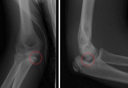
- I: <5mm displacement with no rotation
- II: >5mm displacement with rotation
- III: incarcerated fragment without dislocation
- IV: incarcerated fragment with dislocation
Q27What is the typical age, mechanism and pathoanatomy of medial epicondyle fractures?▸
- Typical age 11-14; fall on outstretched hand
- Associated with elbow dislocation (up to 50%) - fragment may be incarcerated in the joint
- Overuse by overhead throwing athletes (little league elbow)
- Last ossification centre to fuse; posteromedial aspect (ask for oblique view); avulsed via tension from FPO or MCL
Q28What are the indications for surgery and the reduction manoeuvre?▸
- Absolute indication: incarcerated fragment
- Relative indications: >5mm displacement, valgus instability
- Roberts reduction manoeuvre: supination + valgus + wrist extension
- Fix with lag screw, K wire or absorbable pin
Q29What history and examination are important in medial epicondyle fracture?▸
- History: mechanism of injury, chronicity, any dislocation episode
- Examination: check ulnar nerve
- Examination: check wound