37 slides
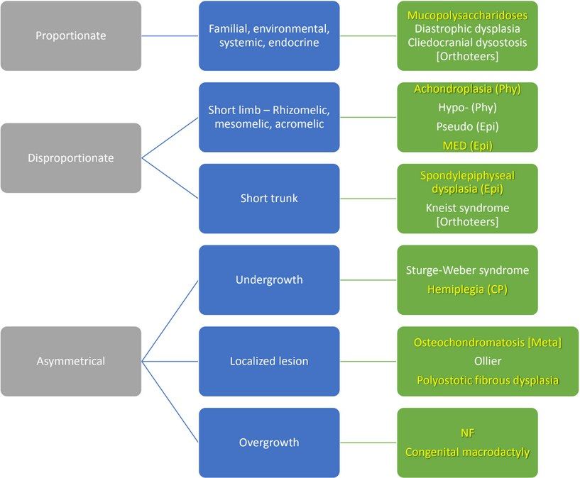
▸ Slide 599 · Short statureShort Stature · 2 questions expand
Question list
Q1-Q22 questions — tap to reveal all answerslist
- How is short stature defined?
- What orthopaedic conditions are associated with short stature?
Answers · Q & A
Q1.How is short stature defined?
- Not covered in the speaker notes - slide image only
Q2.What orthopaedic conditions are associated with short stature?
- Not covered in the speaker notes
▸ Slide 600 · Xray: Tri-foil shaped pelvis, protrusio, lateral bowing of R femur shaft.Short Stature · 16 questions 1 check expand

Question list
Q1-Q1616 questions — tap to reveal all answerslist
- Describe the X-ray features of osteogenesis imperfecta.
- What is the diagnosis and what are the differential diagnoses?
- What clinical features would you look for in suspected osteogenesis imperfecta?
- How is osteogenesis imperfecta diagnosed and what is the treatment goal?
- What medical treatment is used in osteogenesis imperfecta?
- What are the Sillence types of osteogenesis imperfecta?
- What fracture-related findings may be seen in osteogenesis imperfecta?
- What is the genetic basis of osteogenesis imperfecta?
- What is the Rubin classification of osteogenesis imperfecta?
- What are the Sillence types V-VII?
- How do you monitor the effect of bisphosphonates in osteogenesis imperfecta?
- What are the problems with bracing fractures in osteogenesis imperfecta?
- How is limb deformity corrected in osteogenesis imperfecta and why does it progress if untreated?
- When is scoliosis surgery indicated in osteogenesis imperfecta and what are the growing rod generations?
- What are the anaesthetic and orthopaedic difficulties in osteogenesis imperfecta surgery?
- What are the pros and cons of Rush pin vs telescoping rod in osteogenesis imperfecta?
Answers · Q & A
Q1.Describe the X-ray features of osteogenesis imperfecta.
- Tri-foil shaped pelvis, protrusio, lateral bowing of femur shaft
- Erlenmeyer flask femur, gracile or overtubulated bones, anterior bowing of tibia
- Codfish vertebrae or kyphoscoliosis in the spine
- Skull XR: Wormian bones + basilar invagination
Q2.What is the diagnosis and what are the differential diagnoses?
- Dx: OI
- Ddx: rickets/osteomalacia, idiopathic juvenile osteoporosis
- Ddx: non-accidental injury, fibrous dysplasia
Q3.What clinical features would you look for in suspected osteogenesis imperfecta?
- Short stature (proportionate), thin skin
- Eyes: Blue sclera; Ears: Impaired hearing (in type 1); Mouth: Defective detinogenesis. High pitch voice
- Triangular shaped head with broad forehead
- Lateral bowing of femur, anterior bowing of tibia (saber shin), genu varum, acetabular protrusion
- Kyphoscoliosis, congenital radial head dislocation, basilar invagination (myelopathy), generalised ligamentous laxity
Q4.How is osteogenesis imperfecta diagnosed and what is the treatment goal?
- Diagnosis based on radiological and clinical features +/- family history
- Skin biopsy is gold standard (culturing dermal fibroblasts)
- Goals: prevent fracture, deformity correction, optimise function
- Refer to geneticist to establish diagnosis
Q5.What medical treatment is used in osteogenesis imperfecta?
- Bisphosphonates: decrease fracture, 90% increase in cortical diameter, 50% increase in cancellous bone, decrease pain, increase ambulation
- Growth hormone
- Calcium and vitamin D
- Gene therapy
Q6.What are the Sillence types of osteogenesis imperfecta?
- Type I: mildest, AD, most common, blue sclera, abnormal dentinogenesis
- Type II: AD/AR, die, blue sclera
- Type III: AR, white sclera, fracture at birth, limb deformity, short stature, worst survivable type
- Type IV: AD, moderate severity, hearing normal, spine deformity, abnormal dentinogenesis
Q7.What fracture-related findings may be seen in osteogenesis imperfecta?
- Hypertrophic tumoral callus
- Malunion
- Pseudoarthrosis
Q8.What is the genetic basis of osteogenesis imperfecta?
- Hereditary disease with abnormal Type 1 collagen (quantitative/qualitative); 90% have an identifiable genetic mutation
- COL1A1 and COL1A2: substitution of glycine by a bulky amino acid (e.g. cystine) in the procollagen structure, unable to crosslink
- Reduced collagen secretion and abnormal collagen production -> insufficient osteoid production; unable to remodel normally, but initial fracture healing is normal
- Affects bone (woven), tendon, ligament, dentin, sclera
Q9.What is the Rubin classification of osteogenesis imperfecta?
- Diaphyseal hypoplasia
Q10.What are the Sillence types V-VII?
- No COL1 mutation but similar phenotype
Q11.How do you monitor the effect of bisphosphonates in osteogenesis imperfecta?
- Clinical: decreased pain
- XR: vertebral remodelling / Zebra lines
- DEXA: any increase in total body and lumbar spine bone density
- Serum: ALP, C-telopeptide, N-telopeptide, TRAP5b (C telopeptide, N telopeptide , TRAP5b, produced during bone resorption); osteocalcin (synthesised by osteoblasts to control osteoclasts)
- Urine: deoxypyridinoline (a collagen breakdown product)
Q12.What are the problems with bracing fractures in osteogenesis imperfecta?
- Normal healing rate but no remodelling
- Increased risk of further fracture due to immobilisation and/or the stress riser effect at the edges of the cast
- If recurrent fracture: CR + IMN +/- multiple osteotomies to correct deformity at the same time
Q13.How is limb deformity corrected in osteogenesis imperfecta and why does it progress if untreated?
- Sofield-Miller procedure with telescoping IM system such as Bailey-Dubow rod (if medullary canal wide enough) or Rush pin
- Deformity will progress if left untreated due to the HV law (Hueter-Volkmann) leading to selective inhibition of physeal growth
Q14.When is scoliosis surgery indicated in osteogenesis imperfecta and what are the growing rod generations?
- Scoliosis surgery if Cobb >45 (no brace due to rib fragility)
- 1st gen: Sheffield; 2nd gen: Bailey-Dubow; 3rd gen: Fassier-Duval
- 1st and 2nd gen need arthrotomy; 3rd gen allows one proximal insertion with a distal screw
Q15.What are the anaesthetic and orthopaedic difficulties in osteogenesis imperfecta surgery?
- Anaesthesia: malignant hyperthermia, basilar invagination, mitral valve regurgitation
- Ortho: bleeding, abnormal bone quality, minimal autograft, ligamentous laxity
Q16.What are the pros and cons of Rush pin vs telescoping rod in osteogenesis imperfecta?
- Telescoping rod: only a narrow rod can be inserted due to the narrow canal -> increased risk of rod fracture or trapped rod; need to change every 4 years
- Rush pin: easy exchange, reduces implant breakage; change over 2.5 years
Fact check
Skin biopsy is the gold standard for diagnosing OI (culturing dermal fibroblasts) — outdated — OI is now diagnosed by molecular genetic testing of COL1A1/COL1A2; cultured dermal fibroblast collagen analysis has fallen out of favour — source
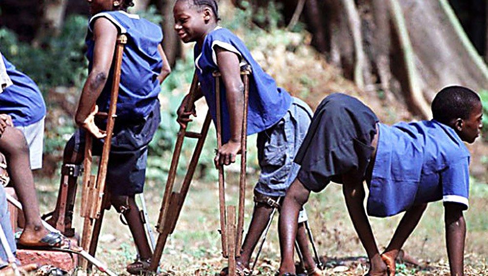
▸ Slide 601 · Poliovirus non-enveloped single-stranded RNA virus, a picornavirus and enterovShort Stature · 8 questions expand
Question list
Q1-Q88 questions — tap to reveal all answerslist
- What is poliomyelitis and how is poliovirus transmitted?
- Describe the four phases of poliomyelitis.
- What is post-polio syndrome and how is it managed?
- What are the two scenarios of late polio presentation?
- How is chronic polio managed by region?
- What is the most common weakness in polio and what adaptive mechanisms occur?
- What is the vaccination history of polio?
- How does the IT band contribute to deformity in polio?
Answers · Q & A
Q1.What is poliomyelitis and how is poliovirus transmitted?
- Non-progressive asymmetrical motor paralysis with sensory sparing
- Poliovirus: non-enveloped single-stranded RNA virus, a picornavirus and enterovirus
- Destroys anterior horn cells in spinal cord and brainstem motor nuclei
- Faecal-oral route; major type (meningitis, encephalitis) and minor type (flu-like)
Q2.Describe the four phases of poliomyelitis.
- Acute: prodromal flu-like, painful muscles, joints flexed
- Paralysis: after 2-3 days; limbs weak, swallowing/breathing affected; ends 7-10 days
- Recovery: within 6 months
- Residual: LMN flaccid residual paralysis with intact sensation, then deformity and growth disturbance
Q3.What is post-polio syndrome and how is it managed?
- Aging phenomenon: remaining neurons degenerate after working extra hard to compensate
- NOT reactivation; affects up to 50%
- Exercise at sub-exhaustion levels to tone affected muscles + periods of rest + light weighted caliper
- Pacing: maintain but do not overuse
Q4.What are the two scenarios of late polio presentation?
- New symptoms after a period of convalescence = post-polio syndrome
- Chronic problems: weakness/flail joint, deformity (posture, muscle imbalance, gravity)
- OA, LLD, vascular dysfunction
- Assessment for post-polio syndrome = rule out other medical, ortho and neuro causes
Q5.How is chronic polio managed by region?
- Look at the following and treat accordingly
- Hip: FABER contracture (Ober procedure, Campbell procedure), Trendelenbeg gait (Trendelenburg gait) (EO transfer, iliopsoas to GT), dislocation, glut max weakness (glut max lurching gait)
- Knee: quads weakness (hand knee gait) (KAFO), FFC (serial casting, soft tissue release, extension osteotomy), recurvatum (triple tenodesis), flail knee (fusion, KAFO)
- Foot and ankle: foot drop and circumduction gait, cavovarus, equinovarus
- LLD (short limb gait); spine/pelvis - determine if pelvic obliquity is infra, supra or combined
Q6.What is the most common weakness in polio and what adaptive mechanisms occur?
- Most common weakness is at the quads
- Weak quads cause knee flexion with CG behind the knee -> flexion moment
- Compensation: hip flexion brings CG forward, ankle plantarflexion (plantarflexion-knee extension couple)
- Alternatively knee recurvatum so ITB acts as extensor, counterbalanced by glut max and ankle dorsiflexors
- Weakness order LL: TA, quads, glut max, gastroc; UL: deltoid, biceps, opponens
Q7.What is the vaccination history of polio?
- 1958: first general use of Sabin live attenuated vaccine by mouth (OPV)
- Salk = inactivated polio vaccine (IPV)
Q8.How does the IT band contribute to deformity in polio?
- IT band is the culprit for many presentations
- Increased lumbar lordosis, pelvic obliquity, FABER hip
- Knee FFC + valgus, LLD, tibial ER
- Secondary foot and ankle problems
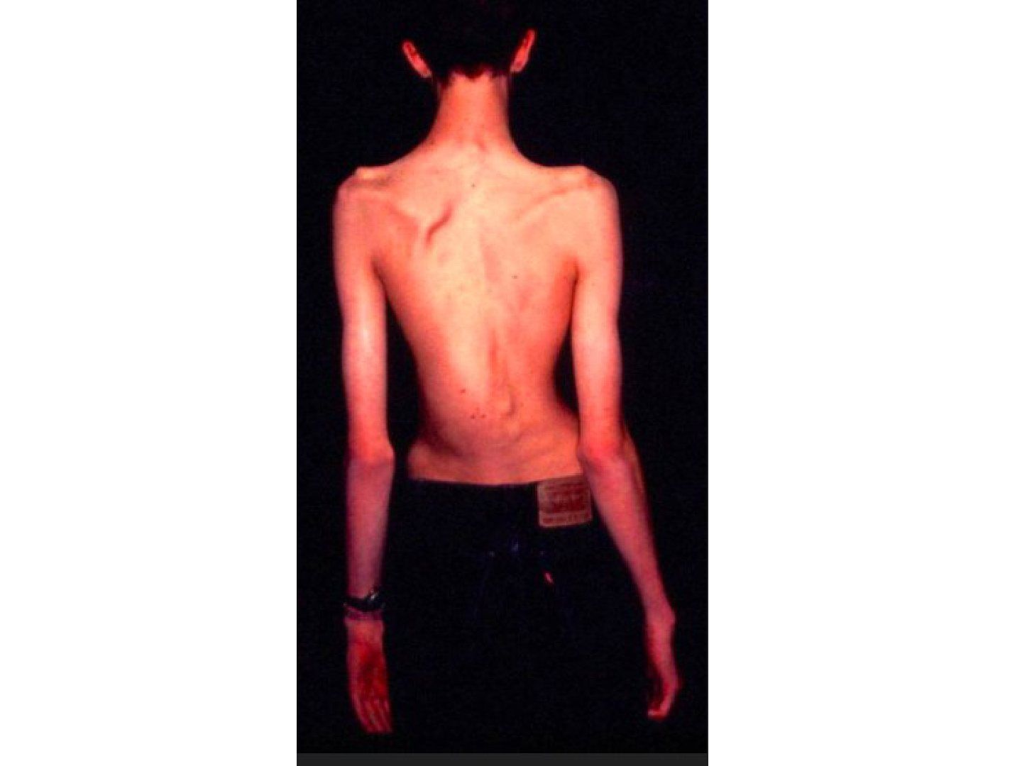
▸ Slide 602 · marfanShort Stature · 8 questions expand
Slide render
Question list
Q1-Q88 questions — tap to reveal all answerslist
- What are the physical examination features of Marfan syndrome?
- What is the genetic basis of Marfan syndrome?
- What are the Ghent nosology criteria for Marfan syndrome when family history is negative?
- What are the Ghent nosology criteria when family history is positive for Marfan syndrome?
- What are the systemic features of Marfan syndrome?
- What imaging findings are expected in Marfan syndrome?
- What is the general management of Marfan syndrome?
- What are the pre-, intra- and post-operative considerations for scoliosis surgery in Marfan syndrome?
Answers · Q & A
Q1.What are the physical examination features of Marfan syndrome?
- Tall stature, long narrow limbs, scoliosis
- Dolichostenomelia: arm span > height (>1.05)
- Arachnodactyly: Steinberg sign (thumb tip extends beyond small finger when clasped in palm) and Walker sign (thumb/index distal phalanges overlap around opposite wrist)
- Ligamentous hyperlaxity, pes planus
Q2.What is the genetic basis of Marfan syndrome?
- Connective tissue disease, Chr 15 (chromosome 15)
- AD, fibrillin 1 (FBN1) - ECM glycoprotein for fibrinogenesis; abnormal mechanical and elastic properties of connective tissue
- 30% sporadic mutation
- Rubin classification: physeal hyperplasia
Q3.What are the Ghent nosology criteria for Marfan syndrome when family history is negative?
- Aortic root dilatation AND ectopia lentis
- Aortic root dilatation AND FBN1 gene
- Aortic root dilatation AND systemic score >=7 points
- Ectopia lentis AND FBN1 gene associated with aortic dilatation
Q4.What are the Ghent nosology criteria when family history is positive for Marfan syndrome?
- Ectopia lentis
- Aortic root dilatation
- Systemic score >=7 points
- Family history (FHx+) of Marfan syndrome
Q5.What are the systemic features of Marfan syndrome?
- Ortho: dolichostenomelia, arachnodactyly, acetabular protrusio, ligamentous laxity (scoliosis, cervical instability, recurrent patella dislocation), dural ectasia
- Face: down-slanting palpebral fissures, malar hypoplasia, retrognathia, enophthalmos, dolichocephaly
- Ocular: lens dislocation; chest: carinatum, excavatum, pneumothorax
- Cardiac: valvular problem, aortic dissection; skin striae
Q6.What imaging findings are expected in Marfan syndrome?
- XR: acetabular protrusion, scoliosis
- MRI: dural ectasia (>60%)
Q7.What is the general management of Marfan syndrome?
- Refer to paediatrician - need monitoring of the cardiac condition
- Scoliosis treatment generally has a worse response than AIS
- Mild: bracing
- Rapidly progressing/large curve in an adult: surgical management with pre-, intra- and post-operative considerations
Q8.What are the pre-, intra- and post-operative considerations for scoliosis surgery in Marfan syndrome?
- Preop: ectopia lentis -> AACG; cardiac -> MVP and aortic root dilatation; recurrent PTX; MRI to rule out dural ectasia
- Intraop: construct needs to span longer (prevent add-on phenomenon); narrow pedicles; higher complication (durotomy)
- Postop: add-on curves; fixation failure (thin laminae, thin pedicles, osteopenia); higher risk of infection
- Postop: pseudoarthrosis
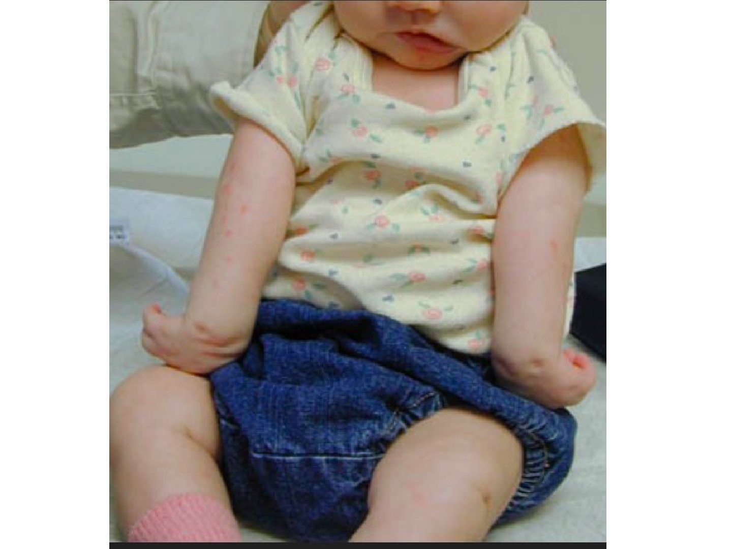
▸ Slide 603 · Describe clinical photo:Short Stature · 6 questions expand
Slide render
Question list
Q1-Q66 questions — tap to reveal all answerslist
- Describe the clinical photo of obstetric brachial plexus injury.
- What risk factors would you identify for obstetric brachial plexus injury in this infant?
- What is the Narakas classification and how is progress monitored?
- What are the favourable and unfavourable prognostic factors in obstetric brachial plexus injury?
- What is the treatment of obstetric brachial plexus injury?
- What is the pathoanatomy of Erb's and Klumpke's palsy?
Answers · Q & A
Q1.Describe the clinical photo of obstetric brachial plexus injury.
- Upper limb abnormal posture: shoulder adducted, internally rotated
- Elbow extended, wrist flexed, forearm pronated
- Fingers flexed, thumb (not seen here) adducted
- Typical of Erb's palsy
Q2.What risk factors would you identify for obstetric brachial plexus injury in this infant?
- High birth weight, breech presentation
- Shoulder dystocia, forceps delivery
Q3.What is the Narakas classification and how is progress monitored?
- Narakas: C5,6 / C5,6,7 / C5-T1 / C5-T1 + Horner's
- Mallet score to monitor progress: 1 active abduction, 2 external rotation, 3 hand to head, 4 hand to back, 5 hand to mouth
- MRI to look for meningocele
Q4.What are the favourable and unfavourable prognostic factors in obstetric brachial plexus injury?
- In general 90% spontaneous resolve
- Favourable: Erb's palsy; +ve twitching (M1) over bicep and deltoid at 2mth old
- Unfavourable: M0 at 3mth old; preganglionic injuries (Horner, dorsoscapular and phrenic nerve involvement)
- Unfavourable: lower nerve root involvement (C5-7, Klumpke)
Q5.What is the treatment of obstetric brachial plexus injury?
- Daily stretching to maintain good ROM
- If no antigravity biceps function return by 2 months (max 3 mth) --> OT
- Primary reconstruction: root avulsion - nerve transfer; lower lesion - nerve repair or nerve graft
- Secondary reconstruction: posterior dislocation (capsulorrhaphy or humeral derotation osteotomy); IR (subscapularis release, LD and teres major transfer to GT, or derotation osteotomy)
Q6.What is the pathoanatomy of Erb's and Klumpke's palsy?
- Erb's: upper trunk C5,6 injury from excessive abduction of head away from shoulder -> traction on plexus
- Associated with glenoid retroversion and flattened, posteriorly subluxed humeral head (persistent shoulder IR), elbow contracture
- C5 deficiency: axillary (deltoid, teres minor), suprascapular (supraspinatus, infraspinatus), musculocutaneous (biceps); C6: radial (brachioradialis, supinator); sensory loss lateral arm/forearm
- Klumpke: lower trunk C8,T1; intrinsic minus, weak wrist and finger flexion, sensory loss medial hand/forearm
▸ Slide 604 · TraumaShort Stature · 4 questions expand
Slide render
Question list
Q1-Q44 questions — tap to reveal all answerslist
- What factors affect remodelling of a paediatric fracture?
- What structural differences make a paediatric fracture different from an adult fracture?
- Why is an arthrogram needed to assess a paediatric articular fracture?
- What are the healing differences between paediatric and adult fractures?
Answers · Q & A
Q1.What factors affect remodelling of a paediatric fracture?
- Age, with >=2 years of remaining growth
- Amount of deformity
- Distance from the physis
- Direction of deformity
Q2.What structural differences make a paediatric fracture different from an adult fracture?
- Thick cartilage cap (need arthrogram to visualise articular fracture)
- Thick periosteum
- More collagen, more ductile
- More cancellous bone, less comminution
- Bone is weaker than ligament/tendon
Q3.Why is an arthrogram needed to assess a paediatric articular fracture?
- The thick cartilage cap makes the articular fracture difficult to visualise
- Arthrogram is needed to visualise the articular fracture
Q4.What are the healing differences between paediatric and adult fractures?
- Paediatric fractures have remodelling power
- They carry a risk of growth disturbance
▸ Slide 605 · 2yo refuse to move one armShort Stature · 5 questions expand

Question list
Q1-Q55 questions — tap to reveal all answerslist
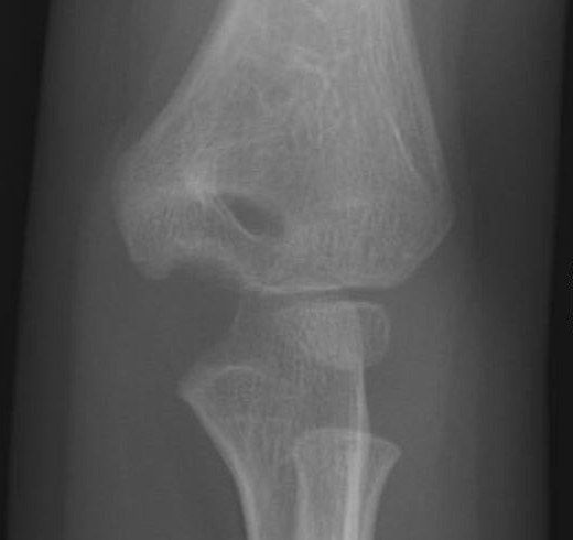
- Describe the X-ray findings and likely diagnosis in this 2-year-old refusing to move the arm.
- How do you differentiate a transphyseal distal humerus fracture from other injuries?
- What is the DeLee classification?
- What is the management of a transphyseal distal humerus fracture?
- What associated features and anatomy are important in transphyseal distal humerus fracture?
Answers · Q & A
Q1.Describe the X-ray findings and likely diagnosis in this 2-year-old refusing to move the arm.
- Skeletally immature patient, bilateral elbow X-ray
- Proximal ulna and radius relationship maintained, but humerus-ulna/radius relationship disrupted with posteromedial displacement
- Likely transphyseal fracture of distal humerus (physis is biomechanically the weakest location)
Q2.How do you differentiate a transphyseal distal humerus fracture from other injuries?
- Radiocapitellar line normal (proximal radius aligns with capitellum); proximal radius and ulna align with each other
- Elbow dislocation: posteromedial displacement favours transphyseal injury
- Lateral condyle fracture: assess lateral condyle-radial head relationship
- Low SCH fracture: transphyseal has wider bone contact and less displacement
Q3.What is the DeLee classification?
- Type A: infant to 7 months; no lateral condyle ossification centre (SH 1)
- Type B: 7 months to 3 years; lateral condyle ossified (SH 1,2)
- Type C: 3-7 years; large metaphyseal fragment exiting laterally
- This fracture almost only occurs <7 years
Q4.What is the management of a transphyseal distal humerus fracture?
- Depends on timing of injury, displacement and DeLee type
- Acute undisplaced: CR + cast in pronation x 3/52
- Acute displaced: CR +/- OR + KWF + cast in pronation x 3/52 (+/- arthrogram)
- Late (>1 week) + displaced: DO NOT manipulate
Q5.What associated features and anatomy are important in transphyseal distal humerus fracture?
- Hyperextension injury (birth injury/abuse/trauma), usually <2 years
- Highly suspicious of NAI; further MRI
- Medial epicondyle involved at young age; physis moves distally with growth, V-shaped cleft
- Large fragment, usually less rotation and tilting; blood supply good but medial crista of trochlea may be disrupted -> cubital varus
▸ Slide 606 · Xray of skeletally immature patient of his kneesShort Stature · 5 questions expand
Slide render
Question list
Q1-Q55 questions — tap to reveal all answerslist
- What do the knee X-rays show and what do they suggest?
- What are the red flag signs of non-accidental injury?
- List the high and moderate specificity fractures for non-accidental injury.
- What history, examination and investigations are needed in suspected non-accidental injury?
- Why must non-accidental injury be diagnosed?
Answers · Q & A
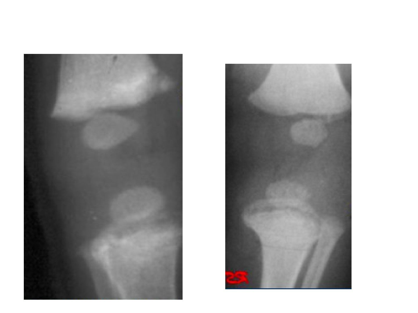
Q1.What do the knee X-rays show and what do they suggest?
- Left X-ray: corner fracture of distal femur
- Right X-ray: bucket handle fracture of proximal tibia
- Bucket handle = corner fracture viewed from a different plane
- In a skeletally immature patient these are very suggestive of non-accidental injury
Q2.What are the red flag signs of non-accidental injury?
- Multiple bruises (most common symptom is skin lesion); multiple fractures in various stages of healing
- Long bone fractures in an infant not yet walking
- Metaphyseal fractures: corner (primary spongiosa), bucket handle; transphyseal separation of the distal humerus
- Posterior rib fractures, scapular fracture, sternal fracture; ruptured frenulum
Q3.List the high and moderate specificity fractures for non-accidental injury.
- High specificity: metaphyseal fractures/bucket handle lesion, scapular fracture, posterior rib fracture, spinous process fracture, sternal fracture
- Moderate specificity: fracture of different ages, multiple fractures especially bilateral, epiphyseal separation
- Moderate specificity: vertebral fracture or subluxation, complex skull fracture, multiple digital fractures
Q4.What history, examination and investigations are needed in suspected non-accidental injury?
- Hx: inconsistent history, fails to explain injury, delayed presentation, multiple fractures
- P/E: systemic - growth, failure to thrive, bruises, burn marks; local - fractures
- Ix: skeletal survey (=8 months of background radiation) +/- bone scan in <5 years
- ADMIT + multidisciplinary approach + child protection service + social worker (remove abuser from environment); mandatory to report
Q5.Why must non-accidental injury be diagnosed?
- Child abuse can be Neglect/ physical/ sexual/ psychological
- If undiagnosed, up to 50% repeated, 10% death
- Ddx: accident, OI, metabolic bone disease
▸ Slide 607 · posterolateral approach or direct posterior approachShort Stature · 2 questions expand
Slide render

Question list
Q1-Q22 questions — tap to reveal all answerslist
- Which approaches are used for elbow arthrogram in children?
- How is the arthrogram performed and what does it show?
Answers · Q & A
Q1.Which approaches are used for elbow arthrogram in children?
- Posterolateral or direct posterior approach
- Direct posterior into olecranon fossa recommended in young children
- Prevents scuffing of articular cartilage seen with the posterolateral portal
Q2.How is the arthrogram performed and what does it show?
- Inject equal parts saline:contrast
- Bring elbow through range of motion
- If pinning needed, aids visualisation of pin starting points on capitellum
- Aids assessment of reduction by seeing anterior humeral line intersecting capitellum
▸ Slide 608 · Describe Xray:Short Stature · 13 questions expand
Slide render

Question list
Q1-Q1313 questions — tap to reveal all answerslist
- How do you describe and classify this supracondylar fracture on X-ray?
- What is the initial assessment of a supracondylar fracture?
- What is the theatre setup and aim of surgery for a supracondylar fracture?
- Describe the closed reduction manoeuvre for supracondylar fracture.
- How is the reduction assessed?
- Describe the pinning technique and post-operative monitoring.
- What are the indications for emergency open reduction after pinning?
- How are nerve injuries managed after a supracondylar fracture?
- Describe the open reduction approach for a supracondylar fracture.
- What wound management is needed in a paediatric supracondylar fracture?
- How do you avoid ulnar nerve palsy when medial pinning, and what are the indications for a medial pin?
- What are the indications for exploration of the AIN?
- What is the evidence for managing a pink pulseless hand after pinning?
Answers · Q & A
Q1.How do you describe and classify this supracondylar fracture on X-ray?
- Age by CRMTOL (around 2-3 years)
- Pattern of fracture (extension, flexion, Gartland)
- Gartland 1 minimal deformity; 2 anterior cortex broken, posterior cortex intact; 3 posterior cortex not intact
- Direction of displacement; this is Gartland type III
Q2.What is the initial assessment of a supracondylar fracture?
- ATLS, ample history; r/o fracture in same limb (especially distal radius)
- Local: wound, pucker sign, neurological deficit, vascular status (radial pulse, CR), compartments
- Nerve deficit: overall most common is AIN; PL - median 75%, PM - radial 77%, flexion - ulnar
- If vascular: emergent CR under conscious sedation then definitive K wire fixation (pulseless pale = emergent OT; pulseless pink = urgent same day, Skaggs JBJS 2015)
- Most common scenario: pulseless, CR <2sec (perfusion of UL is well); I would inform the vascular surgeon on call to be on standby in case the hand perfusion becomes worse after reduction
Q3.What is the theatre setup and aim of surgery for a supracondylar fracture?
- Supine with arm board, image intensifier from the end of the bed parallel to the bed; setup and a good assistant are key
- Aim: restore alignment in all three planes
- Prevents late complications such as cubital varus deformity
Q4.Describe the closed reduction manoeuvre for supracondylar fracture.
- Traction in line with humerus in 20 degrees elbow flexion +/- milking manoeuvre +/- hyperextension to disengage
- Correct valgus/varus and rotation by rotating forearm: supination tenses BR in PL displacement, pronation closes gap in PM displacement
- Direct pressure on distal fragment to correct translational deformity
- Thumb over olecranon to reduce extension deformity while maintaining traction
Q5.How is the reduction assessed?
- Clinically if the hand can touch the shoulder
- XR: AP, lateral, IO and EO views; oblique views to look at the respective columns
- Anterior humeral line cutting middle 1/3 of capitellum
- Baumann's angle: angle between humeral long axis and proximal edge of lateral condyle epiphysis; normal 75-80, compare with normal side (<5 degrees difference)
Q6.Describe the pinning technique and post-operative monitoring.
- AAOS: 2 divergent 1.6mm lateral guide pins, maximum spread at fracture site, bicortical fixation, avoid cross at fracture site
- If unstable add 1 lateral or medial pin; 3 divergent lateral pins at least as stable as crossed pins (Larson JPO2006)
- Cut and bend K wires, cast; monitor perfusion, neurology and pain (compartment syndrome) for 24-48hrs (Robb JBJS 2009)
- If ulnar nerve palsy develops: likely neuropraxia, observe, most return by 3 weeks
Q7.What are the indications for emergency open reduction after pinning?
- Extend elbow and wait 15mins after pinning before deciding
- Still white; pink but pulseless + no anatomical reduction; pre-CR pulse present but post-CR pulse absent
- If hand remains perfused, cast in 40-60deg of flexion to avoid vascular compromise
- Pulseless hand postop: assess reduction and XR; if perfect and pink may observe; if not, release backslab, extend, contact vascular team for exploration +/- repair
Q8.How are nerve injuries managed after a supracondylar fracture?
- CR for all
- Post CR: anatomical reduction -> observe
- Post CR: suboptimal reduction -> explore
Q9.Describe the open reduction approach for a supracondylar fracture.
- Lazy S incision: proximal medial to biceps, distal between PT and BR
- Watch out for the lateral cutaneous nerve of the forearm (2cm lateral to biceps tendon)
- Release bicipital aponeurosis (lateral to medial, risk of AVN); full supination to protect the PIN
- Retract biceps and brachialis medially and brachioradialis laterally
Q10.What wound management is needed in a paediatric supracondylar fracture?
- Inside-out wound
- Gustilo 1 in paediatric cases: debridement/antibiotics not necessary
Q11.How do you avoid ulnar nerve palsy when medial pinning, and what are the indications for a medial pin?
- Avoid: extend elbow, direct visualisation, oscillating mode, protect with drill sleeve
- Indications (Skaggs JBJS 2001): unstable after 2 lateral pins (relative)
- Medial comminution
- Fracture site proximal medial to distal lateral
Q12.What are the indications for exploration of the AIN?
- Postoperative (iatrogenic)
- Fracture site gap
- Open fracture
Q13.What is the evidence for managing a pink pulseless hand after pinning?
- Pink pulseless hand can be managed with observation after pinning
- Carbonell, EPOSNA annual meeting 2017: no long-term sequelae at 20 years
▸ Slide 609 · AAOS guideline 2015Short Stature · 4 questions expand
Slide render

Question list
Q1-Q44 questions — tap to reveal all answerslist
- What are the AAOS 2015 recommendations of Moderate strength for paediatric supracondylar humerus fractures?
- What are the AAOS 2015 recommendations of Weak strength?
- Which AAOS 2015 recommendations were rated Inconclusive?
- Which AAOS 2015 recommendations are Consensus-based?
Answers · Q & A
Q1.What are the AAOS 2015 recommendations of Moderate strength for paediatric supracondylar humerus fractures?
- Nonsurgical immobilization for acute non-displaced fractures (e.g. Gartland Type I)
- Closed reduction with pin fixation for displaced fractures (Type II and III, and displaced flexion)
- Strength of Recommendation: Moderate
Q2.What are the AAOS 2015 recommendations of Weak strength?
- Use two or three laterally introduced pins
- Considerations of potential harm indicate that the physician might avoid the use of a medial pin
- Physician might perform open reduction for displaced fractures with varus or other malposition after closed reduction
- Strength of Recommendation: Weak
Q3.Which AAOS 2015 recommendations were rated Inconclusive?
- Using an open incision to introduce a medial pin
- Time threshold for reduction of displaced fractures without neurovascular injury
- Open exploration of the antecubital fossa in patients with absent wrist pulses but a perfused hand after reduction
- Optimal time for removal of pins and mobilization; routine supervised physical or occupational therapy; optimal time for unrestricted activity after injury
- Optimal timing/indications for electrodiagnostic studies or nerve exploration in nerve injuries; open reduction and stable fixation for adolescent supracondylar fractures
Q4.Which AAOS 2015 recommendations are Consensus-based?
- Emergent closed reduction for patients with decreased perfusion of the hand
- Open exploration of the antecubital fossa for patients who have absent wrist pulses and are underperfused after reduction and pinning
▸ Slide 610 · Complications of supracondylar fractureShort Stature · 5 questions expand
Slide render
Question list
Q1-Q55 questions — tap to reveal all answerslist
- What is the incidence and aetiology of cubital varus deformity?
- What are the sequelae of cubital varus deformity?
- What are the options for corrective osteotomy and when should it be performed?
- What are the complications of corrective osteotomy?
- What is the normal osteology of the humerus?
Answers · Q & A
Q1.What is the incidence and aetiology of cubital varus deformity?
- Incidence: 3% pinning vs 14% with casting
- Malreduction of supracondylar fracture
- Malunion of trochlear in lateral condyle fracture (type I fishtail); trochlear AVN (type II fishtail)
- Lateral overgrowth in lateral condyle fracture; neoplastic (exostosis); skeletal dysplasia (MED)
Q2.What are the sequelae of cubital varus deformity?
- Cosmesis most of the time
- Ulnar nerve palsy
- PL instability
- Deformity: varus, hyperextension, internal rotation
Q3.What are the options for corrective osteotomy and when should it be performed?
- Timing: wait until skeletal maturity
- Lateral closing wedge osteotomy: uniplanar step cut (more stable) or right-angled triangle (superior perpendicular to proximal shaft, inferior: planned angle, lateral spike as step) with lag screw
- French osteotomy: derotation, distal screw lateral anterior, proximal screw lateral posterior, tightened with figure-of-8 loop
- Medial opening wedge osteotomy; dome osteotomy
Q4.What are the complications of corrective osteotomy?
- Recurrence
- Nerve injury (displacement of the radial nerve into osteotomy callus)
- Stiffness, infection, scarring
Q5.What is the normal osteology of the humerus?
- 30 degrees anterior tilt
- 6 degrees valgus
- 5 degrees internal rotation
- Axis of rotation centred at trochlea and capitellum, passes through anteroinferior medial epicondyle
▸ Slide 611 · Describe Xray: Fracture radial neck with >30 deg angulationShort Stature · 5 questions expand

Question list
Q1-Q55 questions — tap to reveal all answerslist
- Describe the O'Brien classification of radial neck fractures.
- What associated injuries and X-ray views are relevant to radial neck fractures?
- How is a radial neck fracture treated according to angulation?
- What are the complications of radial neck fracture?
- What is normal radial neck angulation?
Answers · Q & A
Q1.Describe the O'Brien classification of radial neck fractures.
- I: <30 degrees angulation
- II: 30-60 degrees
- III: >60 degrees
- Median age 9-10 years; usually a valgus loading injury of the elbow
Q2.What associated injuries and X-ray views are relevant to radial neck fractures?
- Associated conditions: elbow dislocation, medial epicondyle fracture
- Greenspan radiocapitellar view: elbow 90 degrees flexion, thumb up, beam 45 degrees from table
Q3.How is a radial neck fracture treated according to angulation?
- <30deg: immoblization in situ (<30 degrees: immobilisation in situ)
- >30 degrees: attempt closed reduction
- Techniques: Patterson (traction, varus stress, thumb pressure), Israeli (supination + flexion, finger pressure, pronate to force radial head through), Chambers (Esmarch circumferential pressure)
- If still >30 degrees: percutaneous K wire reduction (joystick technique); also try Metazeau technique
Q4.What are the complications of radial neck fracture?
- Overgrowth (more common)
- Radial head AVN
- Radioulnar synostosis
- Myositis ossificans
- Open reduction: greater loss of motion, increased osteonecrosis and synostosis vs closed reduction
Q5.What is normal radial neck angulation?
- 0-15 degrees lateral
- 5 degrees posterior to 10 degrees anterior
▸ Slide 612 · Stem: Patient with fall injury + axial loading to UL, lateral side of elbow painShort Stature · 7 questions expand
Slide render
Question list
Q1-Q77 questions — tap to reveal all answerslist
- Describe the X-ray findings and suspected diagnosis.
- What is the mechanism of a lateral condyle fracture?
- What is the Milch classification?
- What are the Jakob and Song classifications?
- What is the definitive management of a lateral condyle fracture?
- Why are lateral condyle fractures prone to non-union and what are the late complications?
- How do you manage a delayed presentation with non-union and deformity?
Answers · Q & A
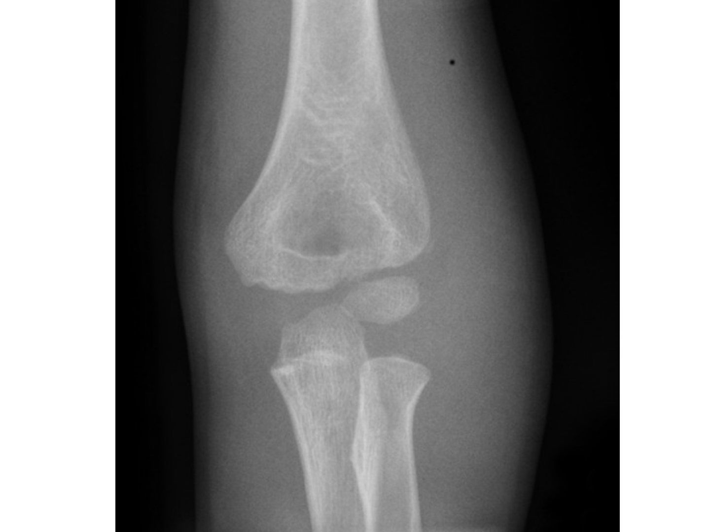
Q1.Describe the X-ray findings and suspected diagnosis.
- Flake of bone over lateral metaphyseal region, no gross malalignment
- Suspected lateral condyle fracture
- Minimally displaced on X-ray; need to see articular surface displacement before planning management
- Get internal oblique XR; urgent MRI to look for articular step; if MRI unavailable, arthrogram (inject from posterior to avoid injury to articular surface and major NV bundle)
Q2.What is the mechanism of a lateral condyle fracture?
- Push off: axial loading by the radial head
- Pull off: common extensor origin
Q3.What is the Milch classification?
- Type 1: fracture lateral to trochleocapitellar groove, SH type 4, more stable
- Type 2: fracture medial to trochleocapitellar groove, SH type 2 (does not cross lateral condyle epiphysis), less stable, worse prognosis
Q4.What are the Jakob and Song classifications?
- Jacob: Jakob 1: <2mm displacement (conservative if articular cartilage confirmed smooth)
- Jakob 2: 2-4mm (OR KWF); Jakob 3: severe displacement +/- rotated fragment (OR KWF)
- Song classification (1-5); 3 or above unstable, needs surgery
- Song: 1 limited metaphyseal fracture line, 2 lateral gap, 3 medial and lateral gap, 4 >2mm displacement no rotation, 5 rotation
Q5.What is the definitive management of a lateral condyle fracture?
- >2mm displacement in any of the 3 views -> OR + KWF
- CR: extension + supination to relax common extensor origin, then varus + anteromedial force
- OR: oblique lateral wound centred at lateral epicondyle (between BR and triceps); avoid posterior dissection to preserve blood supply
- Check anterior and lateral cortex smoothness; fix with 2 divergent pins (joystick reduction)
Q6.Why are lateral condyle fractures prone to non-union and what are the late complications?
- Poor vascular supply, intra-articular, large pull from extensor origin
- Late complications: nonunion, AVN
- Deformity (cubital valgus from lateral condyle AVN, varus from overgrowth or fishtail); PL subluxation of proximal radius and ulna
Q7.How do you manage a delayed presentation with non-union and deformity?
- Yes to OT due to future valgus, instability and tardy ulnar nerve palsy (Masada JBJS 1990)
- Aim: achieve union; debride + bone graft + screw horizontally (K wire gives no compression)
- Only debride anterior, leave posterior intact; avoid aggressive reduction (risk AVN and fishtail); Make sure to have preop imaging to rule out AVN
- Alternative: watchful waiting with later osteotomy; ORIF associated with stiffness and AVN (Jakob), but recent studies suggest surgery is a safe option
▸ Slide 613 · Post lateral condyle fracture deformityShort Stature · 5 questions expand
Question list
Q1-Q55 questions — tap to reveal all answerslist
- What are the main arteries supplying the distal humerus?
- Which structures of the distal humerus are supplied by end arteries, and why does this predispose to AVN?
- What is a fishtail deformity and what are its types?
- What causes cubital varus and cubital valgus after lateral condyle fracture?
- How is non-union defined and managed?
Answers · Q & A
Q1.What are the main arteries supplying the distal humerus?
- Radial recurrent artery (comes from the radial collateral artery)
- Interosseous recurrent artery (comes from the middle collateral artery)
Q2.Which structures of the distal humerus are supplied by end arteries, and why does this predispose to AVN?
- Lateral condyle epiphysis: entry posterolateral to the capsule origin and proximal to the articular cartilage
- Lateral condyle has an end-artery supply; the lateral crista of the trochlea is supplied from it
- Lateral part of the medial crista: crosses the posterior distal humerus metaphysis as an end artery (medial part has multiple vessels)
- Disruption causes lateral condyle AVN (valgus, fishtail) and AVN of the lateral part of the medial crista
Q3.What is a fishtail deformity and what are its types?
- Area between medial ossification centre and lateral condyle ossification centre resorbs/fails to develop
- Type 1 (more common): sharp angled wedge; persistent gap between lateral condyle physis and medial trochlear due to underdevelopment of the lateral crista
- Type 2: smooth edge; AVN of the lateral part of the medial crista of the trochlea
Q4.What causes cubital varus and cubital valgus after lateral condyle fracture?
- Cubital varus: overgrowth (50%)
- Cubital varus from fishtail: malunion of lateral part of medial crista (type 1) or medial crista AVN (type 2)
- Cubital valgus: nonunion leading to lateral condyle AVN
- Cubital valgus: physeal arrest
Q5.How is non-union defined and managed?
- Definition: 3 months
- Displaced >2mm + no K wire fixation -> lateral condyle AVN
- Before physeal closure: osteotomy + BG (displaced <1cm, large fragment)
- After physeal closure: freshen edges + bone graft + compression; severe valgus -> osteotomy; ulnar nerve palsy -> transposition
▸ Slide 614 · Acute and chronic px?Short Stature · 8 questions expand

Question list
Q1-Q88 questions — tap to reveal all answerslist
- What is the Bado classification of Monteggia fractures?
- What is the Monteggia variant (type 1 equivalent) classification?
- What is the acute management of a Monteggia fracture?
- What is the definitive management and what if the radial head does not reduce?
- What nerve injuries and radial head instability complications occur?
- What are the sequelae and management of a late-presenting Monteggia fracture?
- What do the Hubbard (JBJS 2018) review and Nakamura (JBJS 2009) show for chronic missed Monteggia fractures?
- What stabilises the radial head and why treat early?
Answers · Q & A
Q1.What is the Bado classification of Monteggia fractures?
- I: anterior dislocation + mid-proximal 1/3 ulna fracture, apex anterior (hyperpronation or hyperextension; reduce with flexion)
- II: posterior dislocation, apex posterior (axial load with flexed elbow; reduce with extension)
- III: lateral dislocation + ulnar metaphyseal fracture (extension + varus stress; reduce with flexion)
- IV: fracture of both radius and ulna + anterior dislocation (type I plus radial shaft failure; reduce with flexion); children I, adults II
Q2.What is the Monteggia variant (type 1 equivalent) classification?
- Type I: isolated radial head dislocation
- Type II: radial neck fracture + ulna fracture
- Type III: isolated radial neck fracture
- Type IV: elbow dislocation
Q3.What is the acute management of a Monteggia fracture?
- Assess other injury, neurovascular status, open wound
- Reduce ulna fracture: flexion, traction, supination + direct pressure on radial head
- Radius reduces after ulna is reduced and length restored
- Immobilise in supination (tighten IOM) and flexion for type I; extension (<60) for type II
Q4.What is the definitive management and what if the radial head does not reduce?
- Aim: concentric RCJ, anatomical ulnar fixation (length, angulation, rotation)
- Plate or K wire + cast (supination in I/III) for <10 years; ORIF if >10 years, comminuted or length unstable
- If radial head not reduced: confirm anatomical ulnar fixation
- Rule out annular ligament or radial nerve interposition; plastic deformity of ulna
Q5.What nerve injuries and radial head instability complications occur?
- Most common in Bado type II and III; radial or median nerve; PIN (10%), AIN
- Observe 3 months; if no recovery -> explore
- <6 weeks + non-anatomical ulna reduction -> revise fixation + radial head OR
- >6 weeks -> osteotomy
Q6.What are the sequelae and management of a late-presenting Monteggia fracture?
- Sequelae: cubital valgus, valgus/PLRI instability, ulnar nerve and PIN palsy, heterotopic ossification, OA
- Indications: All <12yo; normal concave radial head + convex capitellum
- Ulna osteotomy + open reduction of the radial head +/- annular ligament reconstruction
- Kocher approach for radial head (protect PIN); check stability; Bell-Tawse annular ligament reconstruction (lateral triceps fascia strip); cast 6 weeks
- Complications: residual radial head dislocation, ulnar nerve palsy, heterotopic ossification, radioulnar synostosis
Q7.What do the Hubbard (JBJS 2018) review and Nakamura (JBJS 2009) show for chronic missed Monteggia fractures?
- Hubbard 2018 timing: duration of dislocation <3yr, patient age <12yo
- Osteotomy: crescentic osteotomy without ulnar lengthening
- Radial head reduction: into native annular ligament; if failed piecrust; if still failed incise and repair; reinforce with triceps fascia
- Nakamura 2009: good long-term outcomes after acute reconstruction for chronic missed Monteggia in under 12s within 2 years of initial injury
Q8.What stabilises the radial head and why treat early?
- Static ligamentous: IOL (proximal oblique cord), annular ligament, quadrate ligament
- Static bony: RCJ
- Dynamic: anconeus
- 0.4% of all fractures, peak incidence 4-10 years; delayed treatment >2 weeks much increases complications
▸ Slide 615 · Fracture medial epicondyle with displacement of the fragment into the UHJShort Stature · 4 questions expand

Question list
Q1-Q44 questions — tap to reveal all answerslist
- What is the Watson-Jones classification for medial epicondyle fractures?
- What is the typical age, mechanism and pathoanatomy of medial epicondyle fractures?
- What are the indications for surgery and the reduction manoeuvre?
- What history and examination are important in medial epicondyle fracture?
Answers · Q & A
Q1.What is the Watson-Jones classification for medial epicondyle fractures?
- I: <5mm displacement with no rotation
- II: >5mm displacement with rotation
- III: incarcerated fragment without dislocation
- IV: incarcerated fragment with dislocation
Q2.What is the typical age, mechanism and pathoanatomy of medial epicondyle fractures?
- Typical age 11-14; fall on outstretched hand
- Associated with elbow dislocation (up to 50%) - fragment may be incarcerated in the joint
- Overuse by overhead throwing athletes (little league elbow)
- Last ossification centre to fuse; posteromedial aspect (ask for oblique view); avulsed via tension from FPO or MCL
Q3.What are the indications for surgery and the reduction manoeuvre?
- Absolute indication: incarcerated fragment
- Relative indications: >5mm displacement, valgus instability
- Roberts reduction manoeuvre: supination + valgus + wrist extension
- Fix with lag screw, K wire or absorbable pin
Q4.What history and examination are important in medial epicondyle fracture?
- History: mechanism of injury, chronicity, any dislocation episode
- Examination: check ulnar nerve
- Examination: check wound
▸ Slide 616 · Beware if supracondylar fracture in >10yoShort Stature · 3 questions expand
Slide render

Question list
Q1-Q33 questions — tap to reveal all answerslist
- Why beware of a supracondylar fracture in a child over 10 years old?
- What is the management of Type I and Type II fractures?
- What is the management of Type III fractures and the complications?
Answers · Q & A
Q1.Why beware of a supracondylar fracture in a child over 10 years old?
- Rotatory deformity due to muscle pull
- Relatively intact articular surface compared with osseous displacement
- Large cartilaginous component of the articular surface confers more elasticity
- X-ray: difficult to distinguish from supracondylar fracture
Q2.What is the management of Type I and Type II fractures?
- Type I undisplaced: K wires (1x intercondylar, 2x lateral divergent)
- Type II displaced (>2mm) with intact metaphysis: OR (paratricipital, TRAP, Bryan-Morrey)
- Articular congruity restored by screw fixation
- Supracondylar fixation by K wire
Q3.What is the management of Type III fractures and the complications?
- Type III: displaced + metaphyseal comminution -> OR
- Articular congruity restored with screw
- Supracondylar stabilisation with plate + BG
- Complications: stiffness, trochlear AVN
▸ Slide 617 · Paed forearm fractureShort Stature · 8 questions expand
Slide render

Question list
Q1-Q88 questions — tap to reveal all answerslist
- Describe the X-ray and initial assessment of a paediatric forearm fracture.
- What are the aims of treatment in a paediatric forearm fracture?
- What factors determine acceptable alignment in paediatric forearm fractures?
- What are the acceptable alignment limits (Rockwood 2010)?
- Describe the closed reduction and casting technique.
- What are the principles of K-wire fixation?
- What are the deforming forces by fracture level and how is rotation assessed?
- What are the complications of paediatric forearm fractures?
Answers · Q & A
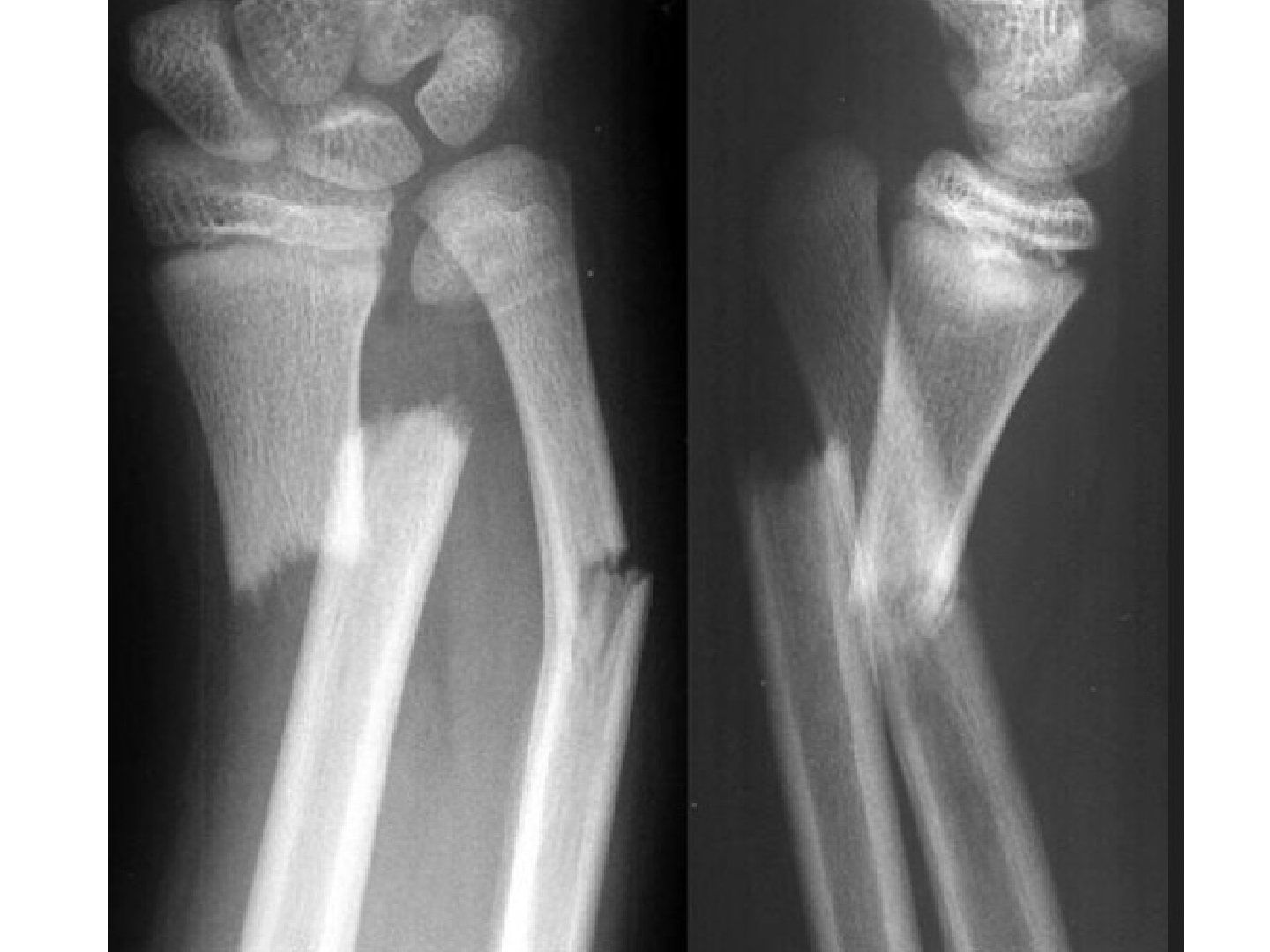
Q1.Describe the X-ray and initial assessment of a paediatric forearm fracture.
- Fracture of both radius and ulna at the shaft with shortening (bayonet) in a skeletally immature patient
- Hx: age, mechanism of injury
- r/o acute complications: compartment syndrome / neurovascular injury
Q2.What are the aims of treatment in a paediatric forearm fracture?
- Restore full rotational range
- Promote union
- Avoid complications: malunion, synostosis, refracture
Q3.What factors determine acceptable alignment in paediatric forearm fractures?
- Site of deformity (affects cast position due to different deforming force)
- Degree of deformity
- Age of patient (affects acceptable deformity)
- NO rotational malalignment accepted
Q4.What are the acceptable alignment limits (Rockwood 2010)?
- <10 years: angulation <15 degrees, shortening and bayonetting <1cm
- >10 years: anatomical reduction (<10 degrees sagittal for 11-13 years; 0-5 for >13 years)
- 0 degrees coronal for >=11 years
Q5.Describe the closed reduction and casting technique.
- Point palm in direction of apex of deformity; traction + angulation + rotation in the direction of angulation
- Bayonet deformity requires more traction to reduce; single bone may need more traction
- Mould with 3-point fixation to correct angular deformity + AP compression (ligamentotaxis by interosseous membrane)
- Sagittal:coronal cast width ratio <0.8
Q6.What are the principles of K-wire fixation?
- K wire should occupy at least 2/3 of the diameter of the medullary isthmus
- Prebend radial wire to 3x the diameter of the medullary canal, apex of bend at fracture level
- Radial styloid entry between EC I and II; ulnar entry at olecranon; confirm correct rotation
- Indication for plating: close to skeletal maturity, meta-diaphyseal junction (width mismatch)
Q7.What are the deforming forces by fracture level and how is rotation assessed?
- Proximal 1/3: proximal fragment flexed (biceps) and supinated (supinator), distal pronated (PT + PQ) -> align in supination
- Middle 1/3: proximal balanced by supinator and PT, distal pronated by PQ -> align in neutral
- Distal 1/3: proximal pronated by PT + PQ -> align in pronation
- AP: radial styloid/ radial tuberosity 180deg; Lat: coronoid process/ ulnar styloid 180deg
Q8.What are the complications of paediatric forearm fractures?
- Immediate: compartment syndrome 1-8%
- Malunion (10 degrees angulation in 1 bone = 20-30 degrees rotational block)
- Refracture (5% in 6 months)
- Synostosis
▸ Slide 618 · Galeazzi fractureShort Stature · 5 questions expand
Slide render
Question list
Q1-Q55 questions — tap to reveal all answerslist
- What is the definition of a Galeazzi fracture?
- What is the assessment and aim of management?
- When is surgery indicated and what technique is used?
- How do you check DRUJ stability and manage instability?
- What is acceptable alignment in distal radius fractures?
Answers · Q & A
Q1.What is the definition of a Galeazzi fracture?
- Displaced fracture of the distal shaft of the radius
- Associated greenstick fracture of the distal ulna and/or distal ulna physeal fracture
- DRUJ dislocation
Q2.What is the assessment and aim of management?
- Age, mechanism of injury; r/o associated injury (X-ray whole UL, look at PRUJ)
- Neurovascular injury, compartment syndrome
- Aim: restore full rotation of the forearm
- Anatomical reduction of radius and concentric reduction of DRUJ
Q3.When is surgery indicated and what technique is used?
- Indications: failed CR, SHII; SHIII/IV (unstable); triplane variant; Galeazzi
- CR under GA; percutaneous K wire pinning (needs to cross the physis to be stable enough)
- Long arm cast in supination
- May need ORIF - volar approach to radius, fix with IMN/plating
Q4.How do you check DRUJ stability and manage instability?
- Method one: elbow 90 degrees, test dorsal/palmar displacement in neutral, pronation, supination and radial deviation
- Method two: compress ulna against radius while passively moving through supination and pronation
- Stable = free mobilisation; unstable in either pronation or supination = immobilise in stable position 4-6 weeks
- Grossly unstable: check radius reduction, fix ulnar styloid if large fragment; open DRUJ, TFCC repair; transfix radius and ulna proximal to DRUJ if still unstable
Q5.What is acceptable alignment in distal radius fractures?
- <10yo 30 deg
- >10 yo 20 deg
▸ Slide 619 · Proximal humerus fractureShort Stature · 4 questions expand
Slide render

Question list
Q1-Q44 questions — tap to reveal all answerslist
- What is the appearance and fusion timing of the proximal humeral ossification centres?
- Describe the Neer classification and the deforming forces.
- What is unacceptable alignment in proximal humerus fractures?
- What is the management of proximal humerus fractures?
Answers · Q & A
Q1.What is the appearance and fusion timing of the proximal humeral ossification centres?
- Head: 3 months
- Greater tuberosity: 1 year; lesser tuberosity: 3 years
- Fusion GT-HT-LT: 6yo
- Fusion to shaft: 18 years
Q2.Describe the Neer classification and the deforming forces.
- Neer (physeal plate fracture): I <5mm displacement; II <1/3 shaft width; III 1/3-2/3; IV >2/3
- Deforming forces: proximal cuff
- Deforming forces: distal deltoid and pec major
Q3.What is unacceptable alignment in proximal humerus fractures?
- Neer III/IV
- <7yo - >75deg
- 7-12yo - >60deg
- >12yo - >45
Q4.What is the management of proximal humerus fractures?
- Rule out NAI
- CR manoeuvre: 90 degrees flexion, 90 degrees abduction + ER
- <12 years: shoulder spica cast; >12 years: pinning or swathe/shoulder immobiliser
- Pahlavan (J Child Orthop 2011, >550 cases): children over 13yo may benefit from anatomic reduction and fixation
- Flexible IM nail vs percutaneous pinning: pinning more complications but shorter surgical time and less blood loss
▸ Slide 620 · Describe Xray:Short Stature · 6 questions 1 check expand
Slide render
Question list
Q1-Q66 questions — tap to reveal all answerslist
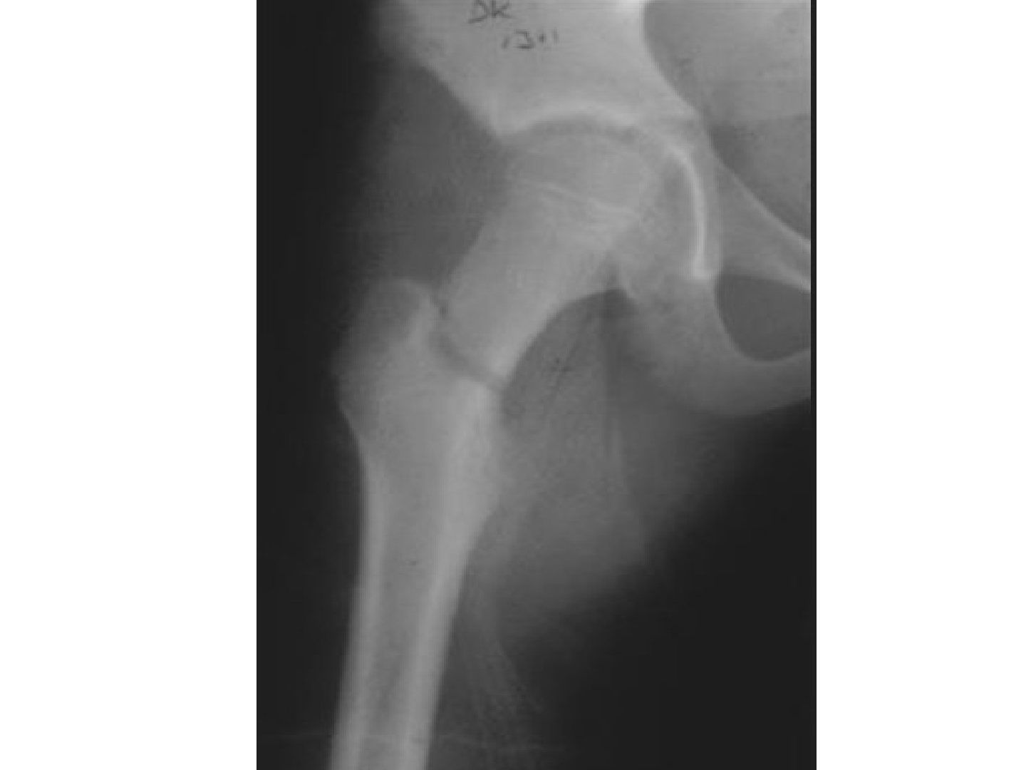
- Describe the X-ray and initial assessment.
- Describe the vascular anatomy of the proximal femur by age.
- Describe the Delbet classification and AVN risk.
- What is the treatment and fixation strategy?
- What is the post-operative rehab and follow-up?
- What are the complications and the Ratliff classification of AVN?
Answers · Q & A
Q1.Describe the X-ray and initial assessment.
- Basal neck of femur fracture in a skeletally immature patient, minimally displaced in this view (see cross table lateral)
- Hx: mechanism of injury (ATLS), r/o fracture in same limb, nerve injury
- Surgical emergency in Delbet type 1-3 to decrease AVN risk
Q2.Describe the vascular anatomy of the proximal femur by age.
- <4 years: MFCA (lateral epiphyseal artery), LFCA, metaphyseal artery
- 4-10 years: MFCA; metaphyseal artery blocked by physis; LFCA regresses
- 10-14 years: MFCA + ligament teres artery
- >14 years: MFCA + metaphyseal artery (anastomosis of metaphyseal and epiphyseal artery redevelops), extracapsular arterial ring, ascending cervical retinacular artery, ligament teres artery
Q3.Describe the Delbet classification and AVN risk.
- Type 1 transphyseal: 40% AVN (100% if positive dislocation of epiphysis from acetabulum)
- Type 2 transcervical: 30%
- Type 3 basal neck: 20%
- Type 4 trochanteric: 5%
Q4.What is the treatment and fixation strategy?
- r/o NAI in non-walking child; conservative: <4 years + non-displaced -> hip spica cast
- Operative: Displaced fracture or > 4 y.o; ORIF for dislocated transphyseal fracture, otherwise CR + pinning/screw/DHS
- Fixation: transphyseal pin (type 1 or proximal type 2 with minimal metaphyseal bone for anchorage; consider post op hip spica if young), screws sparing physis (type 2-4 + young), transphyseal screws near skeletal maturity (avoid posterior cortex protrusion)
- Type 4: paediatric DHS/ locking side plate; + capsulotomy in type 1-3
Q5.What is the post-operative rehab and follow-up?
- TTWB minimum 6 weeks; progression to FWB 6-12 weeks
- Return to activity when radiographically healed
- Removal of implant: old no, young yes
- Need long term FU for physeal arrest and AVN
Q6.What are the complications and the Ratliff classification of AVN?
- AVN risk factors: 3-8 years old, type 1 fracture
- Ratliff I: complete head - epiphyseal artery (all), worst prognosis; II: partial epiphysis - limited epiphyseal artery; III: between physis and fracture line - superior metaphyseal artery, best prognosis
- Coxa vara (neck shaft angle <130 degrees) due to malunion; <3 years likely remodels; <8 years trochanteric apophysis growth arrest; older + Trendelenburg gait -> valgus osteotomy
- Coxa valga in type 4 fracture; physeal arrest + LLD; chondrolysis
Fact check
Coxa vara is defined as a neck-shaft angle <130 degrees — imprecise — Most sources define coxa vara as a neck-shaft angle <120 degrees; the threshold quoted varies with age and author — medium confidence — source
▸ Slide 621 · Femoral head blood supplyShort Stature · 2 questions expand
Question list
Q1-Q22 questions — tap to reveal all answerslist
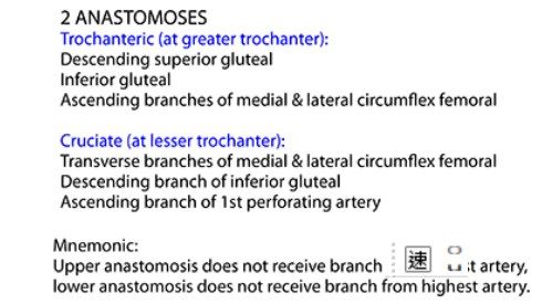
- What is the retinaculum of Weitbrecht?
- How is the blood supply of the femoral head described in the notes?
Answers · Q & A
Q1.What is the retinaculum of Weitbrecht?
- It equals the ascending artery of the cervical retinacular artery
- It contributes to the blood supply of the femoral head and neck
Q2.How is the blood supply of the femoral head described in the notes?
- As a 2 cruciate system
- Together with the ascending artery of the cervical retinacular artery (retinaculum of Weitbrecht)
▸ Slide 622 · Increasing ageShort Stature · 14 questions expand
Slide render
Question list
Q1-Q1414 questions — tap to reveal all answerslist
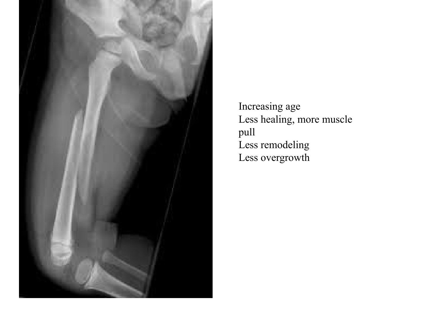
- Describe the fracture and radiographic findings in this skeletally immature femur.
- Why must non-accidental injury (NAI) be ruled out and how?
- What does treatment of a paediatric femoral shaft fracture depend on?
- What are the effects of increasing age on a femoral shaft fracture?
- What are the acceptable deformity limits by age (Rockwood 2010)?
- What is the treatment of a femoral shaft fracture at <6 months and 6 months-6 years?
- What is the treatment of a femoral shaft fracture in a 6-10 year old?
- What is the treatment of a femoral shaft fracture if >10 years, >100lb or length unstable?
- Describe Bryant traction.
- What are the key points of hip spica application?
- How is the hip spica moulded and positioned?
- How is the trunk prepared during hip spica application?
- How is the hip spica completed?
- What is the evidence for management of femur fractures in 4-10 year olds?
Answers · Q & A
Q1.Describe the fracture and radiographic findings in this skeletally immature femur.
- X-ray of a skeletally immature patient
- Mid-shaft femoral fracture, spiral with no comminution
- Shortened with some rotational deformity, no valgus/varus angulation
- Need lateral Xray to see AP (sagittal) angulation
Q2.Why must non-accidental injury (NAI) be ruled out and how?
- Corroborate the history with other family members
- Observe interaction with others
- Look for evidence of neglect
- Ask for a family history of skeletal dysplasia as a cause of bone fragility
Q3.What does treatment of a paediatric femoral shaft fracture depend on?
- Patient factors: age, weight, underlying bone disease
- Disease factors: location, deformity, length stable
- length stable is less relevant now with the advent of endcaps
Q4.What are the effects of increasing age on a femoral shaft fracture?
- Less healing
- More muscle pull
- Less remodeling
- Less overgrowth
Q5.What are the acceptable deformity limits by age (Rockwood 2010)?
- coronal sagittal shorten (Rockwood 2010)
- <2 30 30 20: coronal 30, sagittal 30, shortening 20
- 2-6 15 20 20: coronal 15, sagittal 20, shortening 20
- 6-11 10 15 15: coronal 10, sagittal 15, shortening 15
- >11 5 10 10: coronal 5, sagittal 10, shortening 10
- Will overgrow ~1.5cm, static 2 years from injury (Shapiro 2)
Q6.What is the treatment of a femoral shaft fracture at <6 months and 6 months-6 years?
- <6 months: Pavlik harness
- 6m-6y: skeletal traction if needed then hip spica up to 8 weeks
- Surgery (ex fix/elastic nail if canal >7mm) if alignment unacceptable, open fracture or polytrauma
Q7.What is the treatment of a femoral shaft fracture in a 6-10 year old?
- Flexible nail if not very proximal or very distal and not length unstable
- Length unstable is only relative because of endcaps
- Otherwise ex fix or submuscular plate
Q8.What is the treatment of a femoral shaft fracture if >10 years, >100lb or length unstable?
- Submuscular bridging plating/nailing through a lateral entry
- Avoid piriformis entry - injures the medial circumflex artery -> AVN
- GT entry causes epiphysiodesis, acceptable if >10 years
Q9.Describe Bryant traction.
- Indication: shortening >3cm and a decision for cast
- Overhead skin traction, knees in full extension
- 5-10lbs - just enough to lift the buttock off the mattress
- Until sticky callus seen on X-ray at ~2/52 (16-18kg)
Q10.What are the key points of hip spica application?
- Never traction - muscle contraction after GA will cause a compartment
- Applied after traction and callus formed
- Duration Age + 3week/ most 8weeks; weekly X-ray in the first 3 weeks
- Place the child on a spica table; long leg cast with the knee in 90 deg flexion and generous padding over the popliteal fossa (protects popliteal vessels and peroneal nerve)
Q11.How is the hip spica moulded and positioned?
- Three-point molding centred at the fracture site
- Proximal: may need ORIF due to strong muscle pull
- Middle: valgus moulding for varus displacement
- Distal: flex the knee 20 degrees to reduce gastrocnemius pull
Q12.How is the trunk prepared during hip spica application?
- Folded towel on the anterior thorax and abdomen to create space for inspiration
- 2 layers of stockinette then cotton (Velband) or gortex material over the trunk
- Thick felt belt across the chest just below the nipple line, second felt belt over sacrum, PSIS and ASIS
Q13.How is the hip spica completed?
- One and a half leg spica with hip flexed 90 degrees and 30 degrees abduction (human position), connected with a long leg cast
- Reinforce with a broomstick between the thighs
- Perineal window; double check the reduction on X-ray
Q14.What is the evidence for management of femur fractures in 4-10 year olds?
- 4-10 yrs: Wright et al Lancet 2005 RCT: malunion rates after ex fix vs flexible IM nails
- Improved outcomes with nails
- AAOS guidelines: do not advocate routine use of ex fix in younger children
▸ Slide 623 · PRINCIPLEShort Stature · 6 questions expand

Question list
Q1-Q66 questions — tap to reveal all answerslist
- What is the principle behind flexible intramedullary nailing?
- What are the 4 biomechanical effects of symmetrical splinting (RAFT)?
- What is the indication for flexible IM nailing?
- How is the nail chosen and bent in flexible IM nailing?
- Describe the flexible IM nailing procedure.
- What is the post-operative protocol after flexible IM nailing?
Answers · Q & A
Q1.What is the principle behind flexible intramedullary nailing?
- Based on the double arc secant in equilibrium principle
- load sharing device relying on 3-point fixation of two opposing nails
- The apex of each nail is at the fracture site
- Bending moments of the elastic nails counteract with equal and opposite force to create a stable construct
Q2.What are the 4 biomechanical effects of symmetrical splinting (RAFT)?
- Rotational stability
- Axial stability
- Flexural stability
- Translational stability
Q3.What is the indication for flexible IM nailing?
- Works best at the middle 1/3, length-stable fracture
- Body build <100 lb
Q4.How is the nail chosen and bent in flexible IM nailing?
- Choose a nail 1/3 of the inner canal diameter
- Bend each nail 3 times the inner canal diameter
- Apex at the fracture site so the two nails are maximally separated from each other at the fracture
Q5.Describe the flexible IM nailing procedure.
- Radiolucent table, reduce with the F tool
- Insert the nail retrograde at the metaphysis, then cap to prevent irritation and migration
- Flat tips at the nail end improve metaphyseal anchorage and rotational control; avoid the physis of the femur
Q6.What is the post-operative protocol after flexible IM nailing?
- TDW as tolerated post-op with gentle mobilisation exercises
- Avoid overaggressive knee strengthening (may displace the fracture)
- Usually allow FWB after 6/52
- Follow up with XR for healing/loss of reduction; nail removal in 1 year
▸ Slide 624 · Xray showing tibial eminence fracture, displaced, no hinge in skeletally immaturShort Stature · 8 questions expand
Slide render

Question list
Q1-Q88 questions — tap to reveal all answerslist
- Describe the Meyers and McKeever classification of tibial eminence fractures.
- What is the mechanism and typical age of a tibial eminence fracture?
- What clinical findings must be checked in a tibial eminence fracture?
- What associated injuries occur with tibial eminence fractures?
- How are associated injuries investigated in a tibial eminence fracture?
- How are tibial eminence fractures managed?
- What is used for fixation in a tibial eminence fracture?
- Describe the operative procedure for a tibial eminence fracture.
Answers · Q & A
Q1.Describe the Meyers and McKeever classification of tibial eminence fractures.
- Type 1: undisplaced (<3mm)
- Type 2: hinged
- Type 3: displaced
- Type 4: displaced + comminuted
Q2.What is the mechanism and typical age of a tibial eminence fracture?
- Common mechanism is hyperextension
- Age 8-14 years
- Displaced fracture in a skeletally immature patient, no hinge
Q3.What clinical findings must be checked in a tibial eminence fracture?
- Look for an open wound
- Rule out other injuries
Q4.What associated injuries occur with tibial eminence fractures?
- Association 40%: meniscal injury, collateral ligament injury, capsular damage, osteochondral fracture
Q5.How are associated injuries investigated in a tibial eminence fracture?
- MRI to rule out associated injuries
- Look for interposed meniscus or intermeniscal ligament - needs OT
Q6.How are tibial eminence fractures managed?
- Type 1 and reducible Type 2: immobilise at 20deg
- OT indications: type 3 or 4, mechanical block, entrapped soft tissue, other injuries
Q7.What is used for fixation in a tibial eminence fracture?
- Young with pin, old with screw
Q8.Describe the operative procedure for a tibial eminence fracture.
- Supine with tourniquet, AM and AL portal, diagnostic scope
- Debride haematoma, interposed meniscus and intermeniscal ligament
- 2 strand repair with nonabsorbable sutures, wrap around midsubstance of ACL
- Drill hole with an ACL tibial jig
- Before closing, ensure there is no impingement at full extension
▸ Slide 625 · Lateral knee Xray of skeletally immature patientShort Stature · 8 questions expand
Slide render
Question list
Q1-Q88 questions — tap to reveal all answerslist
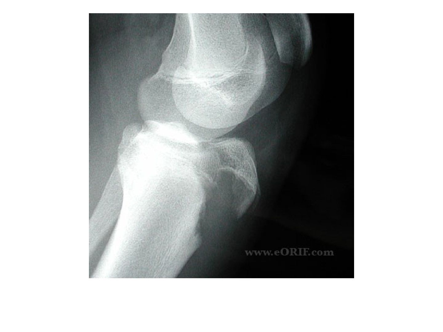
- Describe the injury shown in the lateral knee X-ray.
- What are the clinical concerns and investigations for a tibial tuberosity fracture?
- What is the management of a tibial tuberosity fracture?
- What are the complications of a tibial tuberosity fracture?
- What are the ossification centres of the tibial tuberosity and which is at risk?
- What are the 4 stages of development in the proximal tibia?
- What is the Ogden classification of tibial tuberosity fractures?
- What are the risk factors and mechanism of a tibial tuberosity fracture?
Answers · Q & A
Q1.Describe the injury shown in the lateral knee X-ray.
- Skeletally immature patient
- Fracture over the proximal tibia involving the tibial tuberosity
- Fracture extension into the knee joint
- Ogden Type 3B fracture
Q2.What are the clinical concerns and investigations for a tibial tuberosity fracture?
- Expect extension lag (may still be possible as the medial retinaculum extends beyond the proximal tibial physis)
- Rule out compartment syndrome (proximal tibial recurrent artery)
- Investigate with AP, lateral and oblique X-rays
- +/- CT; +/- MRI for an undisplaced Type 2
Q3.What is the management of a tibial tuberosity fracture?
- Long leg cast in full knee extension for IA and IB with <2mm displacement
- >3 years from closure: smooth pin + TBW
- <3 years from closure: lag screw
- Type III: +/- arthroscopy; 5 soft tissue repair
Q4.What are the complications of a tibial tuberosity fracture?
- Acute: compartment syndrome
- Chronic: recurvatum (anterior growth arrest), stiffness
- Bursitis related to the screw, patellar alta
Q5.What are the ossification centres of the tibial tuberosity and which is at risk?
- Two ossification centres: primary (proximal tibial physis) and secondary (tibial tubercle physis)
- The secondary centre is at risk at 13-16 years
- Primary physis fuses posterior to anterior
- Secondary physis fuses proximal to distal
Q6.What are the 4 stages of development in the proximal tibia?
- 1. Cartilaginous phase - no ossification centre
- 2. Apophyseal phase - 9 years, second ossification centre appears
- 3. Epiphyseal phase - fuses from posterior to anterior to the primary ossification centre
- 4. Bony phase - 16 years, closure of the physis
Q7.What is the Ogden classification of tibial tuberosity fractures?
- Type 1: through the secondary ossification centre
- Type 2: between the primary and secondary centres
- Type 3: crosses the primary physis
- Type 4: through the entire physis
- Type 5: sleeve
- A undisplaced, B displaced
Q8.What are the risk factors and mechanism of a tibial tuberosity fracture?
- Risk factors: patellar baja, tight hamstrings, Osgood-Schlatter disease
- Usually from eccentric contraction of the quadriceps as the patella inserts onto the secondary ossification centre
- Occurs near the end of growth
▸ Slide 626 · 3-10yo valgus force to kneeShort Stature · 6 questions expand
Slide render

Question list
Q1-Q66 questions — tap to reveal all answerslist
- Describe the fracture in this 3-10 year old with a valgus knee injury.
- What is the mechanism of a proximal tibial metaphyseal fracture?
- What must be ruled out clinically in a proximal tibial metaphyseal fracture?
- How is an undisplaced proximal tibial metaphyseal fracture treated?
- What are the associated problems of a proximal tibial metaphyseal fracture?
- What are the deforming forces in a proximal tibia fracture?
Answers · Q & A
Q1.Describe the fracture in this 3-10 year old with a valgus knee injury.
- AP knee X-ray of a skeletally immature patient
- Fracture over the proximal tibia
- Incomplete fracture over the medial metaphyseal area
- No plastic deformity of the fibula
Q2.What is the mechanism of a proximal tibial metaphyseal fracture?
- Valgus bending -> the medial tibial metaphysis fails in tension
- Usually from a trampoline
Q3.What must be ruled out clinically in a proximal tibial metaphyseal fracture?
- Open fracture
- Compartment syndrome
Q4.How is an undisplaced proximal tibial metaphyseal fracture treated?
- Conservative: long leg cast with the knee in full extension and varus moulding
- Known to have overgrowth causing valgus deformity
- OT indication: torn pes anserinus
Q5.What are the associated problems of a proximal tibial metaphyseal fracture?
- Compartment syndrome (recurrent anterior tibial artery)
- Vessel injury (popliteal artery tethered)
- Cozen phenomenon - progressive valgus from medial tibial overgrowth/fracture hyperaemia; 50-90%, most resolve in 24 months
- Recurvatum (anterior closure of physis)
- LLD (average 9mm)
Q6.What are the deforming forces in a proximal tibia fracture?
- Proximal fragment: extension by patellar tendon, varus by pes anserinus
- Distal fragment: valgus by tibialis anterior, flexion by gastrocnemius
▸ Slide 627 · Acceptable alignmentShort Stature · 7 questions expand

Question list
Q1-Q77 questions — tap to reveal all answerslist
- What is acceptable alignment after a paediatric tibial fracture?
- What is a toddler's fracture?
- How is a stable toddler's fracture managed?
- What are the fixation options for an unstable tibial shaft fracture?
- What is the time to healing by age?
- What is the principle of the Sarmiento cast?
- What is the design of the Sarmiento brace?
Answers · Q & A
Q1.What is acceptable alignment after a paediatric tibial fracture?
- >50% apposition of ends
- <1cm shortening
- <10 degrees angulation in all planes
- Comment on any fibular fracture
Q2.What is a toddler's fracture?
- Undisplaced spiral fracture of the tibial shaft
- Mechanism: forceful foot external rotation with the knee fixed
- Age 1-2 years
Q3.How is a stable toddler's fracture managed?
- Long leg cast for 3 weeks, then short leg cast for 3 weeks
- Adolescent: to a patellar bearing/Sarmiento brace at 8 weeks, for a total of 12 weeks
- Casting position: slight ankle plantarflexion (prevents apex posterior angulation) and knee flexion (controls rotation, prevents WB)
- Weekly follow-up for the first 3 weeks
Q4.What are the fixation options for an unstable tibial shaft fracture?
- Too narrow canal: cross K wire
- >=6mm: elastic IMN
- Closed physis: locking nail
Q5.What is the time to healing by age?
- Neonate: 2-3 weeks
- Children: 4-6 weeks
- Adolescent: 8-12 weeks
Q6.What is the principle of the Sarmiento cast?
- Hydraulic mechanism - tissues and noncompressible fluid within a rigid container
- The incompressible fluid around a fracture prevents excessive shortening
- Substantial pressure develops in the soft tissues under load and supports the bone fragments
- Continued function is desirable for osteogenesis; motion between fragments may create an environment conducive to osteogenesis
- Muscle activity stimulates new blood supply for peripheral callus
Q7.What is the design of the Sarmiento brace?
- Patellar tendon bearing for partial weight bearing
- Proximal ears continued as far posteriorly as possible opposite the tibial tubercle without impinging the hamstrings
- Triangular shape to fit the tibial contour
- Coronal plane: large anterior shell and small posterior shell
- Rotational plane: femoral condylar extension with precise infrapatellar moulding (patellar bearing) to hold the tibial plateau and enhance rotational (M-L) stability
- Distal flaring for both malleoli; plastic foot-insert with a hinged ankle component for pistoning
▸ Slide 628 · Distal femur fractureShort Stature · 5 questions expand
Slide render
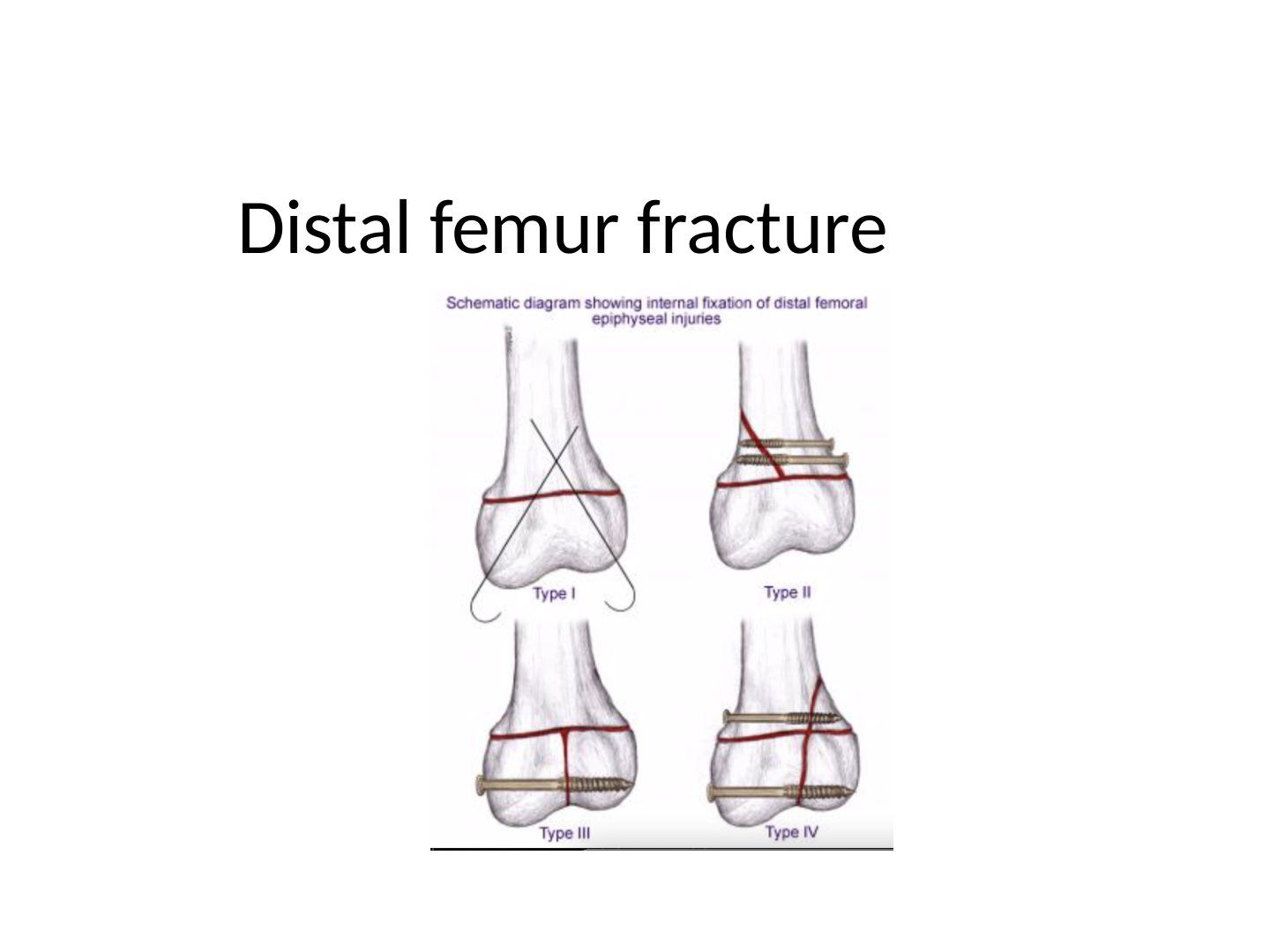
Question list
Q1-Q55 questions — tap to reveal all answerslist
- Describe the Salter-Harris patterns of distal femur fractures.
- What must be assessed in a distal femur fracture?
- How is closed reduction and casting performed for a distal femur fracture?
- What is the operative fixation for a distal femur fracture?
- What are the complications of a distal femur fracture?
Answers · Q & A
Q1.Describe the Salter-Harris patterns of distal femur fractures.
- SH II - commonest; metaphyseal fragment at the compression side; risk of arrest
- SH III - distal femur (MFC most, close to physeal closure); lateral tibial plateau + intra-articular fracture + MCL injury -> OT
- SH IV - risk of partial physeal arrest -> angular deformity
Q2.What must be assessed in a distal femur fracture?
- Popliteal artery injury
- Collateral and cruciate ligament injury
- Peroneal nerve injury
Q3.How is closed reduction and casting performed for a distal femur fracture?
- Reverse the original mechanism
- Hyperextension injury: initial 90 degrees flexion then gradual extension
- Hyperflexion injury: full extension
- Valgus/varus injury: flexion 10-20 degrees
Q4.What is the operative fixation for a distal femur fracture?
- Indication: failed CR, unstable in cast
- Screw parallel to the physis for a large epiphyseal/metaphyseal fragment
- Transphyseal pin for a small fragment or close to maturity (increases stability); bury under skin, otherwise risk of septic knee
Q5.What are the complications of a distal femur fracture?
- 30-50% physeal arrest -> angular deformity
- Physeal arrest does not remodel well
- Tolerable if <2 years of growth remaining
- LLD
▸ Slide 629 · AP and lateral Xray of ankle of a skeletally immature patientShort Stature · 5 questions expand
Slide render

Question list
Q1-Q55 questions — tap to reveal all answerslist
- Describe the triplane fracture on AP and lateral X-ray.
- What is the acute management of a triplane fracture?
- What is the definitive management of a displaced triplane fracture?
- What are the growth and physeal facts of the distal tibia?
- Describe the parts and types of triplane fractures.
Answers · Q & A
Q1.Describe the triplane fracture on AP and lateral X-ray.
- Distal tibia fracture involving the epiphysis
- AP view = SH III, lateral view = SH II
- Distal fragment displaced medial and posterior
- Triplane: fracture courses through the coronal, transverse and sagittal planes
- Intra-articular fracture; MOI: ER (external rotation)
Q2.What is the acute management of a triplane fracture?
- Systemically rule out other injuries
- Locally look for wound, compartment syndrome, and document distal NV status
- RICE, slab, analgesics
- Displacement is >2mm
Q3.What is the definitive management of a displaced triplane fracture?
- Aim: anatomical reduction and stable fixation; CT -> CR +/- OR
- Fix with screws - interfragmental compression with screws parallel to the physis
- CR technique: 2-part - lateral (IR), medial (abduction)
- AL approach: reduce with dorsiflexion + IR +/- pronation or Weber reduction forceps; 4.0 cannulated screw (transmetaphyseal and transepiphyseal)
- OT for 3-4 part (sequence: posterior fragment -> fibula -> AL fragment)
- Advise removal of transepiphyseal screws (JPO 2005 Charlton: increased peak intra-articular contact pressures)
Q4.What are the growth and physeal facts of the distal tibia?
- Distal tibia physis closes at 16 years; distal fibula closes 1 year after
- Contributes 40% of tibia growth and 20% of lower limb growth
- All ligaments attach distal to the physis
- Distal tibial physis fuses age 11-13 in boys; transitional fracture ~18 months
- Medial os subtibiale 20%, lateral os subfibulare 1%
- First site of closure = Kump's bump (central -> anteromedial -> posteromedial -> anterolateral)
Q5.Describe the parts and types of triplane fractures.
- Coronal: crosses the epiphysis; axial: splits the physis; sagittal: exits in the metaphysis
- Can be 2, 3 or 4 parts
- 2 parts (named after the coronal fragment): lateral triplane = supination-external rotation (most common); medial triplane = adduction
- 3 parts: separate Tillaux fragment (SH 3 on AP)
- 4 part: Tillaux + AM fragment
▸ Slide 630 · Ankle Xray of mortise view of skeletally immature patientShort Stature · 5 questions expand
Slide render
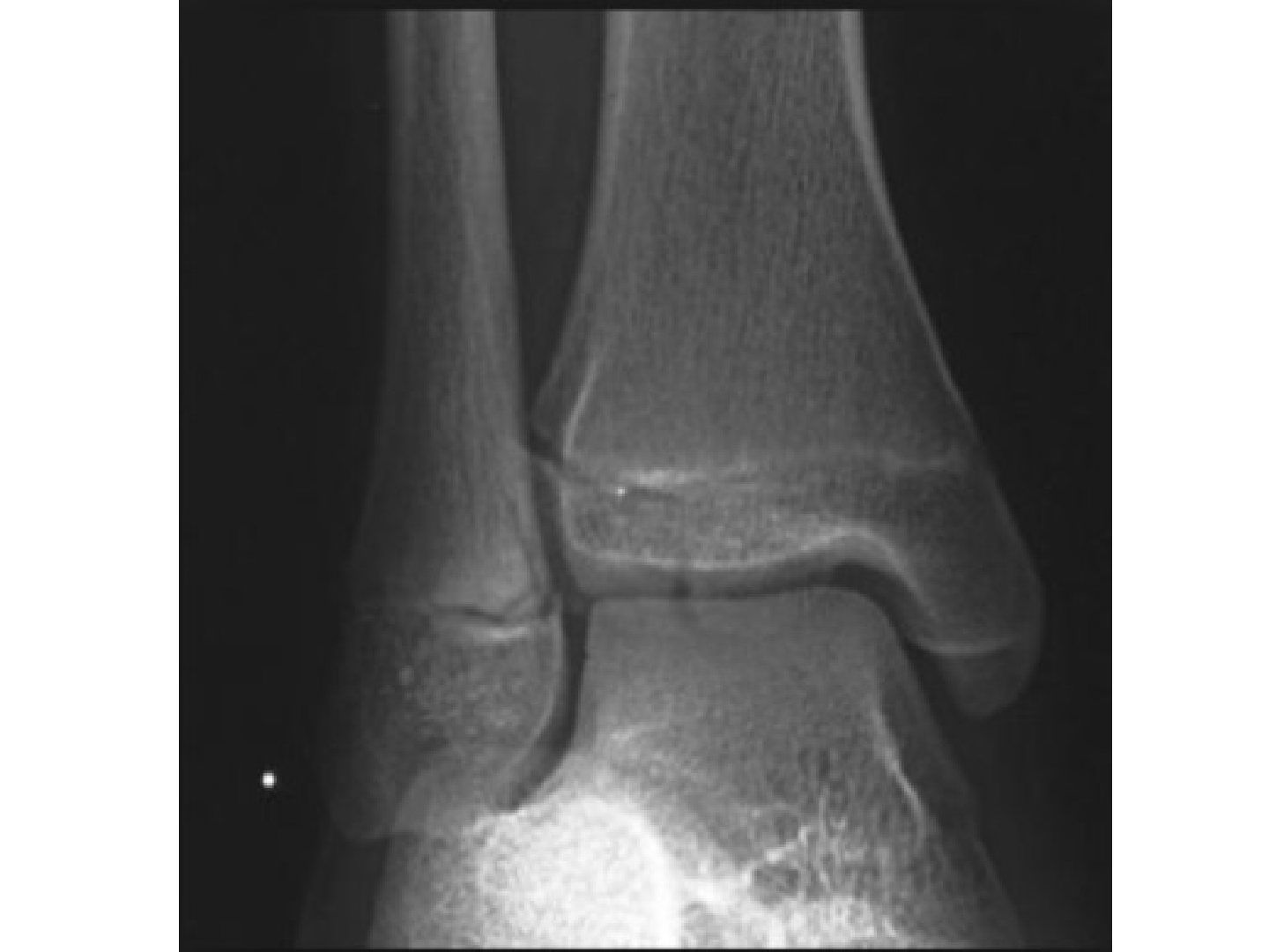
Question list
Q1-Q55 questions — tap to reveal all answerslist
- Describe the Tillaux fracture on the mortise view.
- What is the mechanism and typical age of a Tillaux fracture?
- How is a Tillaux fracture investigated?
- How is a Tillaux fracture managed according to displacement?
- How is a Tillaux fracture distinguished from a triplane injury?
Answers · Q & A
Q1.Describe the Tillaux fracture on the mortise view.
- Skeletally immature patient
- Fracture over the distal tibia epiphysis on the lateral side
- Fibula no fracture
- Tillaux fracture = SH 3, a transitional fracture
Q2.What is the mechanism and typical age of a Tillaux fracture?
- Mechanism: external rotation
- Caused by an avulsion of the anterior inferior tibiofibular ligament
- Near end of growth, older than triplane, ~12-14 years
Q3.How is a Tillaux fracture investigated?
- CT to delineate the fracture pattern and assess displacement
Q4.How is a Tillaux fracture managed according to displacement?
- <2mm: conservative with a long leg cast for rotational control
- Cast position: foot IR + direct anterior pressure + knee 30 degrees flexion; 4/52 long leg cast then 2/52 short leg cast
- Post-cast CT to confirm no displacement
- >2mm: CR +/- OR via AL approach and stable fixation with a percutaneous screw
- Screw can be transphyseal as the patient approaches skeletal maturity
Q5.How is a Tillaux fracture distinguished from a triplane injury?
- Tillaux fracture = SH 3
- A transitional fracture
- Lack of a fracture component in the coronal plane distinguishes it from a triplane injury
▸ Slide 631 · Growth contribution from different physisShort Stature · 4 questions expand
Slide render

Question list
Q1-Q44 questions — tap to reveal all answerslist
- What is the overall growth rate of the leg and which physis contributes most?
- What is the annual growth contribution of each physis of the femur and tibia?
- At what age does a child achieve 1/2 of adult leg length?
- What proportion of longitudinal growth comes from the proximal humerus and distal radius?
Answers · Q & A
Q1.What is the overall growth rate of the leg and which physis contributes most?
- The leg grows 23 mm/year
- Most of that comes from the knee (15 mm/yr)
Q2.What is the annual growth contribution of each physis of the femur and tibia?
- Proximal femur - 3 mm/yr
- Distal femur - 9 mm/yr
- Proximal tibia - 6 mm/yr
- Distal tibia - 5 mm/yr
Q3.At what age does a child achieve 1/2 of adult leg length?
- Female: 3 years old
- Male: 4 years old
Q4.What proportion of longitudinal growth comes from the proximal humerus and distal radius?
- Proximal humerus: 80% of whole humerus
- Distal radius: 60% of whole forearm
▸ Slide 632Short Stature · 2 questions expand
Slide render
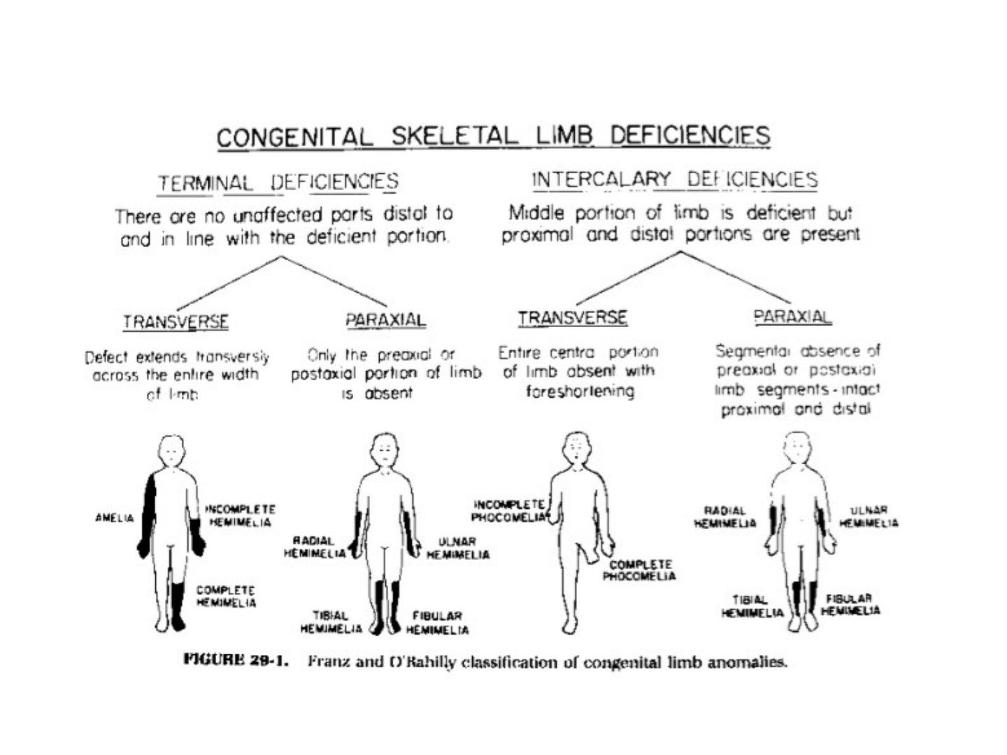
Question list
Q1-Q22 questions — tap to reveal all answerslist
- What is the content of this slide?
- Which topic does this slide belong to?
Answers · Q & A
Q1.What is the content of this slide?
- Not covered in the speaker notes (slide image is the only source)
Q2.Which topic does this slide belong to?
- Topic: Short Stature; no speaker notes available
▸ Slide 633 · Swanson Classification 1976 FDD OU CGShort Stature · 3 questions expand
Slide render
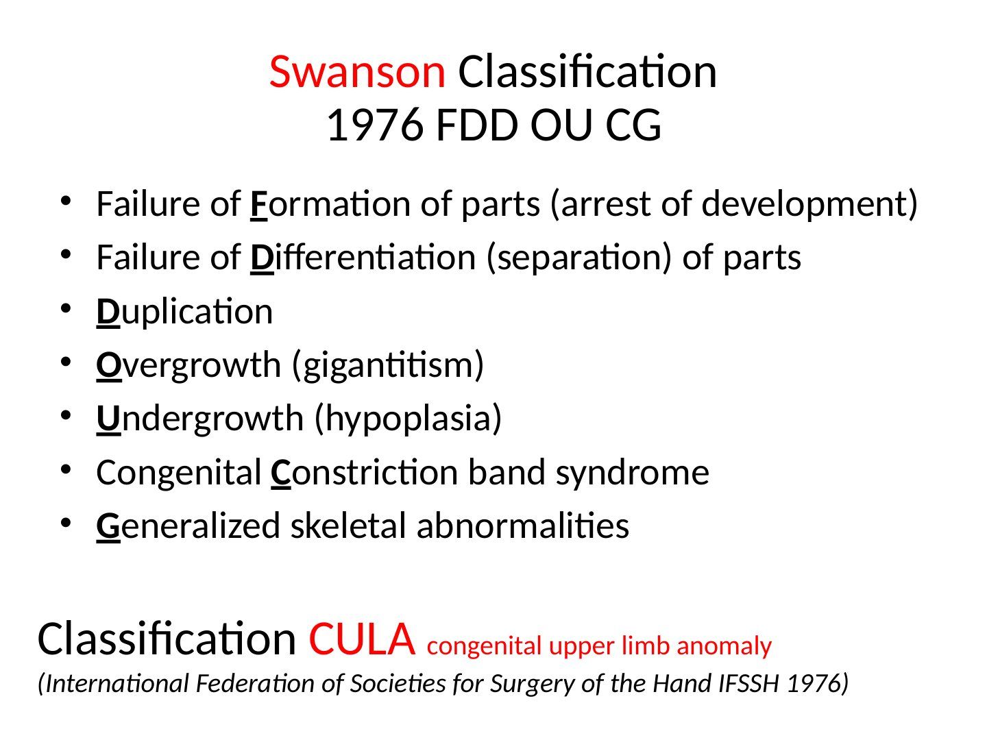
Question list
Q1-Q33 questions — tap to reveal all answerslist
- List the categories of the Swanson classification 1976 (FDD OU CG).
- What do the letters FDD OU CG stand for?
- What is CULA and which organisation introduced it?
Answers · Q & A
Q1.List the categories of the Swanson classification 1976 (FDD OU CG).
- Failure of Formation of parts (arrest of development)
- Failure of Differentiation (separation) of parts
- Duplication
- Overgrowth (gigantitism)
- Undergrowth (hypoplasia)
- Congenital Constriction band syndrome
- Generalized skeletal abnormalities
Q2.What do the letters FDD OU CG stand for?
- Failure of Formation, failure of Differentiation, Duplication
- Overgrowth, Undergrowth
- Congenital constriction band, Generalized skeletal abnormalities
Q3.What is CULA and which organisation introduced it?
- CULA = congenital upper limb anomaly
- International Federation of Societies for Surgery of the Hand (IFSSH) 1976
▸ Slide 634 · Primary GripShort Stature · 2 questions expand

Question list
Q1-Q22 questions — tap to reveal all answerslist
- List the primary grips.
- Which of the primary grips are pinch grips and which are whole-hand grips?
Answers · Q & A
Q1.List the primary grips.
- Precision pinch / fine pinch
- Tripod pinch
- Power grip
- Hook grip
- Key pinch
Q2.Which of the primary grips are pinch grips and which are whole-hand grips?
- Pinch grips: precision/fine pinch, tripod pinch, key pinch
- Whole-hand grips: power grip, hook grip
▸ Slide 635 · Secondary GripShort Stature · 2 questions expand
Question list
Q1-Q22 questions — tap to reveal all answerslist
- List the secondary grips.
- Which grips are secondary rather than primary?
Answers · Q & A
Q1.List the secondary grips.
- Secondary pinch grip
- Lateral interdigital grip
- Extensor pinch grip
Q2.Which grips are secondary rather than primary?
- Secondary: secondary pinch grip, lateral interdigital grip, extensor pinch grip
- Primary: precision/fine pinch, tripod pinch, power grip, hook grip, key pinch