15 slides
▸ Slide 584 · NeuromuscularNeuromuscular · 2 questions expand
Slide render
Question list
Q1-Q22 questions — tap to reveal all answerslist
- Which topic does this slide introduce?
- Which conditions are discussed in this neuromuscular section?
Answers · Q & A
Q1.Which topic does this slide introduce?
- Neuromuscular disorders in paediatric orthopaedics
- Not covered in the speaker notes - section divider slide
Q2.Which conditions are discussed in this neuromuscular section?
- Not covered in the speaker notes
- The slide is an image-only section divider
▸ Slide 585 · ArthrogryposisNeuromuscular · 10 questions expand
Question list
Q1-Q1010 questions — tap to reveal all answerslist
- How does arthrogryposis present and what does the neurological examination indicate?
- What are the other manifestations of arthrogryposis?
- How is arthrogryposis assessed and what is the natural history?
- What is the aetiology and what are the forms of arthrogryposis?
- What is amyoplasia?
- What are the Drummond principles of operative management?
- What are the limb positions and associated deformities in arthrogryposis?
- What are the upper limb operative options in arthrogryposis?
- How is hip involvement in arthrogryposis treated?
- What is the role of physiotherapy in arthrogryposis?
Answers · Q & A
Q1.How does arthrogryposis present and what does the neurological examination indicate?
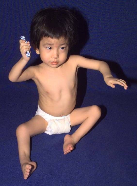
- Non-progressive congenital disease with limited movement involving 2 or more joints
- Limbs: tubular, fusiform, featureless, without skin creases
- Positions: pronated forearm, extended elbow, hand in palm; FABER, foot deformity
- UL: shoulder abducted, IR, elbow extended, wrist UD + flexed, fingers flexed at MCPJ and IPJ, thumb adducted (in palm)
- Normal neuro exam: amyoplasia, distal arthrogryposis, generalised connective tissue disorder or fetal crowding
- Abnormal neuro exam: diminished in-utero movement due to abnormal CNS, PNS, motor endplate or muscle
Q2.What are the other manifestations of arthrogryposis?
- Upper extremity deformity
- Teratologic hip subluxation and dislocation
- Knee contractures
- Foot: clubfoot, vertical talus
- Possible: DDH, dislocated knee, clubfoot
- Neuromuscular C-shaped scoliosis 33%; fractures 25%
- Extraskeletal: haemangioma on forehead, abdominal wall anomalies (inguinal hernia, gastroschisis)
Q3.How is arthrogryposis assessed and what is the natural history?
- Diagnosis: EMG, muscle biopsy, enzyme study, genetic study
- X-ray spine and pelvis
- MDT: paediatrician, ortho, geneticist, physio, P&O, psychologist
- Normal face, IQ and normal life expectancy
- Aim: increase joint mobility and muscle strength to develop adaptive use patterns that allow for walking and independence with ADLs
- 25% non ambulatory
Q4.What is the aetiology and what are the forms of arthrogryposis?
- 1 in 3000 births; the direct factor causing akinesis is unknown
- Intrinsic causes: myopathic, neuropathic, fibropathic (neurogenic 90%, myopathy)
- Extrinsic: decreased IU movement, IU infection, oligohydramnios, multiple pregnancy, amniotic bands
- Maternal disease: diabetes, myasthenia gravis
- Sporadic Arthrogryphosis multiplex congenita (4 limbs); distal arthrogryphosis
- Larsen (4 limbs, knee dislocation, clubfoot)
- Freeman Shelton (hand, foot, scoliosis + face (microstomia))
- Pteryrium syndrome (flexor web)
Q5.What is amyoplasia?
- Classic form of AMC: sporadic multiple contracture syndrome
- Symmetrical involvement of multiple UL and LL joints; CNS normal
- Muscle tissue replaced by fibrofatty tissue; most commonly seen form
- 1 in 10,000 births, 30% of all congenital contractures
- Types: I localised; II full expression (thin limbs, elbows extended, wrists flexed and ulnarly deviated, intrinsic plus hands, adducted thumbs); III adds polydactyly
Q6.What are the Drummond principles of operative management?
- Deformity recurrence is common - dense inelastic soft tissue does not elongate with growth
- Regain muscle balance if muscle is functional
- Delay osteotomy until maturity
- Maximal correction by surgery (tenotomies + capsulectomies) as conservative treatment is ineffective
- Shortening procedure if recurrence (e.g. talectomy)
Q7.What are the limb positions and associated deformities in arthrogryposis?
- Upper limb: shoulder abducted and internally rotated, elbow extended, wrist ulnar deviated + flexed, fingers flexed at MCPJ and IPJ, thumb adducted (in palm)
- Lower limb: hip flexed, externally rotated, abducted (FABER), knee flexed (contracture), equinus
- Also check the spine for scoliosis; possible DDH, dislocated knee, clubfoot; normal face and IQ
Q8.What are the upper limb operative options in arthrogryposis?
- IR contracture of shoulder: external humeral rotational osteotomy
- Elbow extension contracture preventing hand to mouth: posterior capsulotomy and triceps lengthening
- Wrist: dorsal carpal wedge osteotomy +/- FCU transfer to preserve arc of motion
- Correction of the thumb-in-palm deformity
Q9.How is hip involvement in arthrogryposis treated?
- Unilateral: operative reduction (OR)
- Bilateral: controversial - used to be conservative, now a trend towards sequential reduction
Q10.What is the role of physiotherapy in arthrogryposis?
- Passive range will improve
- Active range and muscle power will not improve
▸ Slide 586 · CMT/ hereditary motor sensory neuropathyNeuromuscular · 6 questions expand
Slide render
Question list
Q1-Q66 questions — tap to reveal all answerslist
- What is CMT/hereditary motor sensory neuropathy and how is it inherited?
- Compare CMT type 1 and type 2.
- What is the differential diagnosis of pes cavus?
- What muscle imbalances cause cavus, varus, claw toe and big toe plantarflexion?
- What is the management goal and conservative management of CMT?
- What are the operative options in CMT?
Answers · Q & A
Q1.What is CMT/hereditary motor sensory neuropathy and how is it inherited?
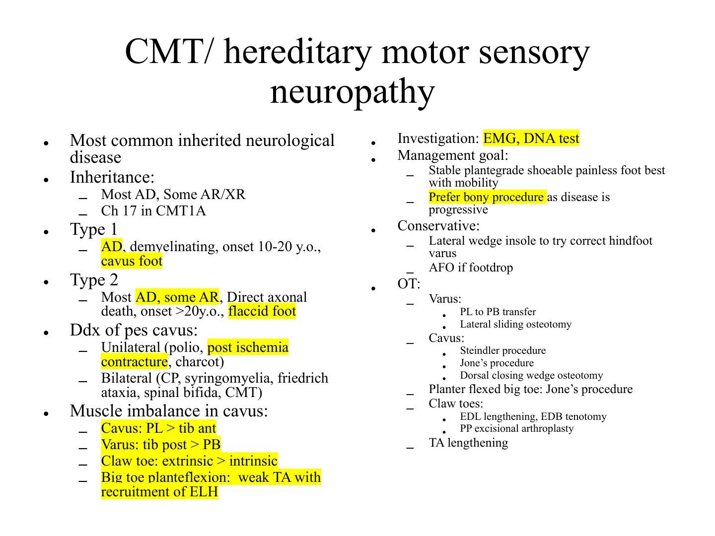
- Most common inherited neurological disease
- Inheritance: most AD, some AR/XR
- Chromosome 17 in CMT1A
Q2.Compare CMT type 1 and type 2.
- Type 1: AD, demyelinating, onset 10-20 y.o., cavus foot
- Type 2: most AD, some AR, direct axonal death, onset >20 y.o., flaccid foot
Q3.What is the differential diagnosis of pes cavus?
- Unilateral: polio, post-ischaemia contracture, Charcot
- Bilateral: CP, syringomyelia, Friedrich ataxia, spinal bifida, CMT
Q4.What muscle imbalances cause cavus, varus, claw toe and big toe plantarflexion?
- Cavus: PL > tib ant
- Varus: tib post > PB
- Claw toe: extrinsic > intrinsic
- Big toe plantarflexion: weak TA with recruitment of ELH
Q5.What is the management goal and conservative management of CMT?
- Investigation: EMG, DNA test
- Goal: stable, plantigrade, shoeable, painless foot best with mobility
- Prefer bony procedures as the disease is progressive
- Conservative: lateral wedge insole to correct hindfoot varus; AFO if footdrop
Q6.What are the operative options in CMT?
- Varus: PL to PB transfer, lateral sliding osteotomy
- Cavus: Steindler procedure, Jone’s procedure, dorsal closing wedge osteotomy
- Planter flexed big toe: Jone’s procedure
- Claw toes: EDL lengthening, EDB tenotomy, PP excisional arthroplasty, TA lengthening
▸ Slide 587 · Peroneal muscle atrophyNeuromuscular · 6 questions expand
Slide render
Question list
Q1-Q66 questions — tap to reveal all answerslist
- What is the epidemiology and genetics of hereditary motor sensory neuropathy?
- What are the three main types of hereditary motor sensory neuropathy?
- How does peroneal muscle atrophy present?
- What are the examination findings in peroneal muscle atrophy?
- What other orthopaedic manifestations are associated with peroneal muscle atrophy?
- How is peroneal muscle atrophy diagnosed?
Answers · Q & A
Q1.What is the epidemiology and genetics of hereditary motor sensory neuropathy?
- 1:2500, most common, affects motor more than sensory
- Usually autosomal dominant (can be AR, X-linked)
- Peripheral myelin protein 22 (PMP22)
- Duplication of chromosome 17
Q2.What are the three main types of hereditary motor sensory neuropathy?
- Demyelinating: 10-20s, AD, cavus foot
- Axonopathy: 20s+, AD, flaccid foot, less disabling
- Both demyelinating and axonal degeneration
Q3.How does peroneal muscle atrophy present?
- Motor deficits: weakness, instability, clumsiness, frequent ankle sprains, difficulty with stairs
- Lateral foot pain
- Sensory deficit
Q4.What are the examination findings in peroneal muscle atrophy?
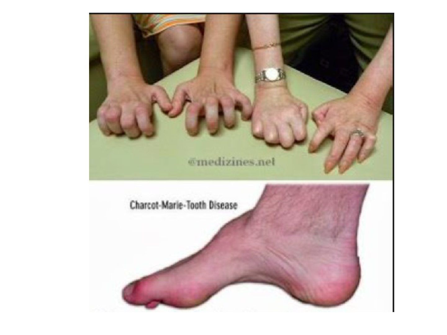
- Cavovarus +/- rigid hindfoot
- Weakened tibialis anterior and peroneal muscles (foot drop during swing phase)
- Hyporeflexia or areflexia
Q5.What other orthopaedic manifestations are associated with peroneal muscle atrophy?
- Hip dysplasia
- Scoliosis
- Hand muscle atrophy and weakness
Q6.How is peroneal muscle atrophy diagnosed?
- NCV / EMG
- DNA - look for PMP22 gene mutation
- Chromosome analysis - duplication of chromosome 17
▸ Slide 588 · Friedreich's AtaxiaNeuromuscular · 5 questions expand
Slide render
Question list
Q1-Q55 questions — tap to reveal all answerslist
- What is the inheritance, genetics and neuroanatomical involvement of Friedreich's ataxia?
- What is the classic triad and gait finding of Friedreich's ataxia?
- What conditions are associated with Friedreich's ataxia?
- How is Friedreich's ataxia diagnosed?
- At what age does loss of ambulation (LOC) occur and what is the life expectancy in Friedreich's ataxia?
Answers · Q & A
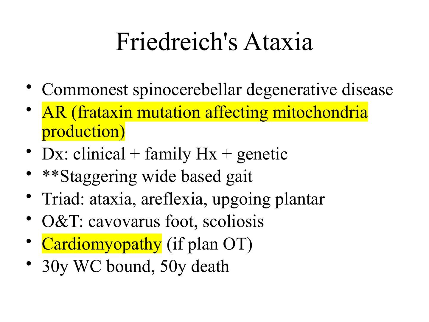
Q1.What is the inheritance, genetics and neuroanatomical involvement of Friedreich's ataxia?
- Commonest spinocerebellar degenerative disease
- Autosomal recessive (frataxin mutation affecting mitochondria production)
- Affects the cerebellar system, dorsal root ganglia, corticospinal tract and sensory peripheral nerve
Q2.What is the classic triad and gait finding of Friedreich's ataxia?
- Ataxia
- Areflexia
- Upgoing plantar response
- Staggering wide based gait
Q3.What conditions are associated with Friedreich's ataxia?
- Cardiomyopathy (if plan OT)
- Cavovarus foot
- Scoliosis
Q4.How is Friedreich's ataxia diagnosed?
- Clinical + family history + genetic testing
Q5.At what age does loss of ambulation (LOC) occur and what is the life expectancy in Friedreich's ataxia?
- Wheelchair bound by 30 y (loss of ambulation)
- Death by 50 y
▸ Slide 589 · XR of this skeletally immature patient showing broken shenton and dislocated lefNeuromuscular · 14 questions 1 check expand
Slide render
Question list
Q1-Q1414 questions — tap to reveal all answerslist
- What are the radiographic findings in this skeletally immature patient with a dislocated left hip?
- What history should be taken in a child with a neuromuscular hip dislocation?
- How is function classified and prognosis assessed in cerebral palsy?
- What is cerebral palsy and how is it classified?
- What are the principles of managing cerebral palsy?
- How is hip dysplasia in cerebral palsy prevented and treated?
- What does the physical examination include in neuromuscular disease?
- How is spasticity defined and treated globally vs focally?
- What is a contracture and how is it treated?
- What are the four types of lever arm dysfunction?
- What are the hemiplegic gait patterns and their management?
- What are the diplegic gait patterns?
- What are the timing and components of surgery in cerebral palsy?
- What soft tissue releases are used for early hip subluxation in CP?
Answers · Q & A
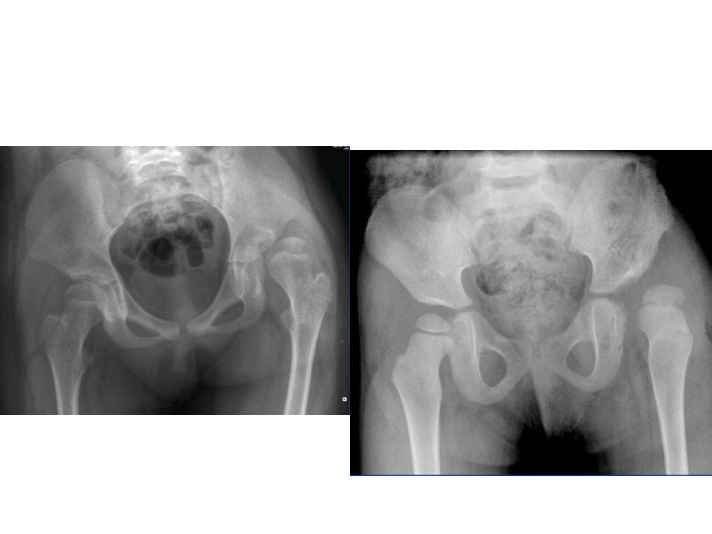
Q1.What are the radiographic findings in this skeletally immature patient with a dislocated left hip?
- Broken Shenton's line and dislocated left hip
- Increased acetabular index and increased neck-shaft angle, perhaps with more anteversion
- Relatively well formed tear drop and femoral head suggest neuromuscular dislocation rather than DDH
- Pelvis looks like an inlet view due to pelvic anteversion from tight rectus; look for VP drain
- Triradiate cartilage fuses at 11-13 years old
Q2.What history should be taken in a child with a neuromuscular hip dislocation?
- Perinatal and birth history
- Developmental milestones (age of sitting and walking)
- Problems faced by patient him/herself; Problems faced by cares (perineal hygiene, pain, transfer, sitting)
- Determine GMFCS level
- Other medical problems and social support
- Prognostication if very young - good: head control by 9 months, sit by 2 years, hemiplegic; bad prognosis: bleck, lack of head control by 20 months, non ambulatory by 7 yr
Q3.How is function classified and prognosis assessed in cerebral palsy?
- GMFCS I no restriction, II stairs with rails, III wheelchair, IV powered wheelchair, V self mobility limited
- If non-ambulatory and >1 year old, test primitive reflexes; 2 or more = poor prognosis for walking (Bleck)
- Gait analysis lab for quantitative measurements
Q4.What is cerebral palsy and how is it classified?
- Non-progressive insult to the immature brain; primary insult non-progressive but musculoskeletal features evolve with growth
- Topographically: diplegic, hemiplegic, quadriplegic
- Neurologically: spastic, hypotonic, mixed, athetoid, ataxic
- Functionally: GMFCS
- Deviations: primary (loss of selective motor control), secondary (growth related), tertiary (compensatory)
- Secondary deviation examples: Anatomic shortening of muscle tendon unit (e.g. myostatic contractures); Persistent bone deformities due to HV principle (e.g. femoral anteversion); Joint subluxations/ dislocations
Q5.What are the principles of managing cerebral palsy?
- Multi-disciplinary approach (MDT); orthopaedics has a role only in spastic type and cannot reverse the primary insult
- Overall aim: maximize patient’s function consistent with the current disease status and prevent deterioration
- Walker aims (Gage) and prevention of early OA; sitter aims: spinopelvic balance, perineal hygiene, mobile knees, plantigrade feet
- Medical spasticity: physio, analgesia, oral or intrathecal baclofen, botox (3-6 months)
- Surgery: delay until >6 years but before tertiary issues; SEMLS vs OSSCS (release/lengthen biarticular muscles, preserve monoarticular antigravity muscles)
Q6.How is hip dysplasia in cerebral palsy prevented and treated?
- Spastic biarticular adductors overpower monoarticular abductors, so the hip displaces; hip dysplasia is preventable
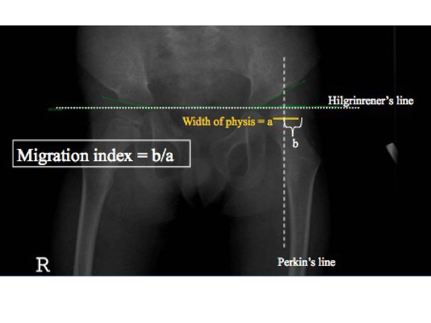
- Surveillance per NICE guidelines and the Swedish CPUP study: X-ray before 3 years; repeat one in 6 y.o; monitor Reimer migration index, hip abduction; refer if abduction <30 degrees or RMI >33%
- Early subluxation RMI 33-50%: botox/soft tissue release (adductor tenotomy, iliopsoas +/- hamstring lengthening) and night abduction splint
- Moderate subluxation RMI >50%: VDRO/pelvic osteotomy; dislocation: <1yr reconstruct, >1 year observe if painless, salvage if painful (Girdlestone, capsule interpositional arthroplasty, McHale)
- Evidence suggests less than 50% of asymptomatic dislocated hips will develop pain in the long run, so observation can be supported
- Psoas spastic, hip flexed, reduced torque on proximal femur, femur cannot derotate, so anteversion; Valgus as non weight bearing
Q7.What does the physical examination include in neuromuscular disease?
- General: IQ, sitting balance, gait (stance and swing phase), orthosis
- Hip: tone, ROM, Thomas or Staheli test for FFC, Phelps test for abduction tightness, Duncan Ely for rectus, Ober for ITB
- Knee: tone, ROM, popliteal angle (hamstring shift more accurate to see if psoas contracture contributes)
- Ankle: tone, ROM, Silverskiold test, bony impingement or impending ulcers
- Spine for scoliosis, pelvic obliquity, LLD
Q8.How is spasticity defined and treated globally vs focally?
- Velocity dependent increase in tone due to loss of supraspinal inhibition
- Global: selective dorsal rhizotomy - decrease afferent input from muscle spindle
- Global: baclofen - GABA agonist, increase K channel opening --> easy hyperpolarization
- Global: gabapentin - VGCC deactivator, decrease nerve excitation and decrease neurotransmitter release
- Focal: botox
Q9.What is a contracture and how is it treated?
- Shortening of the musculotendinous unit due to slowing of growth from lack of tensile loading
- Muscle: release or transfer; joint: release; bone: osteotomy
Q10.What are the four types of lever arm dysfunction?
- Unstable fulcrum (hip dislocation)
- Floppy lever (pes planus)
- Short lever (coxa valga)
- Malrotation (tibial ER)
Q11.What are the hemiplegic gait patterns and their management?
- Foot drop -> hinged AFO / solid AFO
- True equinus (gastroc soleus contracture) -> hinged AFO -> heel cord lengthening
- Jump knee (spastic hamstring + triceps surae) -> hinged AFO -> no OT
- Apparent equinus (iliopsoas contracture + gastroc weakness) -> solid AFO -> psoas lengthening
Q12.What are the diplegic gait patterns?
- True equinus
- Jump knee
- Apparent equinus
- Crouch knee
- Recurvatum knee
- Stiff knee (swing phase problem)
Q13.What are the timing and components of surgery in cerebral palsy?
- Timing: delay OT as long as possible (>6 years) but before tertiary issues - contain the hip before dislocation, modify gait before deviation
- SEMLS vs OSSCS (orthopaedic selective spasticity control surgery)
- OSSCS: correct lever arm dysfunction by releasing or lengthening biarticular muscles and preserving monoarticular antigravity muscles
- Components: balance spasticity, prevent contractures, correct lever arm dysfunction
Q14.What soft tissue releases are used for early hip subluxation in CP?
- Adductor longus transect - protect the anterior branch of the obturator nerve
- Gracilis myotomy and adductor brevis lengthening
- Iliopsoas lengthening +/- hamstring lengthening
- Nighttime abduction splint
Fact check
Triradiate cartilage fuses at 11-13 years old — imprecise age range — Triradiate cartilage closes at about 12 years bone age in girls and 14 in boys (radiographic closure roughly 13-14 years in females and 15-16 in males); overall quoted range is about 12-16 years. — medium confidence — source
▸ Slide 590Neuromuscular · 2 questions expand
Slide render
Question list
Q1-Q22 questions — tap to reveal all answerslist
- What is the topic of this slide?
- What clinical content is shown on this slide?
Answers · Q & A
Q1.What is the topic of this slide?
- Neuromuscular
- Not covered in the speaker notes - slide image only
Q2.What clinical content is shown on this slide?
- Not covered in the speaker notes
▸ Slide 591 · GMFCS (Gross Motor Function classification score)Neuromuscular · 4 questions expand
Question list
Q1-Q44 questions — tap to reveal all answerslist
- What are the GMFCS levels?
- What is the most predictive factor for becoming an independent walker?
- What are the goals for a non-ambulator with cerebral palsy?
- What are the Gage principles for an ambulator?
Answers · Q & A
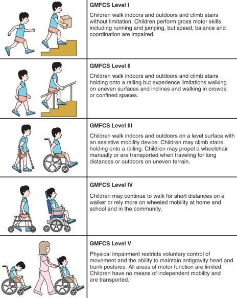
Q1.What are the GMFCS levels?
- Level 1: no restriction in walking, limited in advanced motor skills
- Level 2: no aids, some limitation outdoors or in the community
- Level 3: needs assistive aids, limitation outdoors or in the community
- Level 4: self mobility with limitation, needs transport or powered mobility outdoors
- Level 5: self mobility severely limited even with assistive technology
Q2.What is the most predictive factor for becoming an independent walker?
- independent sitting at 2 y.o
Q3.What are the goals for a non-ambulator with cerebral palsy?
- Good sitting balance, forward gaze, use UL for feeding
- Spine straight, shoulder and pelvis level
- Hip located, stable, pain free with good ROM
- Knee mobile, flexed for sitting, extends in brace for transfer
- Foot plantigrade for standing
Q4.What are the Gage principles for an ambulator?
- Stability in stance
- Good clearance in swing
- Preposition of the foot at end of swing
- Adequate step length
- Energy conservation
▸ Slide 592 · Crouch gaitNeuromuscular · 5 questions expand
Slide render
Question list
Q1-Q55 questions — tap to reveal all answerslist
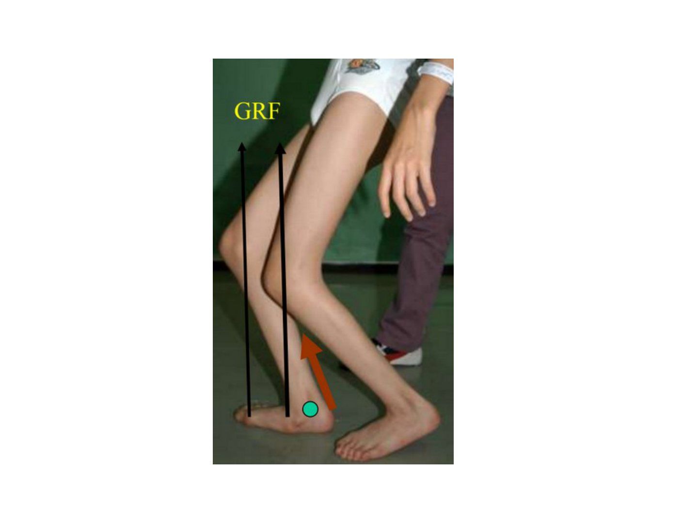
- What are the midstance features of crouch gait?
- What are the causes of crouch gait?
- Briefly explain the mechanism of crouch gait.
- How is crouch gait managed?
- What are the sequelae of hamstring lengthening and why is the semiT transferred?
Answers · Q & A
Q1.What are the midstance features of crouch gait?
- Excess ankle dorsiflexion
- Knee flexion
- Hip flexion
- Overall short stride length and muscle atrophy
Q2.What are the causes of crouch gait?
- Weakness (triceps surae, quads)
- Spasticity (hamstrings, iliopsoas)
- Joint contracture (knee flexion contracture)
- Lever arm dysfunction (increased femoral anteversion, increased tibial torsion, collapse tripod)
Q3.Briefly explain the mechanism of crouch gait.
- Weak gastrocsoleus / excessive ankle dorsiflexion means the knee extension / ankle plantarflexion couple cannot work
- GRF falls posterior to the knee, producing a knee flexion moment
Q4.How is crouch gait managed?
- Weakness: avoid iatrogenic excessive gastrocsoleus release/recession; GRAFO for gastroc; hamstring transfer for quads
- Spasticity: hamstring lengthening (semiM + gracilis fractional +/- biceps femoris) and semiT to adductor tubercle transfer
- Psoas spasticity: release at the pelvic brim or LT
- Joint contracture: extension osteotomy
- Lever arm dysfunction: reconstruct hip, tibia or foot
Q5.What are the sequelae of hamstring lengthening and why is the semiT transferred?
- Sequelae: weak hip extension + anterior pelvic tilt
- SemiT to adductor tubercle transfer converts a biarticular to monoarticular muscle
- Aim: prevent hip extension and decrease anterior pelvic tilt
▸ Slide 593 · Recurvatum gaitNeuromuscular · 3 questions expand
Slide render
Question list
Q1-Q33 questions — tap to reveal all answerslist
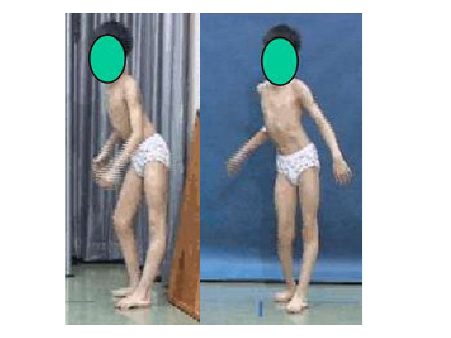
- What compensatory mechanisms maintain balance in recurvatum gait?
- What is the cause of recurvatum gait and how does age guide treatment?
- What operations are used for fixed triceps surae contracture in recurvatum gait?
Answers · Q & A
Q1.What compensatory mechanisms maintain balance in recurvatum gait?
- Knee recurvatum with the ankle in plantarflexion at midstance
- Hip flexion moves the CG anterior to balance the GRF
- Contralateral crouch occurs due to pelvic obliquity
Q2.What is the cause of recurvatum gait and how does age guide treatment?
- Contracture of triceps surae
- <6 years / spasticity: botox, stretching, solid AFO
- Contracture + >6 years: assess with Silverskiold
Q3.What operations are used for fixed triceps surae contracture in recurvatum gait?
- Gastroc only: gastroc recession
- Gastroc + soleus: ETA
- Applied when there is contracture and the patient is >6 years old
▸ Slide 594 · Jump knee gaitNeuromuscular · 4 questions expand
Slide render
Question list
Q1-Q44 questions — tap to reveal all answerslist
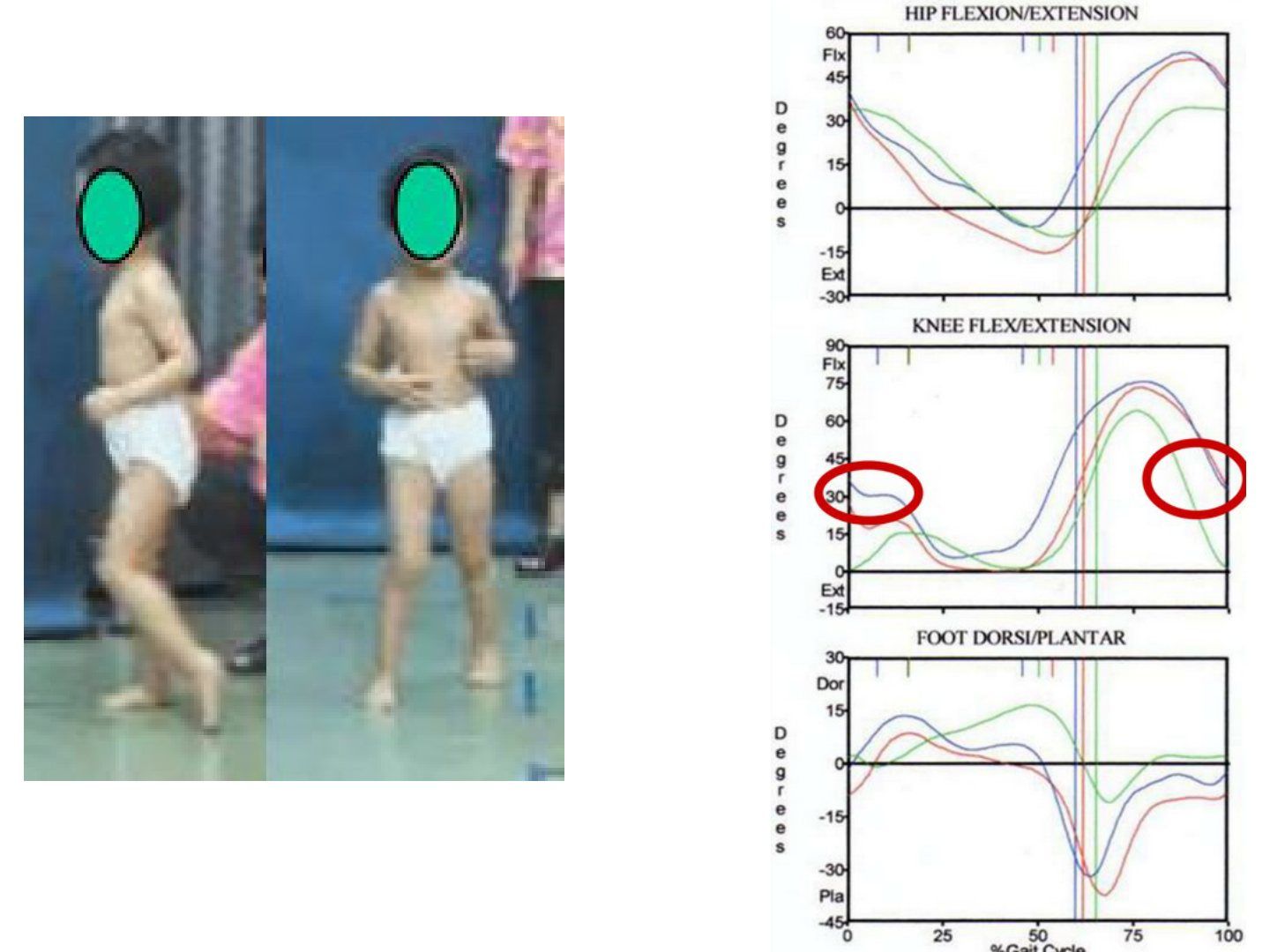
- What are the features of jump knee gait?
- What causes jump knee gait and how is it managed?
- What is gait analysis?
- What is the set-up for gait analysis?
Answers · Q & A
Q1.What are the features of jump knee gait?
- Ankle equines in late stance
- Knee flexion in early stance and terminal swing
- Hip flexion in early stance
- Pelvis normal or anteriorly tilted
Q2.What causes jump knee gait and how is it managed?
- Problem is spastic hamstring and triceps surae
- No OT
- Serial cast, botox, stretching, hinged AFO
Q3.What is gait analysis?
- Systematic description, assessment and measurement of the quantities that characterise human locomotion
- Components: kinematic, kinetic, EMG, energy consumption
Q4.What is the set-up for gait analysis?
- 2D video analysis
- 3D computer analysis using specialised markers on specific bony landmarks
- Force plates to measure GRF
- EMG to look at muscle firing patterns
▸ Slide 595 · Stiff knee gaitNeuromuscular · 3 questions expand
Slide render
Question list
Q1-Q33 questions — tap to reveal all answerslist
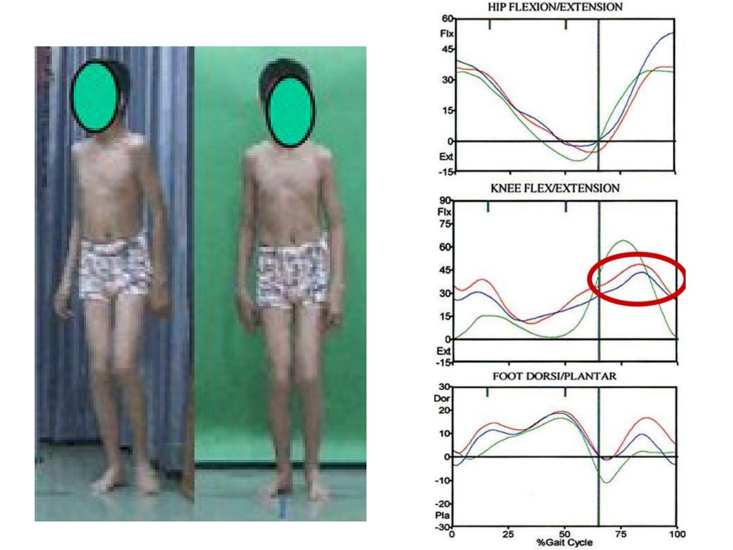
- What causes stiff knee gait and when is knee flexion reduced?
- How is stiff knee gait managed in a child under 6 years?
- What are the indications for rectus to pes transfer?
Answers · Q & A
Q1.What causes stiff knee gait and when is knee flexion reduced?
- Spastic rectus femoris or rectus firing out of phase
- Decreased knee flexion in terminal stance and throughout the swing phase
Q2.How is stiff knee gait managed in a child under 6 years?
- Botox and stretching
Q3.What are the indications for rectus to pes transfer?
- Peak knee flexion <50 degrees
- Peak knee flexion later than 30% of swing
- Active rectus in the mid 3/5 of swing
- Toe dragging
▸ Slide 596 · Winters classificationNeuromuscular · 4 questions expand
Question list
Q1-Q44 questions — tap to reveal all answerslist
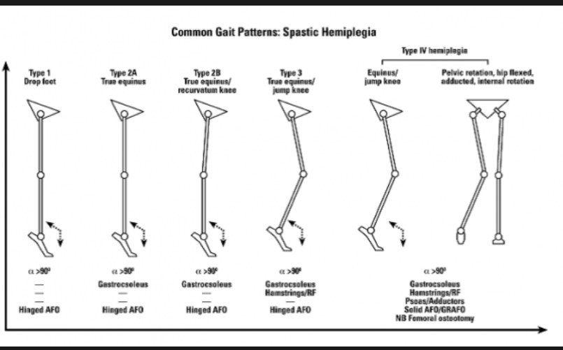
- What is Winters type 1 gait?
- What is Winters type 2 gait?
- What is Winters type 3 gait?
- What is Winters type 4 gait?
Answers · Q & A
Q1.What is Winters type 1 gait?
- Type 1 = 'drop foot' = equinus in swing phase only
Q2.What is Winters type 2 gait?
- Type 2a = 'true equinus' = equinus in the whole gait cycle
- Type 2B = true equinus + recurvatum knee
Q3.What is Winters type 3 gait?
- 'Jump knee' = equinus + tight hamstrings
Q4.What is Winters type 4 gait?
- Equinus + tight hamstrings + tight flexors and adductors
- Results in adduction and internal rotation deformity
▸ Slide 597 · Sutherland classificationNeuromuscular · 4 questions expand
Question list
Q1-Q44 questions — tap to reveal all answerslist
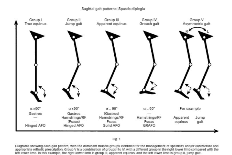
- What is true equinus and how is it managed?
- What is apparent equinus and how is it managed?
- What is crouch gait (equinoplanovalgus) and how is it managed?
- What is jump gait in the Sutherland classification and how is it managed?
Answers · Q & A
Q1.What is true equinus and how is it managed?
- Tight gastrocsoleus complex
- Mx: hinged AFO (allows dorsiflexion while blocking plantarflexion)
Q2.What is apparent equinus and how is it managed?
- Ankle goes into dorsiflexion due to persistent loading with lengthening of the Achilles tendon
- Ankle ROM is still normal
- Mx: solid AFO
Q3.What is crouch gait (equinoplanovalgus) and how is it managed?
- Excessive dorsiflexion due to an overlengthened Achilles
- Can be caused iatrogenically by releasing the Achilles while ignoring hamstring tightness
- Mx: GRAFO
Q4.What is jump gait in the Sutherland classification and how is it managed?
- Tight hamstrings flex the knee and the hip flexes to push the CG in front, with progressively worse equinus
- Mx: hinge AFO
▸ Slide 598 · Strayer --> gastroc release from soleus proximal to conjoint tendon --> 1cmNeuromuscular · 3 questions expand
Question list
Q1-Q33 questions — tap to reveal all answerslist
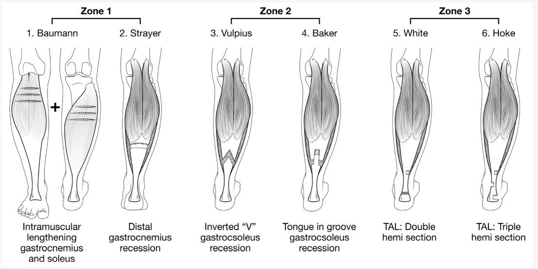
- Describe the Strayer, Vulpius and Baker techniques.
- Describe the White and Hoke PETA techniques.
- Which zone is preferred in cerebral palsy and why?
Answers · Q & A
Q1.Describe the Strayer, Vulpius and Baker techniques.
- Strayer: gastroc release from soleus proximal to the conjoint tendon -> 1 cm
- Vulpius: chevron at the MST junction -> 1.5 cm
- Baker: central fascial tongue
Q2.Describe the White and Hoke PETA techniques.
- White PETA: 1 medial + 1 lateral incision -> 2-4 cm
- Hoke PETA: 2 medial + 1 lateral incision -> 4 cm
Q3.Which zone is preferred in cerebral palsy and why?
- Zone 1 is more preferred in CP
- It allows a more selective release