15 slides
▸ Slide 569 · SpineSpine Paed · 3 questions expand
Slide render
Question list
Q1-Q33 questions — tap to reveal all answerslist
- How would you assess a child with a suspected spinal deformity?
- Which investigations are used for paediatric spine conditions?
- What are the surgical indications in paediatric spinal deformity?
Answers · Q & A
Q1.How would you assess a child with a suspected spinal deformity?
- Not covered in the speaker notes
Q2.Which investigations are used for paediatric spine conditions?
- Not covered in the speaker notes
Q3.What are the surgical indications in paediatric spinal deformity?
- Not covered in the speaker notes
▸ Slide 570 · TorticollisSpine Paed · 14 questions expand
Slide render

Question list
Q1-Q1414 questions — tap to reveal all answerslist
- What are the causes of torticollis?
- Describe the Fielding CT classification of atlantoaxial rotatory instability.
- How does management of AARI depend on timing?
- What are the indications and types of surgery for congenital muscular torticollis?
- What are the dangers of bipolar release?
- What is the evidence for conservative management of CMT (Cheng 2001)?
- What are the clinical features of the torticollis patient shown?
- What history is important in torticollis?
- What examination findings should be sought in torticollis?
- What investigations are used in torticollis?
- What is Grisel's syndrome?
- What is the pathophysiology of congenital muscular torticollis?
- Describe the bipolar release technique.
- Why are children more prone to atlantoaxial rotatory instability?
Answers · Q & A
Q1.What are the causes of torticollis?
- Congenital (usually painless): CMT, Klippel-Feil
- Acquired osseous (usually painful): AARI (trauma, tumour, infection, odontoid hypoplasia in Down's/achondroplasia, JRA/RA)
- Non-osseous: ENT, eye (squint), CNS (Arnold Chiari), postural (usually painless), neck burns
Q2.Describe the Fielding CT classification of atlantoaxial rotatory instability.
- 1: Unilateral subluxation with intact transverse ligament (dens is the pivot)
- 2: Unilateral anterior subluxation 3-5mm (opposite facet acts as pivot)
- 3: Bilateral anterior subluxation >5mm
- 4: Posterior subluxation
Q3.How does management of AARI depend on timing?
- <1 week: soft neck collar, analgesics
- 1-4 weeks: Halter traction (2-3kg), 1hr off/3hr on cycle, until reduced, then SOMI 4wk
- >4 weeks: halo traction 4 weeks, high chance of failure then OT fusion C1-2
- Other OT indications: irreducible >3 months, neurological deficit, recurrent
Q4.What are the indications and types of surgery for congenital muscular torticollis?
- Presentation >1 year
- >15 degree head tilt/lag with tumour
- Failed conservative management x6 months
- Unipolar release: sternal head <3yo, sternal + clavicular head 3-6yo
- Bipolar release >6-10 years (alternative Z plasty)
- Dangers: greater auricular nerve (surface), CNXI (deep), facial nerve (anterior)
Q5.What are the dangers of bipolar release?
- Surface - greater auricular nerve
- Underneath - CNXI (CN XI)
- Anterior - facial nerve
Q6.What is the evidence for conservative management of CMT (Cheng 2001)?
- 800 patients with onset <1 year
- 95% good response to physiotherapy (flexion, rotation, tilting), mean follow-up 4.5 years
- Poor prognostic factors: 15deg rotation deficit, SCM tumour, late presentation (present >1yo) (also stated as >15 degree rotation deficit, SCM tumour, late presentation >1 year)
Q7.What are the clinical features of the torticollis patient shown?
- Head tilting to the right and rotating to the left
- No plagiocephaly or facial asymmetry
- No neck scar, no obvious strabismus
Q8.What history is important in torticollis?
- Perinatal history: oligohydramnios, breech, first born; known congenital conditions (e.g. Klippel-Feil)
- Pain, onset of symptoms, progression, trauma, previous treatment or H&N problems
- Hearing or optical impairment, recent URI symptoms, functional impairment
Q9.What examination findings should be sought in torticollis?
- Plagiocephaly, low hairline, scapula (KF syndrome); facial dysmorphism/asymmetry
- SCM spasm, tumour, cervical LN
- Rotation and tilt lag 15 degrees; squint, hearing; neuro exam
- If newborn, look for other packaging disorders
Q10.What investigations are used in torticollis?
- Bloods: CBC, inflammatory markers
- Xray: lateral mass asymmetry on open mouth view, lateral for ADI, trapezoidal anterior C1 shadow, oval posterior arch sign; exclude fracture or congenital abnormalities
- Dynamic CT is gold standard for AARI (book if failed initial conservative management), 3 sets to look for fixed relationship between C1 and C2
- +/- contrast to rule out retropharyngeal abscess if raised inflammatory markers; +/- MRI to exclude infection or tumour
Q11.What is Grisel's syndrome?
- Drainage of inflammatory mediators through the pharyngovertebral vein to the periodontoid plexus
- Leads to ligamentous laxity
Q12.What is the pathophysiology of congenital muscular torticollis?
- Local compartment syndrome of the SCM
- Remember to look for other packaging disorders
Q13.Describe the bipolar release technique.
- 2cm incision below the mastoid, strip attachment from bone
- 2cm incision above the sterno-clavicular junction, incise platysma, cut both heads of SCM with the deep fascia
- Postop: intensive stretching for 3 months; >2yo add adjustable torticollis brace for 3 months
Q14.Why are children more prone to atlantoaxial rotatory instability?
- Capsular laxity
- Facet more transverse
▸ Slide 571 · Acute curve at TLJ convex to right sideSpine Paed · 8 questions 1 check expand
Slide render
Question list
Q1-Q88 questions — tap to reveal all answerslist
- What is congenital scoliosis and how is it classified?
- What associated anomalies should be screened for in congenital scoliosis?
- What is the natural history of congenital scoliosis?
- What are the surgical options for congenital scoliosis?
- Which types of congenital scoliosis may be observed without surgery?
- What are the clinical and radiological clues in this case?
- What history and examination are needed in suspected congenital scoliosis?
- What is the aim of management in congenital scoliosis?
Answers · Q & A
Q1.What is congenital scoliosis and how is it classified?
- Failure to form normal vertebrae at gestation week 4-6
- Failure of formation / segmentation / mixed (MacEwen classification)
- Failure of formation divided by segmentation (segmented, semisegmented, nonsegmented) and extent (partial vs complete)
- Most are sporadic
Q2.What associated anomalies should be screened for in congenital scoliosis?
- Extraspinal: VACTERL, Klippel-Feil, NF
- Intraspinal: Chiari malformation, tethered cord, syrinx - MRI
- Internal organs: renal USG and echo
Q3.What is the natural history of congenital scoliosis?
- McMaster: 485 patients, 75% need surgical treatment
- Prognosis depends on type of anomaly and anatomical site
- Progression by type: mixed > unilateral bar > fully segmented > partially segmented > incarcerated > nonsegmented
- Position: TLJ worse; also younger age
Q4.What are the surgical options for congenital scoliosis?
- Early in situ fusion for minimal deformity (young: A+P fusion; older: P fusion)
- Hemivertebrectomy for marked truncal imbalance (<5yo, flexible curves <40)
- Convex hemiepiphysiodesis for unilateral failure of formation (<5yo, progressive curves <40-50)
- Spinal column shortening resection for rigid, severe late deformities with decompensation
- Growing rods maximise spinal growth but are controversial
Q5.Which types of congenital scoliosis may be observed without surgery?
- Incarcerated hemivertebrae
- Nonsegmented hemivertebrae
- Some partially segmented hemivertebrae
- Selected because of absence of progression
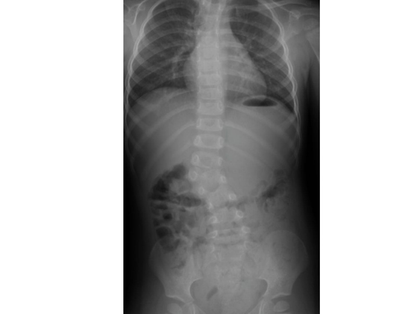
Q6.What are the clinical and radiological clues in this case?
- Acute curve at the TLJ convex to the right
- One pedicle less on the left, seems unsegmented
- Suspect congenital scoliosis with hemivertebra
Q7.What history and examination are needed in suspected congenital scoliosis?
- Hx: birth history, walking age and motor development, associated problems (Klippel-Feil, NF, VACTERL)
- PE: maturity (Tanner-Whitehouse), shoulder balance, rib or loin hump, truncal shift or listing
- Neurology, gait, skin stigmata
Q8.What is the aim of management in congenital scoliosis?
- Arrest progression
- Achieve a balanced spine while preserving as much spinal growth as possible
Fact check
McMaster's natural history paper studied 485 patients, 75% needing surgery — Incorrect patient number — McMaster & Ohtsuka (JBJS 1982) studied 251 patients; 485 does not match the cited source — source
▸ Slide 572 · XR showing scoliosis, with major right thoracic curve. Apex as T8/9. Cobb angle Spine Paed · 13 questions expand
Slide render
Question list
Q1-Q1313 questions — tap to reveal all answerslist
- What are the indications for MRI in scoliosis?
- How do you assess maturity and risk of progression?
- What is the brace treatment and its evidence?
- What are the indications and aims of surgery in AIS?
- Compare anterior and posterior fusion in AIS.
- Why operate in AIS (natural history)?
- Describe the XR findings in this AIS case.
- What history and maturity assessment is needed in AIS?
- What are the components of the general and standing examination in AIS?
- Which classifications are used for AIS curves?
- How is a brace weaned in AIS?
- What are the key elements of scoliosis surgery?
- Describe the Hong Kong scoliosis screening programme.
Answers · Q & A
Q1.What are the indications for MRI in scoliosis?
- Left-sided thoracic curve
- Short angular curve, angled kyphosis
- Neurology, pain
- <10yr + >20 deg (<10 years + >20 degrees)
- Rapid curve progression
Q2.How do you assess maturity and risk of progression?
- Age 9-13 with >2 years before menarche
- growth rate >2cm/yr (>2cm/year)
- Risser 0-1, hand XR capped stage (TW III), open triradiate cartilage
- Risser stages: I prepubertal, II growth spurt, III decreasing growth rate, IV variable
- Limitations: peak height velocity and 2/3 of growth spurt occur before Risser I; curve may not stop at Risser IV
Q3.What is the brace treatment and its evidence?
- The aim of the brace is to slow down curve progression; effect is dosage related (>20 hr/day -> 93% success)
- Keep bracing till skeletal maturity
- Weinstein BRAIST trial (NEJM 2013): 72% success vs 48% observation
- Complications: GI regurgitation, skin irritation/dermatitis/folliculitis, pressure, low self-esteem
Q4.What are the indications and aims of surgery in AIS?
- Curves >45 degrees in a growing patient or >50 degrees when growth has stopped
- Aim: correct deformity, square fusion block with <2cm listing and <20 degree tilt
- Include all structure curves (i.e. all structural curves) while saving fusion segments
- Stable fusion and prevent complications
Q5.Compare anterior and posterior fusion in AIS.
- Anterior: short segment, increase correction power by discectomy, for hypokyphosis; disadvantage difficult exposure
- Posterior: easy exposure; disadvantage long segment, difficult full correction
- Combined indications: stiff curve (>50 residual on FB XR), severe curve (>70), immature (female <10, male <13) to prevent crankshaft
Q6.Why operate in AIS (natural history)?
- Progression >50 degrees -> more likely SOB; >70 deg --> decreased lung function; >100 deg increase risk of death
- Increased mortality and cardiopulmonary compromise
- Truncal imbalance and back pain
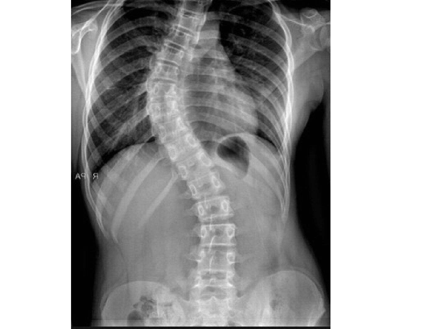
Q7.Describe the XR findings in this AIS case.
- Major right thoracic curve, apex T8/9, Cobb angle 40 degrees
- Shoulders and pelvis level; truncal shift and truncal listing
- No vertebral anomalies such as hemivertebrae or unsegmented bar, ribs normal; Risser sign 3; Triradiate cartilage status
Q8.What history and maturity assessment is needed in AIS?
- Birth history, developmental history and family history of scoliosis
- Current symptoms: age of onset and progression, pain or neurological symptoms, visceral problems
- Assess maturity: menarche, growth spurt
Q9.What are the components of the general and standing examination in AIS?
- General: body height, arm span, sitting height; cutaneous lesions; ligamentous laxity/marfanoid features; spinal dysraphism
- Standing coronal: shoulder/pelvis symmetry, rib or loin hump, decompensation (truncal shift and list)
- Sagittal: hypokyphosis, kyphoscoliosis; bend forward for flexibility; gait (scissoring gait)
- Sit: postural scoliosis; supine: neuro exam + superficial abdominal reflexes; finish by checking secondary sexual characteristics
Q10.Which classifications are used for AIS curves?
- Lenke classification, King classification, Peking Union Medical College (PUMC), Labelle 3D
- This case is a major thoracic curve
Q11.How is a brace weaned in AIS?
- At skeletal maturity: no growth in 18 months, Risser 4, menarche >2 years, DR and DU physis closed
- Done in a gradual manner - no hard science; e.g. spread over 4 months with stepwise decrease in hours/day
Q12.What are the key elements of scoliosis surgery?
- Posterior approach; subperiosteal dissection till transverse processes
- Identify upper and lower instrumented vertebrae (ASF: end to end; PSF: proximal end; distal Lenke I-II: most distal VB touched by CVSL, III-VI: distal end VB)
- Pedicle screw insertion; reduction techniques: Harrington forces, cantilever bending, translation, rod rotation, direct vertebral rotation
- Decortication with bone graft to interlaminar space and into facets
Q13.Describe the Hong Kong scoliosis screening programme.
- 3 tier system starting in P5
- FBT + ATR >=15 -> refer to scoli centre
- 5-14: Moire topography -> >=2 lines -> XR -> >20 degrees -> refer specialist hospital
- <5: re-refer to screening programme
▸ Slide 573 · AISSpine Paed · 6 questions expand
Slide render
Question list
Q1-Q66 questions — tap to reveal all answerslist
- What does the Nash-Moe technique assess?
- How do you describe the X-ray in AIS?
- What is the aim of clinical assessment in AIS?
- What are the key clinical findings on examination in AIS?
- What imaging is required in AIS?
- What is the management principle in AIS and how is a progressing curve predicted?
Answers · Q & A
Q1.What does the Nash-Moe technique assess?
- Vertebral rotation
- A radiographic measure of rotation in scoliosis (AIS)
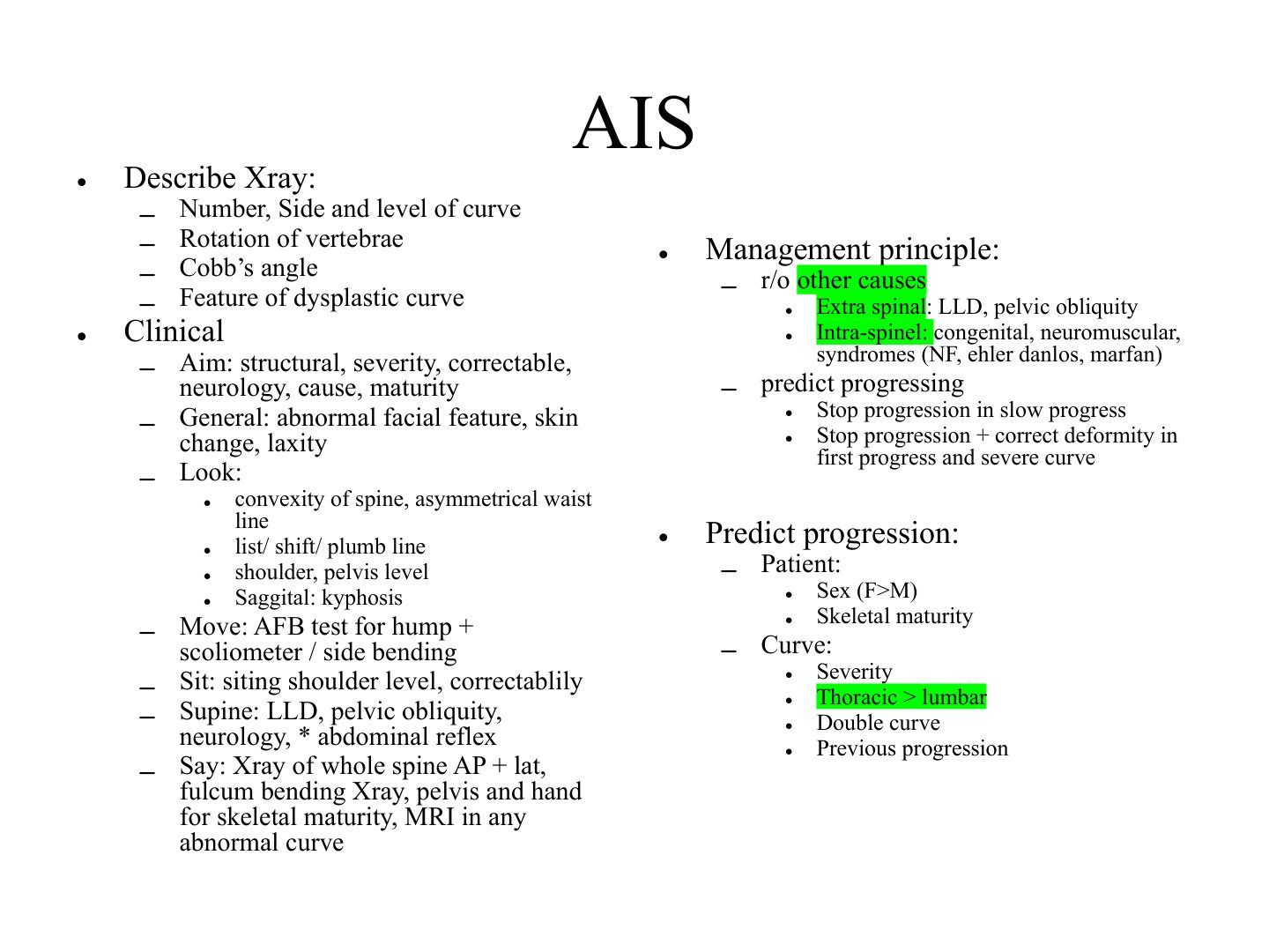
Q2.How do you describe the X-ray in AIS?
- Number, side and level of curve
- Rotation of vertebrae (Nash-Moe)
- Cobb angle
- Feature of dysplastic curve
Q3.What is the aim of clinical assessment in AIS?
- Aim: determine structural, severity, correctable
- Look for neurology, cause, maturity
Q4.What are the key clinical findings on examination in AIS?
- General: abnormal facial feature, skin change, laxity
- Look: convexity of spine, asymmetrical waist line, list/shift/plumb line, shoulder, pelvis level
- Sagittal kyphosis assessment
- Move: AFB test for hump + scoliometer / side bending
- Sit: sitting shoulder level, correctability
- Supine: LLD, pelvic obliquity, neurology, abdominal reflex
Q5.What imaging is required in AIS?
- Xray of whole spine AP + lateral
- Fulcrum bending Xray
- Pelvis and hand X-ray for skeletal maturity
- MRI in any abnormal curve
Q6.What is the management principle in AIS and how is a progressing curve predicted?
- Rule out other causes: extra-spinal (LLD, pelvic obliquity) and intra-spinal (congenital, neuromuscular, syndromes - NF, Ehlers-Danlos, Marfan)
- Stop progression in slow progress; stop progression + correct deformity in fast progress and severe curve
- Predict progressing curve - patient: sex (F>M), skeletal maturity
- Predict progressing curve - curve: severity, thoracic > lumbar, double curve, previous progression
▸ Slide 574 · AISSpine Paed · 4 questions expand
Slide render

Question list
Q1-Q44 questions — tap to reveal all answerslist
- What are the treatment options for AIS?
- What are the indications, principles and complications of bracing in AIS?
- What are the indications and principle of operative treatment in AIS?
- How is the fusion level chosen and what instrumentation is used in AIS?
Answers · Q & A
Q1.What are the treatment options for AIS?
- Treatment option: observe
- Brace above T7 (Milwaukee/Boston)
- Operation
Q2.What are the indications, principles and complications of bracing in AIS?
- Indication: >20 degrees in a skeletally immature patient
- 3 point fixation
- Continue till skeletal maturity with gradual wean off
- Assessment: compliance, complication, progression of curve
- Complications: sore, esophagitis, stiffness
Q3.What are the indications and principle of operative treatment in AIS?
- Indication: >40 degrees if immature, >50 degrees if mature
- Principle: stop progression + correct deformity
- Include all structural curves, achieving a solid fusion block with minimal tilting and shift
Q4.How is the fusion level chosen and what instrumentation is used in AIS?
- Level depends on instrumentation, usually referenced to end vertebrae (mildly tilted) or stable vertebrae (vertebrae bisected by the mid-sacral line)
- Classical: Luque rod, Harrington rod (cannot control rotation), pedicle screws with rod
▸ Slide 575 · AISSpine Paed · 3 questions expand
Slide render

Question list
Q1-Q33 questions — tap to reveal all answerslist
- What is the progression risk for a Risser 2 patient with a curve of 20-29 degrees?
- How do treatment and progression risk vary with Risser grade and curve angle?
- At what curve magnitude does treatment start and when is growth most rapid?
Answers · Q & A
Q1.What is the progression risk for a Risser 2 patient with a curve of 20-29 degrees?
- Risser 2
- Curve 20-29 degrees
- Progression risk 22%
Q2.How do treatment and progression risk vary with Risser grade and curve angle?
- Risser 0-1 (pre-menarche): 10-19 physio (22% curve progression); 20-29 brace (68%); 30-39 brace; 40-49 surgery; >50 surgery
- Risser 2-3 (<1 year after menarche): 10-19 physio (2%); 20-29 physio (22%); 30-39 brace; 40-49 ?surgery (for curve >45); >50 surgery
- Risser 4-5: physio for 10-19, 20-29, 30-39 and 40-49; >50 surgery
Q3.At what curve magnitude does treatment start and when is growth most rapid?
- Start treatment at 20-30 degrees
- Time of most rapid growth is at Risser 0
▸ Slide 576 · ClassificationSpine Paed · 5 questions expand
Slide render

Question list
Q1-Q55 questions — tap to reveal all answerslist
- What does the PUMC classification include?
- What does the King classification describe?
- What does the Lenke classification describe?
- How are curve sites defined in the Lenke classification?
- What defines a structural curve?
Answers · Q & A
Q1.What does the PUMC classification include?
- Peking Union Medical College classification of AIS
- Includes axial rotation
Q2.What does the King classification describe?
- King's - site of curve
- Double, lumbar major
- Double, thoracic major
- Single thoracic
- L thoracic (include L4)
- Double thoracic
- --> Guide usage of Harrington rod
Q3.What does the Lenke classification describe?
- Number and site of structural curve (6)
- Modifiers: central sacral vertical line to lumbar apex, kyphosis
- Curve site: proximal thoracic, main thoracic, TLJ/lumbar
- Types: main thoracic, double thoracic, double major, triple major, thoracolumbar, thoracolumbar/lumbar + main thoracic
Q4.How are curve sites defined in the Lenke classification?
- T: T2-T11/12 disc
- TLJ (thoracolumbar junction): T12 - L1
- L: L1/2 disc - L4
Q5.What defines a structural curve?
- Side bending Cobb's angle ≥25 degrees
- Kyphosis ≥20 degrees
▸ Slide 577 · NM scoliosisSpine Paed · 4 questions expand
Slide render
Question list
Q1-Q44 questions — tap to reveal all answerslist
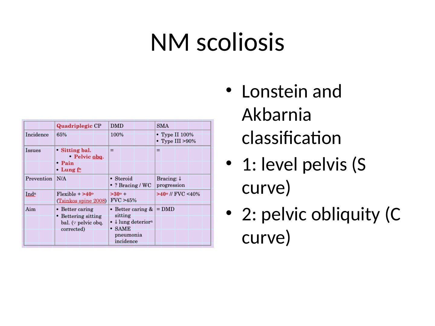
- What is the Lonstein and Akbarnia classification of neuromuscular scoliosis?
- What is the deformity pattern in neuromuscular scoliosis and why?
- How is juvenile scoliosis classified and which group has a poorer prognosis?
- What is the management of neuromuscular/juvenile scoliosis?
Answers · Q & A
Q1.What is the Lonstein and Akbarnia classification of neuromuscular scoliosis?
- 1: level pelvis (S curve)
- 2: pelvic obliquity (C curve)
Q2.What is the deformity pattern in neuromuscular scoliosis and why?
- C curve
- Frontal: primary from erector spinae imbalance; secondary compensatory for hip adductor contracture
- Sagittal: most compensation for hip -> hyperlordosis
- TL/L more involved as more mobile; more listing; long fusion
Q3.How is juvenile scoliosis classified and which group has a poorer prognosis?
- Early onset <5 years vs late onset >5 years
- <5 years has poorer prognosis due to thoracic insufficiency syndrome
Q4.What is the management of neuromuscular/juvenile scoliosis?
- <20 degrees: observation
- Cobb 20-50: brace
- Cobb >50: growth modulation with growing rods or tether if growth potential remains
- If surgery/fusion, may need to consider adding anterior instrumentation
▸ Slide 578 · Other scoliosisSpine Paed · 8 questions expand
Question list
Q1-Q88 questions — tap to reveal all answerslist
- What is juvenile scoliosis and how is it subdivided?
- How is juvenile scoliosis managed according to Cobb angle?
- What is the natural history of infantile scoliosis and why is MRI needed?
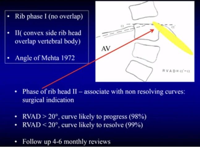
- What is the rib-vertebral angle difference (Mehta angle)?
- What are the three predictors for infantile curve growth?
- How is infantile scoliosis treated according to Cobb angle and RVAD?
- What types of scoliosis are covered under 'other scoliosis'?
- What is the rib phase in infantile scoliosis and what does overlap mean?
Answers · Q & A
Q1.What is juvenile scoliosis and how is it subdivided?
- Early onset (<5 years) vs late onset (>5 years)
- <5 years: poorer prognosis due to Thoracic Insufficiency Syndrome
- Juvenile scoliosis requires MRI
Q2.How is juvenile scoliosis managed according to Cobb angle?
- Cobb <20 degrees: observation
- Cobb 20-50 degrees: brace
- Cobb >50 degrees: growth modulation with growing rods or tether if growth potential remains
- If fusion is needed, consider adding anterior instrumentation
Q3.What is the natural history of infantile scoliosis and why is MRI needed?
- Most resolve spontaneously
- Most common curve: left thoracic (75%)
- MRI needed: 20% present with neuroaxial abnormalities and to rule out tethered cord
Q4.What is the rib-vertebral angle difference (Mehta angle)?
- Angle between a perpendicular line from the apical vertebra endplate and the rib, comparing both sides
- Rib phase: overlap of rib and vertebral body (phase 2) = non-resolving curve
Q5.What are the three predictors for infantile curve growth?
- Cobb >20 degrees
- Rib-vertebral angle difference (Mehta angle) >20 degrees
- Rib-vertebral overlap (rib phase 2)
Q6.How is infantile scoliosis treated according to Cobb angle and RVAD?
- Cobb <25, RVAD <20: observation
- Cobb <35, RVAD >20: Mehta serial casting and bracing
- Cobb >50: surgery with VEPTR or growing rods
Q7.What types of scoliosis are covered under 'other scoliosis'?
- Juvenile scoliosis
- Infantile scoliosis
- Congenital scoliosis
Q8.What is the rib phase in infantile scoliosis and what does overlap mean?
- Rib phase is the overlap of the rib and vertebral body
- Overlap = rib phase 2 = non-resolving curve
▸ Slide 579 · VEPTR (Vertical Expandable Prosthetic Titanium Rib)Spine Paed · 3 questions expand
Slide render

Question list
Q1-Q33 questions — tap to reveal all answerslist
- What does VEPTR stand for?
- What is the role of VEPTR in early-onset scoliosis?
- When is VEPTR used in the lecture?
Answers · Q & A
Q1.What does VEPTR stand for?
- Vertical Expandable Prosthetic Titanium Rib
- Not covered in the speaker notes beyond the title
Q2.What is the role of VEPTR in early-onset scoliosis?
- Not covered in the speaker notes
- Review the slide image and lecture recording
Q3.When is VEPTR used in the lecture?
- Mentioned for infantile scoliosis with Cobb >50 degrees, together with growing rods
- This slide itself has no speaker notes - the title is the only source
▸ Slide 580 · Physical exam:Spine Paed · 10 questions expand
Slide render
Question list
Q1-Q1010 questions — tap to reveal all answerslist
- What is spinal bifida and what are its risk factors?
- List the forms of spinal bifida.
- What causes neurological deterioration in spinal bifida?
- How is the functional level assessed in spinal bifida?
- What are the principles of treatment in spinal bifida?
- How are the hip, knee and ankle managed surgically in spinal bifida?
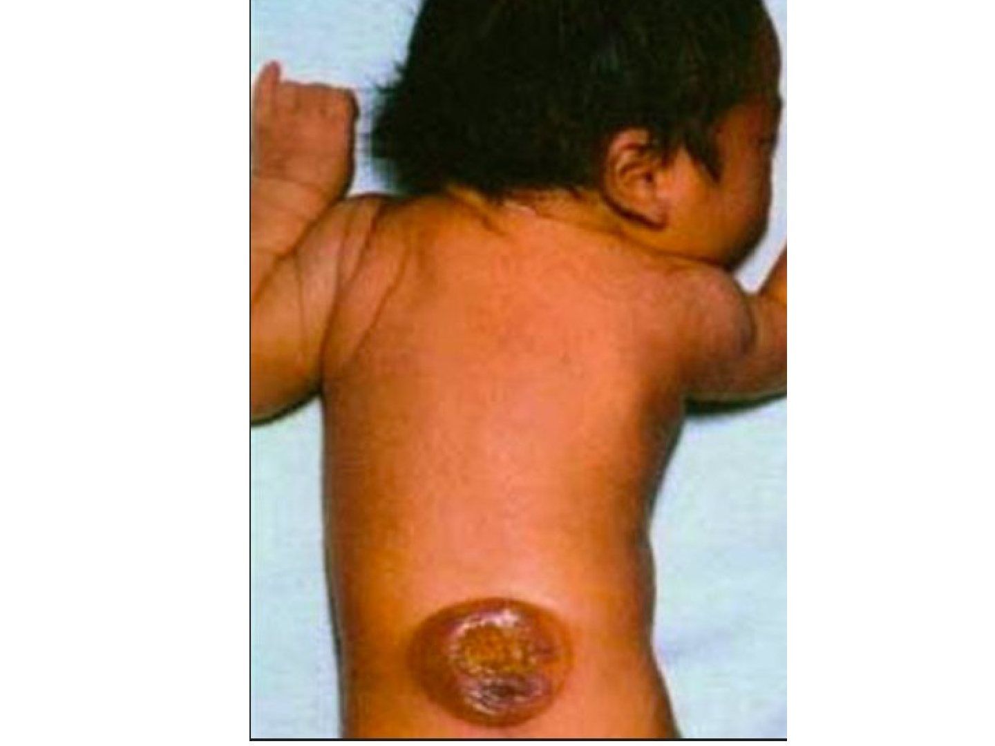
- What are the physical examination findings in spina bifida?
- How does spina bifida present?
- What causes scoliosis in spina bifida?
- At which functional level is hip dislocation the highest risk in spina bifida and why?
Answers · Q & A
Q1.What is spinal bifida and what are its risk factors?
- Fetal neural plate fails to completely close
- Risk factors: folate deficiency, maternal diabetes, chromosomal abnormality
Q2.List the forms of spinal bifida.
- Spina bifida occulta: cord and meninges confined
- Meningocele: protruded sac, no neural elements
- Myelomeningocele: protruded sac with neural elements
- Rachischisis: neural elements exposed
Q3.What causes neurological deterioration in spinal bifida?
- Tethered cord
- Hydrocephalus
- Syringomyelia
Q4.How is the functional level assessed in spinal bifida?
- Walk? yes at least L3
- Ankle equinus? no at least L4
- Hip abduction? yes at least L5
- If not walking: check hip control - no at L1 or above, yes with flexion at L2
Q5.What are the principles of treatment in spinal bifida?
- Treatment depends on the form and level
- Aim: maximise function consistent with the level involved and prevent deterioration
- Multidisciplinary approach with patient and family counselling
- Community-based treatment, minimise hospital admission
- Orthosis and physiotherapy for stretching to prevent contracture and maintain muscle power
- Surgery to balance muscle pull, prevent or correct deformity
Q6.How are the hip, knee and ankle managed surgically in spinal bifida?
- Hip: highest dislocation rate at L3/L4 last functional level due to adductor-abductor imbalance
- Hip surgery is controversial and only applicable with functional quadriceps
- Knee: flexion contracture release if severe and affecting walking
- Ankle: can be equinovarus, cavovarus or calcaneovalgus
Q7.What are the physical examination findings in spina bifida?
- Very short and slim lower limbs, upper limbs normal, AFO
- Scar: longitudinal, mostly at the back
Q8.How does spina bifida present?
- Deformity due to imbalanced muscle pull (hip dislocation, ankle deformity)
- Contracture
- Scoliosis (congenital / muscle imbalance / secondary to unilateral hip dislocation with LLD)
- Functional deficit (walking difficulty)
- Neurogenic bladder
- Foot deformities
Q9.What causes scoliosis in spina bifida?
- Congenital curve
- Muscle imbalance
- Secondary to unilateral hip dislocation with LLD
Q10.At which functional level is hip dislocation the highest risk in spina bifida and why?
- L3 (with L4, the last functional levels) - intact adductor but weak abductor
- L3 patients are household walkers
▸ Slide 581 · L4 calcaneusSpine Paed · 6 questions expand
Slide render

Question list
Q1-Q66 questions — tap to reveal all answerslist
- Using the functional-level table, summarise the hip, knee, ankle and functional findings at each level.
- At which levels is the ankle in equinus and at which level can it dorsiflex?
- Which levels can extend the knee and which level can walk?
- What does the slide title 'L4 calcaneus' refer to?
- Which functional levels have hip flexion with adduction and which can abduct?
- Which levels are Trendelenburg positive and which level has claw toes?
Answers · Q & A
Q1.Using the functional-level table, summarise the hip, knee, ankle and functional findings at each level.
- L1: hip ER + F (budda), knee flex, ankle equinus, flaccid
- L2: hip flex + add, knee flex, ankle equinus, +ve hip flexion
- L3: hip flex + add, knee can extend, ankle equinus, can walk (indoor), hip trdelenberg
- L4: hip flex + add, knee can extend, ankle can dorsiflex, hip tredelenberg, no equinus
- L5: hip flex (can abduct), knee can extend, ankle can dorsiflex with valgus (peronei +, PT weak), hip and knee normal, hindfoot valgus
- S1: hip, knee and ankle OK, but claw toes
Q2.At which levels is the ankle in equinus and at which level can it dorsiflex?
- Equinus at L1-L3
- Can dorsiflex at L4-L5
- L5: dorsiflexion with valgus (peronei +, PT weak)
- L4: no equinus - the slide's 'L4 calcaneus'
Q3.Which levels can extend the knee and which level can walk?
- Knee flex at L1-L2; can extend from L3 onward
- L3: can walk (indoor), hip trdelenberg, equinus
Q4.What does the slide title 'L4 calcaneus' refer to?
- At L4 the ankle can dorsiflex with no equinus (calcaneus deformity)
- Hip is flex + add with tredelenberg; knee can extend
Q5.Which functional levels have hip flexion with adduction and which can abduct?
- L2, L3 and L4: hip flex + add
- L5: hip flex (can abduct)
- L1: hip ER + F (budda)
Q6.Which levels are Trendelenburg positive and which level has claw toes?
- L3 and L4: hip Trendelenburg
- S1: claw toes
▸ Slide 582 · Spine embryologySpine Paed · 5 questions expand

Question list
Q1-Q55 questions — tap to reveal all answerslist
- What are the three embryological layers and their derivatives in spine development?
- When does the neural tube form and what is the metameric shift phenomenon?
- How do somites form the vertebral column?
- What is the timeline of spinal ossification?
- What are the three embryological layers involved in spine development?
Answers · Q & A
Q1.What are the three embryological layers and their derivatives in spine development?
- Ectoderm infolds at the midline to form the neural tube (CNS precursor)
- Neural crest cells dorsal to the neural tube become the PNS
- Mesoderm forms the notochord, which induces neurulation and becomes the nucleus pulposus
Q2.When does the neural tube form and what is the metameric shift phenomenon?
- At 3 weeks the ectoderm aggregates at the midline and infolds to form the neural tube
- PNS migrates between each level of somite = metameric shift phenomenon
Q3.How do somites form the vertebral column?
- At 4 weeks paraxial mesoderm aggregates at the midline to form somites: dermatome, myotome, sclerotome
- Somites unite around the notochord to form the vertebral body and around the neural tube to form the neural arch
- Each somite forms one disc plus the adjoining cranial and caudal halves of the vertebra
- The central part undergoes apoptosis to form the disc
- von Ebner fissure = separation fissure of a single somite
Q4.What is the timeline of spinal ossification?
- 6 weeks: chondrification of the spine
- 9 weeks: primary ossification centres - 1 at the body, 2 at the neural arch
- 2-6 years: arches fuse with the body
- 12 years: secondary ossification centres appear at spinous process, transverse process and apophyseal rings
- 25 years: secondary centres fuse with the primary centres
Q5.What are the three embryological layers involved in spine development?
- Ectoderm, mesoderm and endoderm
- Ectoderm infolds at the midline to form the neural tube (CNS precursor); neural crest becomes the PNS
- Mesoderm forms the notochord, which induces neurulation and becomes the nucleus pulposus
▸ Slide 583 · Scheurmann kyphosisSpine Paed · 12 questions 1 check expand

Question list
Q1-Q1212 questions — tap to reveal all answerslist
- What are the Sorensen criteria for Scheuermann's kyphosis?
- What is the epidemiology and pathophysiology of Scheuermann's kyphosis?
- What are the pathology and associations of Scheuermann's kyphosis?
- What are the differential diagnoses of thoracic hyperkyphosis?
- How is Scheuermann's kyphosis investigated?
- What are the symptoms of Scheuermann's kyphosis?
- What are the clinical findings of Scheuermann's kyphosis?
- What is the conservative treatment of Scheuermann's kyphosis?
- What is the operative treatment of Scheuermann's kyphosis?
- When is anterior release needed in Scheuermann's kyphosis?
- What is the limit of correction in Scheuermann's kyphosis surgery?
- What is a Schmorl node?
Answers · Q & A
Q1.What are the Sorensen criteria for Scheuermann's kyphosis?
- Rigid thoracic hyperkyphosis >45 degrees (normal 20-45 degrees)
- Anterior wedging >5 degrees in 3 consecutive vertebrae
- Irregular endplates
Q2.What is the epidemiology and pathophysiology of Scheuermann's kyphosis?
- Male > female
- Autosomal dominant inheritance, high penetrance with variable expression
- Distribution thoracic > thoracolumbar > lumbar
- Ring apophysis necrosis (occurs at age 12)
- Cartilaginous endplate mechanical weakening
Q3.What are the pathology and associations of Scheuermann's kyphosis?
- Thickened ALL, narrowed vertebral disc, wedged vertebral bodies
- Collagen-to-proteoglycan ratio in the endplate matrix is below normal, causing abnormal end plate ossification
- Spinal associations: spondylolysis and scoliosis
- Pulmonary compromise
Q4.What are the differential diagnoses of thoracic hyperkyphosis?
- Postural
- Compensation for lumbar spondylolisthesis
- Congenital (NF dystrophic curve)
- Infection/ tumour
Q5.How is Scheuermann's kyphosis investigated?
- X-ray: consecutive anterior wedging, endplate irregularity, disc narrowing, spondylolysis and scoliosis
- Supine hyperextension fulcrum view: rules out postural kyphosis and aids preop planning; <50% correction needs anterior release
- MRI for disc pathology, cord compression, epidural cyst and spinal stenosis
- Ask for hyperextension X-ray to see whether the kyphosis is rigid
Q6.What are the symptoms of Scheuermann's kyphosis?
- Thoracic/lumbar pain (compensatory hyperlordosis)
- Pulmonary compromise in severe kyphosis
- Cosmesis
- Ataxic gait
- Knee FFC
Q7.What are the clinical findings of Scheuermann's kyphosis?
- Thoracic kyphosis + compensatory lumbar hyperlordosis
- Tight hamstring
- Neurological deficits are rare
Q8.What is the conservative treatment of Scheuermann's kyphosis?
- Physiotherapy for stretching if kyphosis <60 degrees
- Hyperextension brace - controversial in a growing patient
Q9.What is the operative treatment of Scheuermann's kyphosis?
- PSF +/- Smith-Peterson osteotomy for >75 degrees, neurological deficit or severe pain
- Posterior approach advantages: less blood loss and reduced surgical time
- Posterior approach disadvantage: higher rate of pseudoarthrosis
Q10.When is anterior release needed in Scheuermann's kyphosis?
- When the deformity does not correct to <50 degrees on hyperextension lateral views
Q11.What is the limit of correction in Scheuermann's kyphosis surgery?
- Do not correct more than 50% of the original deformity
- Prevents junctional kyphosis or cord compression
Q12.What is a Schmorl node?
- Herniation of IVD into the vertebral endplate
- Associated with degeneration
- 16% prevalence in SE Asia
Fact check
Sorensen criteria for Scheuermann's kyphosis require rigid thoracic hyperkyphosis >45 degrees — contested threshold — Most sources (Radiopaedia, StatPearls) quote the Sorensen kyphosis threshold as >40 degrees (normal 20-40); some texts use >45. The >5 degree wedging in 3 consecutive vertebrae is agreed. — medium confidence — source