22 slides
▸ Slide 547 · Dysplasia/ SyndromesDysplasia Syndromes · 2 questions expand
Slide render

Question list
Q1-Q22 questions — tap to reveal all answerslist
- What does this slide introduce in the dysplasia/syndromes topic?
- List the common skeletal dysplasias and syndromes relevant to orthopaedics.
Answers · Q & A
Q1.What does this slide introduce in the dysplasia/syndromes topic?
- Not covered in the speaker notes
Q2.List the common skeletal dysplasias and syndromes relevant to orthopaedics.
- Not covered in the speaker notes
▸ Slide 548 · Vit D3 actionDysplasia Syndromes · 3 questions expand

Question list
Q1-Q33 questions — tap to reveal all answerslist
- What are the actions of vitamin D3 on the GI tract?
- What are the actions of vitamin D3 on bone?
- What is the effect of vitamin D3 on PTH and what upregulates vitamin D3?
Answers · Q & A
Q1.What are the actions of vitamin D3 on the GI tract?
- Absorbs Ca
- Absorbs PO4
Q2.What are the actions of vitamin D3 on bone?
- Activates osteoblast matrix production
- Increased osteoclast action through RANK-RANKL
Q3.What is the effect of vitamin D3 on PTH and what upregulates vitamin D3?
- Inhibit PTH
- Increased PTH (hydroxylation in the kidney)
- Low calcium level, low PO4 level
- Decreased vitamin D3 itself (auto-regulation)
▸ Slide 549 · PTH actionDysplasia Syndromes · 3 questions expand
Slide render

Question list
Q1-Q33 questions — tap to reveal all answerslist
- What is the effect of PTH on calcium reabsorption?
- What is the effect of PTH on phosphate reabsorption?
- What is the net effect of PTH on serum calcium and phosphate?
Answers · Q & A
Q1.What is the effect of PTH on calcium reabsorption?
- Increases Ca reabsorption at the bone
- Increases Ca reabsorption at the kidney
Q2.What is the effect of PTH on phosphate reabsorption?
- Increases PO4 reabsorption at the bone
- Decreases PO4 reabsorption at the kidney
Q3.What is the net effect of PTH on serum calcium and phosphate?
- Increase serum Ca
- Decrease serum PO4
▸ Slide 550 · Xray LL of skeletally immature patientDysplasia Syndromes · 11 questions expand

Question list
Q1-Q1111 questions — tap to reveal all answerslist
- Describe the X-ray findings in this skeletally immature patient.
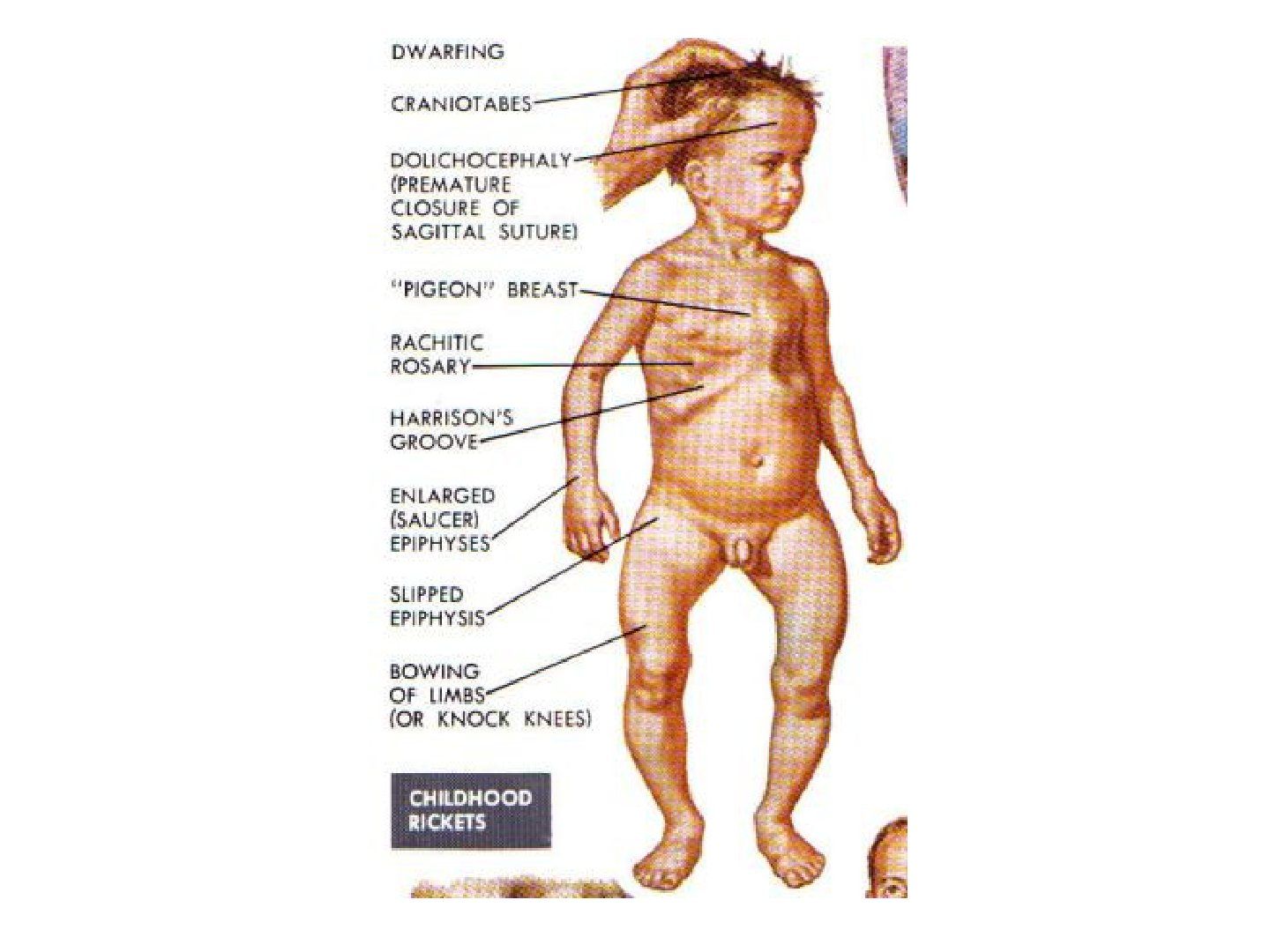
- What is rickets / osteomalacia?
- In a child with widened physes and bowed legs, what history and examination findings should be sought?
- What are the clinical features of rickets?
- What are the biochemical features of rickets?
- Describe the X-ray and histological findings of rickets.
- What are the causes of rickets by category?
- What is the defect in vitamin D dependent rickets type I and type II?
- Describe X-linked dominant familial hypophosphatemic rickets.
- What is hypophosphatasia?
- How is rickets treated?
Answers · Q & A
Q1.Describe the X-ray findings in this skeletally immature patient.
- Increased physeal width in general, cupping of the metaphyseal area
- Bowing of bilateral tibia
- No coxa vara
- Systemic disease affecting the physis; Ddx MED, SED
Q2.What is rickets / osteomalacia?
- Defect in mineralization of osteoid matrix and chondroid (in the zone of calcification)
- Caused by inadequate calcium and phosphate
- Impaired calcification of the cartilage matrix of growing long bones -> increased physeal width, cortical thinning and bowing
Q3.In a child with widened physes and bowed legs, what history and examination findings should be sought?
- History: birth and family history, diet
- PE: hyperlaxity, generalised weakness
Q4.What are the clinical features of rickets?
- Short stature (proportionate)
- Frontal bossing + caput quadratum
- Pigeon chest
- Rachitic rosary
- Harrison's sulcus
- Cat back kyphosis
- LL alignment abnormalities
Q5.What are the biochemical features of rickets?
- Decreased Ca and PO4
- Increased ALP and PTH (except in familial hypophosphatemic rickets and hypophosphatasia)
- Vitamin D level depends on the cause
Q6.Describe the X-ray and histological findings of rickets.
- Long bone bowing
- looser’s zone/ pseudofracture (Looser's zone / pseudofracture): transverse radiolucency from unmineralized osteoid seam; weight-bearing bones on the compression side, non-weight-bearing at the nutrient foramen
- Metaphyseal flaring in paintbrush pattern and cupping; widened physis at the zone of hypertrophy
- +/- osteopenia; cod-fish vertebrae
- Histology: unmineralized osteoid seam
Q7.What are the causes of rickets by category?
- Intake: intake problem, lack of sunlight exposure, GI problem
- Processing: vitamin D dependent rickets
- Recycling: vitamin D independent rickets (X-linked dominant familial hypophosphatemic rickets), hypophosphatasia
Q8.What is the defect in vitamin D dependent rickets type I and type II?
- Type I: defective 1-alpha hydroxylase
- Type II: end organ insensitivity to 1,25-(OH)2 vitamin D
Q9.Describe X-linked dominant familial hypophosphatemic rickets.
- PHEX encodes an endopeptidase cleaving FGF23; cannot chelate FGFR23
- Cannot reabsorb PO4 in the proximal tubules, cannot mineralize
- More diaphyseal bowing rather than metaphyseal bowing
- New drug: Burosumab
Q10.What is hypophosphatasia?
- Mutation in the tissue nonspecific isoenzyme of ALP (tissue non-specific isoenzyme of ALP)
- Reduced ALP, decreased synthesis of phosphate, decreased mineralization
- Increase in urine phosphoethanol-amine (urine phosphoethanolamine)
Q11.How is rickets treated?
- Treat the underlying cause
- Dietary input -> vitamin D3 5000 IU/day
- Vitamin D dependent type 1 -> calcitriol 1-2 micrograms/day; type 2 -> high dose vitamin D
- Hypophosphataemic syndrome -> calcitriol +/- phosphate 1g
- Mild deformity -> watchful observation for remodelling; severe deformity affecting ADL -> deformity correction
▸ Slide 551 · Histology of osteomalacia/ rickets: widened osteoid seams (large amount of unminDysplasia Syndromes · 3 questions expand
Slide render
Question list
Q1-Q33 questions — tap to reveal all answerslist
- What is the histology of osteomalacia / rickets?
- What is the histology of osteoporosis?
- How does tetracycline labelling differ between osteomalacia and osteoporosis?
Answers · Q & A
Q1.What is the histology of osteomalacia / rickets?
- Widened osteoid seams (large amount of unmineralized osteoid)
- Abnormal tetracycline labelling (tetracycline binds the mineralized surface of bone)
Q2.What is the histology of osteoporosis?
- Thinned trabeculae
- Decreased osteon size
- Enlarged Haversian and marrow spaces
- Normal tetracycline labelling
Q3.How does tetracycline labelling differ between osteomalacia and osteoporosis?
- Tetracycline binds to the mineralized surface of bone
- Abnormal in osteomalacia/rickets
- Normal in osteoporosis
▸ Slide 552 · Clinical photo showing patient of short stature which is disproportional (as patDysplasia Syndromes · 9 questions expand
Question list
Q1-Q99 questions — tap to reveal all answerslist
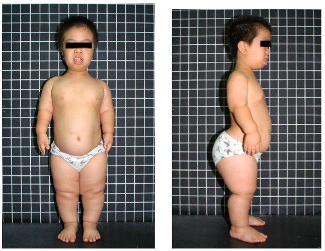
- Describe the clinical findings in this patient with disproportionate short stature.
- What dysmorphic, limb and trunk features are seen in achondroplasia?
- What further examination is needed in suspected achondroplasia?
- What is the differential diagnosis of disproportionate short-limb dwarfism?
- What is the Rubin classification of achondroplasia and how common is it?
- What is the genetics of achondroplasia?
- What is the pathophysiology of achondroplasia?
- What orthopaedic problems occur in achondroplasia?
- Why is spinal surgery difficult in achondroplasia?
Answers · Q & A
Q1.Describe the clinical findings in this patient with disproportionate short stature.
- Hand can only reach the pelvis instead of the thigh; Head also appears large
- Rhizomelic short stature: humerus shorter than forearm, thigh similar length to leg (usually 0.8)
Q2.What dysmorphic, limb and trunk features are seen in achondroplasia?
- Dysmorphism: frontal bossing, hypoplastic midface, otitis media, normal IQ
- Limb: elbow flexion contracture (posterior radial head dislocation), trident hand, genu varum
- Trunk: TL kyphosis, LS hyperlordosis; spinal stenosis (short pedicles), cervical myelopathy (foramen magnum stenosis)
- Protruberant abdomen. Flat chest
Q3.What further examination is needed in suspected achondroplasia?
- Standing and seating height (standing <3rd percentile, sitting normal)
- Measure limb length (rhizomelic shortening)
- Ligamentous laxity
- Check elbow ROM and posterior radial head dislocation
Q4.What is the differential diagnosis of disproportionate short-limb dwarfism?
- Hypochondroplasia
- Pseudochondroplasia
- Leri Wiel Dyschondroosteosis (Leri-Weill dyschondrosteosis)
- MED
Q5.What is the Rubin classification of achondroplasia and how common is it?
- Rubin classification: physeal hypoplasia
- Most common cause of disproportionate dwarfism
Q6.What is the genetics of achondroplasia?
- AD; sporadic mutation >80% (risk with advanced paternal age)
- Mutation of FGFR3 on chromosome 4
- glycine base being substituted by an arginine base -> prolonged receptor activation after ligand binding -> excessive growth limitation
Q7.What is the pathophysiology of achondroplasia?
- Abnormal chondroid production by chondroblasts in the proliferative zone during enchondral bone formation at the physis
- Intramembranous ossification not affected
- Quantitative cartilage defect
- Diagnosed by prenatal USG
Q8.What orthopaedic problems occur in achondroplasia?
- Foramen magnum stenosis
- Spinal stenosis
- Radial head subluxation with elbow flexion contracture
- Genu varum
Q9.Why is spinal surgery difficult in achondroplasia?
- Positioning
- Exposure – increase lumbar lordosis
- Anatomy - small pedicles
▸ Slide 553 · HandDysplasia Syndromes · 4 questions expand
Question list
Q1-Q44 questions — tap to reveal all answerslist
- Describe the hand X-ray findings in this skeletal dysplasia.
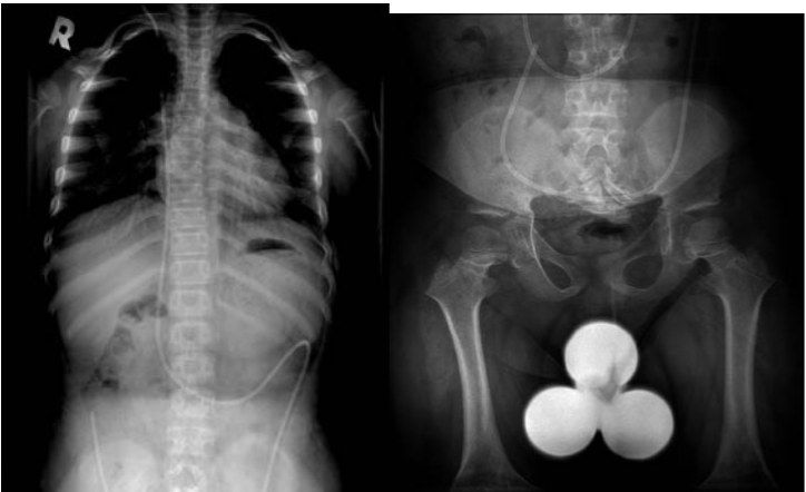
- What are the spinal findings on the scoliosis series?
- What are the pelvic findings?
- What are the lower limb findings?
Answers · Q & A
Q1.Describe the hand X-ray findings in this skeletal dysplasia.
- Increased gap between long and ring finger
- Irregular epiphysis
- Short metacarpals with suspected flaring of the metaphysis; metacarpal also appear short with suspected flaring of metaphysis
Q2.What are the spinal findings on the scoliosis series?
- VP shunt in situ
- Interpedicular distance decreased from cranial to caudal
- Scalloping of vertebrae
- Very flat horizontal sacrum
- Thoracolumbar kyphosis
Q3.What are the pelvic findings?
- Horizontal acetabular roof
- Small squared iliac wings
- Champagne glass pelvic inner contour
- Short SI notches
Q4.What are the lower limb findings?
- Disproportionate long bone development
- Metaphyseal flaring
- V-shaped physis
- Genu varum
- Lateral fibular bowing
▸ Slide 554 · Pseudo --> achondro --> hypoDysplasia Syndromes · 4 questions expand

Question list
Q1-Q44 questions — tap to reveal all answerslist
- What do pseudochondroplasia, achondroplasia and hypochondroplasia have in common, and how is achondroplasia distinguished?
- What are the features of pseudochondroplasia?
- How does pseudochondroplasia differ from SED?
- What are the features of hypochondroplasia?
Answers · Q & A
Q1.What do pseudochondroplasia, achondroplasia and hypochondroplasia have in common, and how is achondroplasia distinguished?
- All are AD with short stature
- Achondroplasia is the only one with an abnormal face and trident hand
Q2.What are the features of pseudochondroplasia?
- AD, COMP defect on chr19 (also in MED); onset usually 2nd decade
- Rhizomelic short limbs, Hip dysplasis (hip dysplasia)
- Windswept LL and knee recurvatum
- Fingers short and broad
- Spine: kyphoscoliosis, platyspondyly, odontoid hypoplasia; normal IQ
Q3.How does pseudochondroplasia differ from SED?
- Mimics SED because of epiphyseal changes
- Difference: later onset, milder spine deformity and normal face in pseudochondroplasia
Q4.What are the features of hypochondroplasia?
- AD, rare form of the same FGFR3 gene
- Milder dwarfism
- Face normal; spine changes rare
- MR; rare need for surgery
▸ Slide 555 · NeurofibromatosisDysplasia Syndromes · 7 questions 1 check expand
Question list
Q1-Q77 questions — tap to reveal all answerslist
- What are the three types of neurofibromatosis?
- What are the NIH consensus diagnostic criteria for NF1?
- What are the orthopaedic manifestations of NF1?
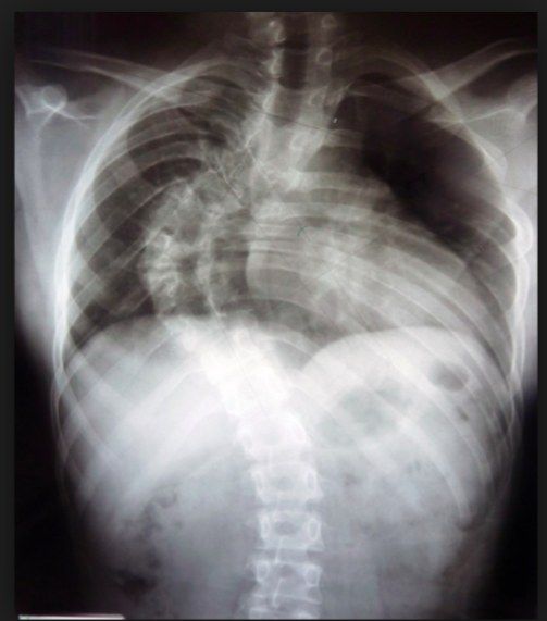
- Describe the features of dystrophic scoliosis in NF1.
- How is NF1 scoliosis managed?
- What are the genetics, prognosis and surgical difficulties in NF1?
- What are the features of malignant transformation in NF1?
Answers · Q & A
Q1.What are the three types of neurofibromatosis?
- NF1 (NIH consensus)
- NF2 with bilateral vestibular schwannomas
- Segmental NF (single body segment)
Q2.What are the NIH consensus diagnostic criteria for NF1?
- 2 of 7 required
- Family history in a 1st degree relative
- Skin: cafe au lait spots >6 (15mm in adults, 5mm in children); axillary and groin freckling (Crowe sign)
- Nerve: neurofibroma x2 or plexiform neurofibroma
- Bone: distinctive osseous lesion (sphenoid dysplasia, long bone cortex thinning +/- pseudoarthrosis)
- Eye: Lisch nodules x2 (iris hamartoma), optic glioma; updated 2021 adds gene analysis and more specific bone/eye criteria
Q3.What are the orthopaedic manifestations of NF1?
- Scoliosis (dystrophic / idiopathic-like form)
- Hemihypertrophy
- Atlantoaxial instability
- Plexiform neurofibroma is pathognomonic for NF1, in 4%, can progress to neurofibrosarcoma
Q4.Describe the features of dystrophic scoliosis in NF1.
- Thoracic kyphoscoliosis with a short segmented, sharp curve and distorted ribs and vertebrae
- AP: acute short segment curve, atypical level, penciling of ribs (>=3 poor prognosis because of rapid progression), TP spindling, hypoplastic pedicles
- Lateral: posterior scalloping and wedging of VB, foramina ectasia
- Transverse: widened canal (increased interpedicular distance)
- MRI: dumbell lesion (dumbbell lesion), dural ectasia (3mm thoracic, 4mm lumbar), meningocele
Q5.How is NF1 scoliosis managed?
- Brace is no use
- Early OT if Cobb >20
- Decompression
- ASF & PSF with instrumentation
Q6.What are the genetics, prognosis and surgical difficulties in NF1?
- AD (50%) / sporadic (50%), 100% penetrance; NF1 gene on chromosome 17q21; abnormal neurofibromin, Affecting RAS which affect osteoclast
- Prognosis: normal life expectancy; high chance of malignancy and hypertension
- Surgery difficulties: poor bone stock, significant kyphosis, small pedicles, incidental durotomy, Bleeding due to extradural venous plexus, rib dislocation during reduction causing nerve injury, may need A+P (nonunion: PSF alone up to 40%, A+P 10%)
Q7.What are the features of malignant transformation in NF1?
- Elephantiasis (large mass with villous skin)
- Plexiform NF lifetime risk 10% ('bag of worms', filariasis)
- Clinical: age 36-50yo, sudden pain and increase in size
- Biopsy to rule out neurofibrosarcoma
Fact check
NF1 gene is on chromosome 17q21 — incorrect locus — The NF1 gene maps to chromosome 17q11.2 (17q21 is the BRCA1 region) — source
▸ Slide 556 · EnchondromatosisDysplasia Syndromes · 7 questions 1 check expand
Question list
Q1-Q77 questions — tap to reveal all answerslist
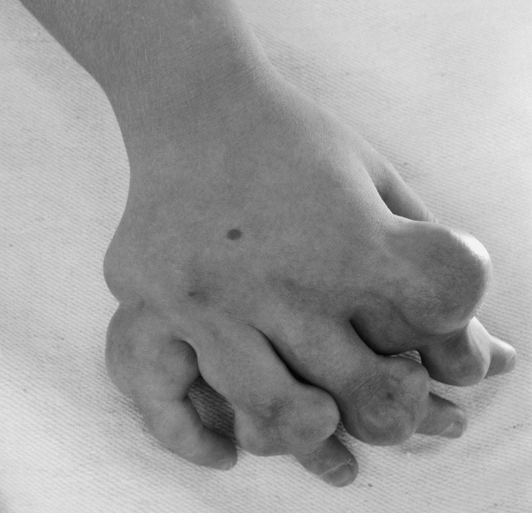
- What is Ollier's disease and how does it present?
- What is Maffucci syndrome and what are its XR features?
- What are the rates of malignant transformation in enchondromatosis?
- Which four planes of deformity are considered in treatment?
- What are the treatment options for deformity in enchondromatosis?
- How does enchondroma differ from osteochondroma?
- What are the XR features of Ollier's disease?
Answers · Q & A
Q1.What is Ollier's disease and how does it present?
- Sporadic defect of enchondral ossification
- Unilateral involvement --> LLD
- Angular deformity
- DDx: polyostotic fibrous dysplasia, OI
Q2.What is Maffucci syndrome and what are its XR features?
- Maffucci = Ollier's + hemangioma
- Expanded bone, angular deformity, cutaneous haemangiomata
- XR: Ollier changes + small rounded calcified phleboliths
Q3.What are the rates of malignant transformation in enchondromatosis?
- Solitary enchondroma <1%
- Ollier's 25-30%
- Maffucci 100%
Q4.Which four planes of deformity are considered in treatment?
- Coronal: valgus/varus
- Sagittal: flexion/extension
- Transverse: torsional
- Longitudinal: LLD
- Aim: well-aligned limbs in all planes
Q5.What are the treatment options for deformity in enchondromatosis?
- Hemiepiphysiodesis or guided growth
- Osteotomy for acute or gradual correction
- Combination of techniques
Q6.How does enchondroma differ from osteochondroma?
- Enchondroma: incomplete enchondral ossification, escape of chondroblast to metaphysis (cartilage in IM canal)
- Enchondroma: Rubin physis hyperplasia
- Osteochondroma: benign chondrogenic lesion from the perichondral ring
- Osteochondroma: metaphysis hyperplasia
Q7.What are the XR features of Ollier's disease?
- Cartilage-containing lesion + irregular translucent area at metaphysis
- Expanded bone
- Polyostotic involvement
Fact check
Malignant transformation occurs in 100% of Maffucci syndrome patients — Overstated - published rates vary widely; not all patients transform — Reported rates range from about 15-40% up to 52-57%; the JBJS 1987 life-table analysis says degeneration is almost a certainty, not 100% — medium confidence — source
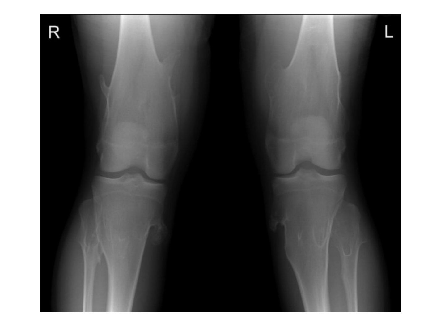
▸ Slide 557 · XR of both knees showing a skeletally mature patient with multiple bony protrusiDysplasia Syndromes · 9 questions expand
Slide render
Question list
Q1-Q99 questions — tap to reveal all answerslist
- Describe the XR findings of multiple hereditary exostosis at the knee.
- How do you differentiate MHE from Trevor disease?
- What is the risk of malignant transformation in MHE?
- Which clinical and radiological features suggest malignant transformation?
- What are the indications for surgery in MHE?
- What is the genetics and pathogenesis of MHE?
- What investigations are required in MHE?
- What complications must be ruled out in MHE?
- What is the growth pattern and natural history of MHE lesions?
Answers · Q & A
Q1.Describe the XR findings of multiple hereditary exostosis at the knee.
- Multiple bony protrusions of the metaphyseal region of distal femur/proximal tibia with metaphyseal tubulation
- Pedunculated, well defined, narrow zone of transition, pointing away from the physis
- Trabeculae within lesion continuous with the rest of the bone
- No cortical destruction, no soft tissue calcification
Q2.How do you differentiate MHE from Trevor disease?
- Clinically: Trevor is younger
- Radiologically: Trevor arise from epiphysis
- Histologically: Trevor cartilage cap has bands of cartilage separating cancellous bone
- Genetics: normal EXT levels in Trevor
Q3.What is the risk of malignant transformation in MHE?
- 5-10% lifetime risk of malignant transformation
- Versus normal lifetime risk of any malignancy of 1 in 3
- 1% lifetime risk per lesion, chondrosarcoma at around the 5th decade
- Cartilage cap >2cm suspicious (normal 2-3mm)
Q4.Which clinical and radiological features suggest malignant transformation?
- Increase in size after puberty
- Acute onset of pain in adults
- Cortical destruction, soft tissue mineralization >2cm
- MRI cartilage cap >2cm, sessile, central lesions more malignant
Q5.What are the indications for surgery in MHE?
- Pain from bursitis
- Deformity (forearm bowing, genu varum and valgum)
- Malignant transformation into chondrosarcoma
- Excise lesion including cartilaginous cap to reduce recurrence; corrective osteotomy +/- lengthening
Q6.What is the genetics and pathogenesis of MHE?
- AD; EXT1 (Chr 8), EXT2 (Chr 11), EXT3 (Chr 19); EXT1 worst
- 10% spontaneous mutation
- Defect in the groove of Ranvier with aberrant cartilage from the perichondral ring
- Mutations affect prehypertrophic chondrocytes; Rubin: metaphyseal hyperplasia
Q7.What investigations are required in MHE?
- Further XR of the scapula and pelvis (regions that cannot be palpated clinically)
- Scannogram for lower limb alignment
- Check the forearms clinically for bony exostosis and associated radial head dislocation
Q8.What complications must be ruled out in MHE?
- Local pressure symptoms (nerve, bursitis, pseudoaneurysm)
- Deformity/growth disturbance
- Malignancy
Q9.What is the growth pattern and natural history of MHE lesions?
- Sessile or pedunculated growth from the physeal plate away from the joint
- Lesions are benign and stop growing at maturity
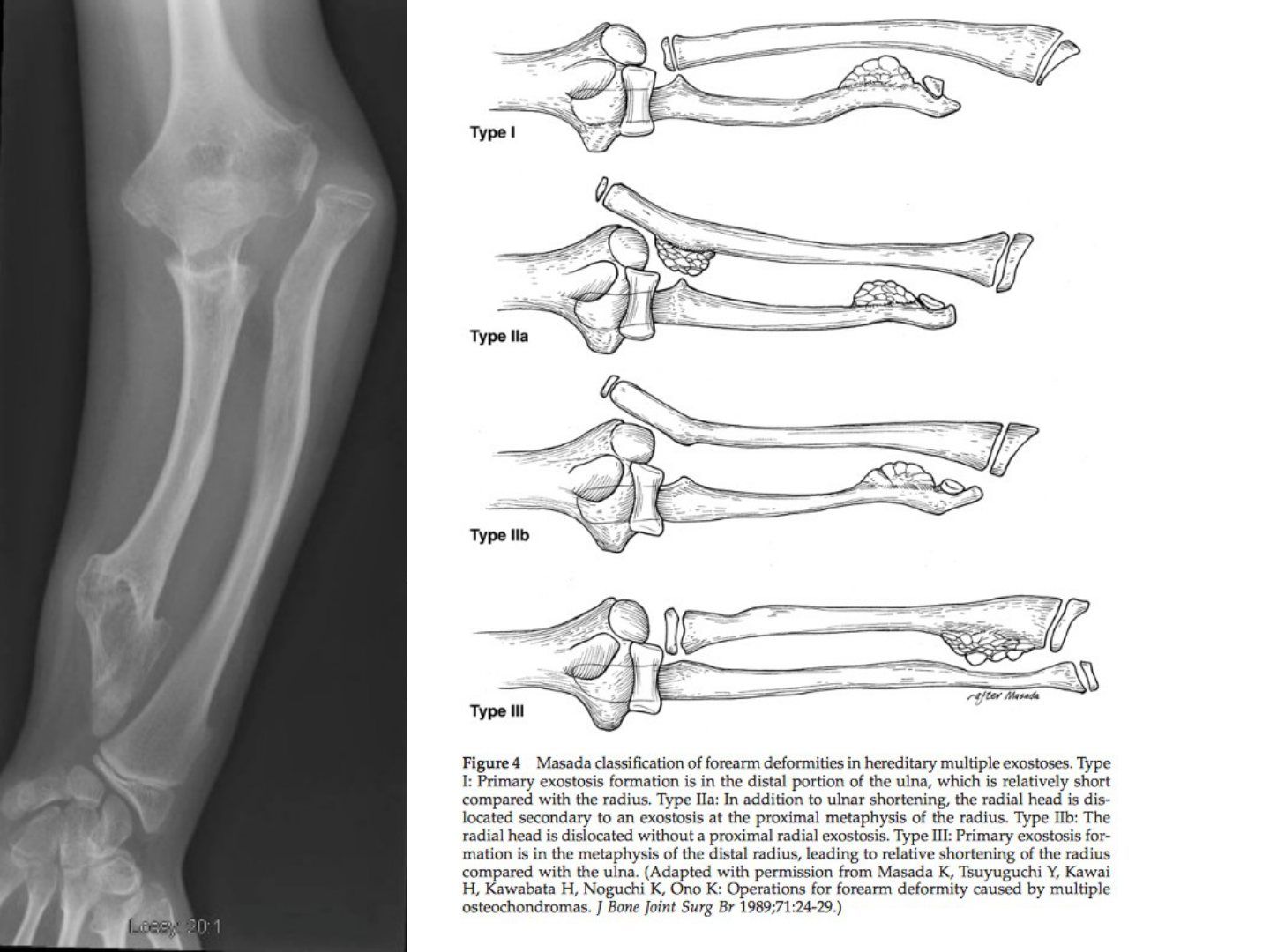
▸ Slide 558 · Xray of skeletally immature patient of forearmDysplasia Syndromes · 9 questions expand
Slide render
Question list
Q1-Q99 questions — tap to reveal all answerslist
- Describe the XR findings in this skeletally immature forearm.
- What are the surgical indications from the deformity point of view?
- What are the surgical indications from the MHE point of view?
- What are the options for radius reconstruction?
- What does the 2018 systematic review conclude about ulnar lengthening?
- What counselling points are given to the family?
- What is the differential diagnosis of the forearm deformity?
- How do you confirm the diagnosis and assess the patient?
- What are the details of surgical treatment?
Answers · Q & A
Q1.Describe the XR findings in this skeletally immature forearm.
- Shortening of the ulna with ulnar negative variance and apex radial bowing
- Abnormal bony enlargement of the ulnar head with exostosis
- Dislocated radial head
- Ulnar shift of the carpus
Q2.What are the surgical indications from the deformity point of view?
- Ulnar shortening >1.5cm
- Radial articular angle >30 degrees
- Carpal translocation >60%
- Painful subluxation of the radial head
- Pronation <60 degrees
Q3.What are the surgical indications from the MHE point of view?
- Enlarging mass after puberty
- Nerve or skin impingement, pain
- Radiologically: cortical erosion
- Cartilaginous cap >2cm, soft tissue mineralisation >2cm
Q4.What are the options for radius reconstruction?
- Proximal radius: unstable - one bone forearm; stable - radial head excision
- Distal: osteotomy/ hemiepiphysiodesis
Q5.What does the 2018 systematic review conclude about ulnar lengthening?
- Restores radiologic anatomy, improves appearance and to a lesser extent objective clinical parameters short to intermediate term
- Poor evidence that gains are maintained long term
- Impact on QOL and function not adequately investigated
- Considerable evidence surgery minimally impacts pre-op function
Q6.What counselling points are given to the family?
- Risk of malignancy
- Risk of further deformity
- Functional impairment may not improve with surgery - it mainly improves cosmesis
Q7.What is the differential diagnosis of the forearm deformity?
- Tumour
- Malunion
- Trevor disease (dysplasia epiphysealis hemimelia)
Q8.How do you confirm the diagnosis and assess the patient?
- Look for other lesions -> ?HME
- Rule out malignancy
- Assess other complications: ROM, nerve compression
- Assess current function
Q9.What are the details of surgical treatment?
- Excise the exostosis to stop differential growth
- Ulnar lengthening (acute vs gradual depends on length)
- Timing of surgery is controversial
▸ Slide 559 · mucopolysaccharidosesDysplasia Syndromes · 6 questions expand
Slide render
Question list
Q1-Q66 questions — tap to reveal all answerslist
- What clinical features suggest mucopolysaccharidosis?
- Describe the XR features of MPS.
- What is the underlying defect and inheritance of MPS?
- How is MPS diagnosed?
- Which MPS is most likely to survive into adulthood and what are the clues?
- How many mucopolysaccharidoses are there and how are they diagnosed?
Answers · Q & A
Q1.What clinical features suggest mucopolysaccharidosis?
- Proportional dwarfism; coarse facies, hypertelorism, short nasal bridge, broad mouth, wide gap between teeth
- Short neck, kyphoscoliosis
- UL: carpal tunnel syndrome
- LL: genu valgum, joint contracture (laxity in Morquio)
- Also pectus carinatum, organomegaly
Q2.Describe the XR features of MPS.
- C spine: odontoid hypoplasia with C1/2 instability
- TL spine: kyphoscoliosis, bullet-shaped vertebra
- UL: bullet-shaped phalanges
- Hips: wine glass pelvis (broad flat ilium, inferior constriction), coxa vara, dysplastic hip
- Knees: genu valgum
Q3.What is the underlying defect and inheritance of MPS?
- Lysosomal enzyme deficiency with aggregation of partially degraded glycosaminoglycan
- Disrupts the hypertrophic zone in the physis
- AR, except Hunter's (X-linked recessive)
Q4.How is MPS diagnosed?
- Urine: excess mucopolysaccharides
- Skin: fibroblast culture to test enzyme activity
- Intrauterine chorionic villous sampling
- Morquio: keratan sulphate
Q5.Which MPS is most likely to survive into adulthood and what are the clues?
- Morquio
- Normal IQ
- Cloudy cornea
Q6.How many mucopolysaccharidoses are there and how are they diagnosed?
- Group of 13 metabolic syndromes
- AR, except Hunter's (X-linked recessive)
- Diagnosed by biochemical analysis of urine (Morquio: keratan sulphate)
▸ Slide 560 · Morquio most likely survive into adulthoodDysplasia Syndromes · 4 questions expand
Slide render

Question list
Q1-Q44 questions — tap to reveal all answerslist
- What enzyme deficiencies cause Morquio and which is more severe?
- What accumulates in Morquio and when does it present?
- What are the clinical features of Morquio?
- Describe the XR features of Morquio.
Answers · Q & A
Q1.What enzyme deficiencies cause Morquio and which is more severe?
- Type A: galactosamine 6 sulphate sulphatase deficiency - more severe
- Type B: beta galactosidase deficiency
- (Gaucher is beta glucocerebrosidase)
Q2.What accumulates in Morquio and when does it present?
- Accumulation of keratan sulphate
- Presents at 18-24 months
- AR inheritance
Q3.What are the clinical features of Morquio?
- Proportionate dwarfism
- Normal IQ
- Waddling gait
- Genu valgum
- Thoracic kyphosis, corneal clouding
Q4.Describe the XR features of Morquio.
- Thickened skull, wide ribs
- Vertebral beaking, thoracic kyphosis with platyspondylia
- Coxa vara with unossified femoral head
- Bullet-shaped metacarpals
- Odontoid hypoplasia leading to cervical instability
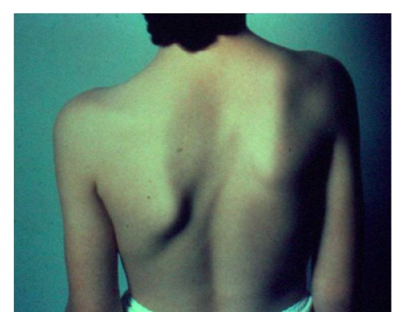
▸ Slide 561 · Clinical photo showing patient with asymmetrical scapulaDysplasia Syndromes · 14 questions expand
Slide render
Question list
Q1-Q1414 questions — tap to reveal all answerslist
- What is Sprengel shoulder and what are its associations?
- What are the XR features of Sprengel shoulder?
- Describe the Cavendish classification.
- What are the principles and options for surgery in Sprengel shoulder?
- What is Klippel-Feil syndrome?
- What are the associations and surgical indications in Klippel-Feil syndrome?
- What are the clinical findings of Sprengel shoulder?
- What should be documented on examination of Sprengel shoulder?
- What further imaging and workup is needed for Sprengel shoulder?
- What is the epidemiology of Sprengel shoulder?
- What are the Woodward and Green procedures for Sprengel shoulder?
- Describe the Klippel-Feil classification.
- What is the activity advice and prognosis in Klippel-Feil syndrome?
- What other features are associated with Klippel-Feil syndrome?
Answers · Q & A
Q1.What is Sprengel shoulder and what are its associations?
- Congenital undescended scapula (4-6th C spine pharyngeal arch; failure of caudal migration to T2-7 during 9th to 12th weeks of gestation (9th-12th weeks); interruption of embryonic subclavian blood supply)
- Associations: Klippel-Feil, congenital scoliosis, cervical spina bifida, VACTERL +/- situ invertus (situs inversus)
Q2.What are the XR features of Sprengel shoulder?
- Omovertebral bar (best seen on lateral/oblique C spine XR)
- Clavicle uptilt
- High riding scapula
- Small scapula (transverse > vertical)
- Inferomedial corner medial tilt + glenoid downward tilt
Q3.Describe the Cavendish classification.
- 1: Very mild, not detectable in clothing
- 2: Mild, 1-2cm higher
- 3: Moderate, 2-5cm higher
- 4: Severe, >5cm higher (superior angle near occiput)
Q4.What are the principles and options for surgery in Sprengel shoulder?
- Excise omovertebral bar + superior trapezius release at C4
- Relocation of the scapula
- Clavicle morselized osteotomy preserving periosteum (decreases brachial plexus injury)
- Aim: improve shoulder abduction (~30 degrees, Caverndish by 1 grade)
- OT if cosmetic problem or abduction < 120 at 3-8y (too late -> more nerve injury)
- Woodward and Green procedures both improve 50 degrees
Q5.What is Klippel-Feil syndrome?
- Fusion of more than two cervical vertebrae; (SGM 1 gene at chromosome 8
- Triad: Low posterior hair line (hairline), short webbed neck, decreased cervical movement (normal F/E, decreased side bending)
- XR triad: unsegmented C spine (commonest C2/3), ADI, degeneration (may have basilar invagination)
Q6.What are the associations and surgical indications in Klippel-Feil syndrome?
- Renal aplasia 33%, deafness 30%, congenital heart disease 15-30%
- Atlantoaxial instability ~50%; adjacent level disease 100%
- OT if pain, instability, neurology; check CV and UG systems before OT
- If C2 involved: no contact sports
Q7.What are the clinical findings of Sprengel shoulder?
- Hypoplastic and undescended scapula (right in the photo)
- Low hairline, short webbed neck
- No scapular winging; check for upward tilting of the clavicle from the front
Q8.What should be documented on examination of Sprengel shoulder?
- Neck: short, stiff; document bar, shoulder movement, abduction power
- Medial wing + supero-medial angle move superiorly -> glenoid pointed inferiorly
- Look for UL abnormalities (VACTERL), periscapular muscle fibrosis/weakness, distal NV status (syringomyelia)
- Look for scoliosis/kyphosis/back pitting (spina bifida)
Q9.What further imaging and workup is needed for Sprengel shoulder?
- CT and MRI spine for the bar and spinal problems such as dural ectasia or diastematomyelia
- Cardiac and renal workup; rule out other C spine problems
- Klippel-Feil associations: C spine failure of segmentation, C1/2 instability, cranial settling
Q10.What is the epidemiology of Sprengel shoulder?
- 10-30% bilateral
- Present in 1/3 of Klippel-Feil
- Association: scoliosis, spinal dysraphism, VACTERL
Q11.What are the Woodward and Green procedures for Sprengel shoulder?
- Woodward: detachment and distal advancement of the midline origin of parascapular muscles
- Green: extraperiosteal detachment, hold scapula in new position with spring wire traction and spica casting for 3 weeks
- Both improve 50 degrees
Q12.Describe the Klippel-Feil classification.
- Type I: extensive fusion
- Type II: only 1 or 2 vertebrae in the cervical spine
- Type III: fusion in part of the thoracic and/or lumbar spine in addition to Type I or Type II
Q13.What is the activity advice and prognosis in Klippel-Feil syndrome?
- If C2 involved: no contact sports
- If lower: usually asymptomatic until adjacent levels degenerate (100%) -> contact sports OK
- Check CV and UG systems before OT
Q14.What other features are associated with Klippel-Feil syndrome?
- Hearing loss, micrognathia, torticolis (torticollis), scoliosis
- Brainstem abnormalities/basilar invagination, congenital cervical stenosis
- MRI to rule out intraspinal cord abnormalities
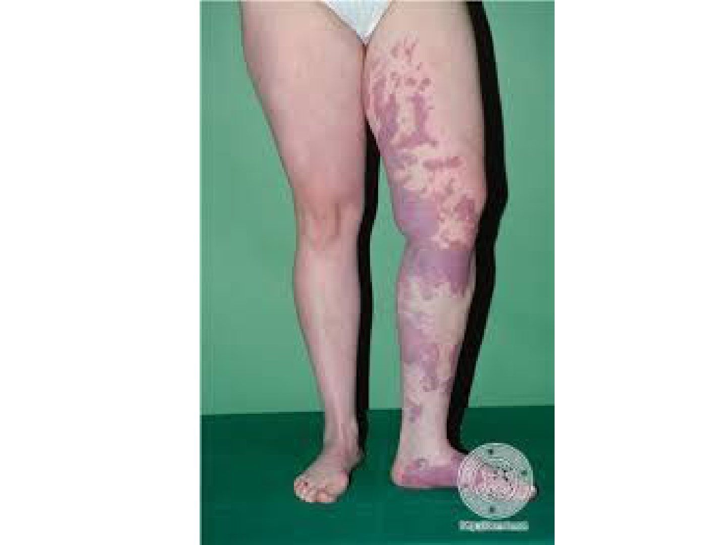
▸ Slide 562 · KlippelTrnaunay syndromeDysplasia Syndromes · 4 questions expand
Slide render
Question list
Q1-Q44 questions — tap to reveal all answerslist
- What is the triad of Klippel-Trenaunay syndrome?
- How do Parkes Weber, Proteus and Maffucci syndromes differ?
- What are the treatment principles for Klippel-Trenaunay syndrome?
- What is the limitation of YAG laser?
Answers · Q & A
Q1.What is the triad of Klippel-Trenaunay syndrome?
- Capillary malformation (port wine stain)
- Venous/ lymphatic malformation
- Soft tissue hypertrophy
Q2.How do Parkes Weber, Proteus and Maffucci syndromes differ?
- Parkes Webers syndrome: capillary, lymphatic and venous malformations with AV fistulas
- Proteus: capillary and venous malformations, macrodactylyl (macrodactyly), hemihypertrophy, lipomas, pigmented nevi, scoliosis
- Maffucci: lymphatic and venous malformation + multiple enchondromas
Q3.What are the treatment principles for Klippel-Trenaunay syndrome?
- Resection may be dangerous; consider embolization
- Ligation of feeding vessels is of no help
- High flow AV malformations difficult; staged partial excision mostly palliative
- Proximal ligation increases collateralization; embolisation risks digital ischemia
- YAG laser in direct contact allows subtotal excision of complicated hemangiomas
Q4.What is the limitation of YAG laser?
- Will not stop bleeding from vessels with lumen diameters greater than 1mm
- Direct contact YAG allows subtotal excision of lesions previously thought untreatable
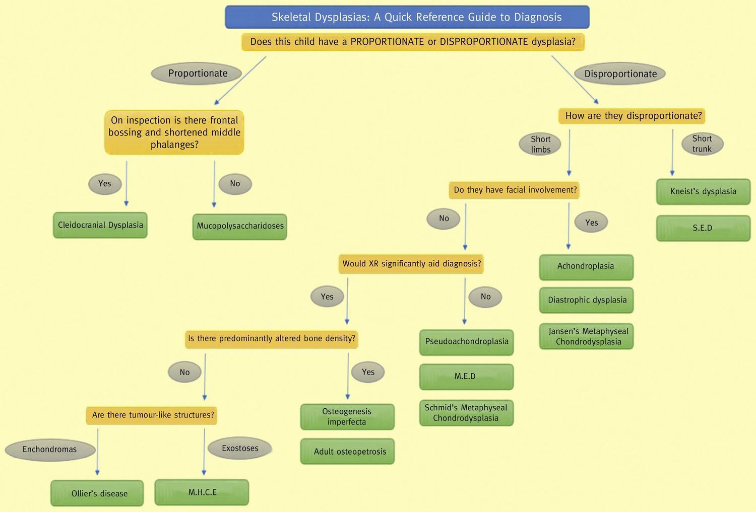
▸ Slide 563 · DwarfismDysplasia Syndromes · 3 questions expand
Question list
Q1-Q33 questions — tap to reveal all answerslist
- What is proportionate dwarfism and what are the examples?
- Classify disproportionate dwarfism with examples.
- What is the difference between proportionate and disproportionate dwarfism?
Answers · Q & A
Q1.What is proportionate dwarfism and what are the examples?
- Symmetric decrease in both trunk and limb length
- MPS
- Cleidocranial dysplasia
Q2.Classify disproportionate dwarfism with examples.
- Short-trunk: Kniest syndrome, spondyloepiphyseal dysplasia
- Short-limb: achondroplasia, diastrophic dysplasia
Q3.What is the difference between proportionate and disproportionate dwarfism?
- Proportionate: symmetric decrease in both trunk and limb length (e.g. MPS, cleidocranial dysplasia)
- Disproportionate: short-trunk (Kniest, SED) or short-limb (achondroplasia, diastrophic dysplasia)
▸ Slide 564 · Epiphyseal hypoplasiaDysplasia Syndromes · 6 questions expand

Question list
Q1-Q66 questions — tap to reveal all answerslist
- What is the genetics of spondyloepiphyseal dysplasia (SED)?
- Which spinal and respiratory complications must be screened for in SED?
- What are the XR findings in SED?
- What is multiple epiphyseal dysplasia (MED) and its genetics?
- What are the clinical and XR features of MED?
- What are the clinical features of SED?
Answers · Q & A
Q1.What is the genetics of spondyloepiphyseal dysplasia (SED)?
- COL2A1 on chromosome 12
- Congenita: onset at birth, AD
- Tarda: onset >5 years, X-linked recessive
Q2.Which spinal and respiratory complications must be screened for in SED?
- Odontoid hypoplasia -> atlantoaxial instability (AADI) - risk of cervical cord injury
- Restrictive lung disease on CXR
- Progressive deformity: coxa vara, genu valgum and kyphoscoliosis
Q3.What are the XR findings in SED?
- Hip dysplasia + coxa vara +/- genu valgum
- Platyspondyly
- Odontoid hypoplasia
- CXR: restrictive lung disease
Q4.What is multiple epiphyseal dysplasia (MED) and its genetics?
- AD
- Type 1: COMP (cartilage mineralization protein; mutation increases proteoglycan in chondrocyte)
- Type 2: COL9A2
- Presents in early adolescence
Q5.What are the clinical and XR features of MED?
- Disproportionate short stature; spine normal; IQ normal
- Limbs: coxa vara, genu valgum, ankle valgus, acromelia (short digits)
- Immature XR: small, flattened, fragmented epiphysis
- Mature XR: flattening of WB joint + incongruity +/- early OA
- Double layered patella
Q6.What are the clinical features of SED?
- General: disproportionate short stature, rhizomelic pattern
- Face: hypertelorism, cleft palate; chest: pectus carinatum
- Limbs: coxa vara, genu valgum, equinovarus
- Spine: kyphoscoliosis, odontoid hypoplasia -> AADI
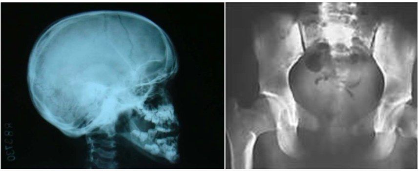
▸ Slide 565 · Cleidocranial Dysplasia (Dysostosis)Dysplasia Syndromes · 3 questions expand
Question list
Q1-Q33 questions — tap to reveal all answerslist
- What is the genetic basis of cleidocranial dysplasia?
- What are the clinical features of cleidocranial dysplasia?
- How is cleidocranial dysplasia managed?
Answers · Q & A
Q1.What is the genetic basis of cleidocranial dysplasia?
- AD, RUNX2/CBFA1 mutation affecting osteoblast
- Skeletal dysplasia affecting bones formed by intramembranous ossification
Q2.What are the clinical features of cleidocranial dysplasia?
- Typical clavicular absence/hypoplasia, hypermobile shoulder
- Proportional short stature
- Midline: delayed suture closure -> frontal bossing; delayed pubic ossification; delayed permanent teeth
- Coxa vara
- Shortened MP of 3-5 fingers
Q3.How is cleidocranial dysplasia managed?
- Only coxa vara requires treatment
- The other features are not described as requiring treatment
▸ Slide 566 · Fibrodysplasia ossificans progressiva is an extremely rare connective tissue disDysplasia Syndromes · 2 questions expand
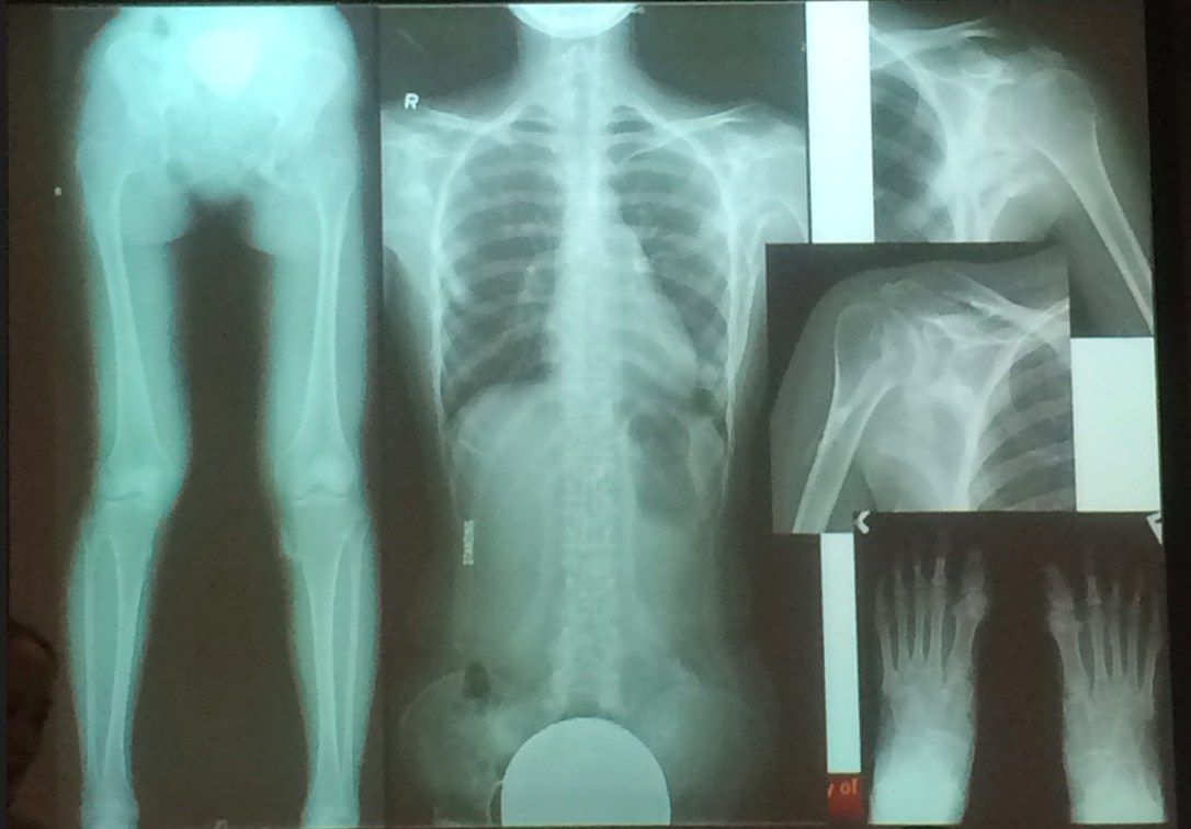
Question list
Q1-Q22 questions — tap to reveal all answerslist
- What is fibrodysplasia ossificans progressiva (FOP)?
- What is the genetics of FOP?
Answers · Q & A
Q1.What is fibrodysplasia ossificans progressiva (FOP)?
- Extremely rare connective tissue disease
- Mutation affecting the body's repair mechanism -> fibrous tissue (muscles, tendons, ligaments) ossifies spontaneously or when damaged
- Deformed big toe + very stiff joints
- No cure; median survival ~40
Q2.What is the genetics of FOP?
- Mutation of the ACVR1 gene (activin A type I receptor, a BMP type-1 receptor)
- AD allele on chromosome 2q23-24
- Variable expression, complete penetrance
- Most are spontaneous mutations
▸ Slide 567 · Duchenne muscular dystrophyDysplasia Syndromes · 8 questions expand

Question list
Q1-Q88 questions — tap to reveal all answerslist
- What is the genetic basis and pathophysiology of Duchenne muscular dystrophy?
- What examination findings are characteristic of DMD?
- How is DMD diagnosed?
- What is the natural history and non-operative management?
- What are the surgical considerations in DMD?
- How does DMD differ from SMA?
- What are the history and gait features of DMD?
- What is the aim of management in DMD?
Answers · Q & A
Q1.What is the genetic basis and pathophysiology of Duchenne muscular dystrophy?
- X-linked recessive - Xp21.2 dystrophin gene
- Absent cytoskeletal dystrophin protein
- Dystrophin provides structural stability to the dystroglycan complex of the muscle cell membrane
Q2.What examination findings are characteristic of DMD?
- Gower sign - climbs up own legs to stand (proximal weakness)
- Calf pseudohypertrophy, tiptoeing, waddling gait
- Scarf and Meryon signs in the floppy baby
- Hyperlordosis (compensates hip FFC), equinovarus foot, hip FABER and knee FFC contractures
Q3.How is DMD diagnosed?
- CPK elevated
- EMG/NCV
- Gold standard: muscle biopsy - absent dystrophin
- Echo for cardiomyopathy
Q4.What is the natural history and non-operative management?
- Wheelchair bound at 10 years, death at 20 years (respiratory difficulty)
- Steroid 0.75mg/kg/day: improves strength, slows weakening, prevents scoliosis, prolongs ambulation
- Delays deterioration of pulmonary function
Q5.What are the surgical considerations in DMD?
- Soft tissue releases to prolong ambulation
- Hip containment NOT beneficial
- Scoliosis surgery: progresses 1-2 degrees per month from age 8-10; indications Cobb >30, FVC <30%; needs spinopelvic fusion
- Malignant hyperthermia common intraop
Q6.How does DMD differ from SMA?
- SMA has absent reflexes (present in DMD)
- SMA has earlier onset
- SMA has no pseudohypertrophy
Q7.What are the history and gait features of DMD?
- FHx; progressive weakness affecting proximal muscles first (gluteus wasting)
- Gait abnormalities: delayed walking, toe walking, waddling gait, difficulty climbing stairs, hopping or jumping
Q8.What is the aim of management in DMD?
- Keep the patient ambulatory as long as possible
- Steroid 0.75mg/kg/day acutely improves strength, slows weakening, prevents scoliosis formation and prolongs ambulation
- Delays deterioration of pulmonary function
▸ Slide 568 · Nail patellar syndromeDysplasia Syndromes · 4 questions 1 check expand
Slide render

Question list
Q1-Q44 questions — tap to reveal all answerslist
- What is the diagnostic tetrad of nail patella syndrome?
- What is the genetics and what associations should be screened for?
- What else should be assessed in nail patella syndrome?
- What other conditions cause congenital patellar dislocation?
Answers · Q & A
Q1.What is the diagnostic tetrad of nail patella syndrome?
- Atrophic nails (usually thumb)
- Hypoplastic/absent patella - empty trochlear groove
- Iliac horns (pathognomonic, 80%)
- Hypoplastic radial head +/- subluxation -> FFC
Q2.What is the genetics and what associations should be screened for?
- AD, LMX1B mutation (Lim homeobox transcription factor 1b)
- Type 5 collagen abnormality (as taught in the lecture; see factcheck)
- Association: glaucoma
- Association: nephropathy
Q3.What else should be assessed in nail patella syndrome?
- External tibial torsion
- Thickened, tight ITB and lateral retinaculum
- Tight quadriceps
Q4.What other conditions cause congenital patellar dislocation?
- Larson syndrome
- Arthrogryposis
- Down syndrome
Fact check
Nail patella syndrome is caused by a type 5 collagen abnormality — Not supported by current evidence — NPS is caused by loss-of-function mutations in LMX1B, a transcription factor; a type V collagen abnormality is not an established cause — medium confidence — source