8 slides
▸ Slide 539 · InfectionInfection · 2 questions expand
Slide render

Question list
Q1-Q22 questions — tap to reveal all answerslist
- What are the key orthopaedic infections in children?
- How would you assess and manage a child with a suspected bone or joint infection?
Answers · Q & A
Q1.What are the key orthopaedic infections in children?
- Not covered in the speaker notes
Q2.How would you assess and manage a child with a suspected bone or joint infection?
- Not covered in the speaker notes
▸ Slide 540 · Septic arthritisInfection · 8 questions 1 check expand
Slide render

Question list
Q1-Q88 questions — tap to reveal all answerslist
- What is the differential diagnosis of unilateral hip pain in a child?
- What are the causes and risk factors for septic arthritis of the hip in a child?
- Describe the pathoanatomy of septic arthritis.
- Describe the Gachter classification of septic arthritis.
- Why is knee septic arthritis uncommon?
- What are the Kocher criteria and the probability of septic arthritis for each score?
- How is septic arthritis of the hip managed?
- How are the sequelae of septic arthritis of the hip classified and treated (Hunka classification)?
Answers · Q & A
Q1.What is the differential diagnosis of unilateral hip pain in a child?
- Transient synovitis / osteomyelitis
- Septic arthritis
- Perthes disease
- SCFE
- Fracture (consider NAI)
- JRA
Q2.What are the causes and risk factors for septic arthritis of the hip in a child?
- Routes: haematological spread, direct inoculation, spread from osteomyelitis
- Neonate with patent transphyseal artery, or the 4 intracapsular physes: proximal humerus, hip, radial head, distal fibula
- Risk factors: prematurity, C-section (C section), history of invasive trauma
- Recent infection (URTI, UTI); vaccine (especially H influenzae)
Q3.Describe the pathoanatomy of septic arthritis.
- Metaphyseal collection from transphyseal vessel: low flow sharp-turn capillary ruptures and spreads to the joint
- Synovial oedema and hypertrophy with fluid production
- Frank pus destroys cartilage within 8hr (marginal and central portions) with joint space narrowing
- Superficial marginal and central bony erosion, then bony ankylosis (chronic)
Q4.Describe the Gachter classification of septic arthritis.
- 1. Synovitis
- 2. Pus
- 3. Cartilage
- 4. Bone erosion
Q5.Why is knee septic arthritis uncommon?
- The metaphyseal collection from the transphyseal vessel spreads to the joint only in intra-articular metaphyseal joints
- The knee is not an intra-articular metaphyseal joint, so knee septic arthritis is uncommon
Q6.What are the Kocher criteria and the probability of septic arthritis for each score?
- Unable to bear weight; fever >38.5; WBC >12; ESR >40
- 1 criterion: 3%; 2: 40%; 3: 93%; 4: 99.6%
- Caird modification (2006 JBJS): CRP >20
Q7.How is septic arthritis of the hip managed?
- History of trauma, onset and duration of pain, constitutional symptoms; check skin for sinus and whether the child can weight bear
- X-ray to rule out SCFE, Perthes, fracture; blood WBC, CRP, ESR; USG for effusion and aspiration
- Drainage: open anterior hip approach, capsulectomy 1cm2, drill holes at metaphysis, thorough irrigation
- Post-op hip spica for 4-6 weeks
- Peltola NEJM 2014: a few days IV (3-5) then a short course of oral antibiotics (3 weeks)
Q8.How are the sequelae of septic arthritis of the hip classified and treated (Hunka classification)?
- I minimal collapse/deformity
- II head deformity (A intact physis, B physeal arrest) -> femoral osteotomy
- III pseudoarthrosis -> head viable: BG + valgus osteotomy; head nonviable: resection of head and neck + GT arthroplasty
- IV complete epiphysis destruction (A neck in acetabulum: adductor tenotomy, B neck not in acetabulum: GT arthroplasty)
- V complete head and neck loss -> conservative
Fact check
Peltola NEJM 2014 showed that only a few days of IV (3-5) then a short course of oral antibiotics (3 weeks) is sufficient for uncomplicated joint sepsis — misattributed and imprecise — The NEJM 2014 Peltola paper is a review of acute osteomyelitis; the septic arthritis RCT (Clin Infect Dis 2009) showed 10 days total therapy (2-4 days IV then oral) was as effective as 30 days — source
▸ Slide 541 · Pathophysiology of acute osteomyelitisInfection · 10 questions expand
Slide render
Question list
Q1-Q1010 questions — tap to reveal all answerslist
- Describe the pathophysiology of acute osteomyelitis.
- Why do bacteria proliferate in the metaphyseal region?
- What is a sequestrum?
- What is an involucrum?
- What is a cloaca?
- Which patients with acute osteomyelitis develop septic arthritis?
- What are the common pathogens of acute osteomyelitis by age group?
- What is the presentation and investigation of acute osteomyelitis?
- What are the indications for surgery in acute osteomyelitis?
- What are the complications of acute osteomyelitis?
Answers · Q & A
Q1.Describe the pathophysiology of acute osteomyelitis.
- Haematological spread; bacteria adhere and proliferate in the metaphyseal region
- Pus formation, drains along Haversian and Volkmann canals, then breaks out as a subperiosteal abscess
- High intraosseous pressure + poor blood supply due to elevated periosteum -> bone ischaemia (vessel thrombosis) -> sequestrum
- Subperiosteal new bone -> involucrum; pus breaks through involucrum -> cloaca
Q2.Why do bacteria proliferate in the metaphyseal region?
- Hairpin turn with sluggish flow
- Low oxygen tension
- Acidic environment
Q3.What is a sequestrum?
- Devitalised bone resulting from high intraosseous pressure and poor blood supply due to the elevated periosteum
- Caused by bone ischaemia (blood vessel thrombosis)
Q4.What is an involucrum?
- Subperiosteal new bone formation
Q5.What is a cloaca?
- Pus breaking through the involucrum
Q6.Which patients with acute osteomyelitis develop septic arthritis?
- Neonate <1.5 years old with a patent transphyseal artery
- Infection in the 4 intra-articular epiphyses: proximal humerus, hip, radial head, distal fibula
Q7.What are the common pathogens of acute osteomyelitis by age group?
- Most common in all ages: Staph aureus
- Neonates: Group B strep, Haemophilus influenzae
- 1-16 years: Strep pyogenes, Haemophilus influenzae
- Infants: also gram-negative rods; consider MRSA
Q8.What is the presentation and investigation of acute osteomyelitis?
- PMHx, recent injury, failure to thrive, refusal to walk, other source of infection
- Bloods: WBC, CRP, ESR and cultures
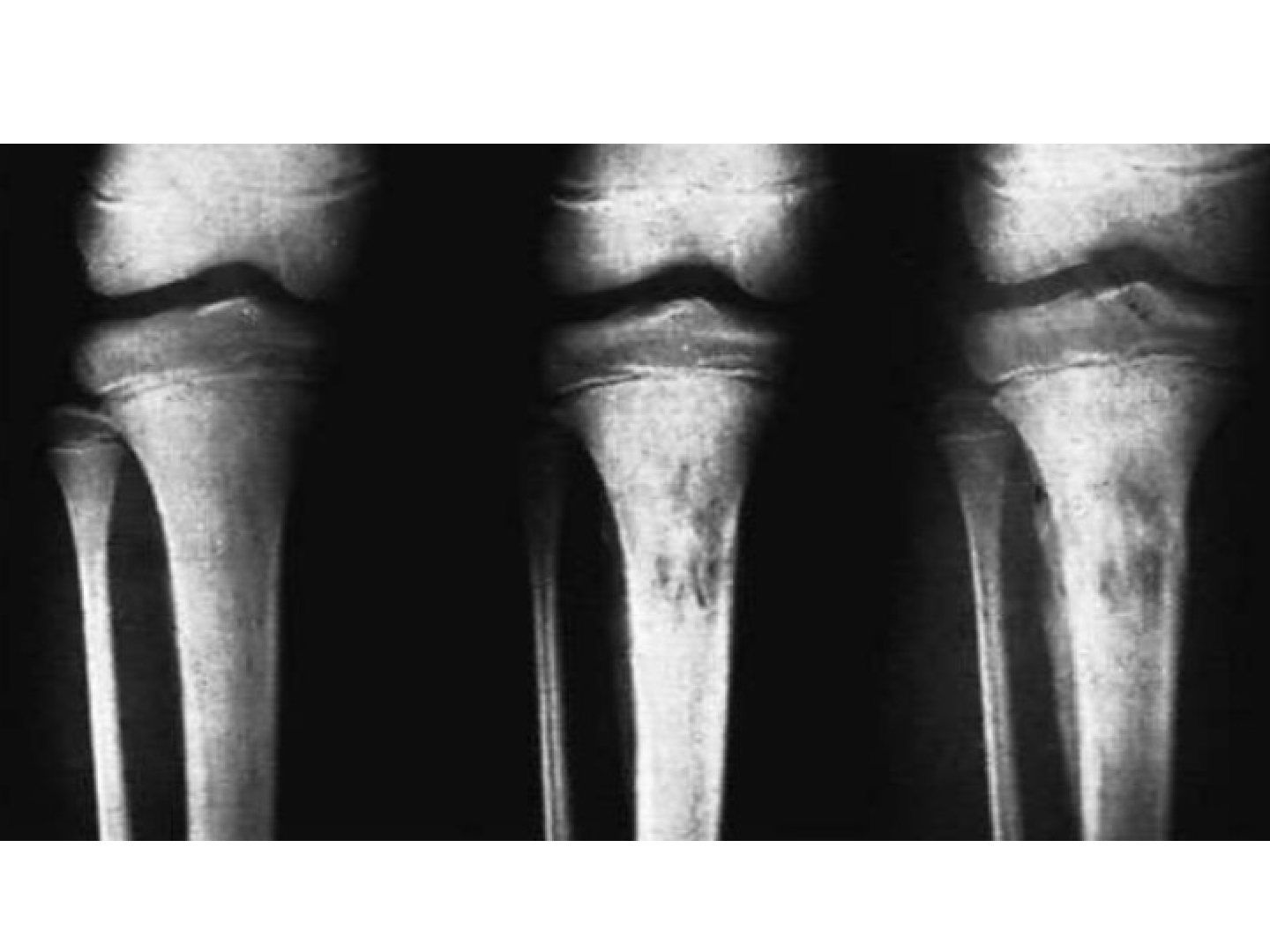
- X-ray: periosteal reaction at 1 week, lytic lesion at 2 weeks (metaphyseal rarefaction)
- MRI: early diagnosis and drainable abscess; bone scan: Indium/Technetium + gallium
- +/- image-guided aspiration
Q9.What are the indications for surgery in acute osteomyelitis?
- No systemic response to antibiotics for 48hrs (fever not settling, CRP not dropping)
- Drainable abscess
Q10.What are the complications of acute osteomyelitis?
- Chronic osteomyelitis (5%)
- LLD: over- or under-growth, arthritis, pathological fracture
- 30% have concomitant septic arthritis (shoulder, elbow, hip, ankle can all spread via transphyseal vessels)
▸ Slide 542 · 14 y.o boy from china, chronic leg pain after trivial injuryInfection · 6 questions expand
Slide render

Question list
Q1-Q66 questions — tap to reveal all answerslist
- Describe the X-ray findings in this case of subacute osteomyelitis.
- What is the differential diagnosis of this diaphyseal lytic lesion?
- What is a Brodie's abscess and how does it arise?
- What are the clinical and investigation findings in subacute osteomyelitis?
- What is the management principle for subacute/chronic osteomyelitis?
- List the stages of acute osteomyelitis.
Answers · Q & A
Q1.Describe the X-ray findings in this case of subacute osteomyelitis.
- Small lytic lesion at the proximal diaphysis of the leg
- Concentric and intramedullary
- Sclerotic rim with a narrow transitional zone
- Significant periosteal reaction
Q2.What is the differential diagnosis of this diaphyseal lytic lesion?
- Tumour: Ewing sarcoma in the diaphysis, osteosarcoma in the metaphysis, chondroblastoma in the epiphysis
- Infection: Brodie's abscess
Q3.What is a Brodie's abscess and how does it arise?
- Chronic abscess surrounded by sclerotic bone and fibrous tissue
- Results from partially treated acute osteomyelitis or decreased virulence of bacteria
Q4.What are the clinical and investigation findings in subacute osteomyelitis?
- PE: discharging sinus, skin erythema, lymph nodes
- Bloods: raised ESR (50% positive), WBC, CRP
- Imaging: MRI, CT; Cierny staging (anatomical + physiological A/B/C)
- May need biopsy to confirm diagnosis before OT
Q5.What is the management principle for subacute/chronic osteomyelitis?
- Eradicate infection, reconstruct bone and soft tissue defects, minimise complications and hospital stay
- Debridement: saucerization and sequestrectomy until healthy bleeding (paprika sign)
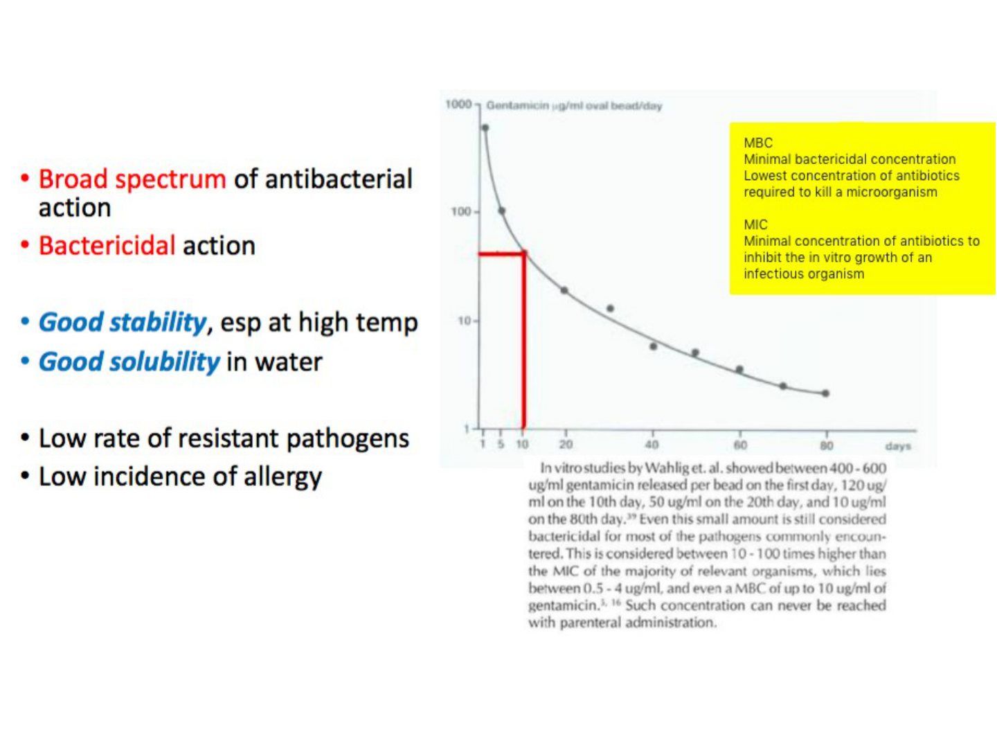
- Dead space management: antibiotic-loaded cement spacer, gentamicin beads, VAC dressing
- Local and systemic antibiotics
- +/- skeletal stabilisation depending on Cierny staging
Q6.List the stages of acute osteomyelitis.
- 1. Inflammation
- 2. Suppuration
- 3. Necrosis
- 4. New bone formation
- 5. Resolution
▸ Slide 543 · Chronic:Infection · 6 questions expand
Slide render

Question list
Q1-Q66 questions — tap to reveal all answerslist
- What are the aims of managing chronic osteomyelitis and how is it assessed?
- Describe the X-ray and clinical findings in chronic osteomyelitis.
- Describe the Cierny classification and its management.
- How is a bone defect after debridement managed?
- Describe the Papineau technique.
- What are the uses of VAC and what is needed for final soft tissue coverage?
Answers · Q & A
Q1.What are the aims of managing chronic osteomyelitis and how is it assessed?
- Aims: control infection, look for complications, skeletal stabilisation if pathological fracture
- PE: skin and soft tissue condition
- Blood: WBC, CRP, ESR; CT to look for collection
- Common bacteria: S aureus, polybacteria
Q2.Describe the X-ray and clinical findings in chronic osteomyelitis.
- Bone destruction at the proximal shaft involving half of the diameter on both AP and lateral
- Clinically a discharging sinus over the arm
- History of previous acute osteomyelitis
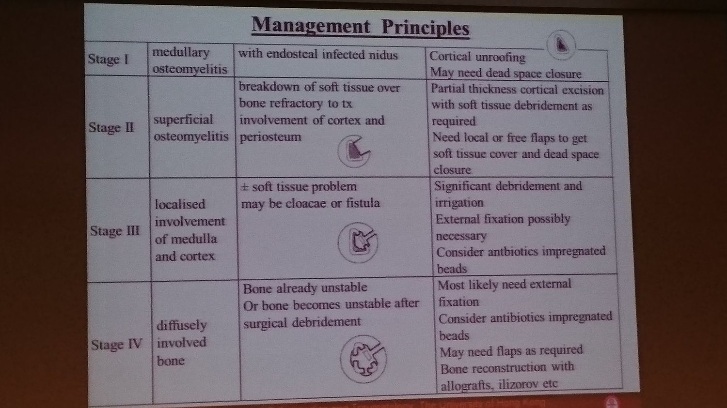
Q3.Describe the Cierny classification and its management.
- I intramedullary -> deroof bone for IM debridement
- II superficial (bone surface) -> debride cortex till paprika sign, may need soft tissue coverage
- III localized (cortex + medullary) -> saucerization, debridement, antibiotic bead +/- ex fix and flap
- IV diffuse -> ex fix, debridement, flap
- Host A/B/C = host immunity
Q4.How is a bone defect after debridement managed?
- Initially: ex fix + antibiotic-loaded beads, repeated debridement till clean
- Small <4cm: Papineau or bypass graft (cross union of tibia/fibula)
- Large >6cm: Masquelet technique, vascularized bone graft, bone transport / distraction osteogenesis
- Bi/trifocal (2 osteotomy sites) bone transport to reduce time to distraction and consolidation, but bone maturation time is longer
Q5.Describe the Papineau technique.
- Bed free of infection with healthy granulation tissue (blood supply)
- Lay morselized bone graft on the defect, not thicker than 2cm
- As it is taken up and forms granulation tissue, add another layer of bone graft
- Continue till dead space obliterated, then VAC +/- split thickness skin graft
Q6.What are the uses of VAC and what is needed for final soft tissue coverage?
- Removes chronic oedema, enhances blood flow, promotes granulation tissue at the floor
- Final coverage by the soft tissue reconstruction ladder
- May need a local or distant muscle flap
- Adequate vascularised soft tissue coverage helps resolution of osteomyelitis
▸ Slide 544Infection · 2 questions expand
Slide render
Question list
Q1-Q22 questions — tap to reveal all answerslist
- What infection-related topic does this slide cover?
- What are the key learning points for this infection slide?
Answers · Q & A
Q1.What infection-related topic does this slide cover?
- Not covered in the speaker notes
Q2.What are the key learning points for this infection slide?
- Not covered in the speaker notes
▸ Slide 545 · `Infection · 2 questions expand
Question list
Q1-Q22 questions — tap to reveal all answerslist
- What is the topic of this slide?
- Outline the classification and management of the condition shown on this slide.
Answers · Q & A
Q1.What is the topic of this slide?
- Not covered in the speaker notes
Q2.Outline the classification and management of the condition shown on this slide.
- Not covered in the speaker notes
▸ Slide 546 · JRAInfection · 7 questions expand
Slide render
Question list
Q1-Q77 questions — tap to reveal all answerslist
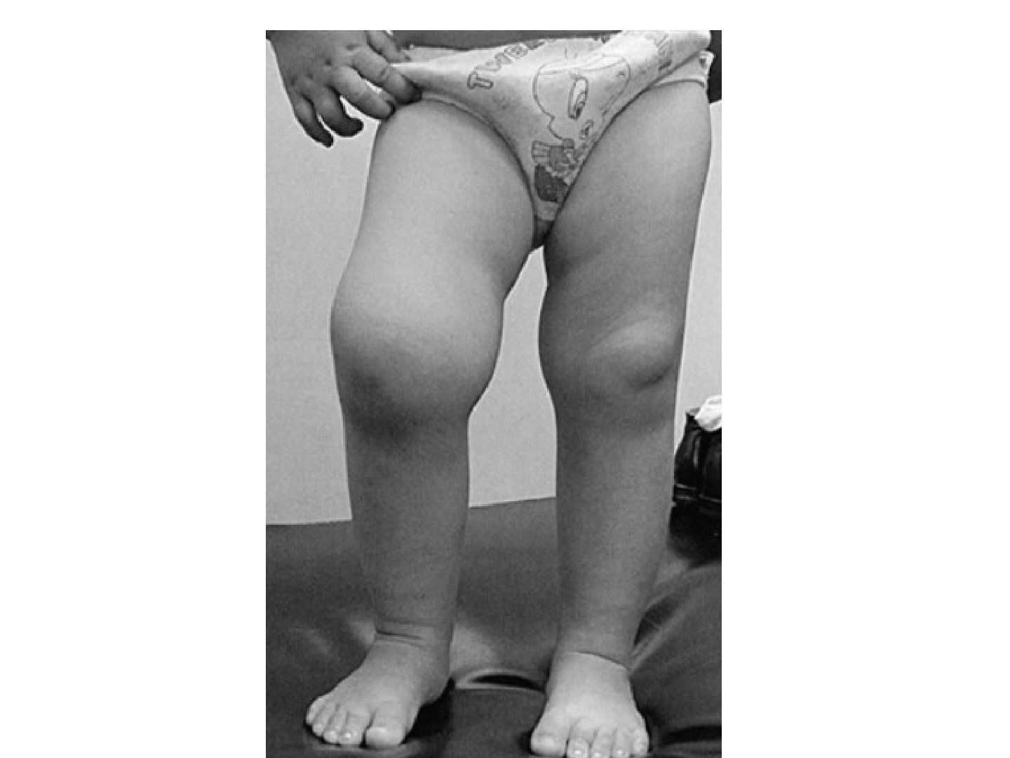
- What is the differential diagnosis of recurrent knee effusion in a child?
- What is juvenile rheumatoid arthritis (JRA) / juvenile idiopathic arthritis?
- What is the genetic association and what are the types of JRA?
- What are the diagnostic criteria for JRA?
- What are the clinical features of JRA?
- What is Still disease and what is the prognosis of JRA?
- What investigations and treatments are used in JRA?
Answers · Q & A
Q1.What is the differential diagnosis of recurrent knee effusion in a child?
- Infection
- Trauma (NAI)
- Inflammatory (JRA)
Q2.What is juvenile rheumatoid arthritis (JRA) / juvenile idiopathic arthritis?
- A persistent autoimmune inflammatory arthritis lasting >6 weeks
- In a patient younger than 16 years of age
- Female > male
Q3.What is the genetic association and what are the types of JRA?
- HLA-DR4 (polyarthritis); HLA-DR8, 5, 2.1; HLA-B27 (pauciarthritis)
- Polyarthritis >5 joints (similar to RA) - 30%
- Oligoarthritis <5 joints (similar to seronegative, commonly eye problems) - 50%
- Systemic (Still disease) - 20%
Q4.What are the diagnostic criteria for JRA?
- Diagnosis by exclusion plus one of the following:
- Rash, presence of RF, iridocyclitis, C-spine involvement
- Pericarditis, tenosynovitis, intermittent fever, morning stiffness
Q5.What are the clinical features of JRA?
- General: intermittent fever, rash, pericarditis, iridocyclitis (inflammation of iris and ciliary body)
- Ortho: C-spine involvement (kyphosis, facet ankylosis, atlantoaxial subluxation), tenosynovitis, morning stiffness
- Osteoporosis with easy fracture; growth retardation (corticosteroid suppression, Shapiro III/V)
- Ankylosis of joints in systemic type
- Deformity: external torsion of tibia, distal ulna dysplasia, underdeveloped mandible (micrognathia), short neck and scoliosis
Q6.What is Still disease and what is the prognosis of JRA?
- Still disease: acute presentation with multiple joint involvement, fever, salmon coloured rash, splenomegaly, usually <3 years old
- Prognosis: 50% spontaneous resolve, 25% slight limitation, 25% severe disability
- Best prognosis pauciarticular > polyarticular > systemic
Q7.What investigations and treatments are used in JRA?
- X-ray: juxta-articular, late osteopenia and joint destruction; C-spine X-ray for atlantoaxial instability
- Blood: RF (worse prognosis if +ve), ANA (+ve is diagnostic), HLA typing
- Physiotherapy to maintain range and power
- DMARD (azathioprine, etanercept) and biological agents (rituximab)
- Surgery: synovectomy, a la carte