Limbs Dysplasia Deformity
Paediatric knee deformity - imaging
Radiographic and clinical assessment of knee deformity in a skeletally immature patient.
16 questions
2 source pages
1 images
2 fact-check flags
Images appear with the first question taken from each source page — tap a question to open it.
Q1Describe the deformity shown in this patient.📷▸
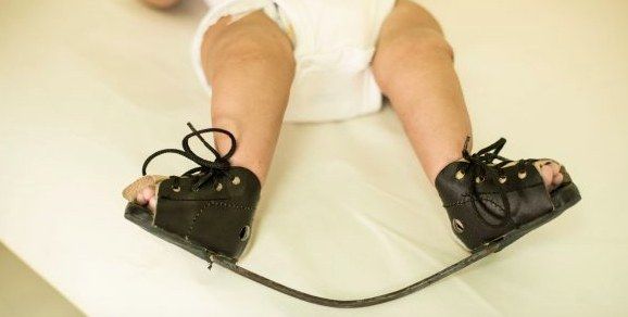
- Paediatric patient with bilateral foot deformity
- Hindfoot: varus and equinus
- Midfoot: cavus
- Forefoot: adduction + pronated position
- Locally assess correctability (?postural) and assess the other side (50% bilateral)
Q2What associated problems must you rule out?▸
- Packaging syndrome: torticollis, DDH, knee dislocation
- Rule out syndromic clubfoot
- Neuromuscular disease (CP)
- Spinal dysraphism
- Teratogenic/syndromes: arthrogryposis, SED, constriction band, Larsen, Pierre Robin, Prune Belly
Q3How would you counsel the parents?▸
- Familial occurrence in 25%; affected child has a 2.5-6.5% chance of a sibling with clubfoot
- PITX gene / chromosome 2 deletion (common genetic pathway with CVT)
- Ponseti treatment: success rate >90%; 70% need PETA; 30% need tib ant transfer for dynamic supination at 4-5 years
- The affected foot will be smaller and shorter regardless of treatment
Q4How is the Ponseti method performed and monitored?▸
- Based on viscoelastic stress relaxation and creep; start at 1 week
- Toe-to-groin POP with knee at 90 degrees, snug but not tight, thin velband, well moulded
- Weekly casts, usually 4-5; assistant holds the big toe and talar head throughout
- Correction sequence: cavus, adductus, varus (corrected with adductus), equinus
- Chart with the Pirani score (high score = high deformity, not prognostic): midfoot and hindfoot components
- Complications of casting: rockerbottom foot (2-5%), flat heel pad, crowded toes, pressure sores
Q5Describe the Ponseti correction sequence in detail.▸
- 1. Cavus: dorsiflex the 1st ray, realign the plantiflexed 1st MT with the other MTs and realign forefoot to hindfoot by supination (warn family the deformity may look worse)
- 2. Adductus: abduct the forefoot and externally rotate at the midfoot using the uncovered head of the talus as fulcrum
- 3. Varus: will be corrected with step 2
- 4. Equinus: pull the calcaneal tuberosity distally while pushing the anterior calcaneus up; when abduction 70deg achieved, able to palpate the anterior process of the calcaneus as it abducts out from beneath the talus (neutral or slight heel valgus)
Q6Describe the pathoanatomy and causes of CTEV.▸
- Primary deformity centred at the talus, with a medially and plantarly deviated neck
- The rest of the acetabulum pedis (calcaneus, navicular, cuboid) rotates around the talus and becomes adducted and inverted
- Secondary soft tissue contracture: plantarflexors/invertors, CFL, deltoid, long/short plantar, spring, plantar aponeurosis
- Causes: majority idiopathic; neuropathic, myopathic, genetic (PITX), mechanical moulding, multifactorial
Q7Would you X-ray every clubfoot?▸
- X-ray is controversial: difficult to position, only calcaneum, talus and MT ossified, and ossification centres do not represent true shape
- Double parallelism
- Dorsiflexion lateral (Turco view): hindfoot parallelism, talocalcaneal angle <35 degrees
- AP view: Kite angle (talocalcaneal) <20, talus-first metatarsal angle negative
Q8How is TA lengthening performed and when is it indicated?▸
- Indicated if everything is corrected but DF still <10deg; needed in 70% of patients
- NA (local anaesthetic), incision medial to TA, transverse cut towards lateral
- Then another cast for 3 weeks at 30 deg ankle DF
Q9Describe the Denis Brown boots.▸
- BOOTS: Strap for 3 point fixation; forefoot open shoe box; midfoot scaphoid and intermetatarsal pad + straight medial lass to keep the foot abducted; hindfoot reverse Thomas heel with stiff heel support for mediolateral stability
- BAR: convex away to keep ankle dorsiflexion 5-10 degrees, shoulder width, ER 70/40 both sides 45 (sum 90-100) to keep the hindfoot in valgus
- Regime: 4 months full time, until 4 years at night time
Q10What if conservative management of clubfoot fails?▸
- Failed if the foot is not plantigrade by 3-6 months
- Timing controversial: early 6 months vs delay to ~1 year to allow weight bearing and avoid recurrence; wait until foot length >=8cm
- OT: Cincinnati incision, a la carte approach
- Do not release: deep talocalcaneal interosseous ligament (risk of lateral translation of calcaneus), deep deltoid (valgus deformity), Posterior tibial-fibular ligament
Q11Describe the X-ray findings and your concern.▸
- Skeletally immature patient
- V-shaped physis of the distal femoral physis
- Suspected connection between metaphysis and epiphysis centrally (cross trabeculation) -> physeal arrest
- Request a long film including the contralateral limb to assess LLD/angular deformity, and a lateral Xray to see angular deformity
Q12How would you assess a child with a suspected physeal bar?▸
- History: age, causes
- Local examination: deformity, length, joint involvement, features suggesting cause
- Systemic examination: maturity
- CT to look for a bony bar; MRI to look for fibrous tissue and the underlying cause
Q13How are physeal arrests classified and what affects prognosis?▸
- Extent (partial/ complete)
- Site: peripheral, central or linear
- Prognosis affected by: cause, extent, site, growth potential
- Sequelae: LLD, angular deformity, epiphyseal deformity
Q14What is the management principle and how does treatment depend on growth remaining?▸
- Aim: equal leg length with no angular deformity, minimize complications and hospital stay
- Treatment depends on growth potential/projected LLD, deformity present, and area involved
- >2 years growth and <50% physis involved: Langenskiold procedure (bar excision + interposition of fat/cement + marker implantation) or physis distraction
- >50% physis involved: hemi/epiphysiodesis + future lengthening
- <2 years growth: hemiepiphysiodesis; no growth: osteotomy; follow up till maturity
Q15How does the cause of the physeal arrest affect prognosis?▸
- Traumatic: responsive to physeal bar excision as only the hypertrophic zone is affected
- Tumour/infection: decreased response as all zones are affected
Q16What factors determine the risk of a physeal bar after fracture (JPO 2009)?▸
- Initial displacement
- Mechanism of injury (high energy)
- Salter-Harris pattern
- Number of reductions - not more than 2
- Residual displacement after closed reduction
- Growth arrest from injury to the germinal matrix (resting zone), Affecting blood supply, or bone bar formation
Fact check
Clubfoot genetics: PITX gene, chromosome 2 deletion — imprecise — PITX1 maps to chromosome 5q31.1; chromosome 2q31-33 deletions involve the HOXD cluster, a separate clubfoot-associated locus — (medium confidence) — source
The Pirani score is not prognostic — contested — Several studies report the initial Pirani score, especially the hindfoot component, predicts tenotomy need and relapse, although a 2022 systematic review/meta-analysis found no significant association with recurrence — (medium confidence) — source