▸ Slide 511 · Limbs dysplasia/deformityLimbs Dysplasia Deformity · 2 questions expand
Q1-Q22 questions — tap to reveal all answerslist
- What limb dysplasia and deformity conditions are covered?
- How are limb deficiencies classified?
Q1.What limb dysplasia and deformity conditions are covered?
- Not covered in the speaker notes
Q2.How are limb deficiencies classified?
- Not covered in the speaker notes
▸ Slide 512 · X-ray elbow showing posterior dislocation of radial headLimbs Dysplasia Deformity · 4 questions expand
Q1-Q44 questions — tap to reveal all answerslist
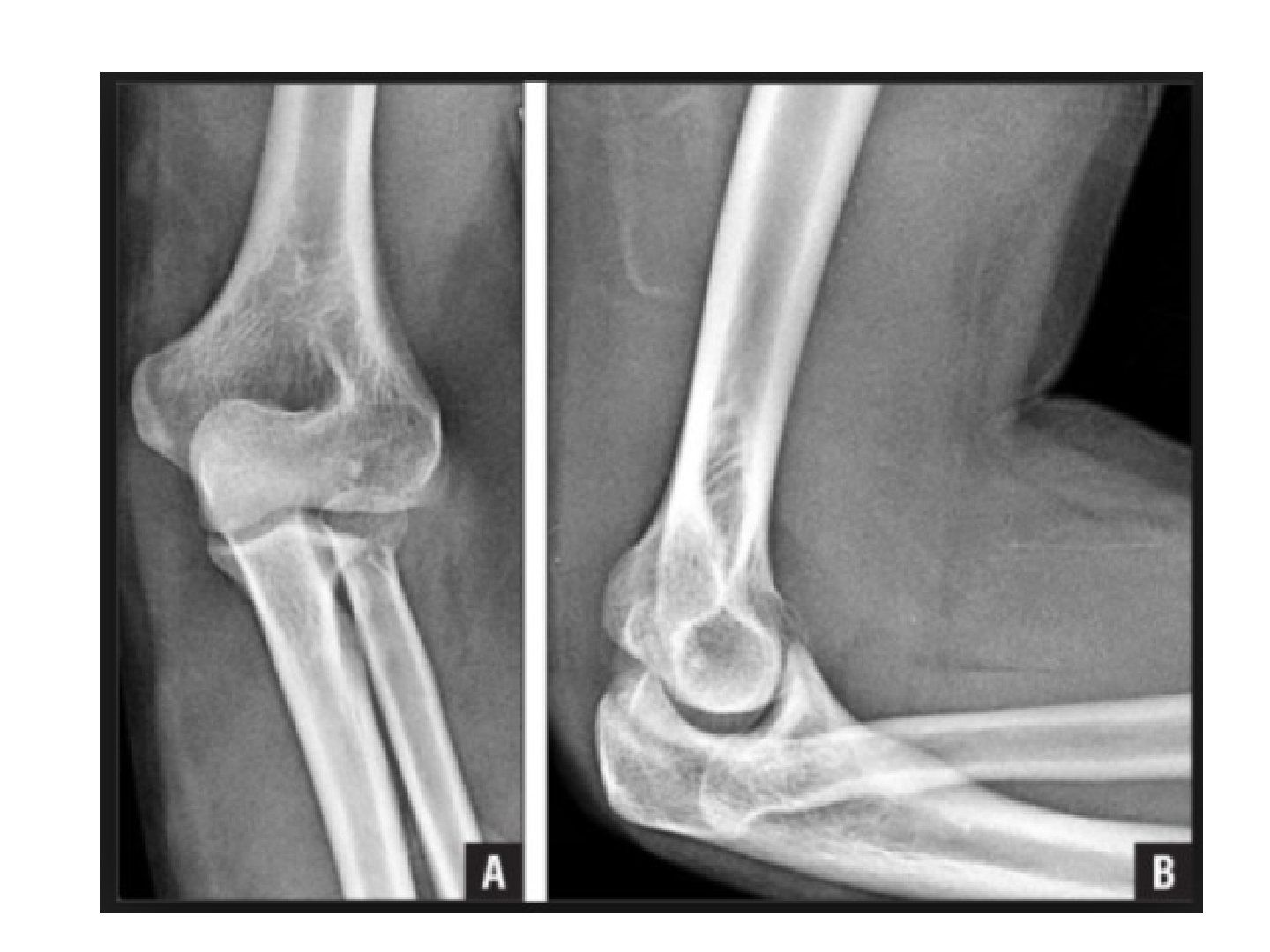
- Describe the X-ray findings in congenital radial head dislocation.
- What clinical clues suggest congenital radial head dislocation?
- What are the causes of radial head dislocation?
- What is the treatment for congenital radial head dislocation?
Q1.Describe the X-ray findings in congenital radial head dislocation.
- Posterior dislocation of the radial head
- Convex radial head with long radial neck
- Hypoplasia of capitellum, valgus deformity
- No fracture of the proximal ulna; no bony exostosis along forearm
- Check whole forearm for bowing/exostosis and the contralateral side (60% bilateral)
Q2.What clinical clues suggest congenital radial head dislocation?
- History of trauma
- Bilateral involvement
- Other congenital abnormalities: achondroplasia, arthrogryposis, Larsen syndrome, nail patella syndrome, HME, ulnar club hand
- Symptoms: cubital valgum, prominence over radial head, decreased range and limited supination
Q3.What are the causes of radial head dislocation?
- Congenital: achondroplasia, nail patella syndrome, Larsen syndrome (and congenital radioulnar synostosis)
- Acquired: HME
- Traumatic (missed Monteggia)
- Paralytic disorders (CP, polio)
Q4.What is the treatment for congenital radial head dislocation?
- Excision when mature if symptomatic
▸ Slide 513 · Xray elbow of skeletally immature patientLimbs Dysplasia Deformity · 11 questions expand

Q1-Q1111 questions — tap to reveal all answerslist
- Describe the X-ray findings and explain why this is likely congenital.
- What would you comment on if shown a clinical photo of this patient?
- What history and examination findings are relevant in radioulnar synostosis?
- What is the pathogenesis of congenital radioulnar synostosis?
- What syndromes and associations should be looked for in congenital radioulnar synostosis?
- What is the differential diagnosis and how do congenital and acquired synostosis differ?
- How does acquired (post-traumatic) radioulnar synostosis differ?
- What is the management of congenital radioulnar synostosis?
- Why is the rotational osteotomy performed at the level of the synostosis?
- Describe the embryological development of the limb.
- What are the 3 axes of limb patterning?
Q1.Describe the X-ray findings and explain why this is likely congenital.
- Radioulnar synostosis bilaterally
- Left anteriorly dislocated radial head with malformation
- Cleary classification type I on the right, type IV on the left
- Likely congenital: bilateral (60%), continuity of IM canal and matrix
- Autosomal dominant with variable penetrance, failure of segmentation
Q2.What would you comment on if shown a clinical photo of this patient?
- Asymmetry in resting position, any compensation with shoulder adduction
- Presence of an elbow crease
Q3.What history and examination findings are relevant in radioulnar synostosis?
- History of forearm fracture; current disability; bilateral involvement
- Look for forearm bowing; assess function - depends on the fused position
- Most are in pronation (>50% >50deg pronation)
Q4.What is the pathogenesis of congenital radioulnar synostosis?
- Failure of segmentation
- Longitudinal segmentation starts at 6 weeks, distal to proximal
- Forearm lies in pronation at the time (limb rotation complete at 9 weeks)
- Always in pronation -> radioulnar growth not in proportion -> radial head dislocation
Q5.What syndromes and associations should be looked for in congenital radioulnar synostosis?
- Look for other associations: Apert syndrome (acrocephalosyndactyly)
- Carpenter's syndrome (acrocephalopolysyndactyly)
- Arthrogryposis
- Klinefelter's syndrome - chromosome analysis for sex-chromosome duplication
Q6.What is the differential diagnosis and how do congenital and acquired synostosis differ?
- Ddx: acquired (post-traumatic) radioulnar synostosis, radiohumeral synostosis
- Congenital: always proximal, from failure of division from distal to proximal
- Continuity of the IM canal; 60% bilateral
- Radial head dislocation: mushroom radial head, hypoplastic capitellum, long radial neck, radial bow rather than ulnar bow (old Monteggia)
Q7.How does acquired (post-traumatic) radioulnar synostosis differ?
- Injury factors: fracture of both bones at the same level, crush/ burn, head injury
- Intervention factors: surgical delay >2 weeks, single incision, disruption of interosseous membrane, infection
- Treatment: physiotherapy, radiotherapy, NSAID, proximal radial excision
Q8.What is the management of congenital radioulnar synostosis?
- No intervention if ADL coped well (wrist usually more mobile than normal)
- Surgery for functional deficit: pronation >60 degrees
- Timing 5yo; rotational osteotomy at the level of the synostosis
- Prophylactic fasciotomy and PIN nerve release; at most 80deg correction in each OT
- Position: unilateral 20 degrees pronation; bilateral dominant 30-45 pronation, non-dominant 20-35 supination
- Complications: PIN injury, compartment syndrome (resection has high recurrence)
Q9.Why is the rotational osteotomy performed at the level of the synostosis?
- Large contact area
- Less compartment syndrome
- Along the axis of rotation
- High power of correction
- Resection of the synostosis has a high chance of recurrence
Q10.Describe the embryological development of the limb.
- 4 weeks: limb bud from 8th to 10th somites (mesoderm + ectoderm), formation of the anlage
- 5 weeks: neural plexus; 6 weeks: differentiation of mesenchyme into cartilage (chondrification) + segmentation (formation of primitive joint)
- 7 weeks: primary ossification centre (complete by 12 weeks); intramembranous at periosteal sleeve, enchondral at central hypertrophied cartilage
- 9 weeks: limbs rotate (UL 90 ER, LL 90 IR); 36 weeks: secondary ossification centre
- 3 axes of limb patterning: AER, ZPA, Wnt pathway
Q11.What are the 3 axes of limb patterning?
- AER (apical epidermal ridge): proximodistal, secretes FGF (e.g. radial club hand)
- ZPA (zone of polarizing activity): anteroposterior, secretes Shh (e.g. mirror hand)
- Wnt pathway: dorsoventral, secretes LMX1 (e.g. nail patella); ventral en-1, dorsal WNT7a
▸ Slide 514 · PFFDLimbs Dysplasia Deformity · 10 questions expand

Q1-Q1010 questions — tap to reveal all answerslist
- Describe the clinical and radiographic features of this child with LLD.
- What history and imaging are important in PFFD?
- What examination findings are important in PFFD?
- What is the pathology and aetiology of PFFD?
- Describe the Aitken and Gillespie classifications of PFFD.
- What is the significance of the acetabular index and epiphysis in PFFD?
- What associated anatomical anomalies accompany PFFD?
- What are the treatment aims and factors guiding management?
- How does the Aitken classification guide reconstruction versus prosthesis?
- What are the principles and complications of limb lengthening?
Q1.Describe the clinical and radiographic features of this child with LLD.
- Femur appears short, flexed + abducted + externally rotated (FABER)
- Knee and foot abnormalities; facial features in the AD type
- X-ray: hip abnormalities + short femur + coxa vara, ?epiphysis
- Acetabular (AI) / tear drop abnormalities; look for fibular hemimelia
- Impression: PFFD - MRI needed to see if the femoral ossification centre is present
Q2.What history and imaging are important in PFFD?
- Birth history, including maternal thalidomide exposure (the classic teratologic agent)
- MRI to see whether the femoral ossification centre is present
- MRI to assess development and version of the femur
Q3.What examination findings are important in PFFD?
- Assess the hip and knee stability
- Is the foot functional?
- Note leg length - at least to the mid tibia for the limb to be salvageable/reconstructable
Q4.What is the pathology and aetiology of PFFD?
- Congenital defect of the proximal femur (dysplasia, coxa vara, retroversion, +/- absent head)
- Defect in the primary ossification centre - an intercalated transverse limb deficiency
- Mainly sporadic; occasional AD inheritance; bilateral in 15%
- Pathology: sclerotome subtraction
Q5.Describe the Aitken and Gillespie classifications of PFFD.
- Aitken classification A-D: based on femoral and acetabular involvement
- AB: femoral head present; CD: femoral head absent
- A to D femur: head present, subtrochanteric pseudoarthrosis, absent head, absent head and proximal femur
- A to D acetabulum: normal, mild dysplasia, severe dysplasia, absent
- Gillespie: I congenital short femur (40-60%, functional hip/knee); II true PFFD (dysplastic hip, non-functional knee)
Q6.What is the significance of the acetabular index and epiphysis in PFFD?
- Look for the femoral epiphysis (ossification centre) on X-ray
- A normal acetabular index suggests the femoral head is present
- Look for acetabular index/tear drop abnormalities
- The defect involves the femoral head, neck and acetabulum
Q7.What associated anatomical anomalies accompany PFFD?
- Hip: coxa vara + retroversion, dysplasia
- Knee: LFC hypoplasia, genu valgum, patellar instability, cruciate (ACL) insufficiency, knee contracture
- Tibia: short + anteromedial bowing
- Fibula: fibular hemimelia
- Ankle: ankle valgus + ball and socket joint
- Foot: tarsal coalition, absent fibular rays
Q8.What are the treatment aims and factors guiding management?
- Aim: allow the patient to ambulate, via the native limb or a prosthesis
- Factors: LLD, Aitken classification, foot stability
- Conservative: shoe raise
- First determine if LLD is too significant for reconstruction: predicted >20cm, foot to mid tibia, femur <50% of the other leg
Q9.How does the Aitken classification guide reconstruction versus prosthesis?
- A/B (femoral head present): reconstruct the hip - valgus derotation osteotomy, PAO, ITB release, excise pseudoarthrosis + bone graft
- C/D (femoral head absent): iliofemoral fusion, then limb lengthening procedures
- Significant LLD: aim for prosthesis; functional foot -> iliofemoral fusion + Van Nes rotationplasty
- Non-functional foot -> knee fusion + amputation
Q10.What are the principles and complications of limb lengthening?
- Roughly 1cm /month; may start around 5 years (consider psychological factors)
- Maximum 20-30% lengthening; related to complications
- Soft tissue: neurovascular; bone: non-union
- Joint: contracture; general: infection
▸ Slide 515 · Clinical photo showing patient standingLimbs Dysplasia Deformity · 10 questions expand
Q1-Q1010 questions — tap to reveal all answerslist
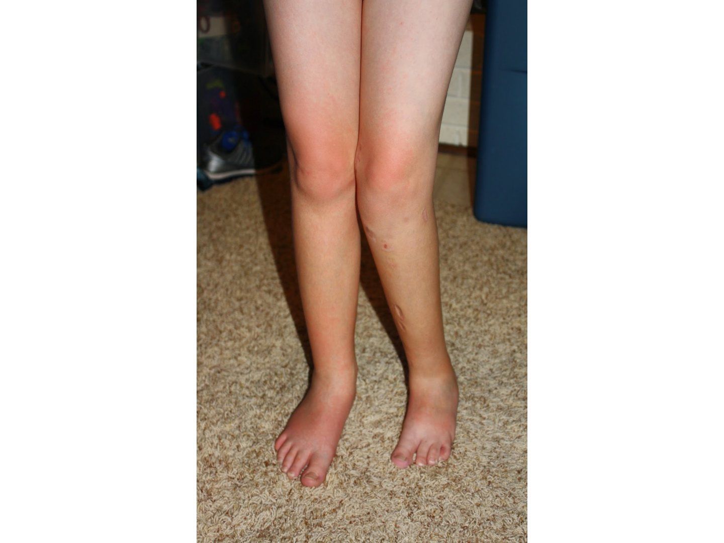
- Describe the abnormalities seen in this standing patient.
- What history would you take in this child with a suspected limb deficiency?
- What associated abnormalities would you look for on examination?
- What is the treatment goal in fibular hemimelia?
- How does the Birch classification guide management?
- What determines ablation rather than reconstruction?
- How do you work up fibular hemimelia?
- What are the components of reconstruction in fibular hemimelia?
- How is the ankle reconstructed in fibular hemimelia?
- Tell me about fibular hemimelia.
Q1.Describe the abnormalities seen in this standing patient.
- Abnormalities over the left lower limb; knee in valgus
- Anterior scars over tibia (?external fixator); medial and lateral scars over proximal tibia and distal femur (?previous epiphysiodesis)
- Missing 5th toe; +/- tibial anteromedial bowing; +/- patella subluxed
- Foot is hypoplastic with missing lateral rays
- Suspect fibular hemimelia; differential includes trauma (tumour and infection unlikely)
Q2.What history would you take in this child with a suspected limb deficiency?
- Establish the diagnosis and rule out associated problems
- Delineate current symptoms
- Find out about previous treatment
Q3.What associated abnormalities would you look for on examination?
- LLD
- Hip: PFFD, coxa vara
- Knee: hypoplastic lateral femoral condyle, genu valgum, patella instability, ACL deficiency
- Tibia: anteromedial bowing
- Foot/ankle: instability from ball and socket ankle, talipes equinovalgus, tarsal coalition, missing lateral toes
Q4.What is the treatment goal in fibular hemimelia?
- Provide a plantigrade foot and a stable ankle
- Achieve equality of leg length
- Correct the tibial deformity to allow more efficient gait
Q5.How does the Birch classification guide management?
- Type 1 = functional foot; type 2 = non-functional foot
- Functional foot (=>3rays, stable ankle) with mild LLD -> shoe raise and brace
- LLD <5cm -> contralateral epiphysiodesis
- LLD >5cm but <30% -> ipsilateral tibial lengthening
- Non-functional foot, unreconstructable ankle or significant LLD -> Syme amputation
Q6.What determines ablation rather than reconstruction?
- Predicted LLD >20cm at maturity
- Foot to the level of mid tibia
- Femur <50% of the other leg
- Radiographic classification by Achterman
Q7.How do you work up fibular hemimelia?
- Scannogram for LLD
- Examination for associated anomalies: hip, knee, tibia, foot/ankle
- Radiographic classification by Achterman
- Treatment depends on LLD at maturity and ankle stability
Q8.What are the components of reconstruction in fibular hemimelia?
- 1. LLD
- 2. Tibial bow correction
- 3. Knee reconstruction
- 4. Ankle reconstruction
Q9.How is the ankle reconstructed in fibular hemimelia?
- Soft tissue: peroneus longus release
- Mild: medial malleolar epiphysiodesis
- Moderate: supramalleolar osteotomy
- Severe: Gruca reconstruction - oblique sliding osteotomy of the tibia to make the lateral fragment the new lateral malleolus
Q10.Tell me about fibular hemimelia.
- Most common congenital long bone deficiency; post-axial longitudinal deficiency (Swanson: failure of formation, problem with AED), occurs in the 1st trimester
- Achterman and Kalamchi classification based on the amount of fibula present
- Type 1A: part of fibula present but proximal fibular epiphysis distal to the proximal tibial physis, distal fibula proximal to the talus
- Type 1B: partial absence (30-50%), distal fibula unable to support the ankle; type 2: absent fibula
- If LLD >30% or unstable ankle -> Symes amputation (88% satisfactory vs ~50% lengthening)
▸ Slide 516 · Birch classificationLimbs Dysplasia Deformity · 2 questions expand

Q1-Q22 questions — tap to reveal all answerslist
- What is the Birch classification?
- How is a functional foot defined in the Birch classification?
Q1.What is the Birch classification?
- A radiological classification of fibular deficiency
- Used to guide management
- Type 1 = functional foot
- Type 2 = non-functional foot
Q2.How is a functional foot defined in the Birch classification?
- 3 or more rays
- Can provide a stable weight-bearing platform
▸ Slide 517 · XR descriptionLimbs Dysplasia Deformity · 3 questions expand
Q1-Q33 questions — tap to reveal all answerslist
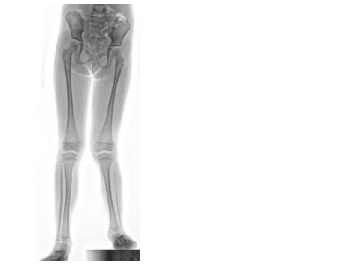
- How do you recognise a leg length discrepancy and fibular hypoplasia on this radiograph?
- What radiographic associations of fibular deficiency should you look for?
- Why is the fibula short? Describe the normal relationships.
Q1.How do you recognise a leg length discrepancy and fibular hypoplasia on this radiograph?
- Pelvis not level despite a block under the short leg -> recognise LLD
- Fibular hypoplasia - the fibula is short
Q2.What radiographic associations of fibular deficiency should you look for?
- Foot: tarsal coalition, absent lateral rays
- Ankle: ball and socket joint, ankle valgus
- Tibia: short + anteromedial bowing
- Knee: LFC hypoplasia, genu valgum, patellar subluxation
- Hip: PFFD
Q3.Why is the fibula short? Describe the normal relationships.
- Normally the fibular head is level with the proximal tibial physis
- The distal fibular physis is level with the tibial plafond
- In fibular deficiency the proximal fibular epiphysis lies distal to the proximal tibial physis
▸ Slide 518 · Clinically photo showing patients right leg with abnormal angulation at level oLimbs Dysplasia Deformity · 10 questions expand

Q1-Q1010 questions — tap to reveal all answerslist
- Describe the clinical and X-ray findings, the diagnosis and its differentials.
- What history and examination findings matter in congenital pseudarthrosis of the tibia?
- Describe the Crawford classification and its union rates.
- What are the general management aims and natural history?
- What is the '4 in 1 osteosynthesis'?
- What did the EPOS 2000 (Gill) study recommend?
- Why is the Ilizarov frame the method of choice and what are the prognostic factors?
- What is the argument for the timing of surgery in congenital pseudarthrosis?
- Bone transport versus acute shortening - what does the EPOS study suggest?
- What is the pathology and the functional outcome at maturity?
Q1.Describe the clinical and X-ray findings, the diagnosis and its differentials.
- Right leg with abnormal angulation at the distal tibia, apex anterior; no significant angulation on the frontal view
- Multiple hyperpigmented skin lesions ?scar/cafe au lait spots
- X-ray: abnormalities of distal tibia and fibula, fracture with ?pseudoarthrosis of the tibia; fibula length normal
- Diagnosis: congenital pseudarthrosis of the tibia
- Differentials: NF, tibial hemimelia (anterolateral bowing), fibrous dysplasia, trauma with fracture callus, Tumor (EWS, osteofibrous dysplasia, adamantimoma)
Q2.What history and examination findings matter in congenital pseudarthrosis of the tibia?
- Know the age
- Associated abnormalities of NF and tibial hemimelia
- Previous treatment
- LLD, angular deformity, joint stiffness/OA
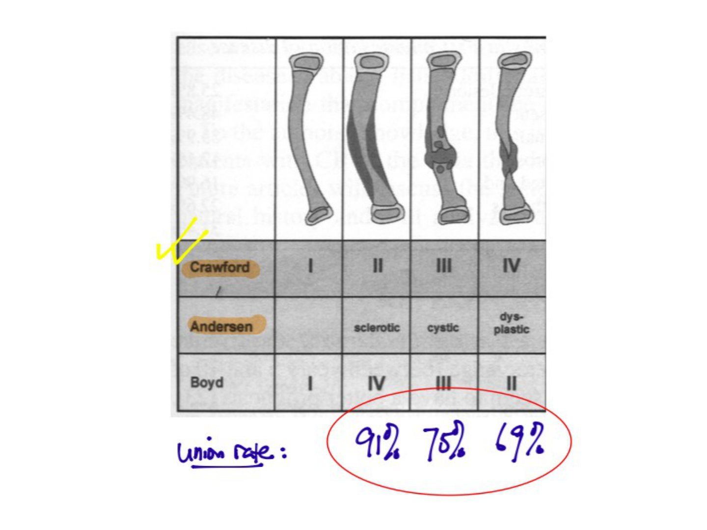
Q3.Describe the Crawford classification and its union rates.
- I: dense IM canal - no brace needed, may never fracture
- II: widened cortex + trabeculation failure - brace; union rate 91%
- III: cystic, pre-fracture - early fixation; union rate 75%
- IV: dysplastic, fracture, cyst, pseudoarthrosis - union rate 70%
Q4.What are the general management aims and natural history?
- Promote union -> prevent fracture; correct deformity + equal leg length
- Natural history: vicious cycle of limb angulation, repeated fracture, LLD, joint stiffness
- Counsel the family from the outset about the possibility of amputation
- If pre-fracture: clamshell brace - no evidence bracing prevents fracture or deformity
- If impending fracture/fracture/pseudoarthrosis: MRI first to see extent and how much to resect
- Serial observation + bracing until maturity
Q5.What is the '4 in 1 osteosynthesis'?
- 1. Resection of pseudoarthrosis + fibrotic periosteum
- 2. Correct angular deformity: bone grafting, acute shortening, or bone transport
- 3. Enhance biology: BMP2,7, periosteal flap, autogenous bone graft +/- bone transport
- 4. Stable fixation with Ilizarov followed by IMN through the ankle joint (often most difficult - William's rod)
Q6.What did the EPOS 2000 (Gill) study recommend?
- 340 patients; pre-fracture clamshell brace
- Fracture -> Ilizarov (provides compression, allows lengthening/bone transport) -> IM nail
- IM nail options: antegrade rush pin, splint the ankle to control the distal fragment, or William's rod
- William's rod through STJ and TTJ if <4 years old; proximalise it 2 years post-op if not grown out of the ankle joint
- Amputate after 3 unsuccessful operations, severe LLD (>15cm) or non-functional limb (Symes, prosthesis protects the pseudoarthrosis)
Q7.Why is the Ilizarov frame the method of choice and what are the prognostic factors?
- EPOS study: excellent stability; allows complete resection of the pseudoarthritic area regardless of segment length (lengthening/bone transport)
- Enables weight bearing, which stimulates healing of bone and soft tissue
- Can transport the fibula distally to avoid valgus ankle deformity
- Poor prognostic factors: young age, advanced Crawford staging, NF (not mentioned: fibular pseudoarthrosis); reasonable success between 3-6 years
- Amputate after 3 unsuccessful operations, LLD >15cm or non-functional limb (Symes)
Q8.What is the argument for the timing of surgery in congenital pseudarthrosis?
- Older child: difficult to provide stable fixation in a small child even with Ilizarov; difficult to weight bear with the frame, which is needed to stimulate bone growth
- Younger child: if delayed, a braced and protected leg cannot develop normally - becomes dysplastic, loses function, shorter from growth retardation
- Based on EPOS, reasonable success between 3-6 years (the fibromatosis in the pseudarthrosis area has greater osteolytic activity in smaller children)
Q9.Bone transport versus acute shortening - what does the EPOS study suggest?
- Bone transport has a lower fusion rate
- Deep muscles and periosteum move with the transported segment but superficial muscle and fascia move very little
- Major vessels lengthen and become tortuous distally, causing kinking
- EPOS suggests acute shortening + LLD correction via proximal metaphyseal lengthening after corticotomy
Q10.What is the pathology and the functional outcome at maturity?
- Fibrous hamartoma in pathological periosteum -> strangulation of blood supply, decrease osteogenicity, increased osteoclastic resorption
- Dysplastic bone -> failure of new bone formation and segmental weakening
- Anterolateral bowing associated with NF (50%), ED syndrome, amnionic band syndrome
- At maturity: FWB 40%, LLD 60%, walk unlimited 70%, no ankle 30%, sports <30%
▸ Slide 519 · Dense cortex, narrowed medullary canal (widened cortex), cystic, dysplasicLimbs Dysplasia Deformity · 2 questions expand
Q1-Q22 questions — tap to reveal all answerslist
- What radiographic patterns are described in the progression of congenital tibial dysplasia?
- Which classification do these features represent?
Q1.What radiographic patterns are described in the progression of congenital tibial dysplasia?
- Dense cortex
- Narrowed medullary canal (widened cortex)
- Cystic lesion
- Dysplastic segment
Q2.Which classification do these features represent?
- The Crawford classification of congenital pseudarthrosis of the tibia
- Types I-IV: dense canal, widened cortex, cystic, then dysplastic pseudoarthrosis
▸ Slide 520 · On the left is a clinical photo of a baby with left leg bowing with apex posteriLimbs Dysplasia Deformity · 4 questions expand
Q1-Q44 questions — tap to reveal all answerslist
- Describe the clinical and radiographic findings.
- What is the diagnosis and its natural history?
- What history and examination findings would you look for?
- What is the management and expected outcome?
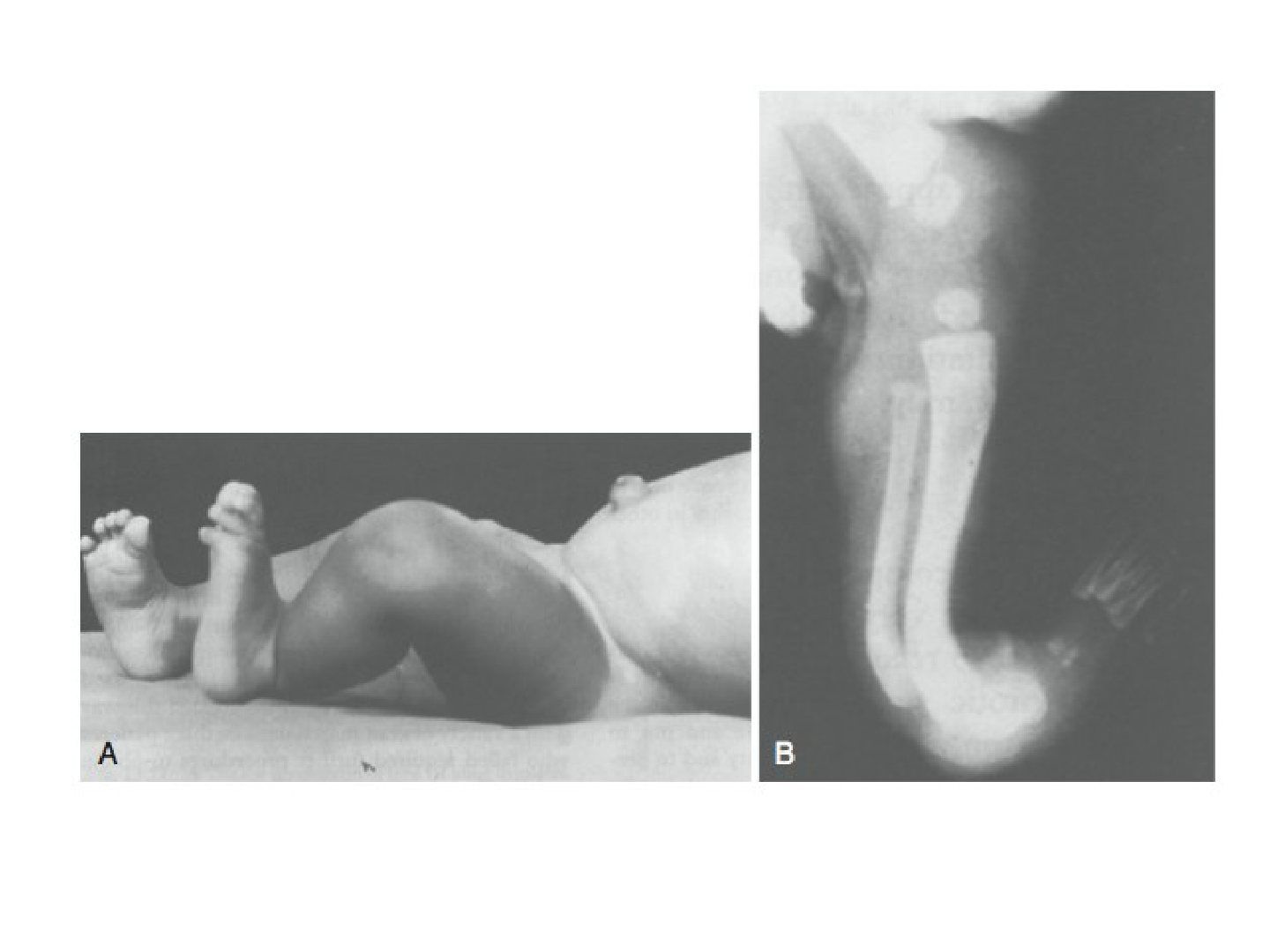
Q1.Describe the clinical and radiographic findings.
- Left leg bowing with apex posterior
- The ankle is not in a normal resting posture
- X-ray: tibia and fibula bowing with apex posterior
- Correlate clinically and obtain an AP film to see whether the bowing is posteromedial
Q2.What is the diagnosis and its natural history?
- Posteromedial bowing of the tibia
- Physiological and commonly associated with intra-uterine malpositioning
- Benign course, usually self-remodels
- No syndromal correlation
Q3.What history and examination findings would you look for?
- Multiple pregnancy, oligohydramnios
- Packaging syndrome: torticollis, DDH, metatarsal adductus
- Check the ankle for calcaneovalgus feet
Q4.What is the management and expected outcome?
- Physiotherapy for stretching
- Monitor for correction and LLD
- Expect LLD of about 3-4cm
▸ Slide 521 · MT adductus and skew footLimbs Dysplasia Deformity · 6 questions expand

Q1-Q66 questions — tap to reveal all answerslist
- What conditions are associated with 'packaging'?
- How does metatarsus adductus present and what is the natural history?
- Describe the Berg classification.
- What is the Bleck classification of metatarsus adductus?
- How is metatarsus adductus managed according to severity?
- What is skew foot and how is it treated?
Q1.What conditions are associated with 'packaging'?
- Torticollis
- DDH
- Knee dislocation
- CTEV
Q2.How does metatarsus adductus present and what is the natural history?
- Intoeing - differential includes femoral anteversion and tibial intorsion
- Must rule out DDH and Blount's
- Natural history (Ponsetti) – 88% spontaneous resolution
Q3.Describe the Berg classification.
- 1. Simple MTA
- 2. Complex MTA = MTA + midfoot lateral shift
- 3. Simple skew = MTA + hindfoot valgus
- 4. Complex skew = simple skew + midfoot lateral shaft
Q4.What is the Bleck classification of metatarsus adductus?
- Bleck classification for MTA uses the values 2,3 / 3 / 3,4 / 4,5
- Berg classification additionally includes skew foot (4 types)
Q5.How is metatarsus adductus managed according to severity?
- Mild (corrects beyond 2nd web): observe
- Moderate (corrects to 2nd web): stretch
- Severe (correct not to 2nd web): casting
- Failed: OT at 4 years - cuboid shortening + 1st MC lengthening
Q6.What is skew foot and how is it treated?
- Deformity: hindfoot valgus, midfoot abduction (TNJ lateral subluxation), forefoot adduction
- Causes: neuromuscular; iatrogenic - improper casting for metatarsus adductus
- Management: cast first
- Refractory: calcaneus lateral opening wedge, medial cuneiform medial closing wedge
▸ Slide 522Limbs Dysplasia Deformity · 2 questions expand

Q1-Q22 questions — tap to reveal all answerslist
- What lower limb dysplasia or deformity is shown on this slide?
- How would you classify and manage the condition shown?
Q1.What lower limb dysplasia or deformity is shown on this slide?
- Not covered in the speaker notes
- Image-only slide; no speaker notes or slide text provided
Q2.How would you classify and manage the condition shown?
- Not covered in the speaker notes
- Image-only slide; no speaker notes or slide text provided
▸ Slide 523 · Genu varum/ valgumLimbs Dysplasia Deformity · 4 questions expand

Q1-Q44 questions — tap to reveal all answerslist
- What are the assessment goals in genu varum/valgum?
- What is the indication for surgery and how is the mature patient managed?
- How is knee malalignment managed in the immature patient?
- What is the difference between irreversible and reversible growth modulation?
Q1.What are the assessment goals in genu varum/valgum?
- Severity - clinical: intercondylar/intermalleolar distance
- X-ray: zoning and tibiofemoral angle
- Delineate the cause
- Rule out complications: patella maltracking, collateral laxity
Q2.What is the indication for surgery and how is the mature patient managed?
- Symptomatic zone 2 or zone 3 malalignment
- Mature patient: osteotomy
Q3.How is knee malalignment managed in the immature patient?
- Hemiepiphysiodesis (hemipiphysiodesis)
- Timing according to the Bowen chart
Q4.What is the difference between irreversible and reversible growth modulation?
- Irreversible: hemiepiphysiodesis, Phemister - advantage is predictable growth
- Reversible: staple, physis bridging plate (guided growth), screw - disadvantage is unpredictable growth and time lag
- Aim to overcorrect because of the rebound phenomenon
- Remove the implant at 2 years, otherwise it will break
▸ Slide 524 · Genu valgumLimbs Dysplasia Deformity · 8 questions expand
Q1-Q88 questions — tap to reveal all answerslist
- How would you assess a child with knee malalignment?
- What are the causes of genu valgum?
- What investigations are indicated in genu valgum?
- What is the aim of treatment in genu valgum?
- When can you reassure the parents?
- When would you follow up?
- When would you operate?
- What are the surgical options?
Q1.How would you assess a child with knee malalignment?
- Delineate the cause: ricket features (caput quadratum, rachitic rosary, Harrison sulcus), mass, dysmorphic features, CP, scars
- Screen: height, body, proportion, symmetry
- Special test: cover and uncover test
- Severity: gait (varus thrusting), TFA and intermalleolar distance, ROM, collaterals, rotational profile
- Complications: LLD, patellar tracking, collateral laxity
- Systems examination: hip, spine and feet
- X-ray: severity (zoning, tibiofemoral angle), features of primary causes
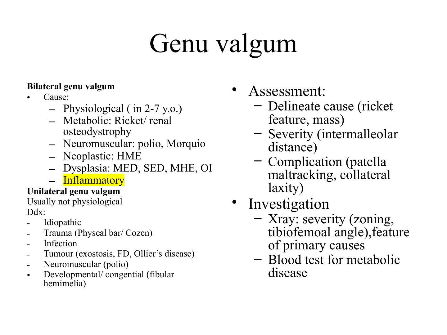
Q2.What are the causes of genu valgum?
- Bilateral genu valgum: physiological (2-7 y), metabolic (rickets/renal osteodystrophy), neuromuscular (polio, Morquio), neoplastic (HME), dysplasia (MED, SED, MHE, OI), inflammatory
- Unilateral genu valgum is usually not physiological
- Unilateral causes: trauma (physeal bar/Cozen), infection, tumour (exostosis, FD, Ollier's disease), neuromuscular (polio), developmental/congenital (fibular hemimelia)
Q3.What investigations are indicated in genu valgum?
- X-ray: severity (zoning, tibiofemoral angle) and features of the primary causes
- Blood test for metabolic disease
Q4.What is the aim of treatment in genu valgum?
- Restore the mechanical axis
- Prevent patellar maltracking and early OA changes
Q5.When can you reassure the parents?
- Age <7 years
- TFA <15 degrees
- Intermalleolar distance <8cm
- Non-progressive
Q6.When would you follow up?
- Age <10 years and:
- Progress after 4 years
- Intermalleolar distance >8cm
- TFA >15 degrees
Q7.When would you operate?
- Age >10 years
- Severe valgus: >15 degrees, intermalleolar distance >8cm, MA zone +3
Q8.What are the surgical options?
- Immature + bilateral: temporary hemiepiphysiodesis at MFC and MTP
- Mature: varus-producing distal femoral osteotomy
▸ Slide 525 · Genu varumLimbs Dysplasia Deformity · 6 questions expand
Q1-Q66 questions — tap to reveal all answerslist
- What is the Salenius curve?
- Why is treatment needed for pathological genu varum?
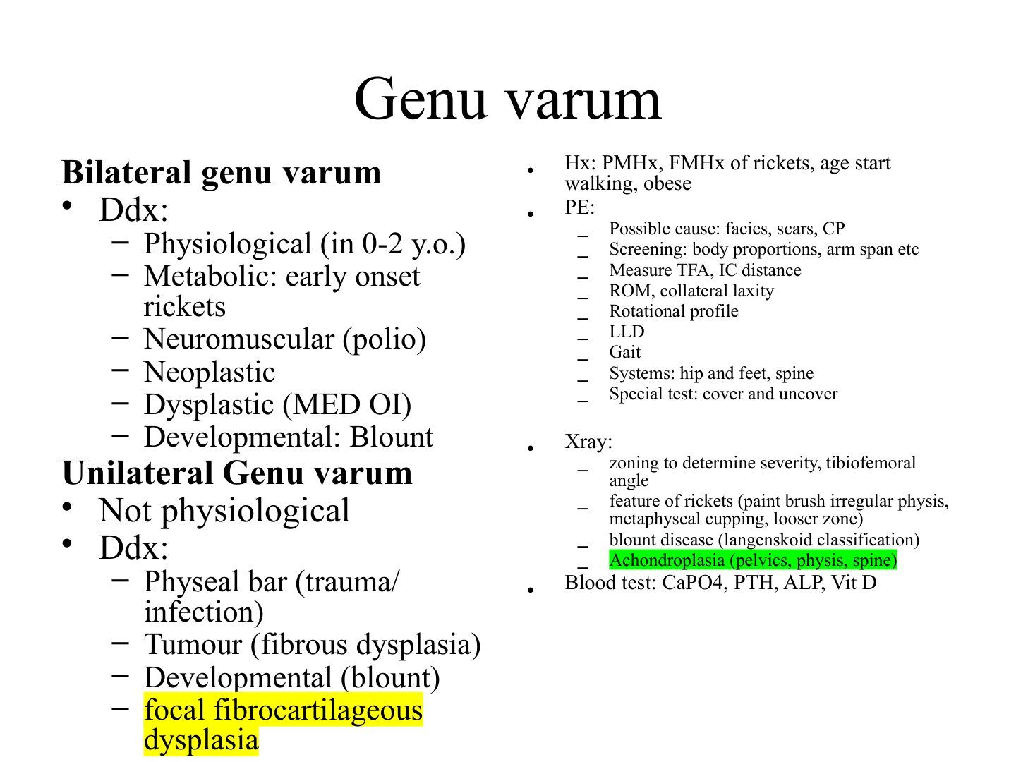
- What is the differential diagnosis of bilateral genu varum?
- What is the differential diagnosis of unilateral genu varum?
- What history and examination findings suggest a pathological cause?
- What is the workup for genu varum?
Q1.What is the Salenius curve?
- 0-2 years: varus
- 2 years: neutral
- 3 years: most valgus
- 7 years: normal valgus
Q2.Why is treatment needed for pathological genu varum?
- Restore the mechanical axis
- Prevent progression of the deformity
- Prevent patellar maltracking and early OA
Q3.What is the differential diagnosis of bilateral genu varum?
- Physiological (0-2 years)
- Metabolic: early-onset rickets
- Neuromuscular (polio)
- Neoplastic; Dysplastic (MED, OI)
- Developmental: Blount
Q4.What is the differential diagnosis of unilateral genu varum?
- Not physiological
- Physeal bar (trauma/infection)
- Tumour (fibrous dysplasia)
- Developmental (Blount)
- Focal fibrocartilaginous dysplasia
Q5.What history and examination findings suggest a pathological cause?
- PMHx/FMHx of rickets, age of starting to walk, obesity
- PE for possible cause: facies, scars, CP
- Screening: body proportions, arm span
- Measure TFA, IC distance, ROM, collateral laxity, rotational profile, LLD, gait
- Systems: hip and feet, spine; special test: cover and uncover
Q6.What is the workup for genu varum?
- X-ray: zoning to determine severity, tibiofemoral angle
- Rickets features: paint-brush irregular physis, metaphyseal cupping, Looser zone
- Blount disease: Langenskiold classification
- Achondroplasia: pelvic, physeal and spinal features
- Blood test: CaPO4, PTH, ALP, Vit D
▸ Slide 526 · Clinical photo showing bilateral genu varumLimbs Dysplasia Deformity · 10 questions expand
Q1-Q1010 questions — tap to reveal all answerslist
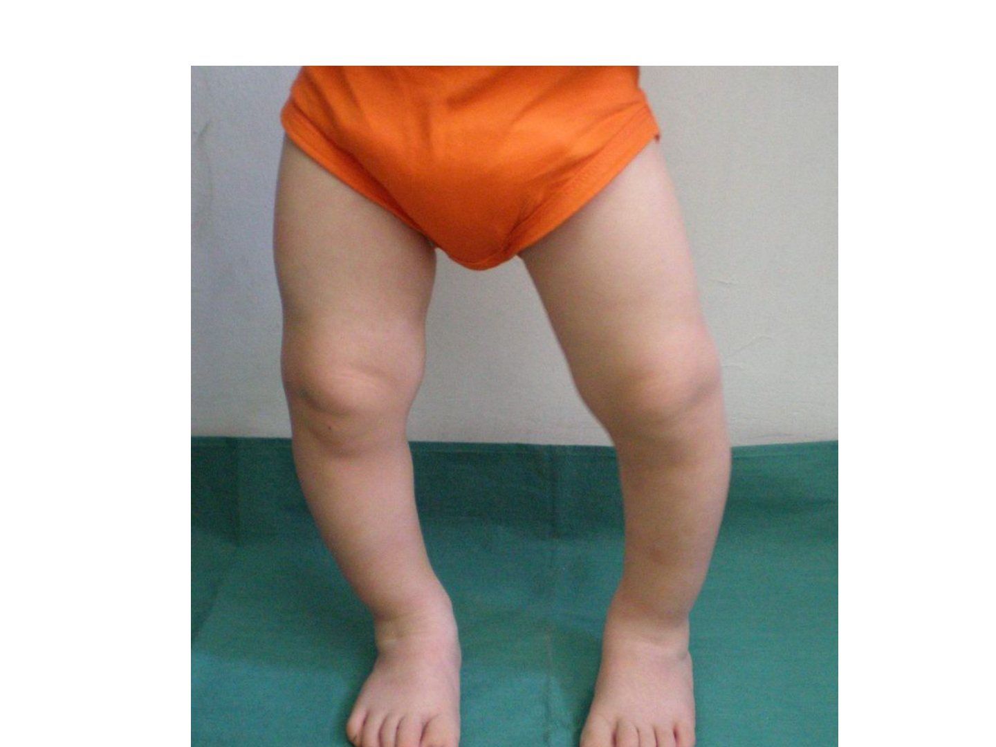
- Describe the clinical findings in this patient with bilateral genu varum.
- What are the causes of pathological genu varum?
- How would you assess a child with genu varum?
- Which radiographic parameters should be measured on the scannogram in Blount's disease?
- What is Blount's disease and what is its pathophysiology?
- What is the problem list in Blount's disease?
- What is the aim of treatment in Blount's disease?
- How is infantile Blount's disease treated?
- How does adolescent Blount's disease differ?
- What are the risk factors for recurrence and the drawbacks of Langenskiold staging?
Q1.Describe the clinical findings in this patient with bilateral genu varum.
- Bilateral genu varum, asymmetrical on both sides
- No scars; some internal rotation/ intoeing of the left lower limb - pathognomonic for Blount's
- Likely pathological
- Can be physiological depending on age (Salenius curve: varus under 2 years can be physiological)
Q2.What are the causes of pathological genu varum?
- Bilateral: metabolic, neoplastic, neuromuscular, dysplasia/developmental (DDNNM)
- Unilateral: infection, tumour (HME, focal fibrocartilaginous dysplasia), trauma, Blount's, iatrogenic, tibial hemimelia
- Physiological depends on age (Salenius curve: varus under 2 years can be physiological)
Q3.How would you assess a child with genu varum?
- History: onset, progression, other deformities; trauma/infection/tumour; rickets risk (diet); Blount's risk (early walking, obesity)
- Look for rickets: caput quadratum, rachitic rosary, Harrison sulcus, catback kyphosis, short stature
- Screen height, body, proportion, symmetry; gait varus thrusting; LLD; ROM and collaterals; TFA and intermalleolar distance
- Rotational profile; systems exam (hip, spine, feet); cover and uncover test
- XR + scannogram: TFA, LDFA, MPTA, mechanical axis zoning, medial slope angle, Drennan angle
- Bloods CaPO, Vita D, PTH ALP; MRI for physeal bar; +/- CT for rotation
Q4.Which radiographic parameters should be measured on the scannogram in Blount's disease?
- TFA, LDFA, MPTA
- Mechanical axis deviation (zoning)
- Medial slope angle, Drennan angle
- LLD, compensatory distal femoral valgus, Tibial-femoral subluxation
- Look for features of rickets, other causes, and classify with Langenskiold
Q5.What is Blount's disease and what is its pathophysiology?
- Dyschondrosis of the medial proximal tibial physis causing progressive pathological genu varum centred at the tibia
- Related to mechanical overload in genetically susceptible individuals
- Excessive medial pressure causes osteochondrosis +/- physeal bar (Heuter-Volkmann law: increased stress = less growth)
- Vs physiological varus: progressive, more abrupt (Drennan angle >11 need close monitoring, >16 = Blount's), physeal abnormality, asymmetrical flaring
- Langenskiold stages: beak, wedge, step, enlarge/fill, double, fused
Q6.What is the problem list in Blount's disease?
- 1. Varus angular deformity
- 2. Medial physeal bar/osteochondrosis
- 3. Depressed joint surface
- 4. Tibial internal torsional deformity
- 5. Femoral anteversion
- 6. Procuvartum
Q7.What is the aim of treatment in Blount's disease?
- Aim of treatment: restore mechanical axis to unload medial physis and prevent further progression (Heuter Vollkman law)
Q8.How is infantile Blount's disease treated?
- Age cut-off 4 years; severity by Langenskiold stage (III-VI) or Drennan angle
- Stage I-II: brace (KAFO with genu varus strap, droplocks to increase corrective force during weight bearing), follow up XR for medial physis reconstitution - operate if progression within 1 year
- Stage III or above: brace likely fails; gold standard RAB (oblique biplanar) osteotomy with overcorrection
- Adjuvant: physeal bar excision, hemiepiphysiodesis, hemiplateau elevation, correct internal rotation
- Other: growth modulation with tension band plate and screws
Q9.How does adolescent Blount's disease differ?
- Onset >10 years; treatment depends solely on the varus angle >10 degrees
- CT/MRI to see whether the physis is closed
- Physis open: lateral tibial epiphysiodesis (permanent or temporary)
- Physis closed: osteotomy without overcorrection - HTO or EF/Ilizarov/TSF +/- prophylactic fasciotomy
Q10.What are the risk factors for recurrence and the drawbacks of Langenskiold staging?
- Recurrence: age >5, body weight >95th percentile, medial physeal slope >60 degrees, Langenskiold stage IV or above
- All stages can occur earlier than the described age
- Stages II and III can progress to VI despite osteotomy (OT does not change the natural history)
- Single osteotomy gives good results only <4 years but not <8 years; original series Only include white population instead of more severe Afro-American population
▸ Slide 527 · Standing XrayLimbs Dysplasia Deformity · 5 questions 1 check expand
Q1-Q55 questions — tap to reveal all answerslist
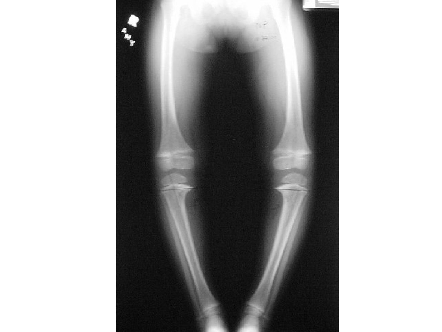
- Describe the findings on this standing radiograph.
- Which angles would you measure?
- What is the Drennan angle and what does it mean?
- What is the medial physeal sloping angle and its significance?
- What other features must you look for on the radiograph?
Q1.Describe the findings on this standing radiograph.
- Skeletally immature; bilateral genu varum (increased femorotibial angle)
- Asymmetrical; mechanical axis zone 3
- Varus centred at the tibial epiphyseal-metaphyseal area
- Widening of the physis, irregularity +/- fragment
- Medial physeal sloping +/- beak, wedge, step; +/- lateral subluxation of the tibia; +/- triangular epiphysis
Q2.Which angles would you measure?
- Tibiofemoral angle
- LDFA (normal 85), MPTA (normal 87)
- Mechanical axis deviation (zoning)
- Drennan angle
- Medial physeal sloping angle
Q3.What is the Drennan angle and what does it mean?
- Used to distinguish physiological bowing from Blount's disease
- <9 degrees: likely physiological
- >11 degrees: suspected Blount's / needs close monitoring
- >16 degrees: consider osteotomy
Q4.What is the medial physeal sloping angle and its significance?
- Angle between tangential lines to the lateral and medial physis
- >60 degrees: high chance of recurrence even after osteotomy
Q5.What other features must you look for on the radiograph?
- Features of trauma, infection, tumour or a systemic cause
- LLD if present
A Drennan (metaphyseal-diaphyseal) angle >11 degrees indicates Blount's disease, and >16 degrees requires osteotomy — imprecise/contested threshold — Levine and Drennan found >11 degrees is associated with Blount's, but Feldman and Schoenecker found 9-16 degrees indeterminate (37% of physiological bowing exceeded 11 degrees); >16 degrees is the more specific threshold used to presume Blount's — medium confidence — source
▸ Slide 528Limbs Dysplasia Deformity · 4 questions expand

Q1-Q44 questions — tap to reveal all answerslist
- Compare the age of onset and laterality of infantile versus adolescent Blount disease.
- Compare the risk factors for infantile and adolescent Blount disease.
- What deformities does each type of Blount disease produce?
- How does each type of Blount disease behave and how is it treated?
Q1.Compare the age of onset and laterality of infantile versus adolescent Blount disease.
- Infantile: 2-5 years, bilateral in >50%
- Adolescent: >10 years, typically unilateral
Q2.Compare the risk factors for infantile and adolescent Blount disease.
- Infantile: Black, early walker, male, obese
- Adolescent: obese
- Infantile disease is more severe
Q3.What deformities does each type of Blount disease produce?
- Both arise at the proximal medial tibial physis
- Infantile: genu varus, flexion, internal rotation, and may have compensatory distal femoral valgus
- Adolescent: proximal tibia physis, and may have distal femoral varus and distal tibial valgus
Q4.How does each type of Blount disease behave and how is it treated?
- Infantile: brace/surgery, staged by Langenskiold
- Adolescent: surgery
- Never resolve spontaneously
▸ Slide 529 · Describe Photo:Limbs Dysplasia Deformity · 6 questions expand

Q1-Q66 questions — tap to reveal all answerslist
- What is the diagnosis and the differential?
- Describe the clinical photograph.
- How would you assess this child?
- What are the X-ray findings and how do you differentiate CVT from oblique talus?
- What is the pathoanatomy of vertical talus?
- How is congenital vertical talus managed?
Q1.What is the diagnosis and the differential?
- Diagnosis: congenital vertical talus (CVT)
- Differential: congenital oblique talus, calcaneovalgus, tarsal coalition, paralytic pes planus
Q2.Describe the clinical photograph.
- Midfoot: rocker bottom deformity
- Equinus hindfoot
- Dorsiflexion and abduction of the forefoot
Q3.How would you assess this child?
- Screen systemically: 50% +ve neuromusclular disease (spina bifida, arthrogryposis), syndromal (Larsen), maltreated CTEV, look for DDH
- 20% positive family history
- Locally confirm a rigid deformity: rocker bottom foot, equinovalgus hindfoot with tight TA and peroneus
- Chopart joint: dorsiflexion with navicular dorsal dislocation; forefoot abduction/dorsiflexion (EDL/EHL, tib ant contractures)
- Prominent talar head plantarly; check lower limb neurology; peg leg gait
Q4.What are the X-ray findings and how do you differentiate CVT from oblique talus?
- Lateral: vertically pointed talus, long axis of the talus below the 1st ray axis (TAMBA - talus axis MT bone angle)
- Dorsally dislocated navicular (navicular ossifies at 3 years old)
- AP: increase in talocalcaneal angle >40 degrees
- Plantarflexion view: if the TNJ reduces it is a congenital oblique talus, not CVT
Q5.What is the pathoanatomy of vertical talus?
- Irreducible dorsal dislocation of the navicular on the talus, producing a rigid flatfoot deformity
- Soft tissue: tight TA; displaced PT and peroneals act as dorsiflexors; attenuated spring ligament
- Bony: dorsally dislocated navicular
- Oblique talus is also treated with TA release
Q6.How is congenital vertical talus managed?
- Counsel: reverse Ponseti may be tried, but the majority eventually need surgery
- Pre-op stretching of dorsolateral structures + reverse Ponseti for 3 months (push forefoot plantar-medially)
- After reduction: talonavicular pinning (MIS) + peroneus/extensor release +/- lengthening +/- spring ligament recon + percutaneous Achilles tenotomy 6-12 months after casting
- Presenting late: talus + navicular excision and lateral column lengthening
- Salvage: triple arthrodesis; Dobbs (JBJS Am 2006) reported good results in 11 patients with stretching, reverse Ponseti, pinning and tenotomy
▸ Slide 530 · Painful flatfoot onset in adolescence: Tarsal coalitionLimbs Dysplasia Deformity · 8 questions expand

Q1-Q88 questions — tap to reveal all answerslist
- Describe the clinical photo and give the diagnosis.
- What is the differential diagnosis for a painful flatfoot in a child?
- What are the general causes of flatfoot by laterality?
- What is the genetics and pathology of tarsal coalition?
- Outline your assessment of a child with a painful flatfoot.
- What imaging would you request?
- How is tarsal coalition treated?
- What does peroneal spasm mean and what is the epidemiology of flatfoot?
Q1.Describe the clinical photo and give the diagnosis.
- Bilateral flatfoot with reduced medial arch
- Medial bulge suggests middle facet coalition
- From behind: hindfoot valgus, too many toes sign positive on the right
- Diagnosis: pes planus
- Must differentiate flexible from rigid
Q2.What is the differential diagnosis for a painful flatfoot in a child?
- Tarsal coalition
- Painful flexible flatfoot (tight Achilles)
- JIA
Q3.What are the general causes of flatfoot by laterality?
- Unilateral: post-traumatic, degenerative, Charcot
- Bilateral: ligamentous laxity, RA, neuromuscular (spastic diplegic, spina bifida)
- Both: idiopathic, PTTD, congenital vertical talus, tarsal coalition
Q4.What is the genetics and pathology of tarsal coalition?
- Autosomal dominant with a high level of penetrance
- NOG (Noggin) gene
- Bilateral in 50%
- Failure of mesenchymal segmentation
Q5.Outline your assessment of a child with a painful flatfoot.
- History: onset, site of pain, trauma, footwear
- Exclude obvious syndromic or neurological causes
- Tenderness: CN - lateral pain at sinus tarsi; TC - medial pain (microfracture of coalition interface, ossification of previously fibrous or cartilaginous coalition, chondral injury, peroneal spasm, subfibular impingement)
- Painful inversion against resistance (peroneal spasm)
- Deformity (hindfoot valgus, forefoot abduction, rigid flatfoot); PTT function
- Limited subtalar movement - reverse Coleman block; examine the contralateral side (50% bilateral, can be associated with fibular hemimelia/ PFFD, carpal coalition, Apert syndrome)
Q6.What imaging would you request?
- CN: oblique view - anteater sign
- TC: lateral view - C sign (subtalar/middle facet coalition), talar neck traction spur
- Harris view may also show coalition
- CT/MRI for size and location of bony/fibrous connection, r/o additional coalitions (5%)
Q7.How is tarsal coalition treated?
- Conservative: arch support/orthosis moulded at current position, walking cast boot, temporary immobilization
- CN coalition: excision via Olliers approach, EDB interposition +/- deformity correction and TA release
- TC coalition: bony union >50% -> fusion; <50% -> excision + interposition (split FHL graft, fat) via medial incision between FDL and PT
- Indications for fusion: malalignment, >50% involvement, degeneration, older patient
- Salvage: triple arthrodesis + deformity correction
Q8.What does peroneal spasm mean and what is the epidemiology of flatfoot?
- Peroneal spasm is a protective mechanism to reduce pain; if absent the subtalar joint is likely okay
- Canadian army foot study: 1/5 have flatfoot; 67% asx (asymptomatic)
- 25% symptomatic flexible flatfoot with tight TA; 8% symptomatic rigid (accessory navicular, tarsal coalition, CVT)
- Wagner JBJS 1989: improvement not related to orthosis; increased laxity showed more improvement
▸ Slide 531 · Accessory navicularLimbs Dysplasia Deformity · 5 questions expand
Q1-Q55 questions — tap to reveal all answerslist
- What is an accessory navicular?
- What causes pain from an accessory navicular?
- How does an accessory navicular cause rigid flatfoot?
- What is the Ray and Goldberg classification?
- What is the management?
Q1.What is an accessory navicular?
- A secondary ossification centre that failed to unite in childhood (usually ossifies at 9 years old)
- Mostly asymptomatic
Q2.What causes pain from an accessory navicular?
- Repeated microfracture of the synchondrosis
- PTT insertion inflammation
Q3.How does an accessory navicular cause rigid flatfoot?
- PTT inserts into the accessory navicular -> change in PTT pull
- Midfoot adduction -> accessory navicular impinges onto the medial malleolus -> PTT pain
- Patient adopts forefoot abduction
Q4.What is the Ray and Goldberg classification?
- Type I: ossicle in PTT
- Type II: synchondrosis
- Type III: synostosis
Q5.What is the management?
- Asymptomatic: no treatment
- Mild: donut-shaped moleskin
- Severe: cast for 6 weeks then UCBL
- OT indication: refractory to cast/brace - Kidner procedure
- Kidner: remove accessory navicular at the synchondrosis + reattach PTT (advance distal and plantar - controversial)
▸ Slide 532 · A.Limbs Dysplasia Deformity · 10 questions 2 check expand
Q1-Q1010 questions — tap to reveal all answerslist
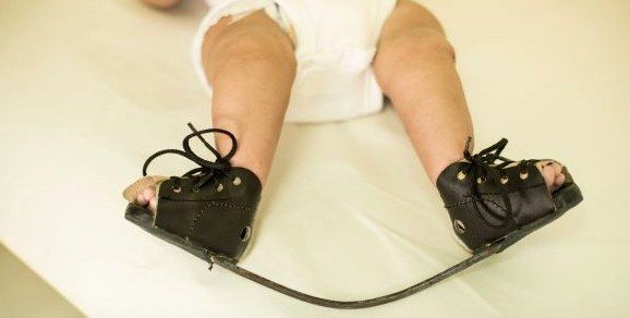
- Describe the deformity shown in this patient.
- What associated problems must you rule out?
- How would you counsel the parents?
- How is the Ponseti method performed and monitored?
- Describe the Ponseti correction sequence in detail.
- Describe the pathoanatomy and causes of CTEV.
- Would you X-ray every clubfoot?
- How is TA lengthening performed and when is it indicated?
- Describe the Denis Brown boots.
- What if conservative management of clubfoot fails?
Q1.Describe the deformity shown in this patient.
- Paediatric patient with bilateral foot deformity
- Hindfoot: varus and equinus
- Midfoot: cavus
- Forefoot: adduction + pronated position
- Locally assess correctability (?postural) and assess the other side (50% bilateral)
Q2.What associated problems must you rule out?
- Packaging syndrome: torticollis, DDH, knee dislocation
- Rule out syndromic clubfoot
- Neuromuscular disease (CP)
- Spinal dysraphism
- Teratogenic/syndromes: arthrogryposis, SED, constriction band, Larsen, Pierre Robin, Prune Belly
Q3.How would you counsel the parents?
- Familial occurrence in 25%; affected child has a 2.5-6.5% chance of a sibling with clubfoot
- PITX gene / chromosome 2 deletion (common genetic pathway with CVT)
- Ponseti treatment: success rate >90%; 70% need PETA; 30% need tib ant transfer for dynamic supination at 4-5 years
- The affected foot will be smaller and shorter regardless of treatment
Q4.How is the Ponseti method performed and monitored?
- Based on viscoelastic stress relaxation and creep; start at 1 week
- Toe-to-groin POP with knee at 90 degrees, snug but not tight, thin velband, well moulded
- Weekly casts, usually 4-5; assistant holds the big toe and talar head throughout
- Correction sequence: cavus, adductus, varus (corrected with adductus), equinus
- Chart with the Pirani score (high score = high deformity, not prognostic): midfoot and hindfoot components
- Complications of casting: rockerbottom foot (2-5%), flat heel pad, crowded toes, pressure sores
Q5.Describe the Ponseti correction sequence in detail.
- 1. Cavus: dorsiflex the 1st ray, realign the plantiflexed 1st MT with the other MTs and realign forefoot to hindfoot by supination (warn family the deformity may look worse)
- 2. Adductus: abduct the forefoot and externally rotate at the midfoot using the uncovered head of the talus as fulcrum
- 3. Varus: will be corrected with step 2
- 4. Equinus: pull the calcaneal tuberosity distally while pushing the anterior calcaneus up; when abduction 70deg achieved, able to palpate the anterior process of the calcaneus as it abducts out from beneath the talus (neutral or slight heel valgus)
Q6.Describe the pathoanatomy and causes of CTEV.
- Primary deformity centred at the talus, with a medially and plantarly deviated neck
- The rest of the acetabulum pedis (calcaneus, navicular, cuboid) rotates around the talus and becomes adducted and inverted
- Secondary soft tissue contracture: plantarflexors/invertors, CFL, deltoid, long/short plantar, spring, plantar aponeurosis
- Causes: majority idiopathic; neuropathic, myopathic, genetic (PITX), mechanical moulding, multifactorial
Q7.Would you X-ray every clubfoot?
- X-ray is controversial: difficult to position, only calcaneum, talus and MT ossified, and ossification centres do not represent true shape
- Double parallelism
- Dorsiflexion lateral (Turco view): hindfoot parallelism, talocalcaneal angle <35 degrees
- AP view: Kite angle (talocalcaneal) <20, talus-first metatarsal angle negative
Q8.How is TA lengthening performed and when is it indicated?
- Indicated if everything is corrected but DF still <10deg; needed in 70% of patients
- NA (local anaesthetic), incision medial to TA, transverse cut towards lateral
- Then another cast for 3 weeks at 30 deg ankle DF
Q9.Describe the Denis Brown boots.
- BOOTS: Strap for 3 point fixation; forefoot open shoe box; midfoot scaphoid and intermetatarsal pad + straight medial lass to keep the foot abducted; hindfoot reverse Thomas heel with stiff heel support for mediolateral stability
- BAR: convex away to keep ankle dorsiflexion 5-10 degrees, shoulder width, ER 70/40 both sides 45 (sum 90-100) to keep the hindfoot in valgus
- Regime: 4 months full time, until 4 years at night time
Q10.What if conservative management of clubfoot fails?
- Failed if the foot is not plantigrade by 3-6 months
- Timing controversial: early 6 months vs delay to ~1 year to allow weight bearing and avoid recurrence; wait until foot length >=8cm
- OT: Cincinnati incision, a la carte approach
- Do not release: deep talocalcaneal interosseous ligament (risk of lateral translation of calcaneus), deep deltoid (valgus deformity), Posterior tibial-fibular ligament
Clubfoot genetics: PITX gene, chromosome 2 deletion — imprecise — PITX1 maps to chromosome 5q31.1; chromosome 2q31-33 deletions involve the HOXD cluster, a separate clubfoot-associated locus — medium confidence — source
The Pirani score is not prognostic — contested — Several studies report the initial Pirani score, especially the hindfoot component, predicts tenotomy need and relapse, although a 2022 systematic review/meta-analysis found no significant association with recurrence — medium confidence — source
▸ Slide 533 · AP Xray of knee of skeletally immature patientLimbs Dysplasia Deformity · 6 questions expand
Q1-Q66 questions — tap to reveal all answerslist
- Describe the X-ray findings and your concern.
- How would you assess a child with a suspected physeal bar?
- How are physeal arrests classified and what affects prognosis?
- What is the management principle and how does treatment depend on growth remaining?
- How does the cause of the physeal arrest affect prognosis?
- What factors determine the risk of a physeal bar after fracture (JPO 2009)?
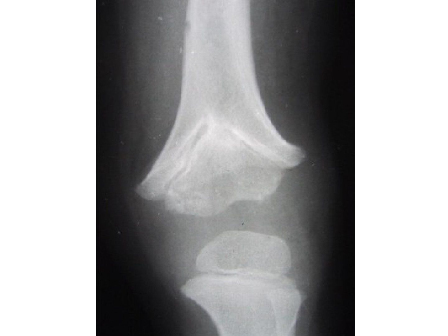
Q1.Describe the X-ray findings and your concern.
- Skeletally immature patient
- V-shaped physis of the distal femoral physis
- Suspected connection between metaphysis and epiphysis centrally (cross trabeculation) -> physeal arrest
- Request a long film including the contralateral limb to assess LLD/angular deformity, and a lateral Xray to see angular deformity
Q2.How would you assess a child with a suspected physeal bar?
- History: age, causes
- Local examination: deformity, length, joint involvement, features suggesting cause
- Systemic examination: maturity
- CT to look for a bony bar; MRI to look for fibrous tissue and the underlying cause
Q3.How are physeal arrests classified and what affects prognosis?
- Extent (partial/ complete)
- Site: peripheral, central or linear
- Prognosis affected by: cause, extent, site, growth potential
- Sequelae: LLD, angular deformity, epiphyseal deformity
Q4.What is the management principle and how does treatment depend on growth remaining?
- Aim: equal leg length with no angular deformity, minimize complications and hospital stay
- Treatment depends on growth potential/projected LLD, deformity present, and area involved
- >2 years growth and <50% physis involved: Langenskiold procedure (bar excision + interposition of fat/cement + marker implantation) or physis distraction
- >50% physis involved: hemi/epiphysiodesis + future lengthening
- <2 years growth: hemiepiphysiodesis; no growth: osteotomy; follow up till maturity
Q5.How does the cause of the physeal arrest affect prognosis?
- Traumatic: responsive to physeal bar excision as only the hypertrophic zone is affected
- Tumour/infection: decreased response as all zones are affected
Q6.What factors determine the risk of a physeal bar after fracture (JPO 2009)?
- Initial displacement
- Mechanism of injury (high energy)
- Salter-Harris pattern
- Number of reductions - not more than 2
- Residual displacement after closed reduction
- Growth arrest from injury to the germinal matrix (resting zone), Affecting blood supply, or bone bar formation
▸ Slide 534 · LLDLimbs Dysplasia Deformity · 18 questions expand

Q1-Q1818 questions — tap to reveal all answerslist
- Describe the findings in the block test photo of this patient with limb length discrepancy.
- List the causes of limb length discrepancy when the affected leg is long versus short.
- What are the steps in the management of limb length discrepancy?
- What are the clinical methods of assessing skeletal maturity?
- What are the radiological methods of assessing skeletal maturity?
- What is the ossification sequence used in the Greulich-Pyle atlas?
- Describe the stages of the Sauvegrain method using the elbow epiphyseal centre.
- What are the general treatment principles for LLD according to the predicted discrepancy at skeletal maturity?
- Which methods calculate LLD at maturity from a single visit versus multiple visits?
- What are the two categories of limb lengthening technique?
- Describe the gradual lengthening procedure in distraction histogenesis.
- What is chondrodiastasis and what are its problems?
- Describe the permanent shortening techniques for the long limb.
- What temporary epiphysiodesis options exist for LLD?
- What is the histology of distraction histogenesis?
- What factors affect distraction lengthening?
- What is the recommended distraction regime?
- What are the complications of distraction lengthening?
Q1.Describe the findings in the block test photo of this patient with limb length discrepancy.
- Block test used to assess limb length
- Right LL shorter by ~5cm
- Both femur and tibia are involved
- Foot and ankle no gross deformity
Q2.List the causes of limb length discrepancy when the affected leg is long versus short.
- Long leg: congenital hemihypertrophy (NF, Klippel-Trenaunay, Beckwith-Wiedemann), physeal stimulation (post-traumatic)
- Short leg, congenital: DDH, fibular hemimelia, PFFD, calcaneovalgus, enchondromatosis
- Short leg, neuromuscular: polio, cerebral palsy
- Short leg, vascular: Perthes, AV fistula
- Short leg, physeal: physeal bar, hyperaemia (trauma, infection, tumour)
Q3.What are the steps in the management of limb length discrepancy?
- Assess cause (femur vs tibia)
- Assess skeletal maturity (clinical, radiological)
- Calculate LLD at maturity
- Assess foot and ankle function (joint mobility and stability)
- Rule out other deformity (angular, rotational, LL contracture)
Q4.What are the clinical methods of assessing skeletal maturity?
- Chronological age
- Tanner staging: 1st sign = enlargement of testis / breast bud; descending phase = menarche / axillary hair
- Menarche
- Growth spurt
Q5.What are the radiological methods of assessing skeletal maturity?
- Tanner-Whitehouse: morphology and fusion of the physis in the hand and wrist
- Greulich-Pyle atlas of the hand and wrist
- Risser sign
- Elbow epiphyseal centre (Sauvegrain)
Q6.What is the ossification sequence used in the Greulich-Pyle atlas?
- Greulich-Pyle atlas assesses skeletal maturity from the hand and wrist
- Ossification sequence: DP -> MC -> PP -> MP -> ulnar -> radius
Q7.Describe the stages of the Sauvegrain method using the elbow epiphyseal centre.
- Double epiphysis -> semi-moon -> quadrangular (tri-cartilaginous closed) -> start fusion -> fused
Q8.What are the general treatment principles for LLD according to the predicted discrepancy at skeletal maturity?
- <2cm: observe
- 2-5cm: shorten the longer limb by epiphysiodesis if physis open, shortening osteotomy if physis closed
- >5cm: lengthen the short limb +/- shorten the long limb
- >20cm: consider amputation
Q9.Which methods calculate LLD at maturity from a single visit versus multiple visits?
- Multiple FU: Moseley chart (needs leg length of both LL and hand X-ray for skeletal maturity), arithmetic method, Green-Anderson chart
- Single FU: Paley multiplier method (LLD x age and sex factor), simplistic method
- Also take into consideration Shapiro's growth pattern
Q10.What are the two categories of limb lengthening technique?
- Acute lengthening
- Gradual lengthening - distraction histogenesis and gradual lengthening procedures
Q11.Describe the gradual lengthening procedure in distraction histogenesis.
- Apply ex fix; low energy corticotomy at metaphysis (2/3 circumference + osteoclasis), preserve periosteum, endosteum, medullary content and nutrient artery
- Corticotomy vs osteotomy (multiple drill holes + osteotome) - no difference in the literature
- Rest 5-7 days, then distract 1mm/day divided into 4 times
- Consolidation ~2x the lengthening time; remove ex fix when interzone closes and 2 side neocorticalisation is seen on AP and lateral films
- Maximum lengthening each time 20% (Michael To tutorial, JPO 2010: % lengthening related to % of complication); >20% increases complications e.g. non-union, NV injury, infection, contracture
Q12.What is chondrodiastasis and what are its problems?
- Gradual distraction of the physis results in hypertrophy of the cellular layers of the physis without separation (Heuter-Volkman)
- Unpredictable results
- Initial lengthening is often followed by growth plate fusion
Q13.Describe the permanent shortening techniques for the long limb.
- Open epiphysiodesis (Phemister): remove a rectangular portion of bone containing metaphysis, physis and epiphysis, flip it around and reinsert to create a bone bridge
- Phemister advantages: predictable outcome; disadvantages: irreversible, long rehab, extensive dissection, angular deformity, exostsosis (exostosis)
- Percutaneous epiphysiodesis (Bowen): stab wound, longitudinal periosteum split + physis penetration 5mm by osteotome, peripheral 1/3 ablated by curette on both medial and lateral sides
Q14.What temporary epiphysiodesis options exist for LLD?
- Staples
- 8-plate
- Transphyseal screw
- Combo procedures (shortening and lengthening combined)
Q15.What is the histology of distraction histogenesis?
- Central fibrous interzone: fibroblasts activated and secrete collagen parallel to distraction force, providing undifferentiated mesenchymal cells that directly transform into osteoblasts -> intramembranous ossification
- Zone of microcolumn formation: osteoblasts align collagen into longitudinal columns and promote blood vessel invasion to lay down osteoid
Q16.What factors affect distraction lengthening?
- Stability of fixation
- Type and site of osteotomy: Ilizarov compared open, percutaneous corticotomy and complete closed - recommends percutaneous corticotomy; metaphysis has more osteogenic potential and soft tissue better adapted to lengthening
- Distraction regime: latency lets the inflammatory phase subside, then distract during the reparative phase
Q17.What is the recommended distraction regime?
- Latency: 3 days for a child, 7-14 days for an adult
- Rate 1mm/day
- Rhythm: distraction applied 4 times/day; increasing frequency increases distraction
Q18.What are the complications of distraction lengthening?
- Bone related: incomplete consolidation, premature consolidation, fracture, growth disturbance of the lengthened bone
- Implant related: NV injury, pin tract infection, joint contracture + subluxation
- Also angular deformity and fracture due to early removal of the ex fix
▸ Slide 535 · LLD scriptLimbs Dysplasia Deformity · 9 questions expand
Q1-Q99 questions — tap to reveal all answerslist
- What is the structure of the clinical assessment (script) for limb length discrepancy?
- What problems are associated with limb length discrepancy?
- What imaging is available for LLD measurement and what are their drawbacks?
- How do the Paley multiplier and simplistic methods predict LLD at maturity?
- Describe the Menelaus arithmetic method of LLD prediction.
- Compare the Green-Anderson and Moseley methods of LLD prediction.
- What are the advantages and disadvantages of the Menelaus arithmetic method?
- What negative findings are noted in this girl with LLD?
- Why is skeletal maturity assessed in a child with LLD?
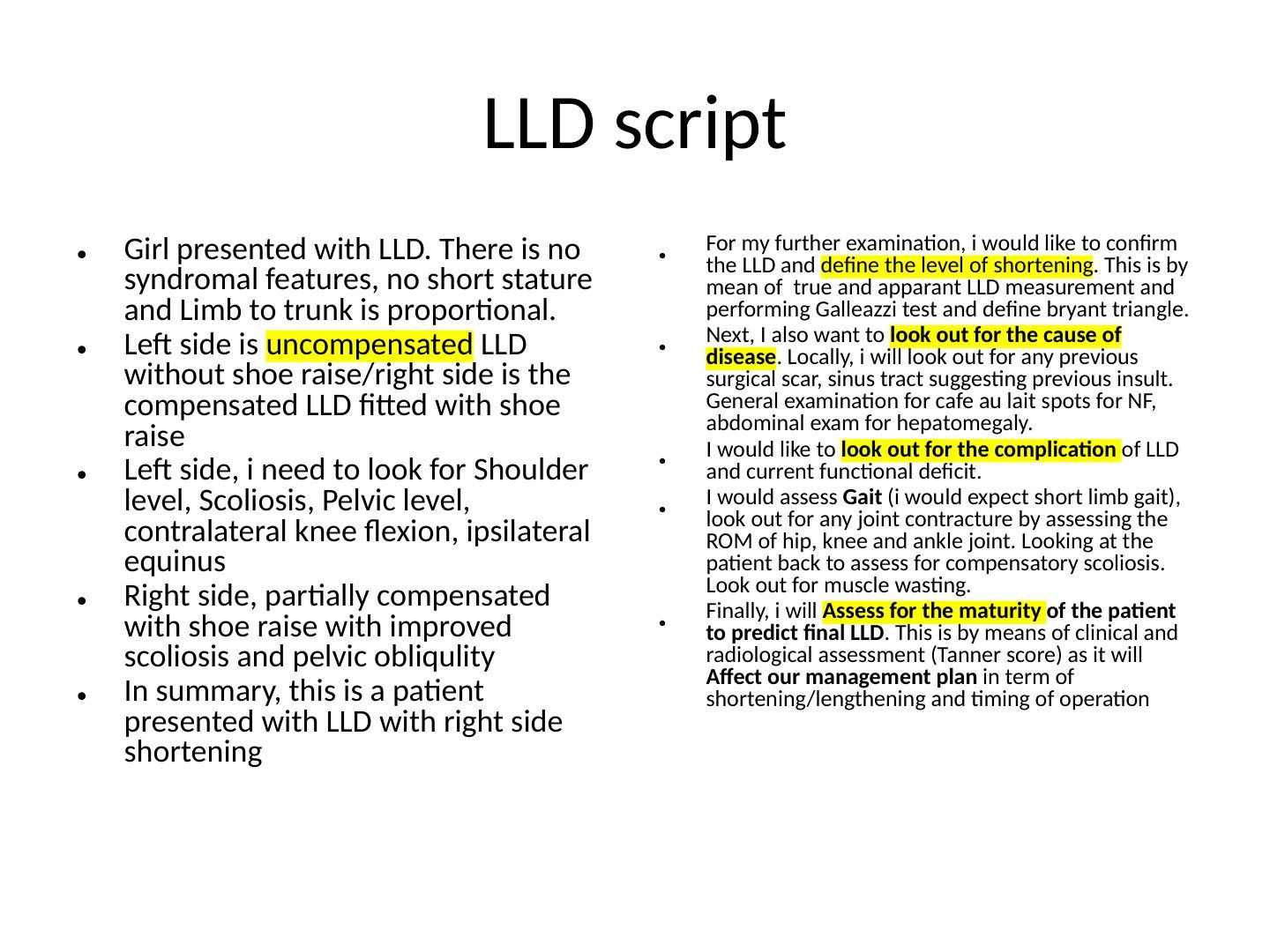
Q1.What is the structure of the clinical assessment (script) for limb length discrepancy?
- 1. Describe the LLD - in this patient a girl with right side shortening: left side uncompensated LLD without shoe raise, right side compensated LLD fitted with shoe raise
- 2. Confirm the LLD and define the level of shortening: true and apparent LLD measurement, Galeazzi test, Bryant triangle
- 3. Pick up signs pointing to the cause of disease: previous surgical scar, sinus tract suggesting previous insult, cafe au lait spots for NF, abdominal exam for hepatomegaly
- 4. Look for complications of LLD and current functional deficit: assess gait (expect short limb gait), joint contracture (ROM of hip, knee and ankle), back for compensatory scoliosis, muscle wasting
- 5. Assess skeletal maturity - clinical and radiological (Tanner score) to predict final LLD and its effect on management (shortening/lengthening, timing of operation)
Q2.What problems are associated with limb length discrepancy?
- Deformity: increased energy of gait, needs shoe raise, cosmetic problem, knee pain
- Compensation: compensatory scoliosis, pelvic tilting, contralateral hip adduction
- Also knee flexion, ipsilateral equinus deformity and OA hip
- Left side (uncompensated, no shoe raise): look for shoulder level, scoliosis, pelvic level, contralateral knee flexion, ipsilateral equinus
- Right side partially compensated with shoe raise: improved scoliosis and pelvic obliquity
Q3.What imaging is available for LLD measurement and what are their drawbacks?
- Teleoroentgenogram: single film, single exposure; inaccurate due to parallax of the X-ray beam
- Orthoroentgenogram: single film, 3 exposures (hip, knee, ankle); more accurate but needs a cooperative patient
- Scanogram: separate films, 3 exposures, ruler fixed on X-ray to avoid magnification
- CT scanogram: especially for angular deformity, accurate measurement
- Standing vs supine: standing film assesses joint line and rotation, which may mask LLD
Q4.How do the Paley multiplier and simplistic methods predict LLD at maturity?
- Paley multiplier: multiplies current discrepancy by a sex and age specific factor
- Applicable at the first visit; most accurate in type I growth pattern; uses chronological age
- Simplistic: LLD x 1.5 (7-8yo), x 2 (4yo), x 2.5 (2yo)
Q5.Describe the Menelaus arithmetic method of LLD prediction.
- Physis closure: boys 16, girls 14
- Growth per year: proximal femur 3mm, distal femur 9mm, proximal tibia 6mm, distal tibia 5mm
- Inhibition is calculated from the discrepancy between 2 visits
- Future discrepancy = current discrepancy + (growth remaining x inhibition); most accurate towards end of growth; uses chronological age
Q6.Compare the Green-Anderson and Moseley methods of LLD prediction.
- Green-Anderson: growth remaining data from 1950s American children; based on skeletal age and growth percentile; Heavy reliance on skeletal age, data from a single population
- Green-Anderson is for epiphysiodesis only, not lengthening; complex, 2 sets of graphs
- Moseley graph: uses skeletal age, accounts for skeletal vs chronological age difference, predicts both epiphysiodesis and lengthening
- Moseley assumes a linear growth pattern
Q7.What are the advantages and disadvantages of the Menelaus arithmetic method?
- Advantages: more applicable in the clinical setting, most accurate towards the end of growth
- Disadvantages: uses chronological age, not accurate for early projection
Q8.What negative findings are noted in this girl with LLD?
- No syndromal features
- No short stature
- Limb to trunk is proportional
Q9.Why is skeletal maturity assessed in a child with LLD?
- To predict the final LLD at maturity
- It affects the management plan: shortening vs lengthening and timing of operation
▸ Slide 536 · Clinical photo showing classic W sitting positionLimbs Dysplasia Deformity · 11 questions expand

Q1-Q1111 questions — tap to reveal all answerslist
- What is the normal foot progression angle?
- Describe the components of the Staheli rotational profile.
- What are the causes of intoeing by age group?
- What are the causes of out-toeing?
- What is the natural history of intoeing and how would you counsel the family?
- What are the indications for surgery in intoeing and what procedures are used?
- What history should be taken in a child with suspected intoeing?
- What should be checked on examination in a child with intoeing?
- What is the mainstay of management of intoeing and which pathological conditions must be ruled out?
- What is the natural history of femoral version?
- What is the natural history of tibial version (transmalleolar-thigh angle)?
Q1.What is the normal foot progression angle?
- Normal -5 to 20 degrees
- The child in the photo shows classic W sitting and is suspected to have in-toe gait
Q2.Describe the components of the Staheli rotational profile.
- Femoral anteversion: hip ER/IR ~ 20/<70 (EFA if IR >70), Craig's test
- Tibial torsion: thigh-foot angle 10 to 20 (ITT if TFA <10)
- Transmalleolar axis (when foot deformity present); any negative value is abnormal
- Heel bisector line
Q3.What are the causes of intoeing by age group?
- Infancy: metatarsus adductus, tibial intorsion
- Toddler: tibial intorsion
- Childhood: femoral anteversion
- Pathological: DDH, CP, Blounts
Q4.What are the causes of out-toeing?
- Pes planus; femoral retroversion
- Infancy: hip ER contracture; later childhood: tibial extorsion
- Pathological: SCFE, Perthes, PFFD
Q5.What is the natural history of intoeing and how would you counsel the family?
- Natural history is out-in-out; maximum intoe at age 11
- Neonate to 11: intoeing due to resolution of intrauterine position
- After 11: femoral anteversion decreases and tibial external torsion increases (2-4 to 10-20)
- The only effective treatment is to cut the femur, rotate it and fix it - major surgery with risks for an essentially cosmetic problem
Q6.What are the indications for surgery in intoeing and what procedures are used?
- Thigh-foot angle < -10
- Femoral anteversion >50 or hip IR >80
- Plus disabling symptoms (rare); timing >10 years
- Femur: intertrochanteric femoral osteotomy; tibia: supramalleolar tibial osteotomy
Q7.What history should be taken in a child with suspected intoeing?
- Birth and perinatal history
- Developmental milestones
- Current problems: pain, limp, frequent tripping
- Onset and progression of intoeing
Q8.What should be checked on examination in a child with intoeing?
- Check spine
- Hips (DDH)
- Knees (Blounts: AVIP)
- Ligamentous laxity
Q9.What is the mainstay of management of intoeing and which pathological conditions must be ruled out?
- Rule out DDH, Blounts, clubfoot, skewfoot, NM disorders
- Once pathological conditions are ruled out, the mainstay is counselling and reassurance
- Family counselling includes the natural history (out-in-out, maximum intoe at age 11)
Q10.What is the natural history of femoral version?
- At birth: 30-40 degrees anteversion
- Adult: 8-15 degrees anteversion
Q11.What is the natural history of tibial version (transmalleolar-thigh angle)?
- At birth: 2-4 degrees ER
- Adult: 10-20 degrees ER
▸ Slide 537 · Congenital knee dislocationLimbs Dysplasia Deformity · 5 questions expand

Q1-Q55 questions — tap to reveal all answerslist
- Describe the Tarek classification of congenital knee dislocation.
- What are the causes of congenital knee dislocation?
- Which other packaging dislocations should be looked for?
- What is the pathoanatomy of congenital knee dislocation?
- How is congenital knee dislocation managed?
Q1.Describe the Tarek classification of congenital knee dislocation.
- Based on the amount of passive knee flexion
- Recurvatum >90
- Subluxation 30-90
- Dislocation <30
Q2.What are the causes of congenital knee dislocation?
- Packaging syndrome
- Arthrogryposis / Larsen syndrome
- Ehlers-Danlos syndrome
Q3.Which other packaging dislocations should be looked for?
- Torticollis
- DDH
- Congenital talipes equinovarus (cTEV)
- Congenital vertical talus
Q4.What is the pathoanatomy of congenital knee dislocation?
- Quadriceps contracture
- Hamstrings subluxed to the anterior
- Overall extension moment at the knee
Q5.How is congenital knee dislocation managed?
- Conservative first line: serial casting with gradual knee flexion
- Hip and knee dislocation: treat the knee first, otherwise it will not fit a Pavlik harness (always associated with Graf 3 DDH)
- OT indication: failed 30 degrees knee flexion after 3 months of casting
- Aim: soft tissue release to obtain 90 degree flexion - quad VY plasty
▸ Slide 538 · Amputation in childrenLimbs Dysplasia Deformity · 3 questions expand

Q1-Q33 questions — tap to reveal all answerslist
- What are the key principles of amputation in children?
- Why is disarticulation preferred over transosseous amputation in children?
- What options exist when a transosseous amputation is required?
Q1.What are the key principles of amputation in children?
- Preserve the growth plate to maintain length
- Disarticulation rather than transosseous amputation
- Stabilise the proximal limb
Q2.Why is disarticulation preferred over transosseous amputation in children?
- Avoids terminal bone overgrowth
- Gives better suspension with the metaphyseal flare
Q3.What options exist when a transosseous amputation is required?
- Fashion a fibula from the amputation limb and wedge it into the tibial/fibular stump
- For revision cases, may use tricortical iliac crest bone graft