Combined Paed
DDH - arthrogram and open reduction
Arthrogram use and normal appearances, open reduction and surgery after 18 months.
24 questions
4 source pages
1 images
Images appear with the first question taken from each source page — tap a question to open it.
Q1What is the use of an arthrogram in DDH?▸
- Assess congruency of reduction
- Define obstacles to reduction
- Delineate optimal position for immobilization
Q2How is an arthrogram performed?▸
- GA, supine, with X-ray control
- Closed reduction first
- Medial sub adductor approach, aiming for ipsilateral scapula
- 22 gauge needle; inject normal saline and look for back flow, then dye to confirm position
- Image at dislocated and reduced positions
- Risks: cartilage damage, injection into growth plate, vessel injury
Q3What structures can obscure reduction?▸
- Intracapsular superior: inverted labrum, neolimbus
- Intracapsular central: pulvinar, ligamentum teres
- Intracapsular inferior: transverse acetabular ligament, constricted capsule
- Extracapsular: tight iliopsoas, tight adductor
Q4What is the safe zone?▸
- Difference (in degrees) between the angle of maximal hip abduction (causes AVN) and the hip adduction at which the femoral head dislocates, with hip examined in 90 deg flexion
- Safety cone lies within 15-20 degree of maximal ROM and dislocation range; satisfactory if at least 40 degrees
Q5When can a closed reduction be accepted and casted?▸
- Successful reduction with <7mm medial pooling
- Check range with stable reduction
- Congruent reduction with safety zone larger than 40 deg
- Hip spica cast + 1 axial cut CT to rule out posterior dislocation
- Otherwise, open reduction
Q6What additional procedures may be required during reduction, and what are the risks?▸
- Adductor tenotomy if post-reduction safe zone is narrow
- Psoas tendon release if the hip tends to sublux as it is extended below 90 degrees
- Risk: injury to the anterior division of the obturator nerve
Q7What two factors determine hip positioning in the spica cast?▸
- Average hip movement in the spica is 15 degrees, so keep the hip ~20 degrees away from the line of dislocation
- Risk of AVN increases at maximum ROM, so stay 20 degrees inside the maximum ROM
Q8What are the abnormal arthrogram findings in DDH?📷▸
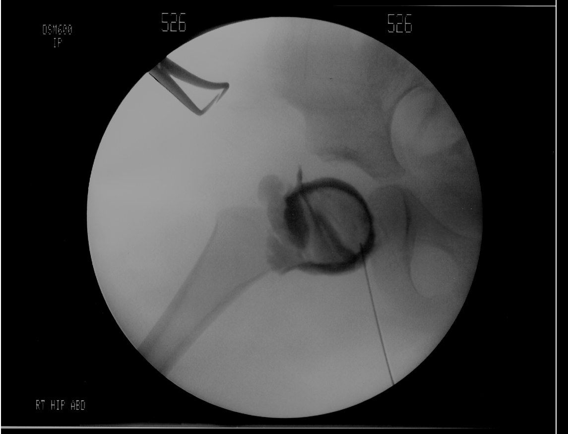
- Medial pooling > 7 mm
- Hourglass constriction of capsule by tight iliopsoas
- Hypertrophied ligamentum teres (head linear defect)
- Pulvinar fat and transverse acetabular ligament
- Inverted labrum (absent rose thorn sign)
Q9What does absence of the rose thorn sign indicate?▸
- Inverted labrum
Q10What is done after a successful reduction on arthrogram?▸
- Spica cast
- Confirm with single cut CT
- Change cast every 6 weeks x 3 months
Q11What are the indications for an anterior open reduction?▸
- High and chronic dislocations with femoral head stuck to ilium (> 1-1.5 yo)
- Perform capsulorrhaphy for very unstable hips
- Plan acetabuloplasty
Q12What are the advantages and disadvantages of the anterior approach?▸
- Advantages: good exposure, can do capsulorrhaphy, can reduce highly dislocated hip
- Disadvantages: large wound, more bleeding
- Iliac crest apophysis and hip abductors at risk
- Lateral cutaneous femoral nerve at risk
Q13What are the key steps of the anterior approach?▸
- Bikini incision
- Plane between sartorius and TFL (protect LCFN)
- Plane between rectus and gluteus medius
- Detach and tag straight and reflected heads of rectus
- Psoas tendon lengthening at the pelvic brim, T capsulotomy, excise intra-articular obstacles
- +/- femoral osteotomy, then capsulorrhaphy
Q14What are the advantages and disadvantages of the medial approach?▸
- Advantages: small wound, less bleeding; can deal with iliopsoas and transverse acetabular ligament; avoids stripping iliac apophysis/abductors; adductor tenotomy through same incision
- Disadvantages: risk to medial circumflex femoral artery -> AVN
- Cannot perform capsulorrhaphy
- Obturator nerve injury
- Not for open reduction within 6 months
Q15What are the dissection planes of the medial approach?▸
- Weinstein: between NVB and pectineus (landmark = anterior obturator nerve; best to reach labrum)
- Ludloff: between pectineus and adductor brevis
- Ferguson: between gracilis and adductor longus, then AM, AB
Q16What structures are at risk in the medial approach?▸
- Obturator nerve (anterior branch between adductor longus and brevis -> adductor contracture)
- Medial circumflex femoral artery (direct vision when performing tenotomy)
- External pudendal artery and femoral vessels
Q17How is the medial approach performed?▸
- GA, supine, affected limb in abduction, flexion and external rotation
- Landmark: 3 cm below the pubic tubercle (attachment of adductor longus)
- Incision: longitudinal just below the pubic tubercle along adductor longus
- Palpate the lesser tuberosity
Q18What is the innervation of the medial compartment of the thigh?▸
- Adductor magnus: posterior branch of obturator nerve + tibial nerve
- Other adductors (longus, brevis, gracilis): anterior branch of obturator nerve
Q19Describe the X-ray findings in DDH presenting after 18 months.▸
- Dislocated left hip
- Small femoral epiphysis and increased neck-shaft angle
- Poorly formed tear drop and ill-defined acetabular margins
- Shenton's line broken
- Further imaging: von Rosen views to check reducibility
Q20What is the aim of treatment in late-presenting DDH?▸
- Stable, congruent, concentric reduction without too much tension
- Painless hip with good ROM
- Prevent complications
Q21Why is an anterior approach chosen for late-presenting DDH?▸
- Better exposure
- Can do capsulorrhaphy and pelvic osteotomy
- Avoids injury to the medial circumflex femoral artery
- Can attack intra-articular obstacles to relocation
- Precautions: iliac apophysis, abductor stripping, more bleeding
Q22When do you choose a femoral vs acetabular osteotomy?▸
- Femoral osteotomy (VDRO +/- shortening) if reduction maintained in internal rotation and abduction
- Acetabular osteotomy if reduction maintained in flexion
Q23What is the choice of osteotomy by age?▸
- 2-10 yrs: redirection (Salter, triple); reshape (Pemberton for AL deficiency)
- 10-14 yrs: open triradiate cartilage -> triple; closed triradiate -> Ganz
- > 14 yrs: Chiari/shelf as no remodelling
- +/- shortening in > 3 yo (contracted soft tissue -> increased tension on head -> AVN)
Q24How do you decide the osteotomy intra-operatively?▸
- Find a stable position of the hip intra-op
- Stable in abduction + IR -> VDRO
- Stable in flexion -> Salter
- Stable in flexion + abduction + IR -> pelvic + femoral osteotomy
- Double acetabulum due to erosion -> Pemberton