Combined Paed
DDH - principles, presentation and diagnosis
Overall treatment principles, ultrasound use and Graf classification, presentation at 6-18 months.
28 questions
4 source pages
3 images
Images appear with the first question taken from each source page — tap a question to open it.
Q1What is the overall treatment principle for DDH by age?📷▸
- < 6 months: Pavlik harness
- Teratogenic irreducible: CR + arthrogram at 4 months
- 6-18 months or < 6 months failing Pavlik: arthrogram + closed reduction or open reduction
- > 18 months: open reduction +/- secondary femoral and acetabular procedures
Q2What are the risk factors and history features of DDH?▸
- Family history, first born, female, breech, oligohydramnios, twin
Q3How do Ortolani and Barlow tests differ?▸
- Ortolani: tests whether the hip is reducible, by elevation and abduction of the flexed femur
- Barlow: tests whether the hip is subluxable, by adduction and depression of the flexed femur
Q4What is Klisic sign?▸
- Connect greater trochanter to ASIS
- Normal if the line points at or above the umbilicus
Q5What is the evidence on universal ultrasound screening for DDH?▸
- Low sensitivity (89%) and poor PPV (62%) leading to overdiagnosis and overtreatment
- Does not meet the WHO/Wilson and Jungner criteria for universal screening
- 2002 JBJS Norwegian group: universal missed 1 case, selective missed 5, no significant difference
- 2009 JBJS international hip dysplasia institute study: selective screening most cost effective
Q6What are the reported test performances in DDH?▸
- USG (static and dynamic): sensitivity/specificity ~90%
- Ortolani/Barlow: sensitivity 70%, specificity 100% in experienced hands
Q7What clinical signs in the photographs suggest DDH?▸
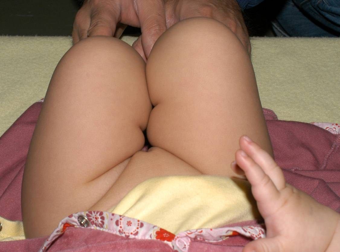
- Asymmetrical proximal skin crease
- Limited abduction of the left thigh
- Galeazzi test shows left femur shortening
- These findings raise suspicion of DDH
Q8What associated conditions are seen with DDH?▸
- Torticollis, congenital recurvatum/knee dislocation
- Calcaneovalgus feet, clubfoot, metatarsus adductus
- Older child: waddling gait with hyperlordosis of the lumbar spine
Q9How is a subluxable or dislocated hip managed in the first weeks of life?▸
- Subluxable <2week can be physiological: double napkin
- Dislocated and reducible, or subluxable > 2 weeks: Pavlik harness
- Dislocated and not reducible: can still try PH x 1 mth
Q10What is the proposed aetiology of DDH?▸
- Genetic
- Mechanical: breech + knee in extension (strong hamstring action on hip)
- Maternal hormone: ligament laxity, especially in the female fetus
- More common over the left side
Q11What are the Wilson and Jungner (1968) criteria for a screening programme?▸
- Disease: important health issue, high enough prevalence, natural history known, early treatment better
- Test: accepted by patient, high sensitivity, available facilities, cost effective
- Population: accepted and effective treatment; willing to undergo further evaluation/treatment
- USG screening for DDH has low sensitivity (89%) and poor PPV (62%), causing overdiagnosis
Q12What is the use of ultrasound in DDH?📷▸
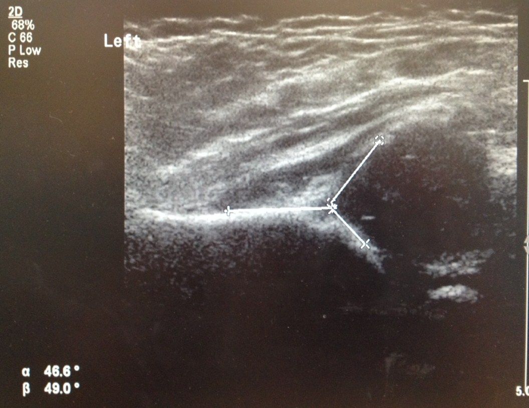
- Confirm reduction
- Baseline of alpha and beta angles
- Perform within 2 weeks of starting Pavlik harness
Q13How do you interpret a hip ultrasound?▸
- Identify landmarks: ilium, bony acetabulum, labrum, femoral head
- Confirm concentric reduction
- Assess coverage: > 50% in vs < 50% in; labrum pointing upwards vs inwards
- Measure alpha and beta angles
Q14What is the alpha angle and its normal value?▸
- Angle between the ilium and bony acetabulum
- Normal > 60 degrees
Q15What is the beta angle and its normal value?▸
- Angle between the ilium and labrum
- Normal < 55 degrees
Q16What is the Graf ultrasound classification?▸
- I: alpha > 60
- II: alpha 43-60 (ABCD), C = critical zone hip, D = decentered (beta > 77)
- III: alpha < 43
- IV: alpha < 43 + interposed labrum
- Harcke = dynamic USG measurement
Q17On static USG, how is the alpha angle used to guide DDH treatment?📷▸
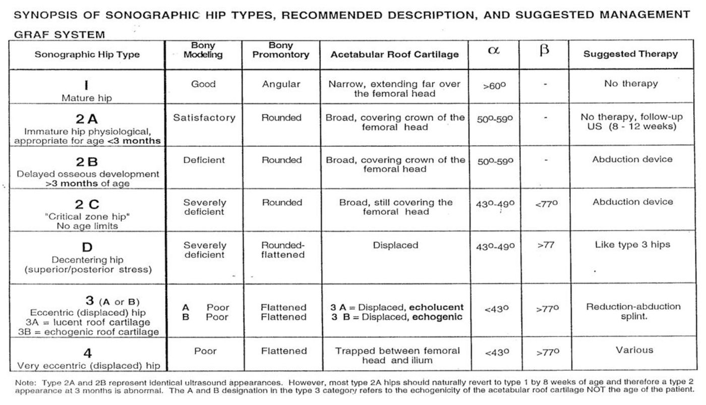
- I: > 60 normal, no treatment needed
- IIA and IIB: 50-59
- < 3 months: follow up with USG
- > 3 months: Pavlik harness
- IIC: < 50: start Pavlik harness
Q18What are the two angles measured on static ultrasound?▸
- Alpha angle and beta angle
- They represent femoral head coverage
Q19What is dynamic USG (Harcke)?▸
- Dynamic ultrasound technique
- Visualises the Barlow test
Q20Describe the X-ray measurements used in DDH at 6-18 months.▸
- Hilgenreiner's line: horizontal line through superior edge of triradiate cartilage
- Perkin's line: perpendicular to Hilgenreiner's line at lateral acetabular roof
- Quadrants and Shenton's line
- Acetabular index: < 2 yo < 30 deg, > 2 yo < 20 deg (take < 25 deg)
Q21What is the Tonnis grading of DDH?▸
- I: below SL margin of acetabulum (SMA), medial to Perkin's line
- II: below SMA line, lateral to Perkin's line
- III: at the level of the SMA line
- IV: above the SMA line
Q22What is the treatment sequence for DDH presenting at 6-18 months?▸
- GA, gentle closed reduction with flexion, traction, abduction
- Determine Ramsey safety zone (cut off 40 degrees)
- Adductor tenotomy first to reduce intracapsular pressure
- Arthrogram, +/- open reduction, hip spica cast
- Postop CT to assess AP congruency
Q23How is a hip spica cast applied?▸
- Spica table; folded towel over anterior chest
- Stockinette over torso, cotton/gore-tex wrap, felt over bony prominences
- Apply in human position: 100 flexion 50 degree abduction
- 6 weeks, then another 6 weeks in human position, then removable abduction brace for 6 weeks
Q24What examination findings are relevant in late-presenting DDH?▸
- Asymmetric groin crease, LLD, Galeazzi test
- Barlow and Ortolani not relevant at this age
- Examine for packaging disorders: torticollis, clubfoot, metatarsus adductus
- Look for neuromuscular conditions e.g. spina bifida, arthrogryposis, CP
- Document NV status
Q25What acetabular morphology should be assessed on the DDH X-ray?▸
- Tear drop morphology (internal and external acetabular wall)
- Acetabular index varies with age: < 2 yo < 30 deg, > 2 yo < 20 deg (take < 25 deg)
- Small femoral ossification centre
Q26What is the caution when measuring acetabular angles?▸
- Do not mix up acetabular angle (of Sharp) and acetabular index
- AA = adult whole acetabulum, ~45 degrees
- AI = measured from Hilgenreiner's line, ~25 degrees
Q27What history should be revisited in late-presenting DDH?▸
- Birth history and breech presentation
- First born child, family history
- Other perinatal complications
Q28What additional X-ray view is needed in DDH?▸
- Von Rosen views