17 slides
▸ Slide 494 · Combined - PaedCombined Paed · 2 questions expand
Slide render

Question list
Q1-Q22 questions — tap to reveal all answerslist
- What does the paediatric combined session cover?
- Which paediatric orthopaedic conditions are examinable in this section?
Answers · Q & A
Q1.What does the paediatric combined session cover?
- Not covered in the speaker notes
Q2.Which paediatric orthopaedic conditions are examinable in this section?
- Not covered in the speaker notes
▸ Slide 495 · HipCombined Paed · 2 questions expand
Slide render

Question list
Q1-Q22 questions — tap to reveal all answerslist
- What are the common paediatric hip conditions?
- How is the paediatric hip assessed clinically?
Answers · Q & A
Q1.What are the common paediatric hip conditions?
- Not covered in the speaker notes
Q2.How is the paediatric hip assessed clinically?
- Not covered in the speaker notes
▸ Slide 496 · Overall treatment principle:Combined Paed · 11 questions expand

Question list
Q1-Q1111 questions — tap to reveal all answerslist
- What is the overall treatment principle for DDH by age?
- What are the risk factors and history features of DDH?
- How do Ortolani and Barlow tests differ?
- What is Klisic sign?
- What is the evidence on universal ultrasound screening for DDH?
- What are the reported test performances in DDH?
- What clinical signs in the photographs suggest DDH?
- What associated conditions are seen with DDH?
- How is a subluxable or dislocated hip managed in the first weeks of life?
- What is the proposed aetiology of DDH?
- What are the Wilson and Jungner (1968) criteria for a screening programme?
Answers · Q & A
Q1.What is the overall treatment principle for DDH by age?
- < 6 months: Pavlik harness
- Teratogenic irreducible: CR + arthrogram at 4 months
- 6-18 months or < 6 months failing Pavlik: arthrogram + closed reduction or open reduction
- > 18 months: open reduction +/- secondary femoral and acetabular procedures
Q2.What are the risk factors and history features of DDH?
- Family history, first born, female, breech, oligohydramnios, twin
Q3.How do Ortolani and Barlow tests differ?
- Ortolani: tests whether the hip is reducible, by elevation and abduction of the flexed femur
- Barlow: tests whether the hip is subluxable, by adduction and depression of the flexed femur
Q4.What is Klisic sign?
- Connect greater trochanter to ASIS
- Normal if the line points at or above the umbilicus
Q5.What is the evidence on universal ultrasound screening for DDH?
- Low sensitivity (89%) and poor PPV (62%) leading to overdiagnosis and overtreatment
- Does not meet the WHO/Wilson and Jungner criteria for universal screening
- 2002 JBJS Norwegian group: universal missed 1 case, selective missed 5, no significant difference
- 2009 JBJS international hip dysplasia institute study: selective screening most cost effective
Q6.What are the reported test performances in DDH?
- USG (static and dynamic): sensitivity/specificity ~90%
- Ortolani/Barlow: sensitivity 70%, specificity 100% in experienced hands
Q7.What clinical signs in the photographs suggest DDH?
- Asymmetrical proximal skin crease
- Limited abduction of the left thigh
- Galeazzi test shows left femur shortening
- These findings raise suspicion of DDH
Q8.What associated conditions are seen with DDH?
- Torticollis, congenital recurvatum/knee dislocation
- Calcaneovalgus feet, clubfoot, metatarsus adductus
- Older child: waddling gait with hyperlordosis of the lumbar spine
Q9.How is a subluxable or dislocated hip managed in the first weeks of life?
- Subluxable <2week can be physiological: double napkin
- Dislocated and reducible, or subluxable > 2 weeks: Pavlik harness
- Dislocated and not reducible: can still try PH x 1 mth
Q10.What is the proposed aetiology of DDH?
- Genetic
- Mechanical: breech + knee in extension (strong hamstring action on hip)
- Maternal hormone: ligament laxity, especially in the female fetus
- More common over the left side
Q11.What are the Wilson and Jungner (1968) criteria for a screening programme?
- Disease: important health issue, high enough prevalence, natural history known, early treatment better
- Test: accepted by patient, high sensitivity, available facilities, cost effective
- Population: accepted and effective treatment; willing to undergo further evaluation/treatment
- USG screening for DDH has low sensitivity (89%) and poor PPV (62%), causing overdiagnosis
▸ Slide 497 · What is the use of USG?Combined Paed · 5 questions expand

Question list
Q1-Q55 questions — tap to reveal all answerslist
- What is the use of ultrasound in DDH?
- How do you interpret a hip ultrasound?
- What is the alpha angle and its normal value?
- What is the beta angle and its normal value?
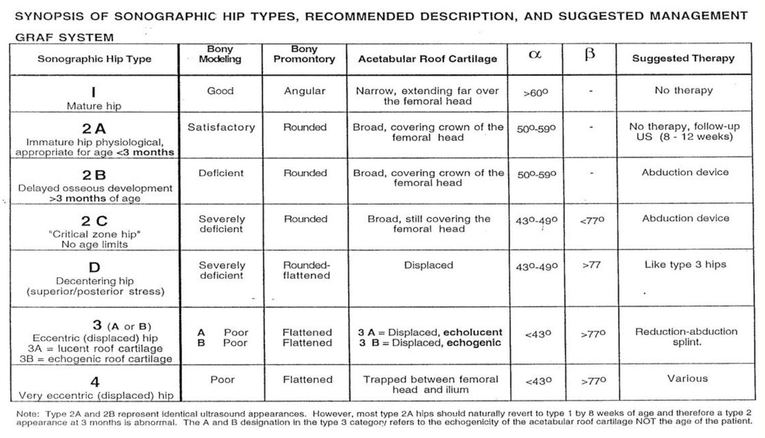
- What is the Graf ultrasound classification?
Answers · Q & A
Q1.What is the use of ultrasound in DDH?
- Confirm reduction
- Baseline of alpha and beta angles
- Perform within 2 weeks of starting Pavlik harness
Q2.How do you interpret a hip ultrasound?
- Identify landmarks: ilium, bony acetabulum, labrum, femoral head
- Confirm concentric reduction
- Assess coverage: > 50% in vs < 50% in; labrum pointing upwards vs inwards
- Measure alpha and beta angles
Q3.What is the alpha angle and its normal value?
- Angle between the ilium and bony acetabulum
- Normal > 60 degrees
Q4.What is the beta angle and its normal value?
- Angle between the ilium and labrum
- Normal < 55 degrees
Q5.What is the Graf ultrasound classification?
- I: alpha > 60
- II: alpha 43-60 (ABCD), C = critical zone hip, D = decentered (beta > 77)
- III: alpha < 43
- IV: alpha < 43 + interposed labrum
- Harcke = dynamic USG measurement
▸ Slide 498 · GrafCombined Paed · 3 questions expand
Question list
Q1-Q33 questions — tap to reveal all answerslist
- On static USG, how is the alpha angle used to guide DDH treatment?
- What are the two angles measured on static ultrasound?
- What is dynamic USG (Harcke)?
Answers · Q & A
Q1.On static USG, how is the alpha angle used to guide DDH treatment?
- I: > 60 normal, no treatment needed
- IIA and IIB: 50-59
- < 3 months: follow up with USG
- > 3 months: Pavlik harness
- IIC: < 50: start Pavlik harness
Q2.What are the two angles measured on static ultrasound?
- Alpha angle and beta angle
- They represent femoral head coverage
Q3.What is dynamic USG (Harcke)?
- Dynamic ultrasound technique
- Visualises the Barlow test
▸ Slide 499 · What is Pavlik harness?Combined Paed · 13 questions expand
Slide render

Question list
Q1-Q1313 questions — tap to reveal all answerslist
- What is a Pavlik harness and when is it used?
- What are the indications for a Pavlik harness?
- Describe the parts and application of the Pavlik harness.
- What advice is given to parents on the Pavlik harness?
- What is the timing of follow-up and ultrasound in Pavlik harness treatment?
- What are the complications of Pavlik harness treatment?
- What is the aim of treatment with a Pavlik harness?
- What is the application sequence of the Pavlik harness?
- What is assessed at follow-up during Pavlik harness treatment?
- What are the risk factors for Pavlik harness failure?
- What is the Pavlik harness success rate and the supporting study?
- What is Pavlik harness disease?
- What are the human and frog positions in an abduction brace?
Answers · Q & A
Q1.What is a Pavlik harness and when is it used?
- Dynamic corrective orthosis maintaining flexion and preventing adduction to keep the hip reduced for dynamic remodelling
- Use up to 6 months old
- 90% success in < 3 month old presentation
- Other abduction brace: von Rosen splint
Q2.What are the indications for a Pavlik harness?
- Ortolani positive
- Barlow positive at 6 weeks
- Fracture femur
Q3.Describe the parts and application of the Pavlik harness.
- Chest strap at nipple line (can place hand in between)
- Shoulder straps at anterior axillary line
- Stirrup at popliteal fossa (not too distal or else knee hyperflexion)
- AM strap maintains 100 degrees flexion, along anterior axillary line
- PL strap prevents adduction; knees able to come together but not touch
Q4.What advice is given to parents on the Pavlik harness?
- Tighten till markers; keep two sets, one for washing
- Assess kicking of leg and upper limb movement (brachial plexus)
- Change napkin in the harness; sponge bath and dry completely
- No self adjustment; regular P&O and ortho follow-up
Q5.What is the timing of follow-up and ultrasound in Pavlik harness treatment?
- Continue until hip stable clinically and radiologically (~6 weeks) + 6 weeks
- USG: within 2 weeks of starting, at the end of the 1st 6 weeks, and before stopping the harness
Q6.What are the complications of Pavlik harness treatment?
- Failure of reduction: inferior dislocation (too much flexion), Pavlik harness disease (splintage in incongruent reduction > 1mth)
- AVN (too much abduction) ~2.4%
- Femoral nerve palsy (too much flexion) ~2.5%; if persisting beyond 3 days, probability of success only 30%
- Murnaghan JBJS 2011: adjust or temporarily suspend, do not completely abandon the harness
Q7.What is the aim of treatment with a Pavlik harness?
- Maintain a stable concentric reduction
- Promote normal growth of the femoral head and acetabulum
Q8.What is the application sequence of the Pavlik harness?
- Supine; fasten the chest strap first
- Fasten the shoulder straps to keep the chest strap at the nipple line
- Feet into the stirrups one by one
- Fasten AM strap to flex the hip
- PL strap last
Q9.What is assessed at follow-up during Pavlik harness treatment?
- Confirm concentric reduction clinically and radiologically
- Adjust size and tension (mainly chest strap and PL strap)
- Compliance
- Complications: loss of reduction, nerve palsy
Q10.What are the risk factors for Pavlik harness failure?
- Graf type IV or radiographically dislocated hips
- Treatment after 3 months of age; non-visible ossific nucleus at start
- Prolonged duration of treatment; significant adductor contracture
- Bilateral disease, low alpha angle, femoral head coverage < 20%
- Initial irreducibility, advanced age, poor parental compliance
- Larger patients are also at risk
Q11.What is the Pavlik harness success rate and the supporting study?
- 85-95% (Omeroglu, Turkey, CORR 2016)
- 90% success in presentation < 3 months old
Q12.What is Pavlik harness disease?
- Hip not reduced in the harness causing superior acetabular erosion
- Occurs with splintage in incongruent reduction > 1mth
Q13.What are the human and frog positions in an abduction brace?
- Human position: 95 deg flexion, 45 deg abduction
- Frog position: extreme flexion and abduction
▸ Slide 500 · Presentation 6-18mthsCombined Paed · 9 questions expand
Slide render
Question list
Q1-Q99 questions — tap to reveal all answerslist
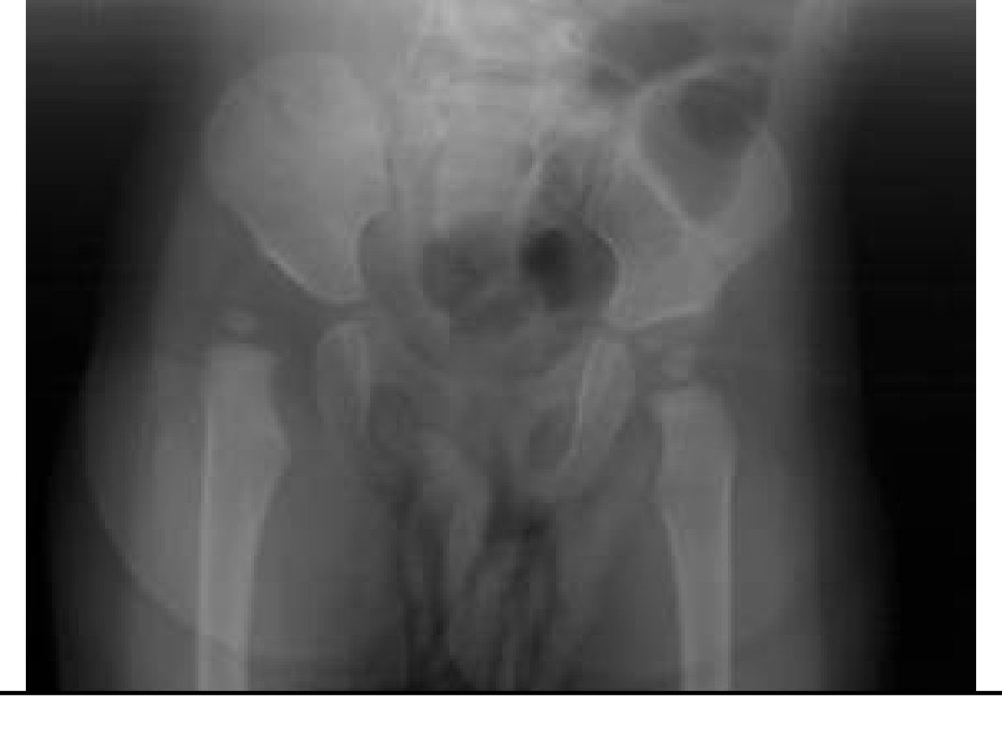
- Describe the X-ray measurements used in DDH at 6-18 months.
- What is the Tonnis grading of DDH?
- What is the treatment sequence for DDH presenting at 6-18 months?
- How is a hip spica cast applied?
- What examination findings are relevant in late-presenting DDH?
- What acetabular morphology should be assessed on the DDH X-ray?
- What is the caution when measuring acetabular angles?
- What history should be revisited in late-presenting DDH?
- What additional X-ray view is needed in DDH?
Answers · Q & A
Q1.Describe the X-ray measurements used in DDH at 6-18 months.
- Hilgenreiner's line: horizontal line through superior edge of triradiate cartilage
- Perkin's line: perpendicular to Hilgenreiner's line at lateral acetabular roof
- Quadrants and Shenton's line
- Acetabular index: < 2 yo < 30 deg, > 2 yo < 20 deg (take < 25 deg)
Q2.What is the Tonnis grading of DDH?
- I: below SL margin of acetabulum (SMA), medial to Perkin's line
- II: below SMA line, lateral to Perkin's line
- III: at the level of the SMA line
- IV: above the SMA line
Q3.What is the treatment sequence for DDH presenting at 6-18 months?
- GA, gentle closed reduction with flexion, traction, abduction
- Determine Ramsey safety zone (cut off 40 degrees)
- Adductor tenotomy first to reduce intracapsular pressure
- Arthrogram, +/- open reduction, hip spica cast
- Postop CT to assess AP congruency
Q4.How is a hip spica cast applied?
- Spica table; folded towel over anterior chest
- Stockinette over torso, cotton/gore-tex wrap, felt over bony prominences
- Apply in human position: 100 flexion 50 degree abduction
- 6 weeks, then another 6 weeks in human position, then removable abduction brace for 6 weeks
Q5.What examination findings are relevant in late-presenting DDH?
- Asymmetric groin crease, LLD, Galeazzi test
- Barlow and Ortolani not relevant at this age
- Examine for packaging disorders: torticollis, clubfoot, metatarsus adductus
- Look for neuromuscular conditions e.g. spina bifida, arthrogryposis, CP
- Document NV status
Q6.What acetabular morphology should be assessed on the DDH X-ray?
- Tear drop morphology (internal and external acetabular wall)
- Acetabular index varies with age: < 2 yo < 30 deg, > 2 yo < 20 deg (take < 25 deg)
- Small femoral ossification centre
Q7.What is the caution when measuring acetabular angles?
- Do not mix up acetabular angle (of Sharp) and acetabular index
- AA = adult whole acetabulum, ~45 degrees
- AI = measured from Hilgenreiner's line, ~25 degrees
Q8.What history should be revisited in late-presenting DDH?
- Birth history and breech presentation
- First born child, family history
- Other perinatal complications
Q9.What additional X-ray view is needed in DDH?
- Von Rosen views
▸ Slide 501 · IHDI classification XR for DDHCombined Paed · 2 questions expand
Slide render

Question list
Q1-Q22 questions — tap to reveal all answerslist
- What is the IHDI classification used for in DDH?
- What are the grades of the IHDI X-ray classification for DDH?
Answers · Q & A
Q1.What is the IHDI classification used for in DDH?
- Not covered in the speaker notes
Q2.What are the grades of the IHDI X-ray classification for DDH?
- Not covered in the speaker notes
▸ Slide 502 · Use of arthrogramCombined Paed · 7 questions expand
Slide render
Question list
Q1-Q77 questions — tap to reveal all answerslist
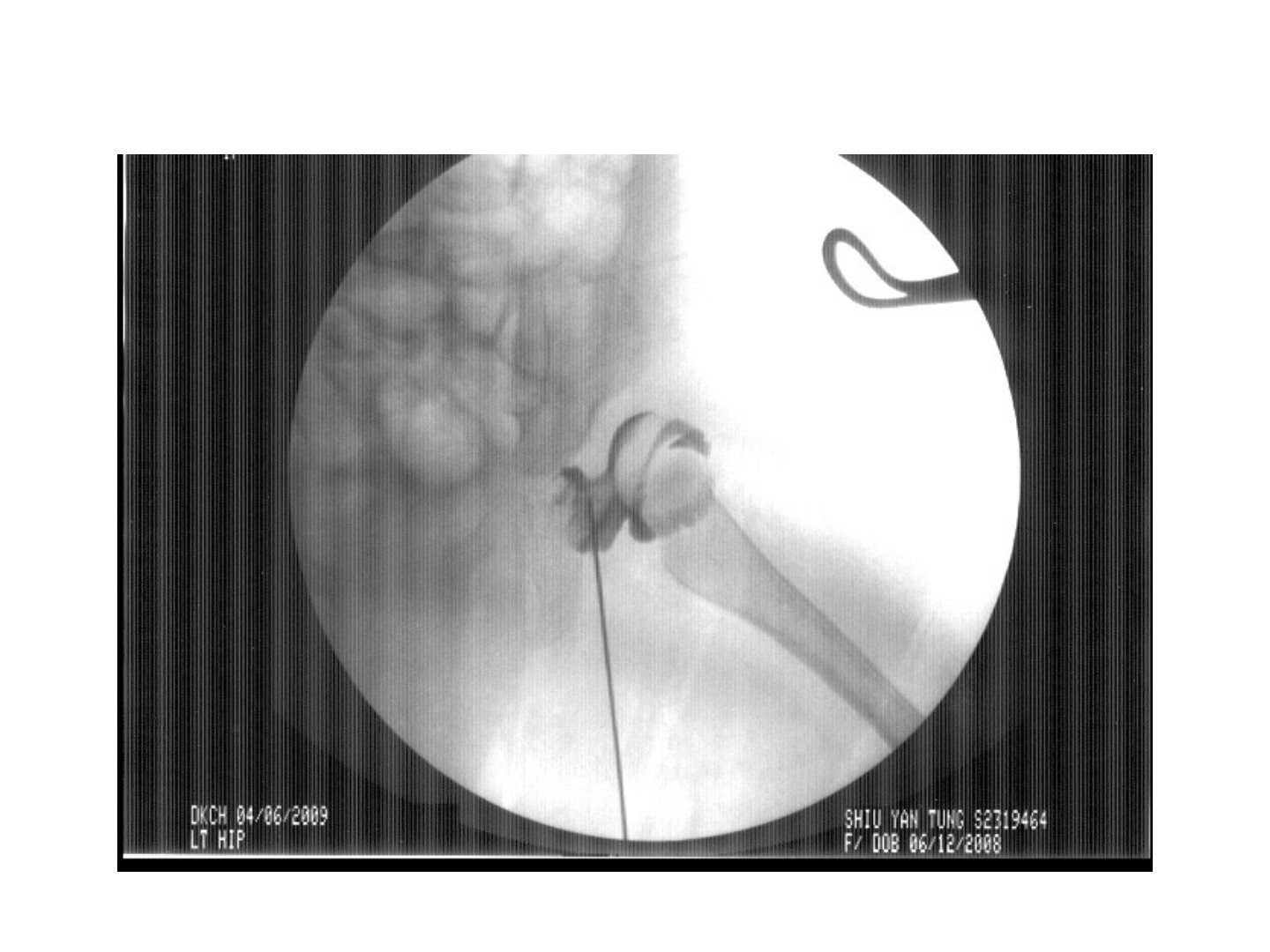
- What is the use of an arthrogram in DDH?
- How is an arthrogram performed?
- What structures can obscure reduction?
- What is the safe zone?
- When can a closed reduction be accepted and casted?
- What additional procedures may be required during reduction, and what are the risks?
- What two factors determine hip positioning in the spica cast?
Answers · Q & A
Q1.What is the use of an arthrogram in DDH?
- Assess congruency of reduction
- Define obstacles to reduction
- Delineate optimal position for immobilization
Q2.How is an arthrogram performed?
- GA, supine, with X-ray control
- Closed reduction first
- Medial sub adductor approach, aiming for ipsilateral scapula
- 22 gauge needle; inject normal saline and look for back flow, then dye to confirm position
- Image at dislocated and reduced positions
- Risks: cartilage damage, injection into growth plate, vessel injury
Q3.What structures can obscure reduction?
- Intracapsular superior: inverted labrum, neolimbus
- Intracapsular central: pulvinar, ligamentum teres
- Intracapsular inferior: transverse acetabular ligament, constricted capsule
- Extracapsular: tight iliopsoas, tight adductor
Q4.What is the safe zone?
- Difference (in degrees) between the angle of maximal hip abduction (causes AVN) and the hip adduction at which the femoral head dislocates, with hip examined in 90 deg flexion
- Safety cone lies within 15-20 degree of maximal ROM and dislocation range; satisfactory if at least 40 degrees
Q5.When can a closed reduction be accepted and casted?
- Successful reduction with <7mm medial pooling
- Check range with stable reduction
- Congruent reduction with safety zone larger than 40 deg
- Hip spica cast + 1 axial cut CT to rule out posterior dislocation
- Otherwise, open reduction
Q6.What additional procedures may be required during reduction, and what are the risks?
- Adductor tenotomy if post-reduction safe zone is narrow
- Psoas tendon release if the hip tends to sublux as it is extended below 90 degrees
- Risk: injury to the anterior division of the obturator nerve
Q7.What two factors determine hip positioning in the spica cast?
- Average hip movement in the spica is 15 degrees, so keep the hip ~20 degrees away from the line of dislocation
- Risk of AVN increases at maximum ROM, so stay 20 degrees inside the maximum ROM
▸ Slide 503 · Normal arthrogramCombined Paed · 3 questions expand

Question list
Q1-Q33 questions — tap to reveal all answerslist
- What are the abnormal arthrogram findings in DDH?
- What does absence of the rose thorn sign indicate?
- What is done after a successful reduction on arthrogram?
Answers · Q & A
Q1.What are the abnormal arthrogram findings in DDH?
- Medial pooling > 7 mm
- Hourglass constriction of capsule by tight iliopsoas
- Hypertrophied ligamentum teres (head linear defect)
- Pulvinar fat and transverse acetabular ligament
- Inverted labrum (absent rose thorn sign)
Q2.What does absence of the rose thorn sign indicate?
- Inverted labrum
Q3.What is done after a successful reduction on arthrogram?
- Spica cast
- Confirm with single cut CT
- Change cast every 6 weeks x 3 months
▸ Slide 504 · Open reductionCombined Paed · 8 questions expand
Slide render
Question list
Q1-Q88 questions — tap to reveal all answerslist
- What are the indications for an anterior open reduction?
- What are the advantages and disadvantages of the anterior approach?
- What are the key steps of the anterior approach?
- What are the advantages and disadvantages of the medial approach?
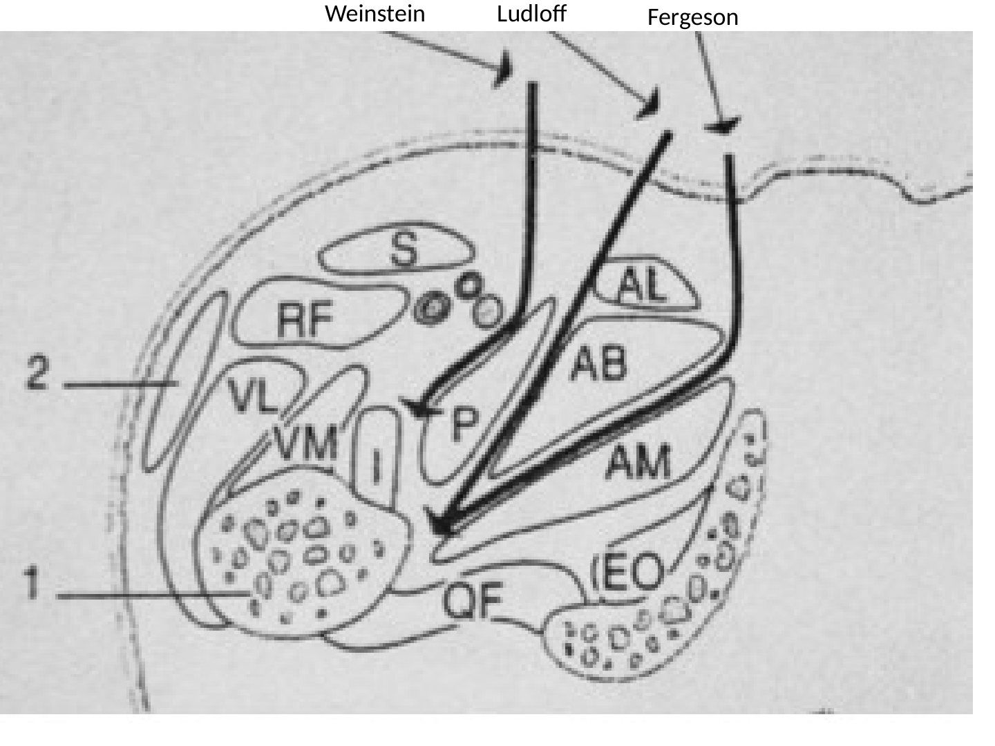
- What are the dissection planes of the medial approach?
- What structures are at risk in the medial approach?
- How is the medial approach performed?
- What is the innervation of the medial compartment of the thigh?
Answers · Q & A
Q1.What are the indications for an anterior open reduction?
- High and chronic dislocations with femoral head stuck to ilium (> 1-1.5 yo)
- Perform capsulorrhaphy for very unstable hips
- Plan acetabuloplasty
Q2.What are the advantages and disadvantages of the anterior approach?
- Advantages: good exposure, can do capsulorrhaphy, can reduce highly dislocated hip
- Disadvantages: large wound, more bleeding
- Iliac crest apophysis and hip abductors at risk
- Lateral cutaneous femoral nerve at risk
Q3.What are the key steps of the anterior approach?
- Bikini incision
- Plane between sartorius and TFL (protect LCFN)
- Plane between rectus and gluteus medius
- Detach and tag straight and reflected heads of rectus
- Psoas tendon lengthening at the pelvic brim, T capsulotomy, excise intra-articular obstacles
- +/- femoral osteotomy, then capsulorrhaphy
Q4.What are the advantages and disadvantages of the medial approach?
- Advantages: small wound, less bleeding; can deal with iliopsoas and transverse acetabular ligament; avoids stripping iliac apophysis/abductors; adductor tenotomy through same incision
- Disadvantages: risk to medial circumflex femoral artery -> AVN
- Cannot perform capsulorrhaphy
- Obturator nerve injury
- Not for open reduction within 6 months
Q5.What are the dissection planes of the medial approach?
- Weinstein: between NVB and pectineus (landmark = anterior obturator nerve; best to reach labrum)
- Ludloff: between pectineus and adductor brevis
- Ferguson: between gracilis and adductor longus, then AM, AB
Q6.What structures are at risk in the medial approach?
- Obturator nerve (anterior branch between adductor longus and brevis -> adductor contracture)
- Medial circumflex femoral artery (direct vision when performing tenotomy)
- External pudendal artery and femoral vessels
Q7.How is the medial approach performed?
- GA, supine, affected limb in abduction, flexion and external rotation
- Landmark: 3 cm below the pubic tubercle (attachment of adductor longus)
- Incision: longitudinal just below the pubic tubercle along adductor longus
- Palpate the lesser tuberosity
Q8.What is the innervation of the medial compartment of the thigh?
- Adductor magnus: posterior branch of obturator nerve + tibial nerve
- Other adductors (longus, brevis, gracilis): anterior branch of obturator nerve
▸ Slide 505 · Presented >18mthsCombined Paed · 6 questions expand
Slide render
Question list
Q1-Q66 questions — tap to reveal all answerslist
- Describe the X-ray findings in DDH presenting after 18 months.
- What is the aim of treatment in late-presenting DDH?
- Why is an anterior approach chosen for late-presenting DDH?
- When do you choose a femoral vs acetabular osteotomy?
- What is the choice of osteotomy by age?
- How do you decide the osteotomy intra-operatively?
Answers · Q & A
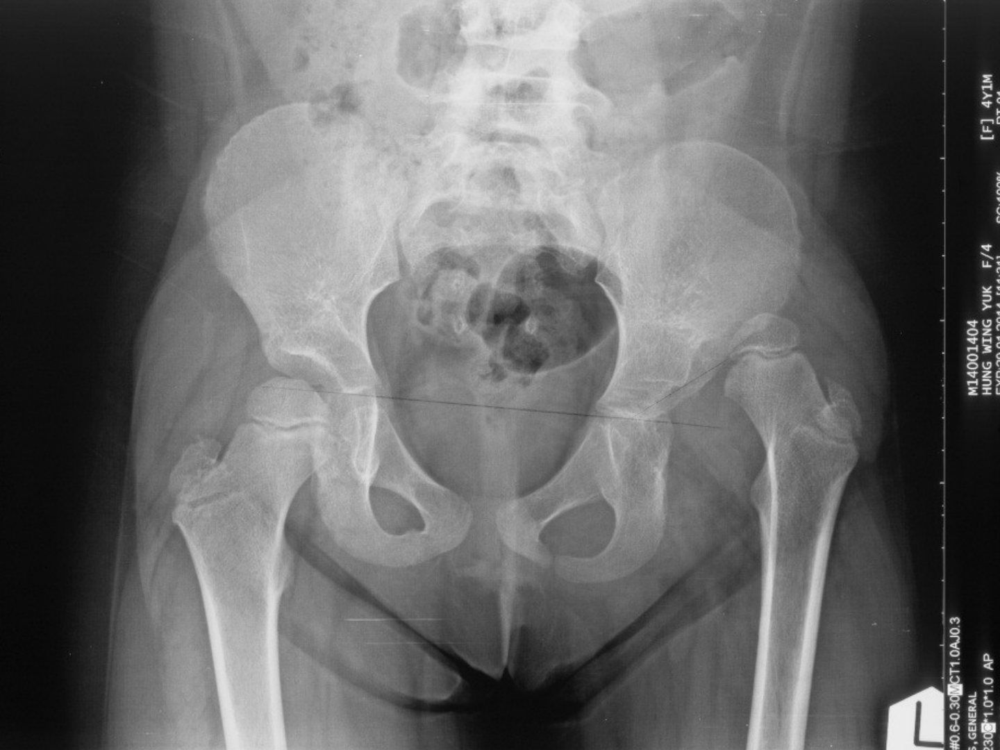
Q1.Describe the X-ray findings in DDH presenting after 18 months.
- Dislocated left hip
- Small femoral epiphysis and increased neck-shaft angle
- Poorly formed tear drop and ill-defined acetabular margins
- Shenton's line broken
- Further imaging: von Rosen views to check reducibility
Q2.What is the aim of treatment in late-presenting DDH?
- Stable, congruent, concentric reduction without too much tension
- Painless hip with good ROM
- Prevent complications
Q3.Why is an anterior approach chosen for late-presenting DDH?
- Better exposure
- Can do capsulorrhaphy and pelvic osteotomy
- Avoids injury to the medial circumflex femoral artery
- Can attack intra-articular obstacles to relocation
- Precautions: iliac apophysis, abductor stripping, more bleeding
Q4.When do you choose a femoral vs acetabular osteotomy?
- Femoral osteotomy (VDRO +/- shortening) if reduction maintained in internal rotation and abduction
- Acetabular osteotomy if reduction maintained in flexion
Q5.What is the choice of osteotomy by age?
- 2-10 yrs: redirection (Salter, triple); reshape (Pemberton for AL deficiency)
- 10-14 yrs: open triradiate cartilage -> triple; closed triradiate -> Ganz
- > 14 yrs: Chiari/shelf as no remodelling
- +/- shortening in > 3 yo (contracted soft tissue -> increased tension on head -> AVN)
Q6.How do you decide the osteotomy intra-operatively?
- Find a stable position of the hip intra-op
- Stable in abduction + IR -> VDRO
- Stable in flexion -> Salter
- Stable in flexion + abduction + IR -> pelvic + femoral osteotomy
- Double acetabulum due to erosion -> Pemberton
▸ Slide 506 · Complication of treatmentCombined Paed · 4 questions expand
Slide render

Question list
Q1-Q44 questions — tap to reveal all answerslist
- What are Salter's criteria for AVN in DDH?
- What is the Kalamchi and McEwen classification of AVN?
- What is the risk of AVN by treatment?
- What is the differential diagnosis of AVN in DDH?
Answers · Q & A
Q1.What are Salter's criteria for AVN in DDH?
- Any one of the following:
- Failure of formation or growth of ossific nucleus 1 year after reduction
- Broad metaphysis
- Increased density and fragmentation of the head
- Residual deformity after ossification
Q2.What is the Kalamchi and McEwen classification of AVN?
- 1: mild with delayed ossification
- 2: lateral, valgus deformity
- 3: central, shortening
- 4: complete, varus deformity
Q3.What is the risk of AVN by treatment?
- Pavlik harness: 1-5%
- Closed reduction + hip spica: 10%
- Open reduction: up to 30%
Q4.What is the differential diagnosis of AVN in DDH?
- MED
- Perthes
- Infection
- DDH with AVN
- PFFD
▸ Slide 507 · Xray showing pelvis of skeletally immature patient with abnormalities over left Combined Paed · 18 questions expand
Slide render
Question list
Q1-Q1818 questions — tap to reveal all answerslist
- What are the X-ray signs of SCFE?
- What are the signs of chronic SCFE?
- How is SCFE classified?
- What is the management and in-situ pinning technique for SCFE?
- When is contralateral (prophylactic) pinning considered?
- What is looked for at follow-up after SCFE?
- What is the definition of SCFE?
- What history points are important in SCFE?
- What is Drehmann's sign?
- What is the Southwick angle classification?
- Which X-ray view is used in SCFE depending on stability?
- What is the natural history of untreated SCFE?
- What radiological markers aid the decision for prophylactic pinning?
- What is the rehabilitation after SCFE pinning?
- How does SCFE present and what deformity results?
- What is the role of capsulotomy in unstable SCFE?
- What does the 2017 J O&T meta-analysis compare for severe slip?
- Which SCFE management decisions remain controversial?
Answers · Q & A
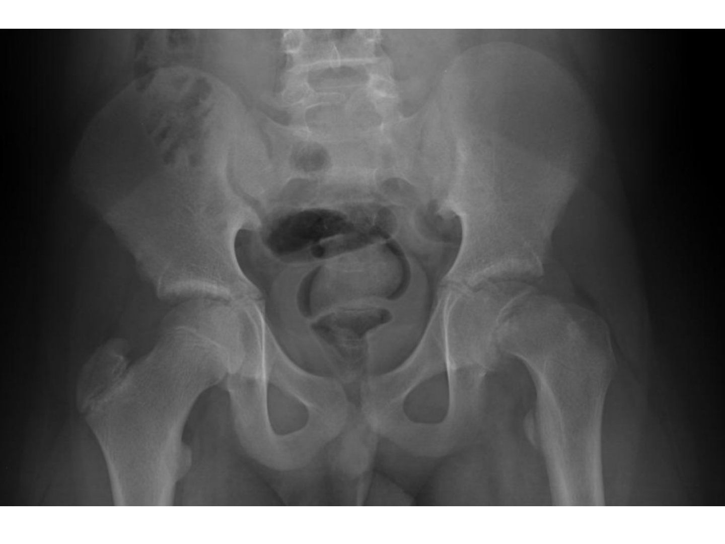
Q1.What are the X-ray signs of SCFE?
- Klein's line (Trethowan's sign)
- Widened physis with decreased epiphyseal height
- Increased distance between the teardrop and femoral neck metaphysis
- Metaphyseal blanch sign of Steel
- Carpenter's sign (less overlap of metaphysis with posterior acetabular wall)
Q2.What are the signs of chronic SCFE?
- Metaphysis smooth and sclerotic with callus formation
- Metaphysis widens (coxa magna)
Q3.How is SCFE classified?
- Chronicity: pre-slip, acute, chronic, acute-on-chronic (no bearing on prognosis)
- Stability (Loder): stable vs unstable (< 10% vs 50% AVN)
- Severity: Southwick angle on frog view; Wilson grade on AP view
- Wilson: I < 33%, II 33-50%, III > 50% of slip over physis
Q4.What is the management and in-situ pinning technique for SCFE?
- Aim: prevent further slip, promote physeal closure, prevent complications
- Unstable = O&T emergency: aim at preventing further slippage and allow physis to fuse in 6hrs; in-situ pinning; stable also pinned, less urgent
- also say whether go for prophylactic pin
- GA, X-ray guidance, supine on traction table; no traction or forceful manipulation
- Triangulation for entry point; one pin center-center, perpendicular to physis, crossing by at least 4 threads
- 6.5 mm cannulated titanium partially threaded screw with reverse cutting flutes; avoid multiple punctures -> chondrolysis
Q5.When is contralateral (prophylactic) pinning considered?
- Young < 10 yo (open triradiate cartilage)
- Systemic disease: hypothyroid, GH deficiency, hypogonadism, renal failure
- Obese/ retroverted neck
- ? Lost to follow-up
- 22% contralateral slip in 18 months
Q6.What is looked for at follow-up after SCFE?
- Fixation of the pathological side; early: recurrent slippage, chondrolysis
- Late: AVN, OA, FAI
- Contralateral side
- Follow up until epiphyseal closure
- Pain + stiffness: rule out infection; chondrolysis if joint space < 3 mm
- Chondrolysis risk factors: manipulation, immobilization, hardware through head
Q7.What is the definition of SCFE?
- Disorder of the proximal femoral physis leading to slippage of the epiphysis relative to the femoral neck
Q8.What history points are important in SCFE?
- Timing (chronicity); able to walk at all (stability)
- Preexisting illness/risk factors: hypothyroidism, GH deficiency, hypogonadism, renal failure
- Symptoms on the other side (20% bilateral, 80% chronic)
Q9.What is Drehmann's sign?
- In chronic SCFE: obligatory external rotation with flexion
- See if patient can stand; check the position of the limb
Q10.What is the Southwick angle classification?
- Measured on frog view: line perpendicular to the base of the epiphysis vs line along the shaft, compared with the contralateral side
- Categories: < 30, 30-60, > 60
- If bilateral, subtract 12 degrees from the corresponding lateral epiphyseal shaft angle
Q11.Which X-ray view is used in SCFE depending on stability?
- Frog lateral if stable
- Cross-table if unstable
Q12.What is the natural history of untreated SCFE?
- Weinstein CORR 1996: 28 untreated patients
- Increasing displacement over time
- Degree of displacement is proportional to the risk of OA
Q13.What radiological markers aid the decision for prophylactic pinning?
- Posterior slope angle (>14deg – 83% risk)
- Modified Oxford bone age (Popejoy 2012 JPO): score femoral head, LT, ilium, GT, triradiate cartilage
- Score range 16-26; < 18 = 85-97% risk; 20 = 5%; > 21 = 0-1%
Q14.What is the rehabilitation after SCFE pinning?
- Stable: weight bear
- Unstable: non-weight bearing
Q15.How does SCFE present and what deformity results?
- May present with knee pain due to radiation by the obturator nerve
- Deformity: extension, external rotation, varus
- Loss of flexion, internal rotation, abduction
- CAM-type FAI and accelerated osteoarthritis from anterior impingement
Q16.What is the role of capsulotomy in unstable SCFE?
- 10. Capsulotomy: theoretical benefit, no strong evidence
- Dodwell JPO 2019 meta-analysis: no statistically significant decrease in odds of AVN
- Still recommend routine capsulotomy for all unstable SCFE - adds little and may minimise devastating insult to the femoral head
Q17.What does the 2017 J O&T meta-analysis compare for severe slip?
- Surgical dislocation (Modified Dunn): 3% AVN but better patient satisfaction
- In-situ pinning: 1.5% AVN and lower satisfaction
Q18.Which SCFE management decisions remain controversial?
- Timing of operation
- Pinning of the contralateral hip
▸ Slide 508 · Any other options for unstable severe slip?Combined Paed · 6 questions expand
Slide render

Question list
Q1-Q66 questions — tap to reveal all answerslist
- Any other options for unstable severe slip?
- When is the Modified Dunn procedure for unstable severe slip performed?
- What are the steps of the Modified Dunn approach for severe slip?
- What is the AVN risk of this procedure?
- What is the anatomical basis of the Modified Dunn procedure?
- How is the slip fixed after surgical dislocation?
Answers · Q & A
Q1.Any other options for unstable severe slip?
- Modified Dunn procedure
Q2.When is the Modified Dunn procedure for unstable severe slip performed?
- Done in the elective setting
Q3.What are the steps of the Modified Dunn approach for severe slip?
- Gibson interval (gluteus medius / gluteus maximus)
- Trochanteric flip osteotomy
- Z capsulotomy (from troch ridge to acetabulum along neck of femur, distal anterior towards calcar, parallel towards posterior)
- Cut ligamentum teres -> ER + adduction -> anterior dislocation
Q4.What is the AVN risk of this procedure?
- AVN up to 25% in inexp hands
Q5.What is the anatomical basis of the Modified Dunn procedure?
- The MFCA is protected by obturator externus
- Avoid obturator externus during the approach
Q6.How is the slip fixed after surgical dislocation?
- Reduction and fixation with multiple pins
▸ Slide 509 · Xray pelvis of skeletally immature patient with left hip pathologyCombined Paed · 17 questions 1 check expand
Slide render
Question list
Q1-Q1717 questions — tap to reveal all answerslist
- Describe the radiographic findings in this case.
- What is the Waldenstrom classification and what are the differential diagnoses?
- What are the clinical and radiological at-risk signs in Perthes disease?
- What is the aim of treatment in Perthes disease and what is the natural history?
- What is the lateral pillar classification and how does it guide management?
- What is the blood supply to the femoral head by age?
- What further investigations are needed in suspected Perthes disease?
- What is the etiology of Perthes disease?
- What are the operative containment options in Perthes disease and their problems?
- What are the prerequisites for surgical containment in Perthes disease?
- Take me through the surgical steps of proximal femoral varus osteotomy.
- What is the postoperative care and follow-up after Perthes surgery?
- What other problems occur in Perthes disease?
- What is the advantage of surgical containment according to B Joseph?
- What were the limitations of Herring's study?
- What did Benjamin Joseph's study recommend for containment?
- What are the limitations of non-operative containment with casts?
Answers · Q & A
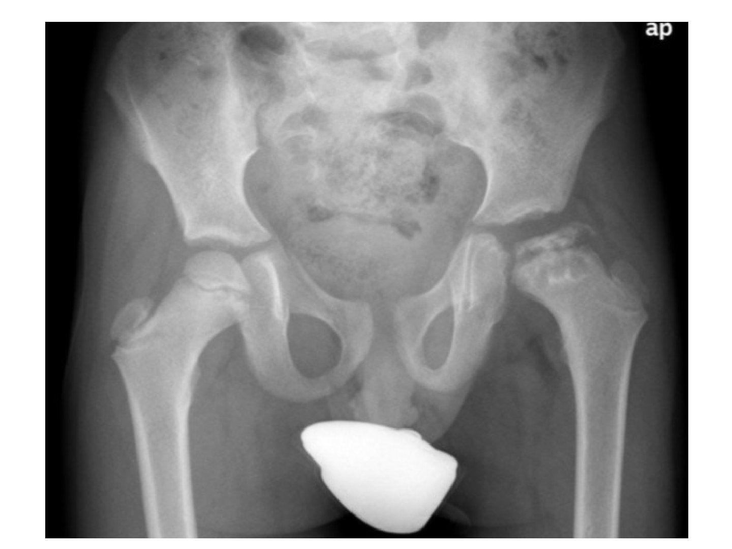
Q1.Describe the radiographic findings in this case.
- Flattened, irregular, fragmented left proximal femoral epiphysis with lateral extrusion
- Cystic changes over the metaphyseal area and lucency over the lateral epiphysis, consistent with Gage sign
- Widened medial joint space
- Contralateral hip not involved (10% bilateral, never same time)
- Working diagnosis: Perthes disease, in the fragmentation phase
Q2.What is the Waldenstrom classification and what are the differential diagnoses?
- Waldenstrom stages: initial, fragmentation, reossification, remodelling
- This case is in the fragmentation phase
- Unilateral Ddx: septic arthritis, Meyer's disease, MED
- Bilateral Ddx: hypothyroidism, lipid storage disease, Meyer's dysplasia, MED, sickle cell
- History/PE focus: Age, gender, body built, onset of symptoms, at-risk signs (FOOBS), r/o Ddx, ROM
Q3.What are the clinical and radiological at-risk signs in Perthes disease?
- Clinical at-risk signs (FOOBS): female, old, obese, bilateral, stiff
- Catterall sign: hip flexion with obligatory external rotation
- Radiological (Catterall): Gage sign (inverted V lucency lateral to the epiphysis)
- Horizontal physis and lateral subluxation of the femoral head
- Calcification lateral to the epiphysis and metaphyseal cyst
Q4.What is the aim of treatment in Perthes disease and what is the natural history?
- Pain control (rest, analgesics)
- Maintain ROM (physio, temporary NWB)
- Contain the hip during the critical period of biologic plasticity during creeping substitution so the head remodels with less incongruity
- Containment position: abduction and internal rotation
- Natural history: We know from the classic Iowa study with a 50yr FU that 40% need arthroplasty if left untreated. Counsel patient regarding containment. Still has a possibility of future OA
- Non-operative containment options: observation, activity restriction, NWB + ROM exercise (abd + IR), Petrie cast, A-frame
Q5.What is the lateral pillar classification and how does it guide management?
- Based on AP X-ray, comparing both sides (Herring); lateral pillar 25%, central 50%, medial 20-35%
- A = normal height; B = > 50%; B/C = exactly 50% or very narrow lateral pillar >50% of original height / very little ossification but > 50%; C = < 50%
- Herring study (2004 JBJS): A and B < 8 yo do well regardless; B and B/C > 8 yo improve with surgery; B/C < 8 and C poor regardless of treatment; no significant effect regardless of treatment in those <8yo
- For this case (lateral pillar B or B/C, > 8 yo): surgical containment improves outcome
- Surgical containment: proximal femoral VDRO (at most 20deg) or ROWO +/- pelvic osteotomy
- Outcome at maturity by Stulberg classification: I normal, II spherical congruent, III mushroom congruent, IV flat congruent, V incongruent
Q6.What is the blood supply to the femoral head by age?
- < 4 yo: equal contribution by MFCA and LFCA
- 4-10 yo: LFCA regresses and supplies metaphysis only
- 10-14 yo: increased contribution from ligamentum teres
- > 14 yo: anastomosis between the three systems
Q7.What further investigations are needed in suspected Perthes disease?
- WBC (infection), lipid (storage disorder), ESR, CRP
- Bone profile (tumour), TSH (hypothyroid), peripheral smear (sickel cell)
- XR frog-leg lateral; +/- USG, aspiration if infection suspected
Q8.What is the etiology of Perthes disease?
- Anatomical
- Hydrostatic pressure
- Thrombophilic
Q9.What are the operative containment options in Perthes disease and their problems?
- Proximal femoral VDRO (at most 20deg) or ROWO - problems: short limb and Trendelenburg gait
- Pelvic osteotomy - re-directional (Salter): potential lengthening increases pressure on the head -> AVN
- Combination with femoral osteotomy increases coverage by 20%
- Shelf to prevent lateral subluxation/no longer containable; Chiari
- Valgus osteotomy for hinged abduction (reposition hinge, correct shortening, improve abductor function)
Q10.What are the prerequisites for surgical containment in Perthes disease?
- Prerequisites include timing: fragmentation phase
- Containable: good range on examination
- > XR – spherical and congruent reduction, containable upon abduction XR/ intraop arthrogram
Q11.Take me through the surgical steps of proximal femoral varus osteotomy.
- Preop plan the amount of varisation; GA; adductor release if tight
- Lateral subvastus approach; elevate periosteum
- Paediatric proximal locking plate: 3.5 mm if < 35 kg, 5 mm if > 35 kg
- Jig angle = plate angle + desired varisation; fix plate proximally then remove it
- Mark rotation, use 2 K-wires above and below the osteotomy, osteotomy with oscillating saw
- Fix plate proximally then distally; Add some internal rotation or extension for better containment
Q12.What is the postoperative care and follow-up after Perthes surgery?
- NWB walking, HKO
- Follow up until maturity
Q13.What other problems occur in Perthes disease?
- LLD, Trendelenburg
- Pain: labral tear, loose body, hinged abduction, FAI
Q14.What is the advantage of surgical containment according to B Joseph?
- The patient will go through the fragmentation phase faster
Q15.What were the limitations of Herring's study?
- Based on AP view only
- 7/12 had surgery before reliable classification
- 1/3 needed upgrade; only 4% in group A on final grading
- Difficult in bilateral disease
Q16.What did Benjamin Joseph's study recommend for containment?
- Factors: age at onset, femoral head extrusion (> 20% Reimer migration index), ROM
- Containment ASAP if: >= 8 yo with extrusion and reduced ROM (restore motion with traction/abduction cast for 6 weeks first)
- < 8 yo with extrusion
- < 8 yo: femoral osteotomy (more remodelling, less Trendelenburg later)
- >= 8 yo: acetabular osteotomy (less shortening)
Q17.What are the limitations of non-operative containment with casts?
- Petrie cast / A-frame must be worn for 6 months to 1 year
- Problem: cannot guarantee abduction
Fact check
Classic Iowa study with 50-year follow-up shows 40% of untreated Perthes disease need arthroplasty — outdated/overstated — The 40% figure comes from a selected historical Iowa cohort (McAndrew & Weinstein, mean 47.7 years follow-up: 15/37 hips). A modern nonoperative cohort (Oslo, mean 48 years) reported only 19% THA, so 40% should not be quoted as the universal natural history. — medium confidence — source
▸ Slide 510 · AP Xray pelvis of skeletally immature patientCombined Paed · 8 questions expand
Slide render
Question list
Q1-Q88 questions — tap to reveal all answerslist
- Describe the X-ray findings of developmental coxa vara.
- What are the causes of coxa vara?
- What are the clinical features of coxa vara?
- What is the prognosis by HE angle and when is surgery indicated?
- What is the pathomechanics of coxa vara?
- What is the diagnosis and inheritance of developmental coxa vara?
- What history is relevant in coxa vara?
- What preoperative planning is needed before VDRO for coxa vara?
Answers · Q & A
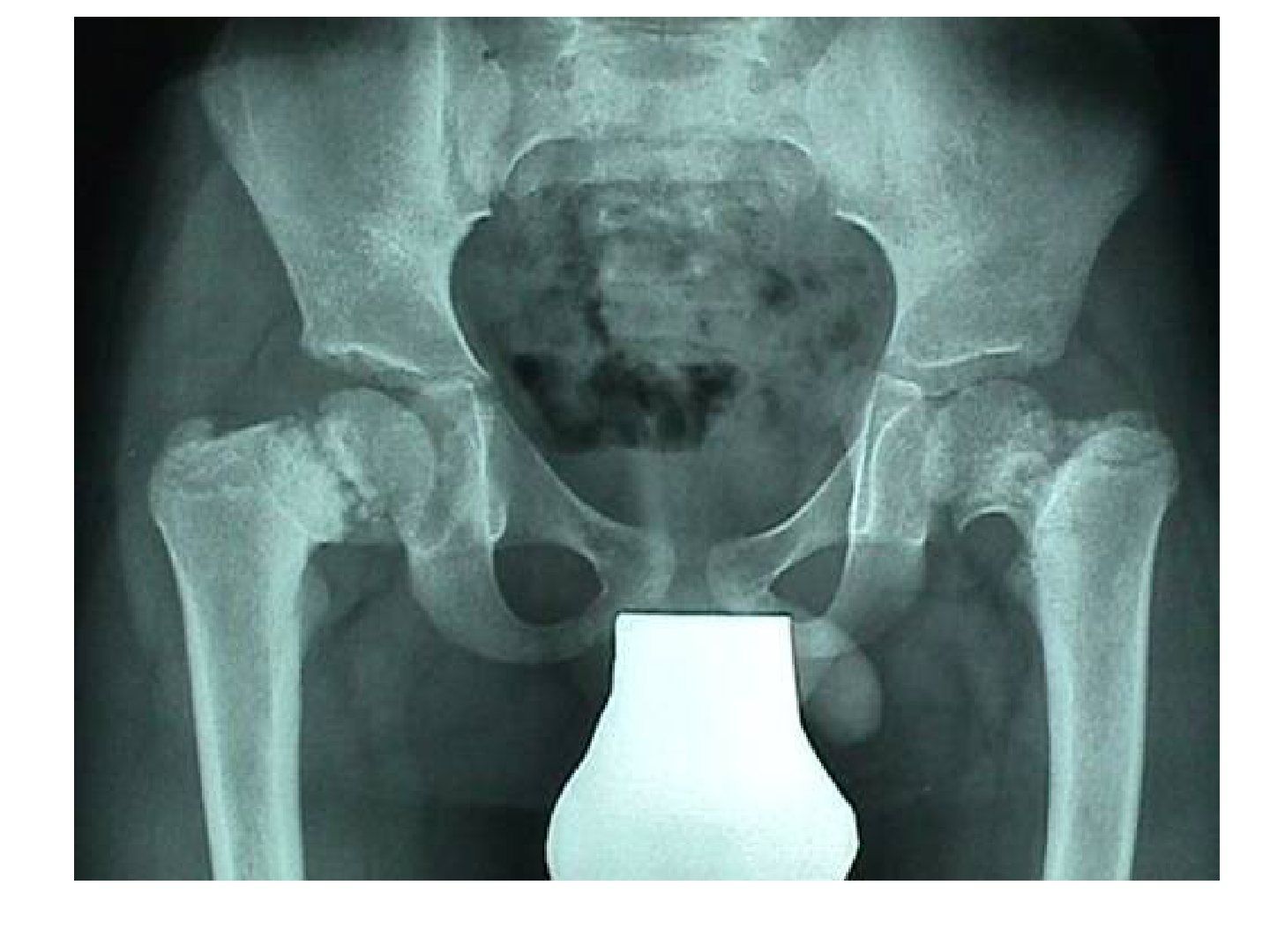
Q1.Describe the X-ray findings of developmental coxa vara.
- Bilateral coxa vara deformity
- Vertical physis with increased HE angle (> 25 abnormal)
- Decreased neck shaft ankle (<125 abnormal)
- Coxa breva
- Fairbank triangle: Inverted Y shape lucency in inferior femoral neck (ossification defect)
- Acetabulum and tear drop normal
Q2.What are the causes of coxa vara?
- Acquired: Perthes, chronic SCFE, trauma, infection
- Developmental
- Dysplasia: MED, SED, OI
- Metabolic: cretinism
- Congenital: PFFD; bilateral in 30-50%
Q3.What are the clinical features of coxa vara?
- Trendelenburg gait
- LLD and high riding greater trochanter
- Excessive lumbar lordosis
- Restricted hip ROM
Q4.What is the prognosis by HE angle and when is surgery indicated?
- < 45: 20% resolve
- 45-60: uncertain
- > 60: will progress
- VDRO if HE > 60 and NSA < 110 (subtrochanteric if < 3 yo, intertrochanteric if > 3 yo)
- Goal: overcorrect NSA, correct LLD and version, restore abductor tension; aim HE < 30
Q5.What is the pathomechanics of coxa vara?
- Decreased neck-shaft angle with an ossification defect in the inferior femoral neck
- -> vertical physis
- -> increased shearing force
Q6.What is the diagnosis and inheritance of developmental coxa vara?
- Developmental coxa vara
- No clear genetic inheritance
- Bilateral in 30-50%
Q7.What history is relevant in coxa vara?
- Previous hip trauma or infection
- Associated skeletal abnormalities
- Perinatal history and family history
Q8.What preoperative planning is needed before VDRO for coxa vara?
- CT PRE OP for associated deformities: coxa breva, femoral retroversion, acetabular dysplasia, GT overgrowth
- Lateral approach, protect physis and periosteum
- Goal: overcorrect NSA, correct LLD, correct version, restore abductor tension; aim HE < 30