▸ Slide 449 · Spine - non traumaSpine Non Trauma · 5 questions expand
Q1-Q55 questions — tap to reveal all answerslist
- Which conditions are listed under 'Spine - non trauma'?
- What spinal manifestations of RA are listed on this slide?
- Which cervical spine conditions are listed?
- Which degenerative/structural topics are listed?
- Which infective, neoplastic and miscellaneous topics are listed?
Q1.Which conditions are listed under 'Spine - non trauma'?
- RA – C1/2 instability, subaxial involvement, cranial settling
- AS – pseudoarthrosis; DISH
- Cervical myelopathy, OPLL, PID, dural tear
- Spinal stenosis, degenerative scoliosis, spondylolysis, sagittal balance
- Vertebroplasty, TB spine, pyogenic spine, spine metastasis, failed back syndrome
Q2.What spinal manifestations of RA are listed on this slide?
- C1/2 instability
- Subaxial involvement
- Cranial settling
Q3.Which cervical spine conditions are listed?
- Cervical myelopathy
- OPLL
- PID
- Dural tear
Q4.Which degenerative/structural topics are listed?
- Spinal stenosis
- Degenerative scoliosis
- Spondylolysis
- Sagittal balance
Q5.Which infective, neoplastic and miscellaneous topics are listed?
- Vertebroplasty
- TB spine
- Pyogenic spine
- Spine metastasis
- Failed back syndrome
▸ Slide 450 · Lateral XR of this patients cervical spineSpine Non Trauma · 8 questions 1 check expand

Q1-Q88 questions — tap to reveal all answerslist
- Describe the lateral flexion/extension cervical X-ray findings.
- What are the risk factors/causes and symptoms of C1/2 instability?
- What do you examine and what further investigations are required?
- What is the natural history of C1/2 instability (Boden's classic paper)?
- What lines and measurements are used to assess cranial settling?
- When do you operate for cranial settling versus C1/2 subluxation?
- What is the pattern of cervical spine involvement in rheumatoid arthritis?
- What is pannus and how does it destroy structures?
Q1.Describe the lateral flexion/extension cervical X-ray findings.
- C1/2 subluxation with increased ADI and decreased PADI
- No associated cranial settling
- No subaxial cervical spine abnormalities
- Cervical height index measured
Q2.What are the risk factors/causes and symptoms of C1/2 instability?
- Inflammatory joint disease or crystal arthropathy; signs of infection
- Previous trauma (odontoid nonunion / os odontoideum)
- Ligamentous laxity (Down's syndrome, MPS)
- Symptoms: pain (neck and occiput), myelopathy, vertebrobasilar insufficiency (vertigo, diplopia, drop attacks)
- Functional grading (Ranawat): I normal, II subjective weakness, IIIA objective weakness (ambulatory), IIIB no longer ambulatory
Q3.What do you examine and what further investigations are required?
- Examination: neurological exam, cranial nerves, RA hand signs, Syndromal features (down, MED, morquio) - e.g. Down's syndrome, MPS
- Open mouth C-spine XR for rotatory instability: rule of Spence >8mm = TAL incompetence; controlled F/E to check if reducible
- CT: bony anatomy for surgical planning
- MRI: compressive pathology; cervicomedullary angle <135deg highly correlated with paralysis; myelomalacia
- Bloods to rule out RA if undiagnosed (RF, anti-CCP, EULAR or ACR criteria)
Q4.What is the natural history of C1/2 instability (Boden's classic paper)?
- ~70 patients with C1/2 instability reviewed
- 60% developed symptoms; best predictor PADI <13mm
- Non-operated: 86% neuro deterioration, all died within 4 years
- Operated: 70% symptom relief, none died
Q5.What lines and measurements are used to assess cranial settling?
- McRae's line: from basion or opisthion; dens tip should be below it
- Ranawat line: centre of C2 pedicle should lie below mid transverse axis of C1 (M 17, F 15; abnormal <13)
- Redlund-Johnell: C2 base to McGregor line (34/30mm)
- Chamberlain's line: hard palate to opisthion; dens tip should be <3mm above
- McGregor's line: hard palate to external occipital protuberance; dens tip <4.5mm above; Clark station: anterior C1 ring level with first third of odontoid
- Pannus: soft tissue mass encircling the eroded dens; stabilization leads to decrease in pannus
Q6.When do you operate for cranial settling versus C1/2 subluxation?
- Cranial settling: neurological compromise, progressive cranial migration >5mm, cephalomedullary angle <135deg -> C0-C2 fusion +/- transoral odontoid resection
- C1/2 subluxation: neurology, SAC <13, ADI >10mm, cord diameter <6mm
- Reducible: C1/2 fusion with transarticular screw, wiring, pedicle/rod or segmental screws; non-reducible: C1 posterior arch excision + O-C2 fusion, or C1 laminoplasty + C1/2 fusion (less morbidity)
- Subaxial: subluxation >4mm or >20% = cord compression; cervical height index <2.0 almost 100% sensitive/specific for neuro compromise; translation >3.5mm, angulation >11deg; operate if >4mm or neurology
Q7.What is the pattern of cervical spine involvement in rheumatoid arthritis?
- Cervical involvement in 90% of RA patients
- Atlantoaxial subluxation 80%: anterior subluxation of C1 on C2 from pannus between dens and C1 ring, destroying the transverse ligament and dens
- Basilar invagination 40%: cranial migration of dens from erosion and bone loss between occiput, C1 and C2
- Subaxial subluxation 20%: pannus and soft tissue instability of facet joints and Luschka joints
Q8.What is pannus and how does it destroy structures?
- Latin for 'cloth'
- Hypertrophied synovium with fibrovascular tissue and inflammatory cells releasing collagenolytic enzymes
- Destroys cartilage, bone, tendons, ligaments and blood vessels
- Stabilization leads to decrease in pannus
90% of RA patients have cervical spine involvement; atlantoaxial subluxation in 80%, basilar invagination 40%, subaxial subluxation 20% — misleading precision — Prevalence varies widely by cohort and imaging: cervical involvement reported up to ~86%, AAS is the most common form (radiographic rates roughly 16-96%), basilar invagination ~5-34% and subaxial subluxation ~10-44%; fixed 80/40/20 figures overstate certainty — medium confidence — source
▸ Slide 451 · PannusSpine Non Trauma · 2 questions expand
Q1-Q22 questions — tap to reveal all answerslist
- What is pannus?
- Where does the term pannus come from and what is its composition?
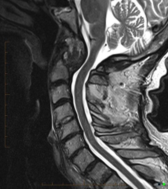
Q1.What is pannus?
- A soft tissue mass
- It encircles the eroded dens
Q2.Where does the term pannus come from and what is its composition?
- Latin for 'cloth'
- Hypertrophied synovium with fibrovascular tissue and inflammatory cells releasing collagenolytic enzymes
- Leads to destruction of cartilage, bone, tendons, ligaments and blood vessels
- Stabilization decreases pannus
▸ Slide 452 · Use of open mouth view in basilar invaginationSpine Non Trauma · 4 questions expand

Q1-Q44 questions — tap to reveal all answerslist
- Which lines are used to assess basilar invagination on this slide?
- What are the normal measurements for Fishgold's digastric line?
- How is Fishgold's bimastoid line interpreted?
- What is the use of the open mouth view in assessing C1/2 instability and basilar invagination?
Q1.Which lines are used to assess basilar invagination on this slide?
- Fishgold's digastric (Biventer) line - connects the digastric grooves on the undersurface of the skull, just medial to the mastoid processes
- Fishgold's bimastoid line - connects the tips of the mastoid processes
Q2.What are the normal measurements for Fishgold's digastric line?
- Odontoid tip normally projects 11 mm below the line
- Atlanto-occipital joint normally projects 12 mm below the line
- Basilar invagination is present when the atlanto-occipital joint projects at or above this line
Q3.How is Fishgold's bimastoid line interpreted?
- Odontoid process should be <10 mm above this line
- More than 10 mm above the line indicates basilar invagination
Q4.What is the use of the open mouth view in assessing C1/2 instability and basilar invagination?
- Open mouth C-spine XR looks for rotatory instability
- Rule of Spence: >8mm = TAL incompetence
- Controlled flexion/extension views to see if the deformity is reducible
▸ Slide 453 · Fusion technique of C1 C2Spine Non Trauma · 4 questions expand

Q1-Q44 questions — tap to reveal all answerslist
- Describe the Gallie fusion technique for C1/2.
- Describe the Brooks and Jenkins fusion.
- What screw-based options are available for C1/2 fusion?
- What are the main categories of C1/2 fusion techniques?
Q1.Describe the Gallie fusion technique for C1/2.
- Interspinous bone graft with sublaminar wiring around the posterior arch of C1 and wiring over the C2 spinous process
- Fusion rate 75%fusion (poor rotational stability)
- Requires post-operative external fixation
Q2.Describe the Brooks and Jenkins fusion.
- Modification of Gallie to overcome rotational deficiency
- Bilateral interlaminar grafts with bilateral sublaminar wiring (C1 and C2)
- Fusion rate 93%
- Also requires post-operative external fixation
Q3.What screw-based options are available for C1/2 fusion?
- Transarticular screw - supplemented with Gallie wiring; stable
- Screw-rod structure - stable and allows reduction
- OC fusion
Q4.What are the main categories of C1/2 fusion techniques?
- Wiring: Gallie; Brooks and Jenkins
- Transarticular screw (supplemented with Gallie wiring) - stable
- Screw-rod structure - stable, allows reduction
- OC fusion
▸ Slide 454 · Landmark of transarticular screwSpine Non Trauma · 2 questions expand

Q1-Q22 questions — tap to reveal all answerslist
- What are the entry landmarks for a transarticular screw (Magerl technique)?
- What is the sagittal trajectory of the Magerl transarticular screw?
Q1.What are the entry landmarks for a transarticular screw (Magerl technique)?
- Axial: 10 degree medial
- Entry point 3-4mm cranial to the C2/3 joint
- Midpoint of the pars mediolaterally
Q2.What is the sagittal trajectory of the Magerl transarticular screw?
- 50 degree cephalad
- Passes through the posterior 1/3 of the C1-2 joint
▸ Slide 455 · C2 pedicle screwSpine Non Trauma · 2 questions expand
Q1-Q22 questions — tap to reveal all answerslist
- What is the entry site for a C2 pedicle screw?
- What is the trajectory of a C2 pedicle screw?
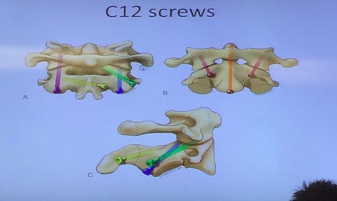
Q1.What is the entry site for a C2 pedicle screw?
- Cranial leading edge of the C2 lamina
- Midpoint of the pars mediolaterally
- Starting point 2mm lateral to this midpoint
Q2.What is the trajectory of a C2 pedicle screw?
- 30-45 degree convergent
- Directed cranially
▸ Slide 456 · AP Xray lumbar spine and pelvisSpine Non Trauma · 10 questions expand
Q1-Q1010 questions — tap to reveal all answerslist
- Describe the AP X-ray findings in ankylosing spondylitis.
- What are the history and examination findings in AS, and how is it diagnosed?
- What are the differentials of multiple syndesmophytes and of SIJ sclerosis?
- What is ankylosing spondylitis and what is the role of HLA-B27?
- What are the discovertebral lesions in AS and the causes of pseudoarthrosis?
- How are deformities in ankylosing spondylitis managed?
- How does SIJ involvement progress radiologically in ankylosing spondylitis?
- What blood tests and further imaging are requested in suspected AS?
- What types of spinal osteotomy are used for AS deformity correction?
- What are the key points of cervical spine osteotomy in AS?
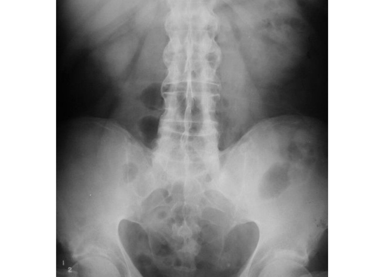
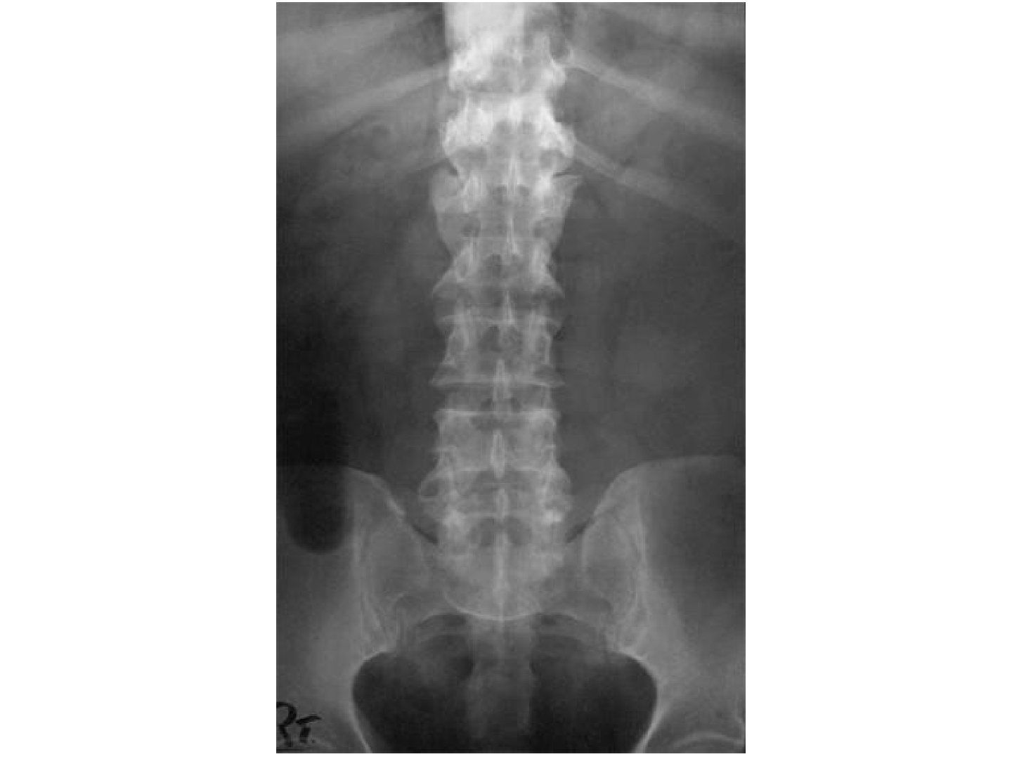
Q1.Describe the AP X-ray findings in ankylosing spondylitis.
- Squaring of vertebrae with marginal syndesmophytes
- Bamboo spine appearance; trolley-track and dagger sign
- Bilateral SIJ ankylosed, hips okay, no discovertebral lesion
- Frontal alignment good, no osteopenia; need lateral XR for facet joint ankylosis
Q2.What are the history and examination findings in AS, and how is it diagnosed?
- Hx: PMHx, FHx, morning stiffness, SOB, pain, other joint or heel pain
- PE: gait, loss of lumbar lordosis with increased thoracic kyphosis, horizontal gaze, chin-brow vertical angle, wall-to-occiput, Schober's test, chest expansion
- SIJ: Faber and Gaenslen tests; neurological exam of LL; listen to heart and lungs, look at eyes and nails
- Diagnosis: modified New York criteria - at least 1 clinical + 1 radiological criterion
- Clinical: inflammatory back pain >3 months improved by exercise/worse with rest, reduced spine ROM, reduced chest expansion
- Radiological: unilateral grade 3-4 or bilateral grade 2-4 SIJ changes
Q3.What are the differentials of multiple syndesmophytes and of SIJ sclerosis?
- Multiple syndesmophytes/ossification: DISH - non-marginal syndesmophytes, disc and SIJ spared, more common in older patients (DM)
- SIJ sclerosis: osteitis condensans ilii (young women, iliac side)
- Fibrodysplasia ossificans progressiva
Q4.What is ankylosing spondylitis and what is the role of HLA-B27?
- A systemic seronegative arthritis mainly affecting the axial skeleton
- More common in young male patients with family history (HLA-B27)
- Presents with back pain not relieved by rest, spine/hip stiffness, stooped posture, fracture
- HLA-B27 is an MHC presenting intracellular protein to T cells, initiating an immune response through TNF
- Patho hypotheses: misfolding -> homodimer triggering a pro-inflammatory response; arthritogenic peptide presented to autoreactive cytotoxic T cells
Q5.What are the discovertebral lesions in AS and the causes of pseudoarthrosis?
- Romanus lesion (50%): erosion at the anterolateral corner with surrounding sclerosis, posterior column spared
- Andersson lesion (4%): erosion at the mid-endplate (anterior + middle column), posterior column spared
- Pseudoarthrosis: secondary to fracture/Andersson lesion with posterior column involvement - all 3 columns
- Causes (D Fang): acute fracture through fused segment, stress fracture, escape fusion -> need circumferential fusion
Q6.How are deformities in ankylosing spondylitis managed?
- Aim: posture (horizontal gaze), visceral function, prevent complications (fracture, pseudoarthrosis)
- Medical: physio, NSAIDs, TNF-alpha antagonists (infliximab, adalimumab, etanercept)
- Preop: anaesthetic/airway, biologics, lung function test, echo, steroid coverage, PPI; osteotomy site usually lumbar (wider canal, end of conus, no ribs, large correction power)
- Osteotomies: SPO, Ponte, PSO, VCR; cervical osteotomy for chin-on-chest at C7/T1, aim CBVA within 10 degrees; PSO closing wedge preferred over SPO opening wedge
- Complications: neurologic injury, SMA, aortic rupture, non-union, residual deformity
- Instrumentation: U3L3; C8 root decompression; C7 laminectomy + partial lateral mass resection +/- C6 and T1 partial laminectomy
Q7.How does SIJ involvement progress radiologically in ankylosing spondylitis?
- X-ray SIJ progression: erosion, sclerosis and ankylosis
- Assess with the Ferguson view (~20 degrees cranial)
- First radiological sign is squaring of the vertebrae
Q8.What blood tests and further imaging are requested in suspected AS?
- Bloods: CBC, ESR, CRP, HLA-B27
- XR of the thoracic and cervical spine; whole spine sagittal alignment
- Need lateral XR for facet joint ankylosis; watch for vertebral collapse (osteoporosis)
Q9.What types of spinal osteotomy are used for AS deformity correction?
- SPO: laminectomy + pars
- Ponte: SPO + facets
- PSO: posterior elements + pedicle
- Vertebral column resection (VCR)
- Site usually lumbar: wider canal, end of conus, no ribs, large correction power
Q10.What are the key points of cervical spine osteotomy in AS?
- Indications: chin-on-chest; residual upper cervical kyphosis causing downward gaze after lumbar osteotomy
- Aim to correct CBVA to within 10 degrees
- Level C7/T1: wide canal, mobile root (C8), vertebral artery not entering lateral mass
- PSO closing wedge better than SPO opening wedge: instability; oesophageal stretching causing dysphagia
- Instrumentation U3L3; C8 root decompression; C7 laminectomy + partial lateral mass resection +/- C6 and T1 partial laminectomy
▸ Slide 457 · AS spine fracturesSpine Non Trauma · 6 questions expand
Q1-Q66 questions — tap to reveal all answerslist
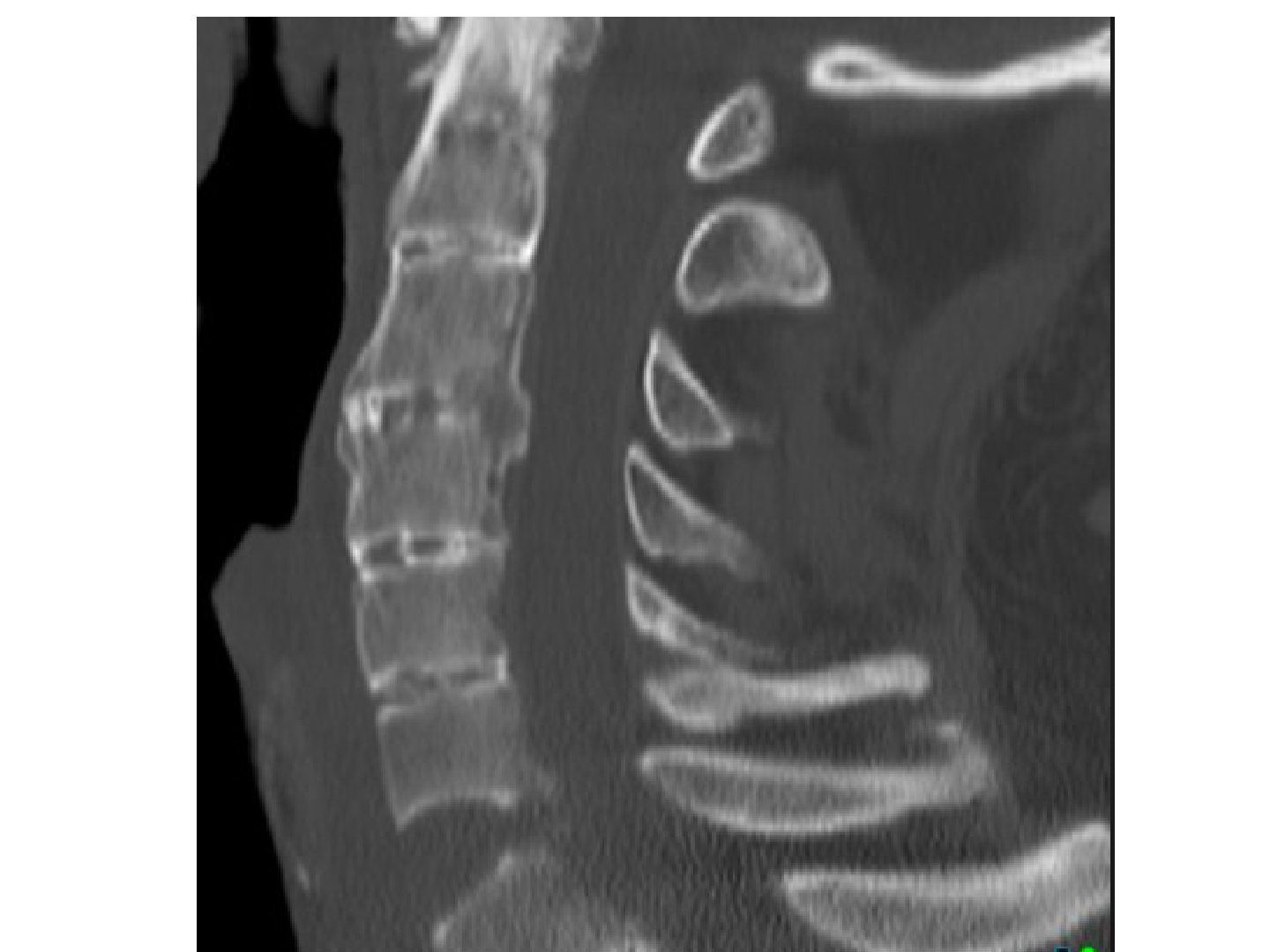
- What does the sagittal CT show and what should you suspect?
- How do you initially manage a suspected AS spine fracture?
- What is the definitive management and why is healing difficult?
- What are the pre-operative challenges in AS spine fracture surgery?
- What are the intra-operative challenges in AS spine fracture surgery?
- What are the post-operative complications and epidemiology?
Q1.What does the sagittal CT show and what should you suspect?
- Most of the spine ankylosed with a break at the C6/7 level
- No obvious soft tissue swelling; posterior element not involved in this CT cut
- Suspect ankylosing spondylitis with fracture/pseudoarthrosis
Q2.How do you initially manage a suspected AS spine fracture?
- Start with ATLS and immobilize the spine
- Expect shock - at this level it can be neurogenic shock; rule out other causes, give fluid resuscitation and vasopressor
- Once stabilized, document neurology
- Screen the rest of the CT for noncontiguous fractures
- MRI to look for epidural haematoma
Q3.What is the definitive management and why is healing difficult?
- Fractures heal poorly as the spine is stiff with high stress
- Offer posterior decompression and instrumented fusion
Q4.What are the pre-operative challenges in AS spine fracture surgery?
- Risk of GA due to cardiorespiratory comorbidities; adjust medications
- Difficult airway and ventilation - may need awake fibreoptic intubation
Q5.What are the intra-operative challenges in AS spine fracture surgery?
- Positioning in functional/natural position (sandwich prone), do not reduce, fix in situ/original kyphosis, avoid pressure points
- Posterior extensile approach; loss of landmarks - need CT navigation and neuromonitoring
- Osteoporotic bone (cement - not in the cervical spine); bleeding
- Need decompression? If no neurology, no need as want to save more bone bed for fusion; fixation in situ as heroic reduction risks neuro compromise
- Long segment fusion; defer anterior reconstruction and deformity correction
Q6.What are the post-operative complications and epidemiology?
- Pseudoarthrosis (may need secondary anterior procedure), infection, instrumentation failure
- Wound breakdown (tight skin from kyphosis)
- SCI incidence 21.1%; multiple and noncontiguous fractures in 13.1%
- ASAS classification for AS (modified from modified NY criteria due to delay in diagnosis)
▸ Slide 458 · DISH Forestier diseaseSpine Non Trauma · 4 questions expand
Q1-Q44 questions — tap to reveal all answerslist
- What is DISH (Forestier disease)?
- What are the radiological features of DISH?
- How is DISH treated?
- What is the association between DISH and total hip replacement?
Q1.What is DISH (Forestier disease)?
- Diffuse idiopathic skeletal hyperostosis - common disorder of unknown etiology with back pain and stiffness
- XR: flowing candle wax; associated with HLA-B8
- Defined by non-marginal syndesmophytes over 4 continuous vertebral bodies (3 IVD)
- 10%, more common in middle-aged males, equal in old; mainly anterolateral
- Risk factors: DM, hyperlipidaemia, gout
Q2.What are the radiological features of DISH?
- Distribution T (R > L) > C > L
- Preserved disc space, no osteopenia, no degenerative changes
- SIJ not involved; posterior elements not involved (no facet ankylosis, no intra-articular osseous fusion/sclerosis)
Q3.How is DISH treated?
- Activity modification, physical therapy, brace wear
- NSAIDs and bisphosphonate therapy
Q4.What is the association between DISH and total hip replacement?
- Patients with DISH have double the chance of heterotopic ossification (HO)
- This applies to patients undergoing total hip replacement (THR)
▸ Slide 459 · Describe CT/XR:Spine Non Trauma · 14 questions expand

Q1-Q1414 questions — tap to reveal all answerslist
- Describe the CT/XR findings in cervical myelopathy.
- Describe the MRI findings in cervical myelopathy.
- What are the causes of cervical myelopathy and its poor prognostic factors?
- What is the natural history of cervical myelopathy and OPLL?
- How do you choose the operative approach (PAINS)?
- What are the complications of laminoplasty, and how does it compare with laminectomy and fusion?
- What history is taken in cervical myelopathy?
- What does the physical examination include in cervical myelopathy?
- What investigations are performed in cervical myelopathy?
- What is the pathology of OPLL?
- What is the neurological recovery after surgery for cervical myelopathy (Cheung 2008)?
- What is the pseudoarthrosis rate after anterior cervical surgery?
- What is the incidence of adjacent segment disease and the evidence for ADR?
- What are the criteria, technique, advantages and disadvantages of ADR?
Q1.Describe the CT/XR findings in cervical myelopathy.
- Degenerative changes with osteophyte formation and mixed-type OPLL; kyphosis (K line/ C2-C7 angle)
- Canal stenosis: absolute <10mm, relative 13mm
- Pavlov ratio <0.8; Penning's jaw diameter <12mm suggests a congenital stenosis component
- Occupancy ratio >0.4 = poor prognosis
- Mizuno XR classification for OPLL: segmental, continuous, mixed, circumscribed/localized (at disc)
Q2.Describe the MRI findings in cervical myelopathy.
- Multi-level stenosis with compression from both anterior and posterior (disc, bulging, ossified PLL/yellow ligament)
- Kyphosis
- Cord edema
Q3.What are the causes of cervical myelopathy and its poor prognostic factors?
- Causes: spondylosis (anterior PID, calcified/buckled PLL, Luschka osteophytes; posterior calcified/buckled YL), OPLL, instability (e.g. C1/2), worsened by congenital narrowing (Pavlov <0.8)
- Poor prognosis: age >70, chronic symptoms >6 months, JOA <9, multiple levels, canal <30mm2, cord signal (T1 hypo/T2 hyper), compression ratio <0.4
Q4.What is the natural history of cervical myelopathy and OPLL?
- Clark: 70% stepwise, 20% gradual, 10% sudden deterioration then plateau
- Matsunaga Spine 2012 OPLL: 20% myelopathy at presentation, 20% develop symptoms, 70% asymptomatic
- Risk of myelopathy: >60% canal stenosis by OPLL and laterally deviated-type OPLL on CT
- Bad CT: C sign = high risk of dural tear
Q5.How do you choose the operative approach (PAINS)?
- Assess: axial neck pain/ radiculopathy, alignment (rigid kyphosis, K line), instability, number of levels, site of pathology
- 1-2 levels: ASF or ASD if no instability
- >3 levels without rigid kyphosis: posterior (laminectomy + fusion if axial neck pain/instability, else laminoplasty)
- >3 levels with kyphosis: anterior + posterior (anterior to realign/decompress, posterior to decompress/fuse)
- K line: K+ no instability = laminoplasty; K+ instability = laminectomy + fusion; K- = A+P
- Conservative: analgesics for neck pain, physio; advise operation if JOA <=13 as symptoms usually progress
Q6.What are the complications of laminoplasty, and how does it compare with laminectomy and fusion?
- Complications: fracture hinge, spring back of hinge, C5 palsy, axial neck pain, late kyphosis
- Laminoplasty advantages: motion preserving (30-40% motion loss quoted), less morbidity, no instrumentation, shorter rehab
- Laminectomy + fusion advantages: may relieve neck pain; OPLL may resolve after stabilisation
Q7.What history is taken in cervical myelopathy?
- Age, demand, history of DM, chief complaint and progress
- Axial neck pain and radicular pain
- Function by JOA: use of chopsticks/spoon, walking on level ground/stairs/walking aids, sphincter condition, numbness of UL, trunk and LL
- Trauma and red flag signs
Q8.What does the physical examination include in cervical myelopathy?
- Power, sensation, jerks, myelopathy hand signs, long tract signs
- Lhermitte, Spurling, Romberg/tandem walking
- PR examination and jaw jerk
Q9.What investigations are performed in cervical myelopathy?
- XR oblique views and flexion/extension views
- CT for extent of OPLL and the double layer sign
- MRI for cause, level and severity of stenosis
Q10.What is the pathology of OPLL?
- Mass of woven and lamellar bone plus fibrocartilage
Q11.What is the neurological recovery after surgery for cervical myelopathy (Cheung 2008)?
- Cheung WY Int Orthop 2008: neurological recovery in 71% of patients
- Plateau at 6 months
- Best in upper limbs, then lower limbs; worst with sphincter dysfunction
Q12.What is the pseudoarthrosis rate after anterior cervical surgery?
- Without anterior plate: one level 0-5%, two level 10-20%, three level 30-60%
- With anterior plate: one level 0-5%, two level 0-3%, three level 0-7%
Q13.What is the incidence of adjacent segment disease and the evidence for ADR?
- Adjacent segment disease: 25% within the first 10 years
- BJJ Findlay 2018: meta-analysis of 14 RCTs, follow-up 2-10 years
- TDR as effective as ACDF and superior for some outcomes; TDR > ACDF at 2 years and also 4-7 years
- Reduces the risk of adjacent segment disease (ACDF ASD 1.6-4.2% per year)
Q14.What are the criteria, technique, advantages and disadvantages of ADR?
- Strict criteria: no significant neck pain, no facet arthritis, anterior compression only, no instability, good bone stock (no osteoporosis)
- Technique: alignment of COR in coronal and sagittal planes, accurate sizing and endplate preparation
- Advantages: preserve motion, prevent ASD
- Disadvantages: implant failure/migration; easy to overstuff -> posterior neck pain (increased facet loading)
▸ Slide 460 · OPLLSpine Non Trauma · 3 questions expand

Q1-Q33 questions — tap to reveal all answerslist
- What are the classifications of OPLL?
- Why is OPLL difficult to treat, and what is the anterior strategy?
- What is the indication for treatment of OPLL?
Q1.What are the classifications of OPLL?
- XR (Mizuno): focal, segmental, continuous, mixed
- CT (Hirabayashi): square, mushroom, hill
Q2.Why is OPLL difficult to treat, and what is the anterior strategy?
- Difficult: multi-level, at the level of the vertebral body, high chance of dural tear
- If anterior approach needed: decompress the surrounding bone; may need to leave OPLL on the dura (floating technique)
- Corpectomy may be required
Q3.What is the indication for treatment of OPLL?
- Indication for treatment is the same as for cervical myelopathy
- No separate OPLL-specific indications are given in the notes
▸ Slide 461 · Functional classification of cervical myelopathySpine Non Trauma · 3 questions expand
Q1-Q33 questions — tap to reveal all answerslist
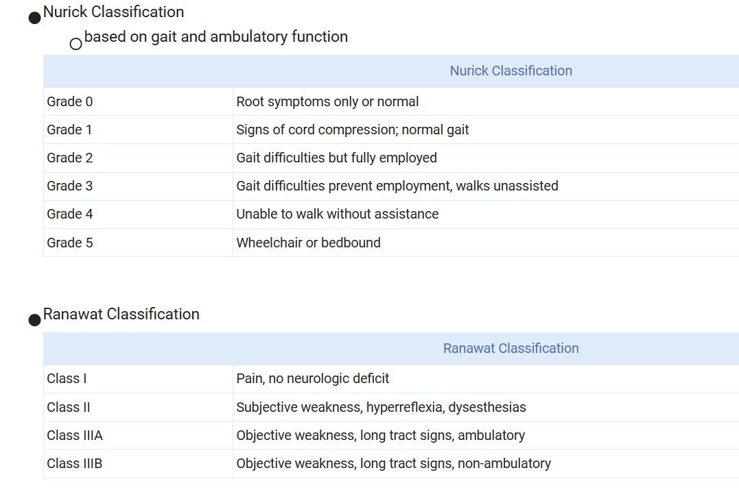
- Which functional classifications of cervical myelopathy are shown?
- Describe the Nurick classification (grades 0-5).
- Describe the Ranawat classification.
Q1.Which functional classifications of cervical myelopathy are shown?
- Nurick classification - based on gait and ambulatory function
- Ranawat classification - based on pain, weakness and ambulatory status
Q2.Describe the Nurick classification (grades 0-5).
- Grade 0: root symptoms only or normal
- Grade 1: signs of cord compression, normal gait
- Grade 2: gait difficulties but fully employed
- Grade 3: gait difficulties prevent employment, walks unassisted
- Grade 4: unable to walk without assistance
- Grade 5: wheelchair or bedbound
Q3.Describe the Ranawat classification.
- Class I: pain, no neurologic deficit
- Class II: subjective weakness, hyperreflexia, dysesthesias
- Class IIIA: objective weakness, long tract signs, ambulatory
- Class IIIB: objective weakness, long tract signs, non-ambulatory
▸ Slide 462 · JBJS 1987 by OnoSpine Non Trauma · 2 questions expand

Q1-Q22 questions — tap to reveal all answerslist
- What is the finger escape sign classification (Ono, JBJS 1987)?
- What is the source of this classification and how is the test performed?
Q1.What is the finger escape sign classification (Ono, JBJS 1987)?
- Cannot hold
- Cannot do
- 2 finger / cannot do full extension
- 3 finger
- Closing the eyes is not necessary for the test
Q2.What is the source of this classification and how is the test performed?
- JBJS 1987 by Ono
- Finger escape sign classification
- Patient does not need to close the eyes
▸ Slide 463 · Lumber stenosisSpine Non Trauma · 12 questions expand

Q1-Q1212 questions — tap to reveal all answerslist
- Describe the MRI findings in lumbar stenosis.
- What must be excluded and assessed in back pain with suspected stenosis?
- What are the red flag signs in lumbar stenosis?
- How do you differentiate the nature of pain and claudication?
- What are the treatment aims and indications for fusion?
- What is the evidence for decompression with or without fusion, and the natural history of degenerative spondylolisthesis?
- What else should be delineated on MRI in lumbar stenosis?
- What are the borders of the lateral recess, midzone and intervertebral foramen?
- How can fusion relieve back pain and why choose interbody fusion?
- What are the anterior and posterior approaches and operations used to relieve stenosis?
- What are the complications of fusion and of each surgical approach?
- What is degenerative spondylolisthesis and what do the natural history studies show?
Q1.Describe the MRI findings in lumbar stenosis.
- Sagittal T2 image: severe stenosis and grade 1 anterolisthesis at L3/4 and L4/5
- Disc bulging and buckling of the thickened ligamentum flavum
- No endplate destruction, no collapse
Q2.What must be excluded and assessed in back pain with suspected stenosis?
- Rule out infection/tumour, rule out cauda equina syndrome, rule out upper spinal involvement (brisk jerk)
- Assess severity of axial symptoms and neurology
- Assess instability
Q3.What are the red flag signs in lumbar stenosis?
- Extremes of age; history of trauma
- History of cancer/drug abuse; constitutional symptoms
- Rest pain / night pain waking the patient
- Saddle anaesthesia / sphincter disturbance
Q4.How do you differentiate the nature of pain and claudication?
- Discogenic: worse with increased intradiscal pressure - flexion, sitting, heavy lifting, sneezing
- Facet: pain in extension, radiating to posterior thigh and buttock; instability: forward flexion + extension catch
- Inflammatory: relief with exercise, morning stiffness; pathological: persistent/night pain
- Vascular claudication: exertion-related, worse walking/upslope, relieved by rest even upon standing, diminished peripheral pulses
- Neurogenic claudication: exertion + posture, worse walking/downslope, relieved by sitting, not standing (flexion reduces ligament buckling)
Q5.What are the treatment aims and indications for fusion?
- Aims: pain relief (disc, instability, facet), decompression of neural elements, prevent progression
- Conservative: anti-lordotic brace, flexion-based exercises
- Fusion if: pain (discogenic, facet effusion, foraminal stenosis), severe deformity (degenerative scoliosis), instability (dynamic X-ray, anterolisthesis, iatrogenic >50% facet removal, extensive discectomy)
- In anterolisthesis with a very narrowed disc (autostabilization) and no dynamic instability, decompression alone may suffice
Q6.What is the evidence for decompression with or without fusion, and the natural history of degenerative spondylolisthesis?
- SPORT trial: surgery better SF-36 and ODI vs non-operative up to 8 yrs
- Pro-fusion: Herkowitz 1991, Kornblum 2004, Martin 2007 (revision 28% vs 17%), Ghogawala NEJM 2016 (SLIP) - stable slips – fusion group had better functional outcomes and lower revision rate
- Against: Forsth NEJM 2016 - fusion group had significantly higher blood loss, surgical cost, length of stay, no difference in outcomes; authors did not distinguish between stable and unstable slips. Repoperation rate of 20% due to ASD, restenosis
- Weinstein commentary 2016: decompression alone reasonable for many; fusion may prevent slip progression and reduce reoperation
- Degenerative spondylolisthesis (Johnson): 70% stable, 15% improve, 15% deteriorate
- Matsunaga: 30% progressive, no X-ray/clinical correlation; instability risk: translation >5mm, disc height <6.5mm, Facet angle >50 deg
Q7.What else should be delineated on MRI in lumbar stenosis?
- Neuroforamen stenosis and which side of the lateral recess/central canal is more stenotic
- Facet effusion and facet orientation
Q8.What are the borders of the lateral recess, midzone and intervertebral foramen?
- Lateral recess: posterior SAP, anterior disc, medial sac, lateral pedicle
- Midzone: posterior pars, anterior VB, medial canal, lateral foramen, superior pedicle
- IVF: posterior SAP, anterior disc, superior/inferior pedicle
Q9.How can fusion relieve back pain and why choose interbody fusion?
- PL and interbody fusion have similar fusion rates (Levin Spine 2018 meta-analysis: PL 84% vs TLIF 94% radiological fusion; weak data for clinical improvement)
- Interbody fusion: compression side gives theoretically better fusion and eliminates the disc pain generator
- Better at reducing listhesis and restoration of sagittal balance
- Corrects disc height (helps neuroforamen stenosis) and restores ligament tension (flagpost theory)
Q10.What are the anterior and posterior approaches and operations used to relieve stenosis?
- Anterior: less muscle dissection and infection, bigger cage, better deformity correction, indirect decompression
- Posterior: avoids great vessels, allows foraminal decompression
- Central stenosis (claudication): laminectomy, which can selectively increase decompression on the more stenotic side
- Radiculopathy (lateral recess/neuroforamen): undercut/medial facetectomy; restore disc height to increase neuroforamen space
Q11.What are the complications of fusion and of each surgical approach?
- Fusion complications: pseudoarthrosis, cage migration/subsidence, implant failure
- Posterior approach: durotomy and traction injury to the nerve root, posterior muscle pain and spasm
- Anterior approach: vascular injury, sympathetic dysfunction, transpsoas injury (ureter, lumbar plexus e.g. genitofemoral nerve)
Q12.What is degenerative spondylolisthesis and what do the natural history studies show?
- Result of disc degeneration with decreased disc height, ligament buckling and microinstability
- Commonest at L4/5; PE shows stepping at L4/5
- 76% of patients with no neuro deficits initially remained without deficits; 83% with neuro symptoms experience deterioration
▸ Slide 464Spine Non Trauma · 2 questions expand

Q1-Q22 questions — tap to reveal all answerslist
- What topic does slide 464 cover?
- What are the key teaching points of slide 464?
Q1.What topic does slide 464 cover?
- Not covered in the speaker notes
- The slide image is the only source for this slide
Q2.What are the key teaching points of slide 464?
- Not covered in the speaker notes
- No content is provided beyond the slide number
▸ Slide 465Spine Non Trauma · 11 questions expand

Q1-Q1111 questions — tap to reveal all answerslist
- Describe the radiographic assessment of isthmic spondylolisthesis.
- What are the clinical features of isthmic spondylolisthesis?
- What is the natural history of spondylolisthesis (Fredrickson 1984)?
- What are the risk factors for progression of an isthmic slip?
- What are the treatment options for spondylolisthesis?
- What are the causes of symptoms and the etiological classification of spondylolisthesis?
- What is the Spinal Deformity Study Group classification for reduction in high-grade spondylolisthesis?
- What are the problems of in-situ fusion for high-grade spondylolisthesis?
- What is the Gaines procedure and what is its risk?
- What are the subgroups of isthmic spondylolisthesis?
- What fusion technique is preferred in spondylolisthesis surgery?
Q1.Describe the radiographic assessment of isthmic spondylolisthesis.
- Lateral: L5 pars defect with spondylolisthesis; slip severity (Meyerding); L5/S1 disc status; bone morphology (trapezoidal VB, dome sacrum)
- Lumbar lordosis (normal 20-45), lumbosacral kyphosis (slip angle), sacral inclination (normal >30)
- AP: spina bifida occulta; inverted Napoleon's hat in spondyloptosis
- Dynamic lateral: 4.5mm translation or 22deg angulation vs the adjacent motion segment
- Scannogram: PI = SS + PT
Q2.What are the clinical features of isthmic spondylolisthesis?
- PE (L5/S1): stepping at L4/5, lumbar hyperlordosis, pelvic retroversion + hip flexion contracture + hamstring tightness + knee flexion contracture
- Phalen-Dixon sign in severe slip
- Phalen-Dixon crisis - high grade slip: sudden severe sciatic pain (L5/S1 root irritation), tight hamstrings, flat sacrum, pelvic waddling gait, cauda equina compression with ischiocrural spasm, scoliosis
Q3.What is the natural history of spondylolisthesis (Fredrickson 1984)?
- 500 patients at age 6 with 45-year follow-up
- M>F
- No slip if unilateral; most frequent progression in the adolescent
- No association between slip progression and back pain
- Conclusion: benign course, with only a small percentage developing symptomatic progression
Q4.What are the risk factors for progression of an isthmic slip?
- Patient factors: female, young age at presentation, frequent back extension
- Morphology: dysplastic pars, sacral dome, wedging of L5, sacral inclination <30, PI >70, large slip angle (>45)
- Slip: high grade
Q5.What are the treatment options for spondylolisthesis?
- Conservative: flexion-based exercises, anti-lordotic brace, hamstring stretching, core strengthening
- Surgery if: slip progression, high grade, Phalen-Dixon crisis, failed conservative treatment (back pain/radiculopathy)
- Pars repair in young patients with no slip and normal disc: Buck's screw (cortical bone trajectory, directly across the fracture), Scott wire, Morscher hook screw, pedicle hook screw
- Fusion +/- reduction +/- decompression
- Reduction controversial: consider in high grade (>50%) + retroverted pelvis + poor sagittal balance; risk of neuro impairment ~33% (L5 stretch injury), needs neuromonitoring
- Reduction advantages: restores sagittal alignment, reduces gait abnormalities, larger fusion bed placing fusion in compression rather than shear
Q6.What are the causes of symptoms and the etiological classification of spondylolisthesis?
- Wiltse and Newman: dysplastic, isthmic (lytic, elongated, pars fracture), degenerative, traumatic, pathological, post-surgical
- Back pain causes: discogenic, pars defect, instability, facet degeneration (remember asymptomatic slip is common)
- Radiculopathy: L5/S1 slip compresses the L5 root (foramen enlarged; root encroached by pedicles/fibrocartilage of the pars defect); L5/S1 PID gives S1 radiculopathy
- Isthmic: most common at L5/S1; risk factors repetitive hyperextension (gymnasts, dancers), Eskimo populations, first-degree relative
- Reduction: passive (muscle relaxant, prone, facet + discectomy, disc height restoration); active (reduction screws, neuromonitoring, fluoroscopy)
Q7.What is the Spinal Deformity Study Group classification for reduction in high-grade spondylolisthesis?
- High grade, balanced pelvis = no need for forceful attempts at reduction
- High grade, retroverted pelvis but balanced spine = attempt postural (passive) reduction
- High grade, retroverted pelvis and unbalanced spine = reduction/realignment mandatory (active), under neurological monitoring
Q8.What are the problems of in-situ fusion for high-grade spondylolisthesis?
- Pseudoarthrosis
- Progressive slippage
- Persistent lumbosacral deformity
- Cauda equina syndrome
Q9.What is the Gaines procedure and what is its risk?
- Anterior L5 vertebrectomy + posterior L4 to S1 fusion
- 75% neurological injury
Q10.What are the subgroups of isthmic spondylolisthesis?
- Elongated pars
- Fatigue fracture
- Acute fracture
Q11.What fusion technique is preferred in spondylolisthesis surgery?
- Posterolateral (PL) vs interbody fusion
- Circumferential fusion is preferred
▸ Slide 466 · SpondylolysisSpine Non Trauma · 4 questions 1 check expand

Q1-Q44 questions — tap to reveal all answerslist
- What is spondylolysis and what is spondylolisthesis?
- What are the radiographic findings of spondylolysis?
- What is the pathophysiology and who is affected?
- What is the treatment and prognosis of spondylolysis without listhesis?
Q1.What is spondylolysis and what is spondylolisthesis?
- Spondylolysis: defect in the pars interarticularis
- Spondylolisthesis: forward slippage of one vertebra over another
Q2.What are the radiographic findings of spondylolysis?
- Defect in the neck of the 'Scottie dog'
- Notes state 80% seen on lateral, 15% on oblique, 5% subtle defect on CT/SPECT
Q3.What is the pathophysiology and who is affected?
- Fatigue fracture from repetitive hyperextension stress
- Examples: gymnasts, football linemen
Q4.What is the treatment and prognosis of spondylolysis without listhesis?
- Activity restriction, flexion exercise, bracing
- Nonunion is common
- Unilateral defect never progresses to listhesis
Spondylolysis on XR: 80% seen on lateral, 15% on oblique, 5% are subtle defects on CT/SPECT — contested — The oblique 'Scotty dog' view is classically the standard projection for spondylolysis, though sensitivity of lateral vs oblique is debated (coned lateral may detect ~85% of bilateral L5 defects). Early stress reactions are commonly invisible on plain XR (detection ~11-23% in early lesions), so more than 5% require CT/SPECT/MRI — medium confidence — source
▸ Slide 467Spine Non Trauma · 2 questions expand
Q1-Q22 questions — tap to reveal all answerslist
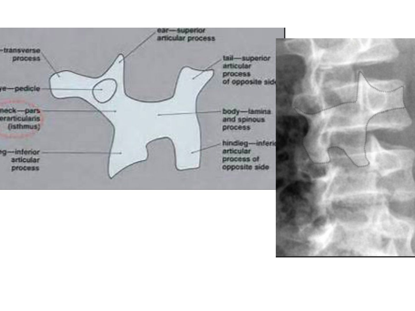
- On the oblique lumbar radiograph, which structure forms the 'neck' of the Scottie dog?
- Why is the oblique view used in suspected spondylolysis?
Q1.On the oblique lumbar radiograph, which structure forms the 'neck' of the Scottie dog?
- The pars interarticularis (isthmus) of the vertebra
- Ear = superior articular process, foreleg = inferior articular process, body = lamina and spinous process
Q2.Why is the oblique view used in suspected spondylolysis?
- Profiles the pars interarticularis - the neck of the Scottie dog
- The radiograph shows the outlined defect: a break in the neck = pars defect (spondylolysis)
▸ Slide 468 · Degenerative scoliosisSpine Non Trauma · 6 questions expand
Q1-Q66 questions — tap to reveal all answerslist
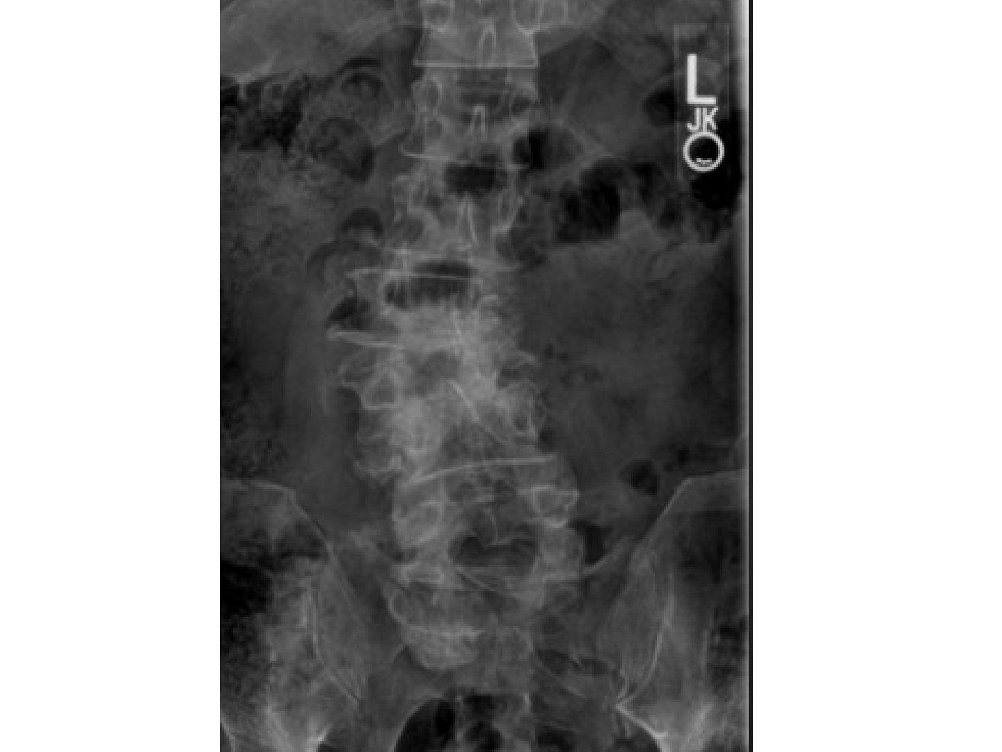
- Describe the X-ray findings in degenerative scoliosis.
- What are the causes of degenerative scoliosis?
- What is the degenerative cascade of the spine?
- What is the management approach for degenerative scoliosis?
- How does de novo degenerative scoliosis differ from pre-existing/neglected scoliosis?
- What is the rate and what are the risk factors for curve progression?
Q1.Describe the X-ray findings in degenerative scoliosis.
- Convexity to the right at the mid lumbar spine with a rotational component
- Marked degenerative change at multiple facet joints; pedicles and endplate intact
- Need lateral + whole spine XR (coronal and sagittal balance), F/E XR, prone traction views for vertical instability, bending and fulcrum views
- MRI also required
Q2.What are the causes of degenerative scoliosis?
- Osteoporotic collapse
- Spondylolisthesis
Q3.What is the degenerative cascade of the spine?
- Disc degeneration with decreased water content and decreased disc height
- Buckling of ligaments, decreased stability
- Increased stress on facet joints -> osteoarthritis and facet hypertrophy
- Facet cannot resist shear -> anterior translation (especially with sagittal-oriented facets)
- Can cause spinal stenosis/deformity; some may auto-stabilize
Q4.What is the management approach for degenerative scoliosis?
- Define the patient's complaint clearly; assess neurology, spinal stability, sagittal balance
- Osteoporosis treatment if related to osteoporotic collapse
- Decompression only: leg pain, minimal/no back pain, Cobb <30, <2mm subluxation, no thoracic hyperkyphosis, acceptable balance, poor premorbid
- Short fusion: back and leg pain, scoliosis <30, segmental instability, no significant global imbalance, extensive decompression
- Long fusion: back and leg pain, scoliosis >45, >2mm subluxation, significant global imbalance (determine UIV and LIV)
- Severe deformity with poor balance: corrective osteotomy + long fusion (high OT risk, outcome not guaranteed)
Q5.How does de novo degenerative scoliosis differ from pre-existing/neglected scoliosis?
- De novo has less coronal deformity (less Cobb, less L4 tilt, less rotational deformity) but more sagittal pelvic malalignment
- Fewer vertebral segments involved; affects the lumbar spine
- Disc space wedging, osteoporotic collapse, less bone maldevelopment (pedicles)
Q6.What is the rate and what are the risk factors for curve progression?
- 3 degrees per year
- Prior history of progression; unstable segment (asymmetrical disc degeneration, lateral disc wedging, >5mm lateral osteophyte difference)
- Cobb >30 degrees, lateral listhesis >6mm
- Intercrestal line through L4/5 disc or lower, rotatory subluxation, loss of lumbar lordosis
▸ Slide 469 · Osteoporotic collapseSpine Non Trauma · 16 questions expand
Q1-Q1616 questions — tap to reveal all answerslist
- Describe the X-ray findings of an osteoporotic vertebral collapse and the initial workup.
- What are the aims of management in osteoporotic collapse?
- How is osteoporotic collapse managed systemically?
- How is osteoporotic collapse managed locally?
- When is vertebroplasty considered for an osteoporotic collapse?
- What are the prerequisites for vertebroplasty?
- What are the complications of vertebroplasty?
- Why is kyphoplasty preferred over vertebroplasty?
- What did the two NEJM 2009 RCTs show about vertebroplasty and how were they criticised?
- What did the VAPOUR and VERTOS IV trials show about vertebroplasty?
- What is the AAOS 2010 guideline on treating osteoporotic collapse?
- How can adjacent level collapse and non-union be decreased after augmentation?
- What other options exist apart from vertebroplasty/kyphoplasty and what are the problems with them?
- How can failure at the bone-implant interface be reduced?
- What is the mechanism of calcitonin for pain relief?
- What is Kummel disease and what are its imaging features?
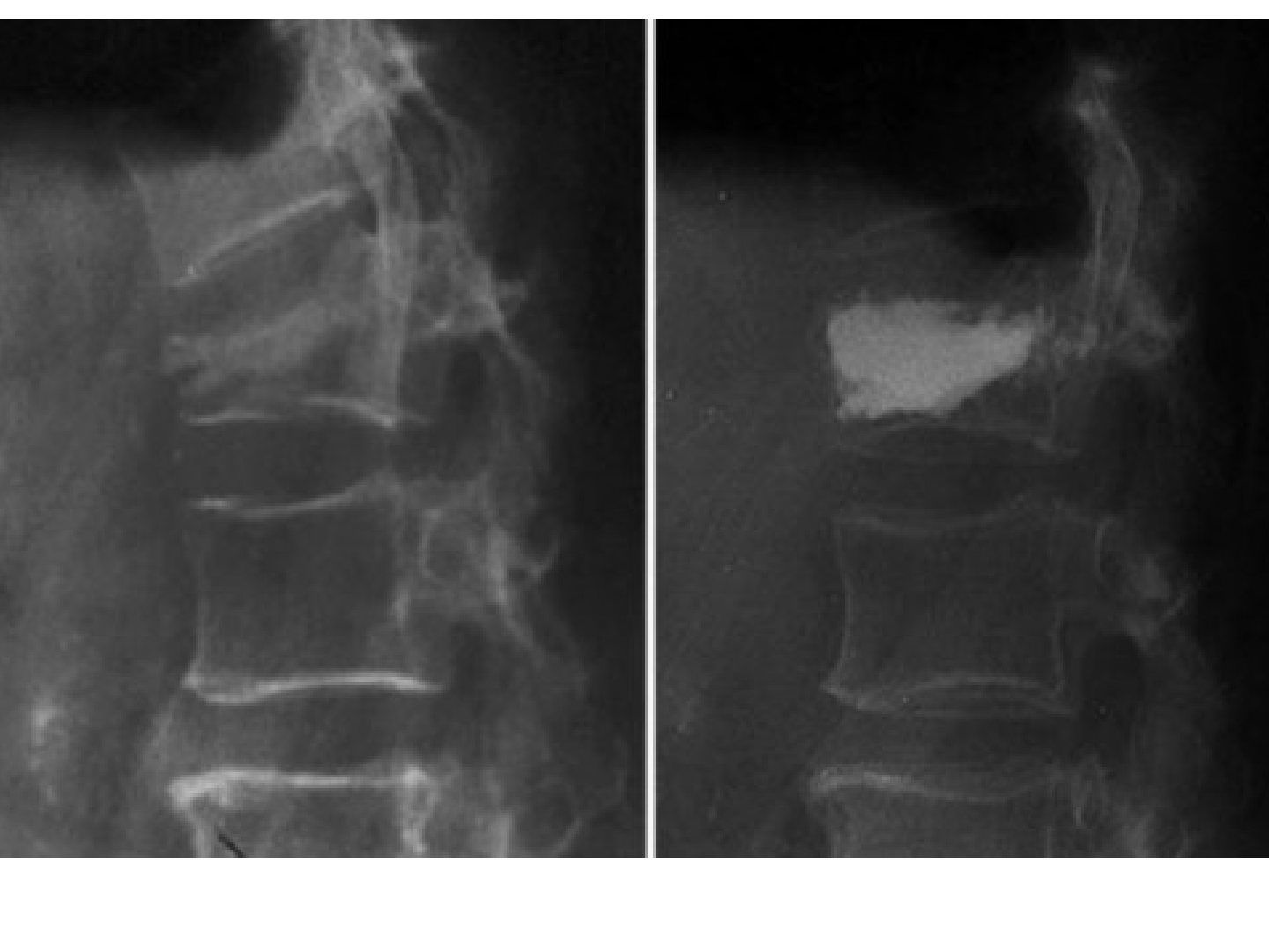
Q1.Describe the X-ray findings of an osteoporotic vertebral collapse and the initial workup.
- Anterior wedge collapse of T12 with no retropulsion
- Request AP X-ray to look for widening of the interpedicular distance
- Ask about mechanism of injury, cancer history, features of infection/TB, neurological deficit
- Bloods: bone profile, tumour markers, SEP and inflammatory markers
Q2.What are the aims of management in osteoporotic collapse?
- Pain relief and early mobilisation
- Prevent deformity and neurological deficit
- Prevent future collapse
Q3.How is osteoporotic collapse managed systemically?
- Assess osteoporosis severity and calculate future fracture risk
- Quit smoking and alcohol, give calcium and vitamin D, weight-bearing exercise
- Anti-osteoporotic medication
Q4.How is osteoporotic collapse managed locally?
- Analgesics, calcitonin +/- brace
- If failed conservative management or non-union, consider kyphoplasty
Q5.When is vertebroplasty considered for an osteoporotic collapse?
- Ongoing pain more than 2 weeks after occurrence of a new fracture despite treatment
- Progressive compression fractures of one or multiple vertebrae with subsequent loss of posture
- Non-union with persisting instability
Q6.What are the prerequisites for vertebroplasty?
- Intact posterior wall
- Neurologically intact
Q7.What are the complications of vertebroplasty?
- Anaesthesia-related complications
- Cannula placement: infection, spinal cord injury, fracture of pedicles/transverse process, lung injuries
- Cement injection: cement leakage into the spinal canal causing neurological deficits, pulmonary cement embolism, fat embolism
- Late adjacent segment collapse
Q8.Why is kyphoplasty preferred over vertebroplasty?
- Better evidence on pain relief and potential kyphosis correction
- Fewer complications than vertebroplasty due to decrease in insertion pressure
- Mechanism: reduces kyphosis, low-pressure injection, increased viscosity of injection
- Greater reduction in kyphotic angle and less extraosseous cement leakage than vertebroplasty
Q9.What did the two NEJM 2009 RCTs show about vertebroplasty and how were they criticised?
- Buchbinder et al.: pain, QoL and function same as placebo surgery up to 6 months
- Kallmes et al.: pain similar to placebo surgery at 3 months
- A JBJS 2009 publication criticised their methodology, mainly the cement volume
Q10.What did the VAPOUR and VERTOS IV trials show about vertebroplasty?
- VAPOUR trial (Lancet 2016, Clark): vertebroplasty vs sham - great reduction in pain at 3 and 6 months
- It is the only RCT to support vertebroplasty; patients had more severe pain, fracture age <6 weeks, and more PMMA cement was used
- VERTOS IV (BMJ 2018): no significant pain relief vs sham
- AAOS changed stance in 2014, suggesting limited evidence
Q11.What is the AAOS 2010 guideline on treating osteoporotic collapse?
- Moderate evidence: calcitonin for pain relief
- Weak evidence: ibandronate and strontium, kyphoplasty
- Inconclusive: bed rest, brace, exercise programme, electrical stimulation
- Not advised: vertebroplasty
- Avoid in fractured posterior wall and neurologically compromised patients
Q12.How can adjacent level collapse and non-union be decreased after augmentation?
- Treat osteoporosis
- Watch for fracture patterns that increase non-union: split type fracture / large angular motion
- Beware of endplate deficit leading to intradiscal cement leakage
- Avoid aggressive restoration of vertebral height
Q13.What other options exist apart from vertebroplasty/kyphoplasty and what are the problems with them?
- Problems with more extensive surgery: medically fragile patients, poor bone quality at the bone-implant interface
- Approach: anterior vs A+P vs posterior
- Decompression; reduction?
- Instrumentation: short or long, construct type (screw-rod, plate, sublaminar hooks and wiring), augmentation
- Fusion and rehabilitation; removal of implant: no
Q14.How can failure at the bone-implant interface be reduced?
- Preop: treat osteoporosis with a bone-forming agent such as teriparatide
- Intraop: screw type (HA coated, PMMA augmented), diameter, length - pullout strength 60% depends on cortical bone of the pedicle, 20% on trabecular bone of the body; advancing to 80% of the vertebral body is adequate
- Self tap screws (stronger pullout force), or undertap (self-tapping screws); diverging screws aimed towards subchondral bone, straightforward technique better than anatomical trajectory
- Expandable pedicle screws; circumferential fusion
- Postop bracing
- Crosslink and increasing the length of fixation only increase construct stiffness, they do not decrease failure at the bone-implant junction
Q15.What is the mechanism of calcitonin for pain relief?
- Binds receptors in the CNS/PNS
- Releases endorphin and decreases prostaglandins
Q16.What is Kummel disease and what are its imaging features?
- Post-traumatic osteonecrosis of a vertebral body
- XR: sclerosis of cleft margin and vertebral body, pedicle ossification, stress fracture of spinous process, paravertebral callus
- MRI: double line sign
- Vertebroplasty more likely to fail due to lack of interdigitation of cement
▸ Slide 470 · AP Xray T spine of patient showing soft tissue fusiform swellingSpine Non Trauma · 20 questions expand
Q1-Q2020 questions — tap to reveal all answerslist
- What did the plain X-rays show in this case of spinal TB?
- What are the typical X-ray and MRI features of spinal tuberculosis?
- What is the clinical assessment in suspected spinal TB?
- How is spinal TB diagnosed?
- What are the aims of management in spinal TB?
- How does the spinal level affect the management of TB spine?
- How is TB spine managed when there is no neurology and no instability?
- How is TB spine with neurological involvement or instability managed?
- What is the epidemiology of TB spine?
- What is the pathophysiology of spinal TB?
- What is Pott's paraplegia and what are its early causes?
- What are the Rajasekaran types of paediatric spine deformity?
- What are the features of healed TB disease?
- What was the design of the MRC trial of TB spine?
- What was a favourable outcome in the MRC trial and what did it conclude?
- What did Prof Luk criticise about the MRC trial?
- What are the absolute indications for surgery in spinal TB?
- What are the late complications of spinal TB?
- What are the surgical approaches and deformity correction options in TB spine?
- What are the spine-at-risk signs of Rajasekaran?
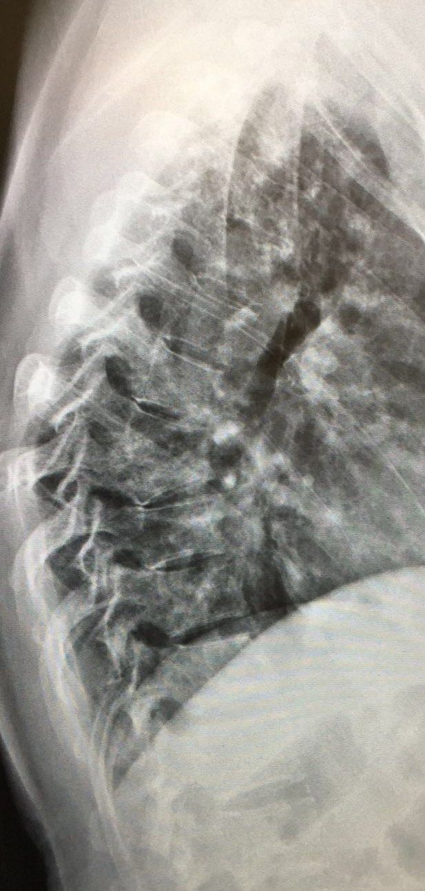
Q1.What did the plain X-rays show in this case of spinal TB?
- AP T spine: soft tissue fusiform swelling
- Lateral T spine: collapse of T7 and T8, obliterated disc space, acute kyphosis
- Gibbus = acute structural thoracolumbar kyphosis
- No skipped lesion
Q2.What are the typical X-ray and MRI features of spinal tuberculosis?
- X-ray: anterior vertebral body collapse, disc sparing, multiple levels +/- skip lesions, kyphosis, +/- calcification
- MRI: skip lesions 15%, paravertebral abscess 50%, disc sparing (multicentric and multilevel), subligamentous spread
- Ddx: infection (TB > pyogenic > fungus) and tumour (lymphoma)
Q3.What is the clinical assessment in suspected spinal TB?
- Symptoms: deformity, pain, neurology
- Risk factors of infection: immunocompromised, previous TB infection, contact history
- Red flag signs
- Rule out other differentials: pyogenic infection, carcinoma, lymphoma
Q4.How is spinal TB diagnosed?
- Morning urine and sputum TB PCR
- CXR abnormal in 2/3 of cases
- Biopsy (low sensitivity)
- MRI with gadolinium: low T1, bright T2
Q5.What are the aims of management in spinal TB?
- Control infection
- Control back pain
- Salvage neurology
- Prevent/correct deformity
Q6.How does the spinal level affect the management of TB spine?
- C spine: low threshold for surgery
- Lumbar spine: conservative first
- T spine: depends on neurology
Q7.How is TB spine managed when there is no neurology and no instability?
- Isoniazid + rifampicin, +/- streptomycin
- Expected kyphosis = total vertebral involvement x 30.5 + 5.5 (Rajasekaran)
Q8.How is TB spine with neurological involvement or instability managed?
- Debridement only: expected kyphosis 10 degrees
- HK operation: anterior radical debridement (extirpation) + fusion with strut graft
- No instrumentation if <3 levels; instrumentation if >=3 levels
- Advantages: direct visualisation of the lesion, fusion under compression anteriorly
Q9.What is the epidemiology of TB spine?
- Extrapulmonary TB in 3% of cases
- Skeletal TB in 10% of extrapulmonary TB
- Spinal TB in 50% of all skeletal TB
- Site: TLJ > T > LS > C
Q10.What is the pathophysiology of spinal TB?
- Granulomatous spondylitis with a delayed hypersensitivity reaction
- Starts in the metaphysis of the vertebral body
- Subligamentous spread under the ALL: contiguous multilevel, skip lesions, paraspinal abscess
- Disc is a natural barrier and is usually spared
- Acute kyphosis may progress in children due to growth spurt
Q11.What is Pott's paraplegia and what are its early causes?
- Neurological involvement found in up to 10% of patients
- Inflammatory tissues/sequestrum
- Epidural abscess
- Prolapsed disc/retropulsed fragment
- Pachymeningitis (poor prognosis)
- Spinal subluxation due to non-union level
Q12.What are the Rajasekaran types of paediatric spine deformity?
- Retropulsion
- Lateral translation
- Toppling
- Facet separation
- More than 2 = unstable and should be fused
Q13.What are the features of healed TB disease?
- Direct compression by a transverse bone ridge
- Kyphus: sequestrum >5mm, adjacent segment stenosis
- Cord atrophy (syringomyelia)
- Reactivation
Q14.What was the design of the MRC trial of TB spine?
- Series of studies since 1965 in multiple centres worldwide; follow-up up to 15 years
- Korea - Masan: inpatient 6 months bed rest vs outpatient; Pusan: POP jacket vs no jacket
- Hong Kong: radical debridement + graft vs debridement; Bulawayo: nonoperative vs operative debridement
- Inclusion: clinical and radiological TB, T1-S1 disease
- Exclusions: significant neurological deficit (unable to walk room length), significant extraspinal disease, >3 levels of destruction, C spine, anti-TB drugs >1 year
- All had anti-TB drugs (PASINAH)
Q15.What was a favourable outcome in the MRC trial and what did it conclude?
- Favourable outcome: no symptoms, full physical activity, no CNS involvement, no sinus/abscess, radiological healing; assessed at 5, 10, 15 years
- POP jacket not required (Pusan)
- Outpatient antibiotics without bedrest are OK (Masan)
- HK procedure: anterior decompression + radical debridement + uninstrumented autogenous strut graft fusion > debridement alone
- Quicker pain relief, earlier abscess/sinus resolution, higher fusion rate (85% at 5y, 95% at 15y), Kyphosis (3deg@5 maintained @15)
- Nonoperative vs operative: all had favourable outcomes
Q16.What did Prof Luk criticise about the MRC trial?
- Patients had mild disease
- Only 15-year follow-up
- Surgery gave quicker resolution of sinus and abscess, higher fusion rate, less late deformity (kyphosis ~10 degrees)
Q17.What are the absolute indications for surgery in spinal TB?
- Failed conservative treatment (3-4 weeks)
- Neurological involvement
- Instability/deformity: kyphosis more than 60 degrees causes significant morbidity, or spinal instability score >2
- Non-compliance or non-diagnostic biopsy
Q18.What are the late complications of spinal TB?
- Gibbus due to non-union after central necrosis
- Pott's paraplegia: reactivation, adjacent segment disease, cord atrophy
- Kyphus with sequestrum >5mm and adjacent segment stenosis
- Cord atrophy/syringomyelia
Q19.What are the surgical approaches and deformity correction options in TB spine?
- Approach - anterior: above T4 equal from either side (usually from the side with the large abscess); below T4 from the left; thoracotomy with rib resection 2 levels above
- Decompression: mild - posterior decompression; severe - costotransversectomy + internal kyphus excision
- Stabilisation: anterior fusion with strut graft
- Deformity correction: mild - closing wedge osteotomy; severe - halo pelvic traction
Q20.What are the spine-at-risk signs of Rajasekaran?
- Spine toppling sign
- Lateral translation
- Posterior facet widening
- Retropulsion
- More than 2 signs = unstable and should be fused
▸ Slide 471Spine Non Trauma · 2 questions expand
Q1-Q22 questions — tap to reveal all answerslist
- What topic does this slide cover?
- What key teaching point is illustrated on this slide?
Q1.What topic does this slide cover?
- Spine Non Trauma (from the slide topic)
- Not covered in the speaker notes
Q2.What key teaching point is illustrated on this slide?
- Not covered in the speaker notes
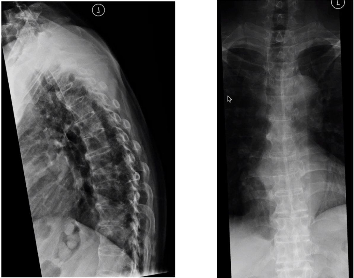
▸ Slide 472 · XR showing T9 collapseSpine Non Trauma · 6 questions expand
Q1-Q66 questions — tap to reveal all answerslist
- What is the initial X-ray finding in this case?
- What history and examination points are important in a patient with a collapsed vertebra?
- What is important about the pain history in a collapsed vertebra?
- What investigations are used for suspected TB spine (T9 collapse)?
- How is an open biopsy of the spine performed?
- What are the indications for surgery in TB spine and what is the medical regimen?
Q1.What is the initial X-ray finding in this case?
- T9 vertebral collapse
Q2.What history and examination points are important in a patient with a collapsed vertebra?
- PMHx and premorbid mobility
- Neurology and sphincter function
- Costal margin pain
- PE: neurological examination
Q3.What is important about the pain history in a collapsed vertebra?
- Nature of pain - rest/nocturnal vs mechanical
- Onset, progression and alleviating factors
Q4.What investigations are used for suspected TB spine (T9 collapse)?
- Routine lymphocytosis
- Bone profile and tumour markers
- Urinary TB and sputum TB
- CT to see the extent of bony destruction
- Tissue biopsy: caseating granuloma, AFB, giant cells
Q5.How is an open biopsy of the spine performed?
- Transpedicular approach
- Costotransversectomy
Q6.What are the indications for surgery in TB spine and what is the medical regimen?
- Indications: debilitating pain, deformity, instability
- Nonoperative: TLSO brace
- DOT: isoniazid, rifampicin, ethambutol, pyrazinamide (intensive phase and continuation phase)
- Follow-up to monitor for complications; MRC trial
▸ Slide 473 · jjSpine Non Trauma · 12 questions expand
Q1-Q1212 questions — tap to reveal all answerslist
- Describe the X-ray findings in spondylodiskitis.
- What are the risk factors and assessment findings in spondylodiskitis?
- What investigations establish the diagnosis and extent of spondylodiskitis?
- What MRI features favour infection over tumour?
- What are the goals of management in spondylodiskitis?
- What are the goals and indications for surgery in spondylodiskitis?
- What is the nonoperative management and expected success rate in spondylodiskitis?
- What is the pathophysiology of spondylodiskitis?
- What is the anatomical distribution of spondylodiskitis?
- Which organisms cause spondylodiskitis and how long are antibiotics given?
- What are the operative approaches and strut graft options in spondylodiskitis?
- What is the role of instrumentation in active spinal infection?
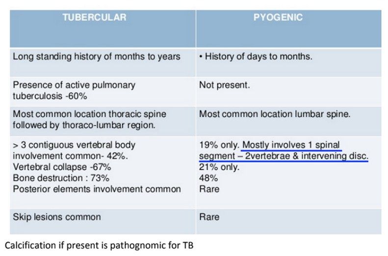
Q1.Describe the X-ray findings in spondylodiskitis.
- L4 lower and L5 upper endplate destruction
- Decreased disc height and decreased lordosis
- Ddx: spondylodiskitis (pyogenic/TB) and malignancy
- Less likely TB because the disc is usually spared
Q2.What are the risk factors and assessment findings in spondylodiskitis?
- Immunocompromised: IVDA, DM, steroids, malnutrition
- Recent systemic infection: UTI, GI, respiratory; recent spine operation
- Neurology and pain
- Femoral stretch and psoas stretch = psoas abscess
- Constitutional symptoms suggest malignancy
Q3.What investigations establish the diagnosis and extent of spondylodiskitis?
- CBC, ESR, CRP
- Blood cultures positive in ~33%; if positive, ~85% accurate for isolating the correct organism
- CT-guided biopsy sent for aerobic, anaerobic, fungal and acid-fast culture
- Contrast MRI: loss of T1 body contrast, T2 disc enhancement, contrast-enhancing paravertebral exudate, epidural/psoas abscess
Q4.What MRI features favour infection over tumour?
- Disc space involvement
- end plate erosion
- Significant inflammation
- Look for epidural abscess, paraspinal collection and psoas abscess
Q5.What are the goals of management in spondylodiskitis?
- Optimise host
- Establish microbiological diagnosis
- Control of infection
- Monitor and prevent neurological deficit, instability and late deformity
Q6.What are the goals and indications for surgery in spondylodiskitis?
- Operative goals: eliminate infection (debridement), prevent/improve neurologic deficits (decompression), maintain stability (instrumentation +/- fusion)
- Failure to obtain microbiological diagnosis from closed needle biopsy (e.g. C spine)
- Poor response to conservative treatment, abscess, neurological deficit
- Extensive disease: 2 adjacent VB or single VB with more than 50% collapse, +/- deformity
Q7.What is the nonoperative management and expected success rate in spondylodiskitis?
- Nonoperative treatment is the mainstay
- IV antibiotics + corset if collapse
- Success rate 60-90%
Q8.What is the pathophysiology of spondylodiskitis?
- Haematogenous seeding of the endplates (low-flow vascular anastomosis) -> direct extension into the disc -> second endplate
- Arterial: diffusion (children/adolescents via intermetaphyseal artery, adults via metaphyseal artery) and septic emboli
- Venous via the Batson plexus
- Contiguous spread from local infection (retropharyngeal and retroperitoneal abscesses)
- Direct inoculation
Q9.What is the anatomical distribution of spondylodiskitis?
- Lumbar spine 60% > thoracic 30% > cervical 10%
Q10.Which organisms cause spondylodiskitis and how long are antibiotics given?
- Staphylococcus aureus and epidermidis; Gram-negative organisms
- E. coli, Pseudomonas in immunocompromised patients
- Salmonella in sickle cell anaemia
- Duration: 6 weeks vs 12 weeks - no difference (Bernard, Lancet 2015)
Q11.What are the operative approaches and strut graft options in spondylodiskitis?
- Single stage: anterior - debridement + reconstruction with strut graft
- Two stage: delayed posterior instrumentation and fusion after anterior debridement
- Epidural abscess: laminectomy
- Strut graft: autogenous tricortical iliac crest, rib or fibula are safe and effective in acute infection
- Titanium mesh cages filled with autograft improved deformity correction (Carragee, Spine 2008), followed by posterior instrumentation)
Q12.What is the role of instrumentation in active spinal infection?
- Spinal instrumentation in active infection is controversial
- Studies show stability of the spine is important for suppression and eventual elimination of infection
- Titanium is preferred over stainless steel
▸ Slide 474 · Gadolinium contrast MRISpine Non Trauma · 2 questions expand
Q1-Q22 questions — tap to reveal all answerslist
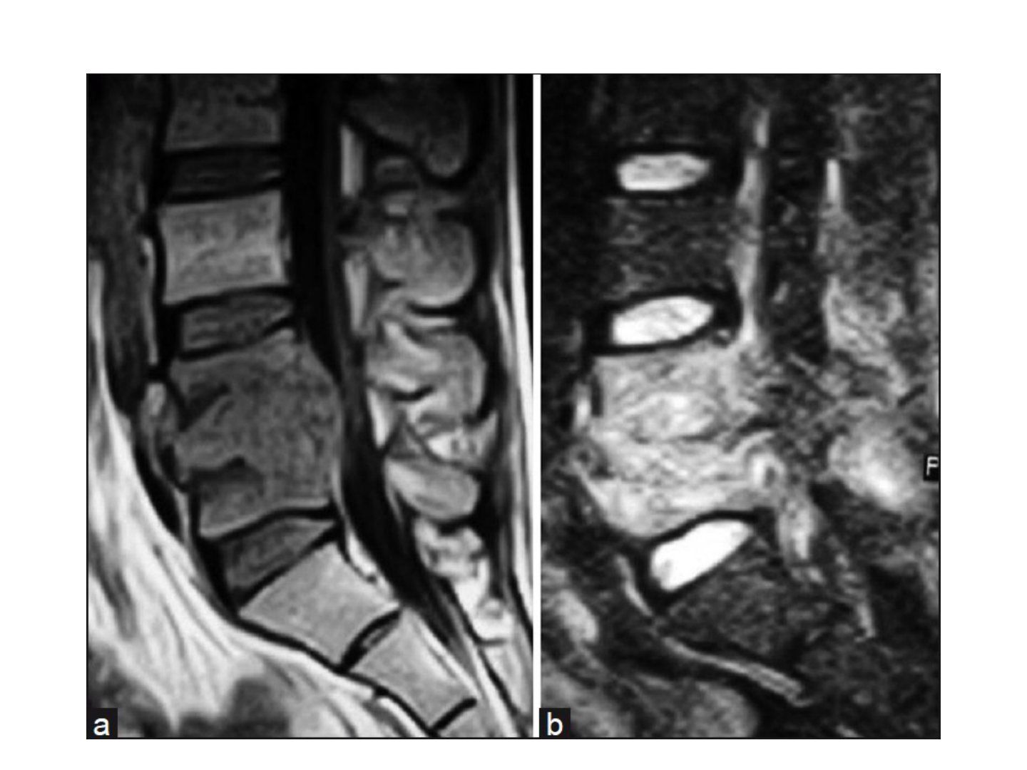
- What is the role of gadolinium contrast MRI in spinal infection?
- How is an epidural abscess managed?
Q1.What is the role of gadolinium contrast MRI in spinal infection?
- Differentiate infection from tumour
- Differentiate epidural abscess from CSF - both are high signal on T2
- Gadolinium enhance pus in T1, CSF remains low signal
Q2.How is an epidural abscess managed?
- Laminectomy and drainage because pus is predominantly posterior
- Anterior and posterior decompression if concomitant vertebral osteomyelitis
▸ Slide 475 · XR:Spine Non Trauma · 14 questions 1 check expand
Q1-Q1414 questions — tap to reveal all answerslist
- What X-ray signs suggest spinal metastasis?
- What history is important in suspected spinal metastasis?
- What examination is needed in suspected spinal metastasis?
- What is the Bilsky ESCC score used for?
- What imaging and classification systems are used for metastatic spine disease?
- What are the treatment goals and principles in metastatic spinal disease?
- What medical treatment is given for metastatic cord compression?
- What is the evidence for surgery plus radiotherapy in metastatic cord compression?
- What is the epidemiology of spinal metastasis?
- What is the difference between osteolytic and osteoblastic metastases?
- How is hypercalcaemia of malignancy managed?
- What are the components and interpretation of the Tokuhashi score?
- What are the components of the Tomita score?
- What is the principle of tumour separation surgery?
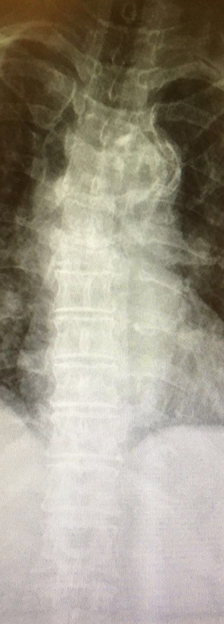
Q1.What X-ray signs suggest spinal metastasis?
- Collapse and kyphosis
- Winking owl sign (pedicle destruction)
- Fusiform swelling
- Lung mass
Q2.What history is important in suspected spinal metastasis?
- Premorbid status
- History of malignancy
- History of HIV, IVDU, travel to endemic countries
- Nature of pain: tumour infiltration, cortical expansion, instability/fracture, neurocompression
- Neurology: direct invasion, epidural seedling, fracture, deformity; also expectations
Q3.What examination is needed in suspected spinal metastasis?
- Full neurological examination
- Systems exam: lungs, abdomen, PR, breast, thyroid
Q4.What is the Bilsky ESCC score used for?
- Grading epidural spinal cord compression on MRI
- 0: bone only
- Ia/b/c: out of bone, deformation of thecal sac, touching cord
- II: cord compression with CSF still visible; III: cord compression with CSF not visible
- Determines high grade (2,3) vs low grade (1) compression
Q5.What imaging and classification systems are used for metastatic spine disease?
- Whole spine MRI with the Bilsky ESCC score
- Weinstein, Boriani, Biagini (WBB) classification
Q6.What are the treatment goals and principles in metastatic spinal disease?
- Treatment is palliative in intent: salvage neurology, stabilise spine, maintain ambulation, pain relief
- Always biopsy if no confirmed bone met
- Look for other spinal/skeletal mets, other primaries and other ddx (infection, TB, benign tumour, MM)
- Manage by the NOMS framework with a multidisciplinary approach
Q7.What medical treatment is given for metastatic cord compression?
- Dexamethasone for neurocompression (50mg loading then 4mg Q6H, with PPI coverage)
- Treat hypercalcemia
- Bisphosphonate can reduce skeletal events: Zometa 4mg IV every month
Q8.What is the evidence for surgery plus radiotherapy in metastatic cord compression?
- Patchell, Lancet 2005: better outcomes with surgery + RT vs RT alone
- Adjunct treatment includes preoperative embolisation for vascular tumours
Q9.What is the epidemiology of spinal metastasis?
- Bone metastasis is common after lung and liver primary
- Spine is the most common site, then proximal femur and humerus (Batson's valveless venous plexus allows seeding like red marrow)
- Primary sites: breast > lung > prostate/renal/thyroid; liver also common
- Survival is variable (6 months to 5 years)
Q10.What is the difference between osteolytic and osteoblastic metastases?
- Osteolytic: osteolysis caused by tumour-induced activation of osteoclasts (RANK, RANKL and OPG)
- Osteoblastic: tumour secreted endothelin 1 (90% prostate, 60% breast)
Q11.How is hypercalcaemia of malignancy managed?
- A metastatic emergency
- Hydration
- Loop diuretics
- Bisphosphonate
Q12.What are the components and interpretation of the Tokuhashi score?
- General condition
- Primary tumour
- Number of vertebral body foci
- Number of extraspinal foci
- Major organ involvement and neurology (palsy)
- Score 0-15; 9-11 predicts >=6 months, >=12 predicts >1 year survival for excisional surgery; PF FONG mnemonic
Q13.What are the components of the Tomita score?
- Primary tumour
- Mets to vital organs (treatable/untreatable)
- Bone mets
- Score 2-10; >6 = palliative decompression/no surgery
Q14.What is the principle of tumour separation surgery?
- Circumferential decompression to allow a safety margin for tumoricidal radiation doses without toxicity to the cord
Dexamethasone 50mg loading dose for metastatic spinal cord compression (slide notes) — non-standard dose, likely error — Guidelines recommend an initial 10mg IV loading dose followed by 4mg (or 6-10mg) every 6 hours; a 50mg bolus is not a recognised regimen — medium confidence — source
▸ Slide 476 · XR: C6 collapse with vertebral plana deformity. Acute kyphosis and increase in vSpine Non Trauma · 6 questions expand
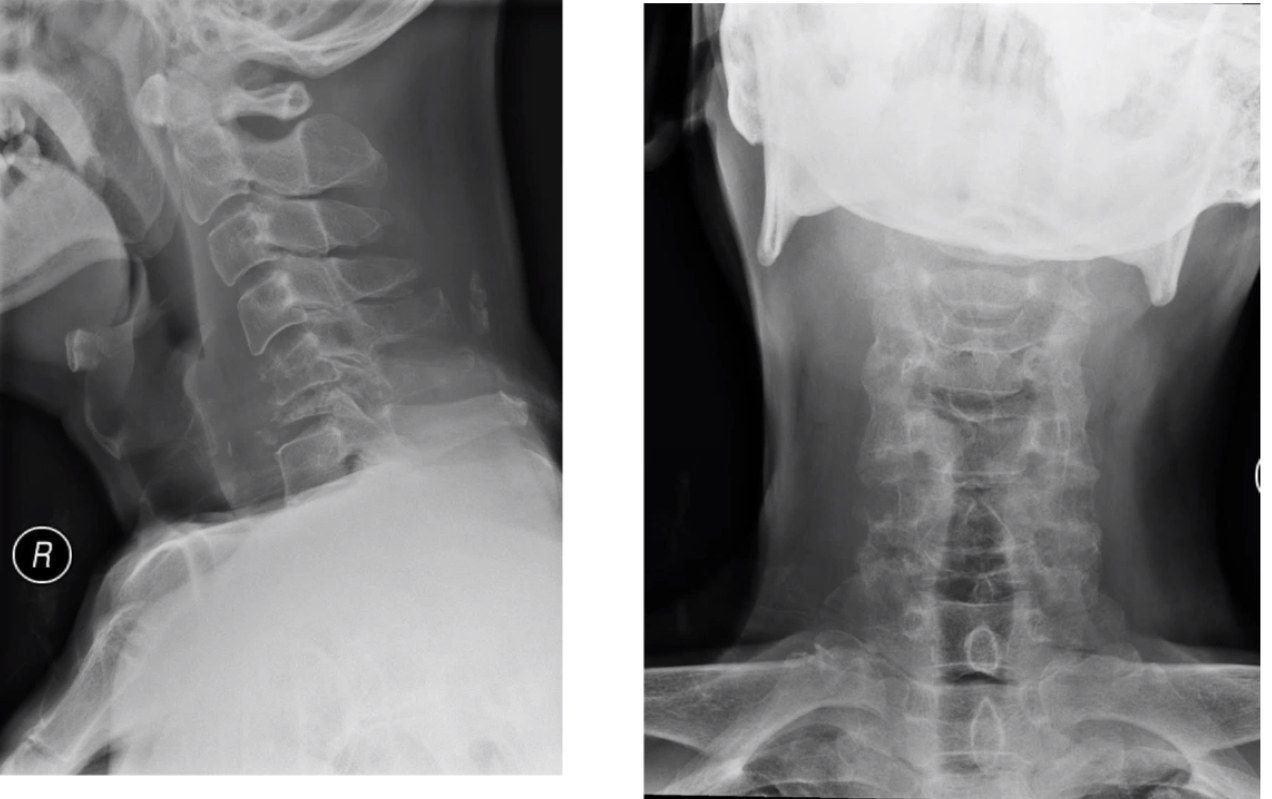
Q1-Q66 questions — tap to reveal all answerslist
- What are the X-ray findings in this case?
- How do you differentiate infection from malignancy on X-ray of the spine?
- What history, examination and blood tests are needed for a C6 collapse?
- What staging is required once malignancy is confirmed?
- What are the four components of the NOMS framework as applied to this case?
- What are the components of the Tokuhashi score?
Q1.What are the X-ray findings in this case?
- C6 collapse with vertebral plana deformity
- Acute kyphosis
- Increase in vertebral shadow
Q2.How do you differentiate infection from malignancy on X-ray of the spine?
- Infection starts in the disc, malignancy in the vertebral body
- More endplate erosion in infection
- Posterior elements less commonly involved in infection
Q3.What history, examination and blood tests are needed for a C6 collapse?
- Red flags, recent infection (e.g. UTI), risk factors for infection, pain, neurology
- Systems screening: breast, abdomen, thyroid, chest, PR
- Local tenderness and neurological examination
- Bloods: WCC, CRP, CaPO4, ALP, tumour markers
Q4.What staging is required once malignancy is confirmed?
- Local and systemic staging
- MRI and CT
- Managed in a multidisciplinary team according to the NOMS framework
Q5.What are the four components of the NOMS framework as applied to this case?
- Neurology: clinical motor deficits; radiological Bilsky stage on MRI - high or low grade cord compression
- Oncologic: radioresistant or radiosensitive tumour
- Mechanical stability: SINS
- Systemic: traditionally Tokuhashi score, now NESM or SORG nomogram
Q6.What are the components of the Tokuhashi score?
- General condition
- Number of spine metastases
- Number of extraspinal metastases
- Internal organ metastases
- Tissue of origin and neurology
- Score 0-15; >9 predicts surgery
▸ Slide 477 · Primary spine tumorsSpine Non Trauma · 3 questions expand
Q1-Q33 questions — tap to reveal all answerslist
- How are benign primary spine tumours classified by Enneking stage?
- What are the malignant primary spine tumours?
- Which other classification systems are used for spine tumours?
Q1.How are benign primary spine tumours classified by Enneking stage?
- Enneking 1 (self-limiting) = osteoid osteoma, ABC, haemangioma
- Enneking 2 (active) = osteoblastoma
- Enneking 3 (aggressive) = giant cell tumour
Q2.What are the malignant primary spine tumours?
- Chordoma
- Osteosarcoma
- Chondrosarcoma
- Ewing's sarcoma (EWS)
Q3.Which other classification systems are used for spine tumours?
- Tomita
- Weinstein, Boriani, Biagini (WBB)
▸ Slide 478 · Posterior approachSpine Non Trauma · 7 questions expand
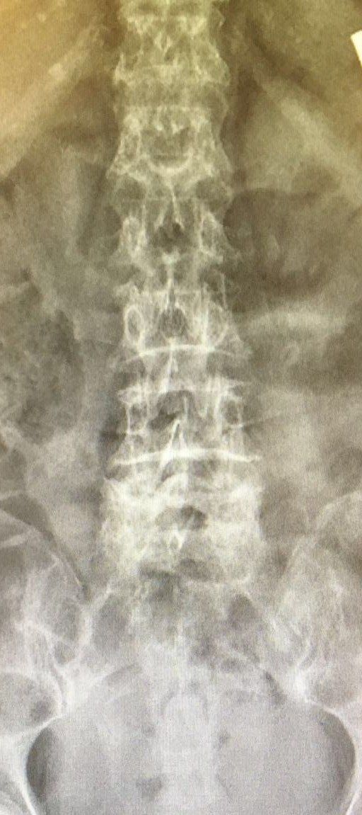
Q1-Q77 questions — tap to reveal all answerslist
- How is the patient positioned for a posterior lumbar approach?
- How is the level confirmed and the incision/dissection performed?
- Describe the decompression steps in a posterior lumbar approach.
- What are the boundaries of Kambin triangle?
- What is the traditional safe triangle for nerve root injection?
- What are the steps of discectomy in the posterior approach?
- What does the 2014 Cochrane review show for minimally invasive versus open discectomy?
Q1.How is the patient positioned for a posterior lumbar approach?
- Prone on an OSI table
- Wilson frame or 4 posters
- Pressure areas well padded
Q2.How is the level confirmed and the incision/dissection performed?
- Surface landmark: intercrestal line = L4/5
- Midline incision along the spinous processes through the lumbar fascia
- Subperiosteal dissection laterally to the facet joints (to the transverse process if fusion, but not beyond the intertransverse ligament)
- Cobb used to strip laterally along the lamina until facet capsules identified but not violated
Q3.Describe the decompression steps in a posterior lumbar approach.
- Remove spinous processes of operative levels with rongeur
- Blunt dissection between the lamina of the level above and the ligamentum flavum with an angled curette to create a working window
- Thin the cephalad lamina with a burr, complete with a Kerrison rongeur to expose the ligamentum flavum, leaving at least 7mm pars
- Extend caudally to the superior most 3mm of the inferior lamina
- Dissect the ligamentum flavum from the medial facet edge and remove; facetectomy (medial 3mm vs total)
Q4.What are the boundaries of Kambin triangle?
- Exiting nerve root
- Medial margin of the superior articular process (SAP)
- Superior endplate of the caudal vertebra
- Also a landmark for nerve root injection
Q5.What is the traditional safe triangle for nerve root injection?
- Inferior margin of the pedicle
- Exiting nerve root
- Lateral margin of the vertebral body
Q6.What are the steps of discectomy in the posterior approach?
- Retract the traversing nerve root
- Epidural vein hemostasis
- Confirm the level with a needle
- Annulotomy with a discectomy knife
- Discectomy
Q7.What does the 2014 Cochrane review show for minimally invasive versus open discectomy?
- MIS: higher leg pain
- Reoperation/readmit rate due to recurrent disc herniation higher (18 vs 13%)
- Lower risk of infection
- Long-term functional and clinical outcomes show no difference
▸ Slide 479Spine Non Trauma · 3 questions expand
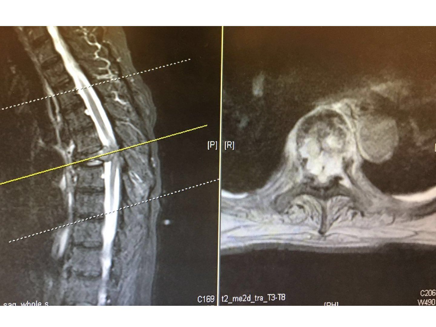
Q1-Q33 questions — tap to reveal all answerslist
- Which MRI study and planes are shown on this slide?
- What abnormality is demonstrated on the axial T2 image?
- What is the purpose of the dashed and yellow lines drawn on the sagittal image?
Q1.Which MRI study and planes are shown on this slide?
- Sagittal T2-weighted MRI of the whole spine (printed label: whole-spine survey)
- Axial T2 MRI labelled T3-T8, cut at the plane marked by the yellow line
Q2.What abnormality is demonstrated on the axial T2 image?
- A discrete rounded mass (intermediate-to-high T2 signal) posterolateral to the thoracic vertebral body
- It lies in the paraspinal/epidural region; the diagnosis is not stated in the speaker notes (read from the slide image)
Q3.What is the purpose of the dashed and yellow lines drawn on the sagittal image?
- The yellow line marks the plane of the axial cut shown on the right
- The dashed white lines mark other reference levels across the whole-spine survey
▸ Slide 480Spine Non Trauma · 3 questions expand
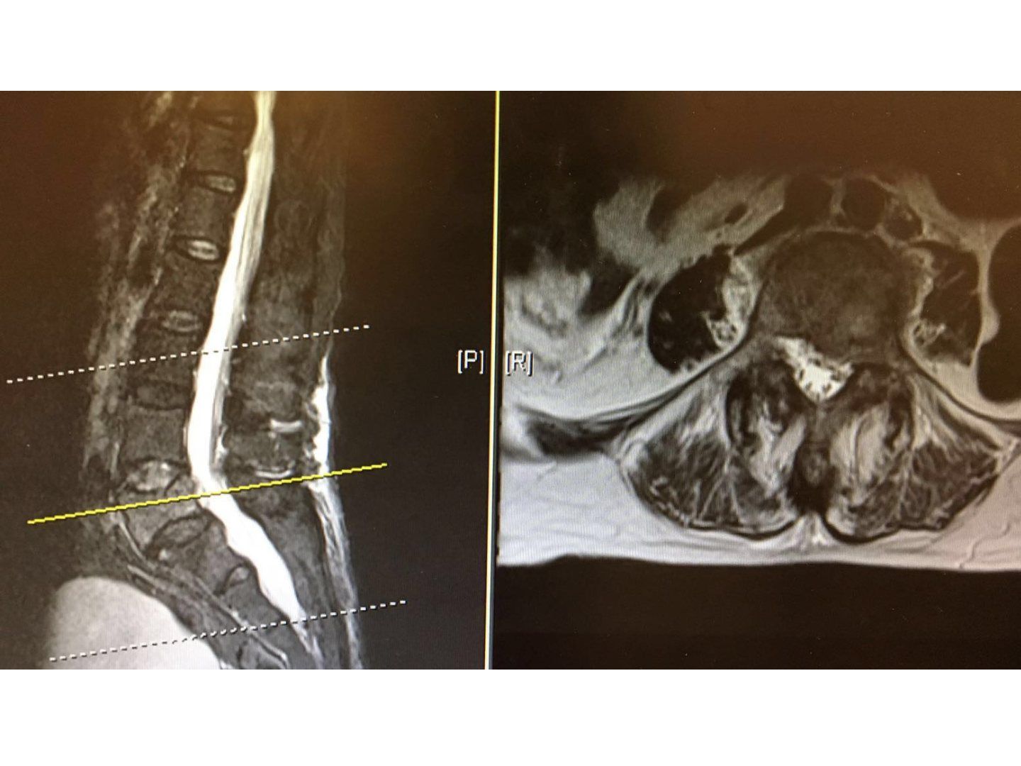
Q1-Q33 questions — tap to reveal all answerslist
- Which spinal region and MRI views are displayed on this slide?
- What normal structures can be identified in the spinal canal on the axial T2 image?
- Why is sagittal imaging combined with an axial image when reporting a lumbar MRI?
Q1.Which spinal region and MRI views are displayed on this slide?
- Sagittal T2-weighted MRI of the lumbosacral spine
- Paired axial T2 image at the level marked by the yellow line
Q2.What normal structures can be identified in the spinal canal on the axial T2 image?
- The canal contains bright (high T2) CSF between the vertebral body and the posterior elements
- Dark dots within the bright CSF are the nerve roots traversing the canal
- Findings are read from the slide image; the speaker notes do not cover this slide
Q3.Why is sagittal imaging combined with an axial image when reporting a lumbar MRI?
- Sagittal shows the level and cranio-caudal extent of disease
- Axial shows cross-sectional relationships to the canal, foramina and nerve roots
▸ Slide 481 · What is fail back syndrome?Spine Non Trauma · 8 questions expand
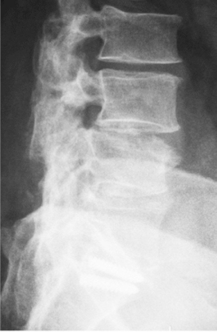
Q1-Q88 questions — tap to reveal all answerslist
- What is failed back syndrome?
- What history and risk factors are important in failed back syndrome?
- What is the 4W+1 approach if pain never resolved after surgery?
- What is the RIPIE differential if pain recurred after initial relief?
- How can symptoms of failed back syndrome be classified?
- How do leg pain and back pain differ in their causes after lumbar surgery?
- What specific postoperative diagnoses can present after lumbar surgery?
- How are failed back syndrome and adjacent segment disease investigated?
Q1.What is failed back syndrome?
- Recurrent or residual back pain following previous surgery on the lumbar spine
- Causes are multifactorial, often the result of poor patient selection
- Contributors: patient (physical/psychological), disease (recurrent disease), surgical (indication/technical)
Q2.What history and risk factors are important in failed back syndrome?
- Initial indication for surgery, preoperative symptoms, perioperative complications
- Postoperative pain relief and current nature of pain; background history
- Risk factors for poor outcome: IOD, smoking, obesity, psychiatric history
Q3.What is the 4W+1 approach if pain never resolved after surgery?
- Wrong patient (Waddell signs - DONTS) or wrong diagnosis
- Wrong indication
- Wrong level
- Wrong procedure done
- Poor surgical technique - inadequate decompression, iatrogenic instability, implant malposition
Q4.What is the RIPIE differential if pain recurred after initial relief?
- Recurrent disease (recurrent disc, adjacent segment)
- Infection
- Pseudoarthrosis
- Instrumentation failure
- Epidural fibrosis
Q5.How can symptoms of failed back syndrome be classified?
- Mechanical symptoms: recurrent disc herniation, discogenic pain, instability, spinal stenosis
- Non-mechanical symptoms: local scar tissue formation (arachnoiditis, epidural fibrosis), psychosocial issues, abdominal/pelvic/systemic disease
Q6.How do leg pain and back pain differ in their causes after lumbar surgery?
- Leg pain: prolapsed intervertebral disc (PID), stenosis
- Back pain: discogenic pain, instability
Q7.What specific postoperative diagnoses can present after lumbar surgery?
- Arachnoiditis: inflammation of the arachnoid layer, no effective treatment
- Epidural fibrosis (3 months): responds poorly to re-exploration
- Diskitis (3-6 weeks postoperatively): rapid onset severe back pain - bedrest, bracing, antibiotics
Q8.How are failed back syndrome and adjacent segment disease investigated?
- Bloods to rule out infection; long film
- CT: fusion status, implant loosening, bone regrowth, incomplete decompression
- MRI: recurrent disc, perineural scar; Scarring can be differentiated from recurrent HNP with a gadolinium-enhanced MRI
- ASD risk factors: obesity, preop disc protrusion/adjacent disc degeneration, poor restoration of sagittal balance, facet joint violation, long segment fusion (incidence 5-19%; JBJS article)
▸ Slide 482Spine Non Trauma · 3 questions expand
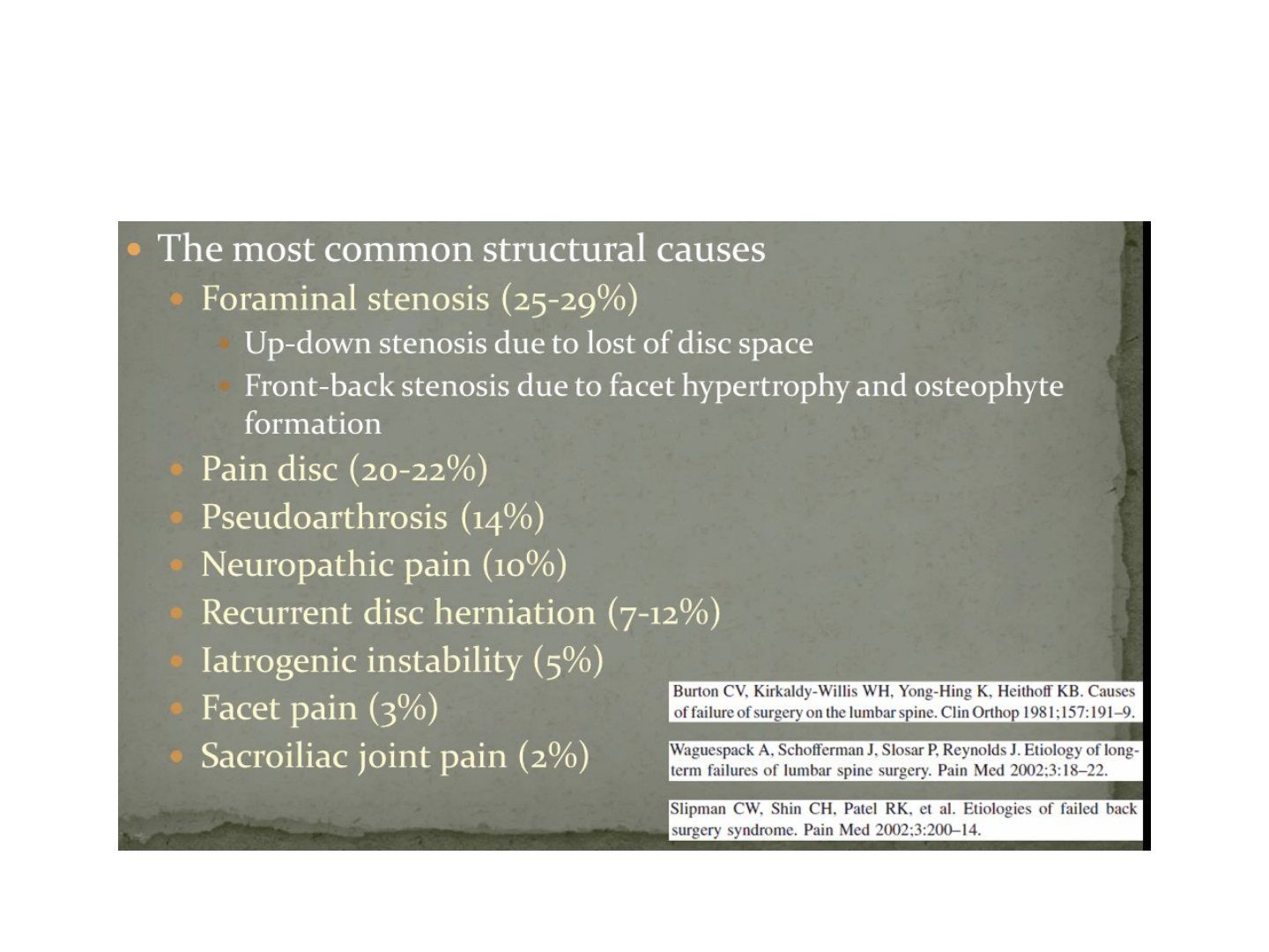
Q1-Q33 questions — tap to reveal all answerslist
- According to this slide, what is the most common structural cause of failed back surgery, and how does it arise?
- What frequency does the slide quote for pain disc, pseudarthrosis and neuropathic pain?
- Which structural causes are listed at 12% or less, and at what rates?
Q1.According to this slide, what is the most common structural cause of failed back surgery, and how does it arise?
- Foraminal stenosis (25-29%) - the single commonest structural cause
- Up-down stenosis from loss of disc space; front-back stenosis from facet hypertrophy and osteophyte formation
Q2.What frequency does the slide quote for pain disc, pseudarthrosis and neuropathic pain?
- Pain disc: 20-22%
- Pseudoarthrosis: 14%
- Neuropathic pain: 10%
Q3.Which structural causes are listed at 12% or less, and at what rates?
- Recurrent disc herniation: 7-12%
- Iatrogenic instability: 5%; facet pain: 3%; sacroiliac joint pain: 2%
▸ Slide 483 · Non organic causes suggested if 3 or above +veSpine Non Trauma · 3 questions expand
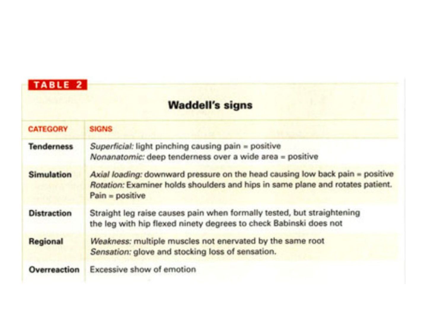
Q1-Q33 questions — tap to reveal all answerslist
- How many positive non-organic signs suggest a non-organic cause?
- List the five non-organic signs.
- Which mnemonic is used for the five non-organic (Waddell) signs?
Q1.How many positive non-organic signs suggest a non-organic cause?
- 3 or more positive signs suggest non-organic causes
Q2.List the five non-organic signs.
- Distraction
- Overreaction
- Non-dermatomal distribution
- Tenderness
- Simulation
Q3.Which mnemonic is used for the five non-organic (Waddell) signs?
- DONTS
- Distraction, Overreaction, Non-dermatomal distribution, Tenderness, Simulation
▸ Slide 484 · Paintbrush/ meniscusSpine Non Trauma · 2 questions expand
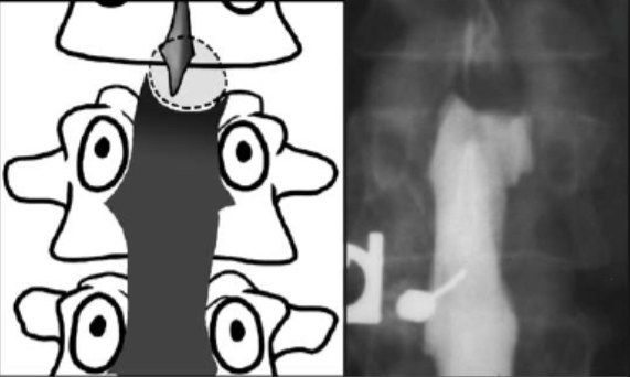
Q1-Q22 questions — tap to reveal all answerslist
- Which two named signs are listed on this slide?
- What do these signs represent?
Q1.Which two named signs are listed on this slide?
- Paintbrush
- Meniscus
Q2.What do these signs represent?
- Not covered in the speaker notes
▸ Slide 485 · MyelogramSpine Non Trauma · 3 questions expand
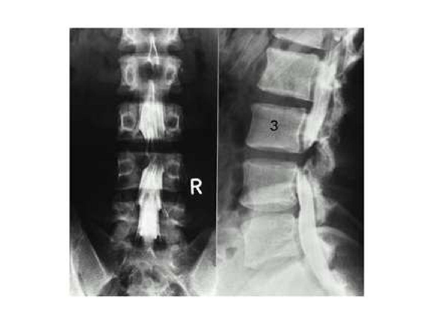
Q1-Q33 questions — tap to reveal all answerslist
- How is a myelogram performed?
- How do you interpret an extradural space-occupying lesion on myelogram?
- How do you interpret an intradural, extramedullary lesion on myelogram?
Q1.How is a myelogram performed?
- Under fluoroscopy
- Spinal needle at L3/4, withdraw CSF, inject contrast
- Table tilt to allow contrast to fill up and down
- Then XR/CT
Q2.How do you interpret an extradural space-occupying lesion on myelogram?
- Examples: PID, metastasis, TB abscess
- Paintbrush appearance
Q3.How do you interpret an intradural, extramedullary lesion on myelogram?
- Examples: neurofibroma, meningioma
- Meniscal sign (can outline the SOL)
▸ Slide 486 · PI is the angle formed from two lines. One from centre of sacrum to femoral headSpine Non Trauma · 3 questions expand
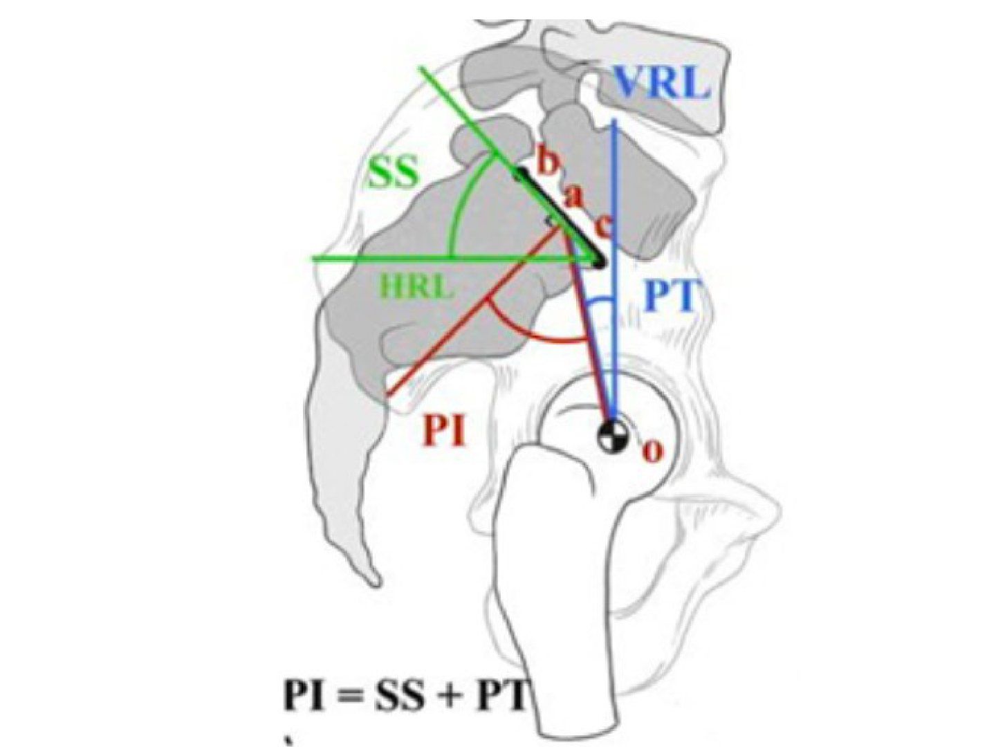
Q1-Q33 questions — tap to reveal all answerslist
- How is pelvic incidence (PI) measured?
- What is the relationship of pelvic incidence to spondylolisthesis?
- How does the body compensate when lumbar lordosis reduces?
Q1.How is pelvic incidence (PI) measured?
- Angle between a line from the centre of the sacrum to the femoral head and a line perpendicular to the S1 endplate through its midpoint
- PI = PT + SS
Q2.What is the relationship of pelvic incidence to spondylolisthesis?
- Direct relationship to the severity of spondylolisthesis
- High PI: more shearing across L5/S1 and less lumbar lordosis
- Body tries to keep sagittal balance by increasing lumbar lordosis
Q3.How does the body compensate when lumbar lordosis reduces?
- Pelvis rotates posteriorly, reducing the sacral slope
- When the sacrum is vertical, the knee flexes to further compensate
▸ Slide 487 · Diagram showing measurements for spinal sagittal balanceSpine Non Trauma · 4 questions expand
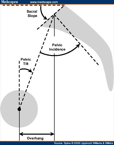
Q1-Q44 questions — tap to reveal all answerslist
- How is sagittal vertical axis (SVA) measured?
- What is the relationship between sacral slope, pelvic tilt and pelvic incidence?
- What are the target values for spinopelvic harmony?
- Why does sagittal imbalance matter?
Q1.How is sagittal vertical axis (SVA) measured?
- On standing long film X-ray
- Line from the centre of C7 vertebral body perpendicular to the ground; measure horizontal distance from the posterosuperior corner of S1
- Normal = within 5cm
Q2.What is the relationship between sacral slope, pelvic tilt and pelvic incidence?
- Sacral slope + pelvic tilt (positional) = pelvic incidence (morphology)
Q3.What are the target values for spinopelvic harmony?
- Lumbar lordosis = PI +/- 9 degrees
- PT < 25
- SVA < 50mm
- Spinal curvature (spinopelvic harmony / cone of economy)
Q4.Why does sagittal imbalance matter?
- Imbalance leads to pain and progressive deformity
▸ Slide 488 · When to use this table?: Think of the following question.Spine Non Trauma · 3 questions expand
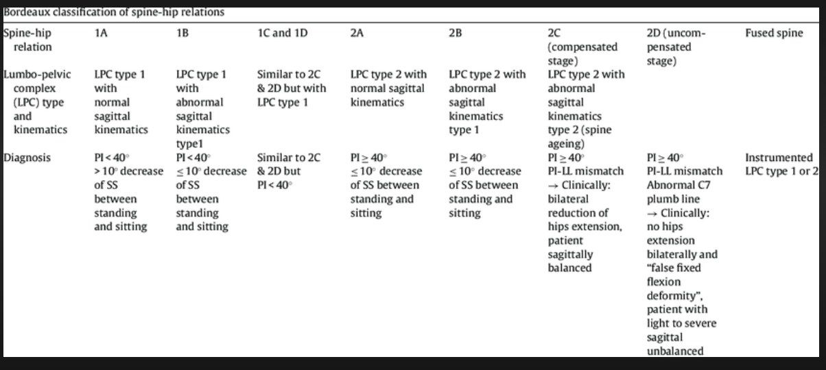
Q1-Q33 questions — tap to reveal all answerslist
- What clinical question is the Bordeaux classification used to answer?
- What determines the type in the Bordeaux classification?
- How does the Bordeaux classification guide implant choice?
Q1.What clinical question is the Bordeaux classification used to answer?
- How to decide acetabular position/implant in a patient with spinal deformity
Q2.What determines the type in the Bordeaux classification?
- Type of lumbo-pelvic complex (LPC): type 1 = PI <40, type 2 = PI >40
- Then subclassified A/B/C/D
Q3.How does the Bordeaux classification guide implant choice?
- Increasing constraint required for types B, C and D
- Based on LPC type: PI <40 (type 1) or PI >40 (type 2)
▸ Slide 489 · SRS (scoliosis research society) Schwab classification for Adult Spine DeformitySpine Non Trauma · 3 questions 1 check expand
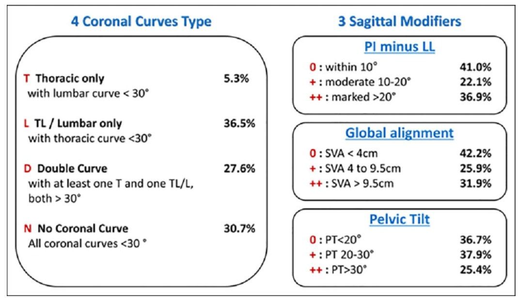
Q1-Q33 questions — tap to reveal all answerslist
- What is the SRS (Schwab) classification used for?
- What does the Ascani 1986 natural history study report for adult scoliosis?
- What features indicate a poor prognosis in adult spine deformity?
Q1.What is the SRS (Schwab) classification used for?
- Classification for adult spine deformity (Scoliosis Research Society)
- Includes parameters relevant to OLIF/degenerative scoliosis surgery
Q2.What does the Ascani 1986 natural history study report for adult scoliosis?
- 3 degrees per year progression (per slide notes)
- All curves increase after skeletal maturity
Q3.What features indicate a poor prognosis in adult spine deformity?
- >30 degree Cobb angle
- >30% apical rotation
- 6mm lateral listhesis
- Poor quality bone
Ascani 1986 Spine natural history study: adult scoliosis progresses 3 degrees per year — mismatch with cited source — Ascani 1986 reported an average progression of 0.4 degrees per year after skeletal maturity; most curves progress at less than 1 degree per year, though larger curves progress faster — source
▸ Slide 490 · SDSG L5-S1 slip classificationSpine Non Trauma · 3 questions expand
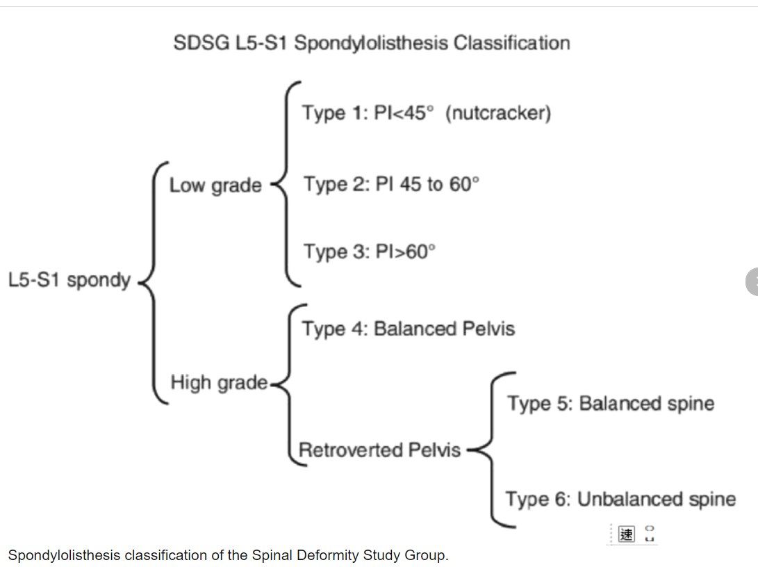
Q1-Q33 questions — tap to reveal all answerslist
- What is the SDSG L5-S1 slip classification used for?
- How are high and low grade slips defined in the SDSG classification?
- What is the management principle for high grade dysplastic slip?
Q1.What is the SDSG L5-S1 slip classification used for?
- The current recommended simple classification for dysplastic slip
- Balanced pelvis = low pelvic tilt, high sacral slope
Q2.How are high and low grade slips defined in the SDSG classification?
- High grade = >50% slip
- Low grade = <50% slip
Q3.What is the management principle for high grade dysplastic slip?
- Consider partial reduction in high grade slip
- Restore normal lumbosacral/pelvis relationship
▸ Slide 491 · Sacral agenesisSpine Non Trauma · 4 questions expand
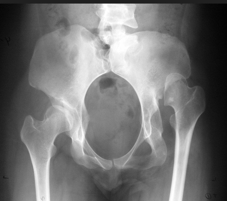
Q1-Q44 questions — tap to reveal all answerslist
- What is sacral agenesis associated with?
- What is the Renshaw classification of sacral agenesis?
- How is Renshaw type 1/2 sacral agenesis managed?
- How is Renshaw type 3/4 sacral agenesis managed?
Q1.What is sacral agenesis associated with?
- Rare; associated with maternal diabetes
- Part of caudal regression syndrome
- Gastrointestinal disorders e.g. imperforate anus
- Genitourinary and cardiovascular disorders
- Lower extremity deformities and progressive kyphosis
Q2.What is the Renshaw classification of sacral agenesis?
- Type I: unilateral
- Type II: partial, bilaterally symmetrical
- Type III: ilium articulating with vertebra present
- Type IV: fused both ilia or iliac amphiarthrosis
Q3.How is Renshaw type 1/2 sacral agenesis managed?
- Conservative physical therapy for Renshaw type 1/2
- Most become community ambulators
- Foot and knee deformities to be addressed
Q4.How is Renshaw type 3/4 sacral agenesis managed?
- Operative management for Renshaw type 3/4
- Spinal stabilization procedures for progressive kyphosis and/or scoliosis
- Limb amputation for non-functional lower limb deformities
▸ Slide 492 · Bartolotti syndromeSpine Non Trauma · 4 questions expand

Q1-Q44 questions — tap to reveal all answerslist
- What is the Castellvi classification of Bertolotti syndrome?
- Which Castellvi type is associated with lumbar disc herniation?
- What are the reported rates of sacralization and lumbarization?
- In the Castellvi classification, what do the suffixes a and b mean?
Q1.What is the Castellvi classification of Bertolotti syndrome?
- Type I: enlarged transverse process >= 19 mm (a unilateral, b bilateral)
- Type II: pseudoarticulation of transverse process and sacrum (a/b)
- Type III: transverse process fuses with sacrum (a/b)
- Type IV: type IIa on one side and type IIIa contralateral
Q2.Which Castellvi type is associated with lumbar disc herniation?
- Type II
- Not associated with types I, III or IV
Q3.What are the reported rates of sacralization and lumbarization?
- Sacralization: 7%
- Lumbarization: 5%
Q4.In the Castellvi classification, what do the suffixes a and b mean?
- a = unilateral
- b = bilateral
▸ Slide 493 · Cervical cord injury rehabSpine Non Trauma · 7 questions expand

Q1-Q77 questions — tap to reveal all answerslist
- What is the ventilator weaning success rate after cervical spinal cord injury?
- Why does vital capacity decrease on sitting in cervical cord injury?
- What is the motor recovery prognosis after traumatic spinal cord injury?
- What is the ambulatory potential by ASIA grade?
- What are the functional goals at C5 and C6?
- How does sphincter function differ in suprasacral vs cauda equina lesions?
- Which 1999 consensus published the traumatic spinal cord injury rehabilitation potential data?
Q1.What is the ventilator weaning success rate after cervical spinal cord injury?
- C3: 51%
- C4: 78%
Q2.Why does vital capacity decrease on sitting in cervical cord injury?
- Increased dead space as the abdominal organs drop down when sitting
- Can be improved with a corset
Q3.What is the motor recovery prognosis after traumatic spinal cord injury?
- Muscles with some motor power below an antigravity muscle have a better prognosis than muscles with no motor power
- Grade 1 or 2: 90% reach grade 3 by 1 year
- Grade 0: 64% reach grade 3 by 2 years
- Recovery is faster in incomplete injuries
- Median time to antigravity strength: 2 months motor complete vs 2 weeks motor incomplete (Ditunno 1992)
Q4.What is the ambulatory potential by ASIA grade?
- 3% / 50% / 75% / 95% in ASIA A / B / C / D (van Middendorp, Global Spine J 2011)
Q5.What are the functional goals at C5 and C6?
- C5: use of power wheelchair
- C6: manual wheelchair, feeding well
- Consider tenodesis grasp/transfer at C6
Q6.How does sphincter function differ in suprasacral vs cauda equina lesions?
- Suprasacral: detrusor-sphincter dyssynergia, spastic bladder, reduced volume, may develop reflux/HN, finally incontinence
- Cauda equina: flaccid bladder that stretches out, can develop stress incontinence
Q7.Which 1999 consensus published the traumatic spinal cord injury rehabilitation potential data?
- Consortium of Spinal Cord Medicine 1999