Spine
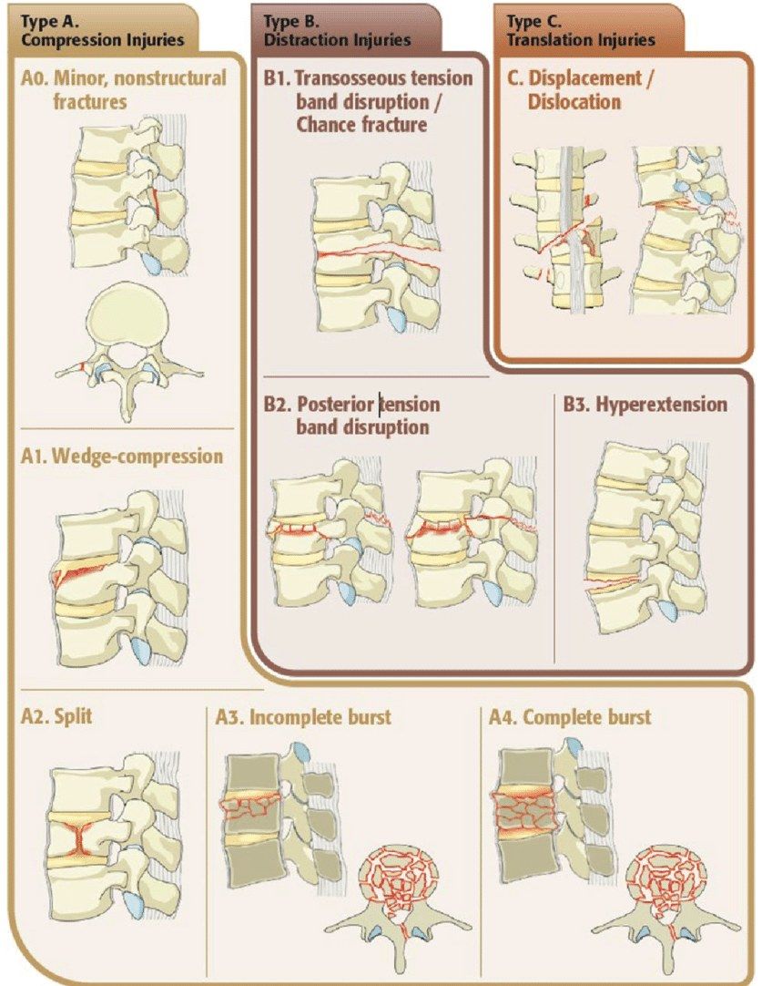
Thoracolumbar trauma - burst and chance fractures
Thoracolumbar junction vulnerability, burst and chance fracture management with radiograph features
29 questions
4 source pages
1 images
1 fact-check flags
Images appear with the first question taken from each source page — tap a question to open it.
Q1Describe the X-ray findings and classification of this L1 burst fracture.▸
- Anterior wedging of L1, some retropulsion, widening of interpedicular space
- Assess for posterior element widening/fracture on CT and MRI
- AO A3 or A4; Denis 2 column fracture
Q2What is the initial management of a thoracolumbar burst fracture?▸
- High energy trauma - manage according to ATLS
- C spine immobilisation + pelvic binder; primary and secondary survey
- Assess neurology
- CT and MRI (whole spine) - no posterior element involvement, no nerve compression
Q3What is the TLICS and how is it scored?▸
- Thoracolumbar injury classification system
- Calculate the TLICS for further mx
- Injury morphology (1-4), neurology (0-3), PLC injury (0-3)
- Provide scoring (1-10, </=3 conservative, >/= 5 operative )
- Total score 1-10; 3 or less conservative, 5 or more operative
Q4What are the indications for surgery in a thoracolumbar burst fracture?▸
- TLICS score 5 or higher
- Neurological deficit needing decompression
- Unstable pattern: injury to the posterior ligament complex (PLC), progressive kyphosis
- 30 deg kyphosis (controversial)
- 50% loss of vertebral body height (controversial)
- 50% canal compromise (controversial)
Q5What is the timing and approach, and why is posterior chosen?▸
- Early surgery (within 48hrs) if neurocompromise - Fehlings 2021 Lancet
- Posterior decompression + instrumentation from T10 to L4 +/- fusion
- Posterior: easier/familiar, shorter OT, less blood loss; If PLC disrupted, I can keep the intact ALL, avoiding further destabilisation of the spine; ligamentotaxis; three-column purchase; later implant removal avoids fusion
- Anterior if McCormack and Gaines >6, disc pathology, poor bone, chronic fracture, pincer fracture
Q6What is the McCormack and Gaines load sharing classification?▸
- Comminution (<30, 30-60, >60%)
- Apposition of fragments (minimal, <50%, >50%)
- Reducibility of sagittal deformity (<=3, 4-9, >=10 deg)
- >6/9 = add anterior approach
Q7What is the aim of management in a thoracolumbar burst fracture?▸
- Relieve pain
- Prevent neurological deficit and deformity
- Allow early mobilisation
Q8When is conservative management appropriate for a thoracolumbar burst fracture?▸
- No neurology, relatively stable (PLC intact), acceptable deformity (size of anterior collapse)
- Protect with a brace
Q9What are the indications for decompression in a thoracolumbar burst fracture?▸
- Neurological deterioration
- When deformity correction is needed (e.g. shortening osteotomy)
- Indirect decompression by ligamentotaxis, or facetectomy for the retropulsed fragment +/- expandable cage; directly deal with an entrapped nerve root
Q10How is reduction achieved and what counts as good reduction?▸
- Distraction restores vertebral height and tenses the PLL to reduce the retropulsed fragment
- Lordolisation by cantilever rod reduction corrects deformity
- Good reduction: vertebral height restored, lumbar lordosis restored
Q11What instrumentation is used and what determines the number of levels?▸
- Posterior pedicle-rod system able to engage all three columns
- Levels depend on fracture site, fracture age, deformity to correct and bone quality
- USS (universal spine system) reduction tools help lordolisation
Q12When is fusion performed and why must implant removal be planned?▸
- Not fusing preserves the mobile segment
- Fuse if decompression destabilises the spine
- Plan removal of instrumentation, otherwise fatigue fracture or loosening
- Rehab: TLSO, FWB, DVT prophylaxis
Q13Why is the thoracolumbar junction prone to injury?📷▸
- Transition from kyphosis to lordosis
- Transition from rigid to mobile - more shearing
- Smaller vertebral body compared with the lower lumbar spine
- Increase bending moment
- Facet orientation changes from coronal to sagittal
Q14Describe the X-ray findings and diagnosis of this Chance fracture.▸
- Lateral: L1 fracture involving all three columns, vertebral height loss >50%, kyphosis >30 deg
- Mechanism RTA - Chance fracture: fulcrum anterior, distraction from posterior to anterior column, tensile failure of all three columns
- AP: widening of interpedicular distance
- AO B1, Denis three column fracture
Q15What associated injuries must be assessed in a Chance fracture?▸
- ATLS, prevent secondary cord injury; spine precaution, bed rest
- PR + neuro examination
- Palpate abdomen (intraabdominal injury 40%) + assess LL pulses; bloods, urine for blood
- XR whole spine (non-contiguous fracture in 20%)
- CT for canal compromise by retropulsed fragment
- MRI not absolutely indicated: neuro compromise, suspected soft tissue Chance, high risk epidural haematoma (e.g. AS)
Q16What is the principle and timing of surgery?▸
- Principle: pain relief, stabilise the spine for early mobilisation, prevent neurological deficits and future deformity
- Timing: early (within 24-48hrs) if neurocompromise - Fehlings 2021 Lancet
Q17What determines the approach in a Chance fracture?▸
- Consider neurology and PLC
- Posterior: easier/familiar, shorter OT, less blood loss; keeps intact ALL if PLC disrupted; later implant removal avoids fusion - but decompression may be inadequate
- Anterior indications: severe collapse and kyphosis needing height restoration (McCormack and Gaines), disc pathology, poor bone quality, chronic fracture, failure of posterior surgery
Q18How is reduction achieved and what is good reduction?▸
- Reduction by ligamentotaxis - needs to be done within 5-6 days
- Distraction restores vertebral height and tense up PLL to reduce the retropulsed fragment; lordolisation by cantilever rod reduction
- Good reduction: vertebral height restored, lumbar lordosis restored
- Fusion: not fuse, preserve mobile segment; fuse if decompression destabilises the spine; tend to fuse in soft tissue Chance as ligamentous healing is unpredictable
Q19What are the implications of a lamina fracture?▸
- Dural tear
- Nerve root injury
- Need posterior decompression
Q20What are the decompression and instrumentation options in a Chance fracture?▸
- Decompression for neurological deterioration or when deformity correction (shortening osteotomy) is needed
- Indirect decompression by ligamentotaxis, or facetectomy +/- expandable cage for the retropulsed fragment; Directly deal with entrapped nerve root if present
- Posterior pedicle screw-rod engages all three columns; levels depend on fracture site, age, deformity, bone quality
- Anterior: bone graft, cement or expandable cage (transpedicular from posterior)
Q21What rehabilitation and implant planning follow surgery for a Chance fracture?▸
- Plan removal of instrumentation, otherwise fatigue fracture or loosening
- Rehab: TLSO, FWB
- Fusion not routine - preserve the mobile segment; fuse if decompression destabilises, and soft tissue Chance tends to be fused
Q22Describe the X-ray findings in this subaxial cervical injury.▸
- Lateral C spine with exposure adequate C1-T1
- Kyphotic alignment at C5/6; C5/6 anterior translation <25% with angulation and disruption of 4 lines
- Loss of facet stacked parallelogram; C5 perched on C6; +/- bow tie sign
- Prevertebral soft tissue swelling; no obvious OC dissociation
- AP: upper spinous processes displaced towards the side of dislocation
- 10% of patients have a fracture elsewhere
Q23What is the initial management of a perched facet injury after high energy trauma?▸
- ATLS - ABC, primary and secondary survey
- Protect whole spine, neck collar, spinal board; rule out other spine fractures; watch for neurogenic shock
- Assess GCS and neurology
- CT to look for fractures, may need to direct OR
- Pre-CR MRI if obtunded, or no neuro deficit + conscious
- MRI looks for large PID, transdiscal injury, epidural haematoma, PLC, adjacent level injury
Q24Will you perform urgent closed reduction and what are the contraindications?▸
- Prerequisite of safe CR is a fully conscious and cooperative patient
- Order urgent MRI before reduction to r/o PID (bilateral facet dislocation: PID incidence 10-40%)
- CR in the presence of PID causes further neurological deterioration that may be irreversible
- Contraindications: PID, OC dissociation, fracture dislocation, skull fracture
- PID -> straight to OT for anterior discectomy; no PID -> closed reduction under X-ray guidance
Q25How do you perform closed reduction?▸
- Controlled sequential traction with monitoring of X-ray and neurology; fluoroscopy suite, reverse trendelenberg position (reverse Trendelenburg) with head spoon
- Insert halo ring (3 ppl minimum, i.e. 3 people minimum, 1-2cm clearance); anterior pins at lateral 1/3 and 1cm above eyebrow (insert with eye closed), posterior pins above mastoid process; tighten 8 pound-inch
- Traction in flexion using a towel under the occiput
- Start 5kg (~weight of head), then add 5-10lb every 30min; min weight = 10lb (5kg) for head + 5lb (~2kg) per level (Crutchfield 1954); max = min x 2
- Monitor X-ray for disc space widening/OC dissociation and check neurology each time; additional manoeuvres: unilateral - rotate 30 deg to side; bilateral - anterior directed force
- Once facet out of perched position, extend neck and decrease to minimal maintaining weight; end points: neuro deficit/pain/decrease GCS, excessive distraction, max weight, successful reduction
Q26After successful closed reduction, what are your management options?▸
- No neurology + unilateral facet dislocation: can treat conservatively
- Persistent neurological deficit after reduction: posterior decompression + instrumentation
- Bilateral facet dislocation: gross posterior ligament destruction - circumferential fixation
- PID: anterior discectomy first, indirect reduction with Caspar pins (max 1cm); if fails, posterior reduction then ASF; low threshold for 360 deg fusion
- Postop: neck collar, sit out and walk as tolerated, DVT and pressure ulcer prophylaxis
Q27What is the Allen and Ferguson classification of subaxial cervical injuries?▸
- Flexion-distraction - facet dislocation
- Flexion-compression - large anterior (flexion type) teardrop; unstable, needs corpectomy + ASF
- Extension-distraction - small anterior (extension type) teardrop
- Extension-compression - facet fracture
- Vertical compression - burst fracture
- Lateral flexion
Q28What are the pros and cons of anterior versus posterior subaxial cervical surgery?▸
- Anterior adv: fusion in compression; saves fusion levels; direct decompression for burst fracture or PID
- Anterior disadv: posterior tension band not restored; difficult to reduce complex facet dislocation
- Posterior adv: preserves mobility (implant removal after healing); restores tension band; easier exposure; stronger construct (pedicle screw > lateral mass screw)
- Posterior disadv: VA injury (pedicle/lateral mass); cord injury (pedicle screw); difficult to correct deformity; indirect decompression
Q29What is the halo pin retightening schedule after insertion?▸
- Retighten pins after 24hrs, 3 days then weekly (AO guideline)
Fact check
Crutchfield rule: minimum weight = 10lb (5kg) for the head plus 5lb (~2kg) per level, and maximum weight = minimum x 2 — misleading/inverted — Crutchfield's 'rule of fives' is usually cited as a MAXIMUM: 10lb for the head + 5lb per level (e.g. C4/5 = 30lb). Traction is typically started at 5-10lb and increased in 5-10lb increments with serial X-ray and neurological checks — (medium confidence) — source