▸ Slide 419 · SpineSpine · 2 questions expand
Q1-Q22 questions — tap to reveal all answerslist
- What spinal topic does this slide cover?
- What are the key learning points for this slide?
Q1.What spinal topic does this slide cover?
- Slide title is Spine
- Not covered in the speaker notes
Q2.What are the key learning points for this slide?
- Not covered in the speaker notes
- No answers derivable from the slide title alone
▸ Slide 420 · C spine ImmobilizationSpine · 3 questions 1 check expand

Q1-Q33 questions — tap to reveal all answerslist
- What are the degrees of immobilization provided by a rigid collar, SOMI and halo?
- Which cervical orthosis provides the greatest immobilization?
- What are the complications of cervical spine immobilization?
Q1.What are the degrees of immobilization provided by a rigid collar, SOMI and halo?
- Rigid collar: F/E 70%, LF/R 30% (7-3-3)
- SOMI: F/E 80%, LF 60%, R 30% (8-6-3)
- Halo: F/E, LF, R 90% (9-9-9)
Q2.Which cervical orthosis provides the greatest immobilization?
- Halo provides the greatest immobilization
- F/E, LF, R 90% (9-9-9)
Q3.What are the complications of cervical spine immobilization?
- Skin problems
- Compliance
- Increase ICP
- Mandibular palsy
- Aspiration
A rigid collar restricts lateral flexion/rotation by only 30%, and halo immobilization is 90% in all planes — contested/imprecise — Reported restriction varies widely with collar design and study; classic data on the Philadelphia collar show ~70% flexion-extension restriction but ~44% lateral bending and ~66% rotation, and halo values ~90% or more — medium confidence — source
▸ Slide 421 · Blood supply to spinal cordSpine · 4 questions expand

Q1-Q44 questions — tap to reveal all answerslist
- What are the vertical arteries supplying the spinal cord?
- What are the segmental and radicular arteries of the spinal cord?
- What is the artery of Adamkiewicz?
- Describe the venous drainage of the spinal cord.
Q1.What are the vertical arteries supplying the spinal cord?
- One anterior and two posterior spinal arteries
- Arise from the vertebral artery/PICA
- Form the vertical arteries that run along the length of the spinal cord
Q2.What are the segmental and radicular arteries of the spinal cord?
- Segmental arteries: CS from VA, TS from posterior intercostal artery, LS from lumbar and iliac artery
- Enter via intervertebral foramen and divide into posterior and anterior radicular arteries
- Follow the anterior and posterior rami of each nerve root; anastomose with the ASA and PSA
Q3.What is the artery of Adamkiewicz?
- A segmental medullary artery coming directly off the segmental artery to anastomose with ASA/PSA
- From left posterior intercostal artery; 60% left
- T9-L2, supplies lower 2/3 of spinal cord; joins the ASA
Q4.Describe the venous drainage of the spinal cord.
- Anterolateral and posterolateral spinal veins (x4); 1 anterior median vein and 1 posterior median spinal vein
- Drain into radicular veins --> internal vertebral venous plexus
- --> external vertebral venous plexus
- --> ascending lumbar veins and azygous system (right side)
▸ Slide 422 · ApproachSpine · 5 questions expand
Q1-Q55 questions — tap to reveal all answerslist
- What are the indications for the anterolateral/retroperitoneal approach?
- How is the patient positioned and the incision placed for the anterolateral approach?
- Describe the dissection of the anterolateral/retroperitoneal approach.
- What are the risks of the anterolateral/retroperitoneal approach?
- Which spinal levels can be approached anteriorly, and what are the level-specific caveats?
Q1.What are the indications for the anterolateral/retroperitoneal approach?
- Infection, especially TB
- Delayed presentation of retropulsed VB
- Scoliosis
- Upper thoracic spine: consider costotransversectomy approach
Q2.How is the patient positioned and the incision placed for the anterolateral approach?
- Left side up (aorta side; IVC is fragile)
- Break table centering at level of interest
- Incision from 12th rib posterior half to rectus lateral border, centred at target VB
Q3.Describe the dissection of the anterolateral/retroperitoneal approach.
- Split EOM; divide IOM in line with incision
- Split transversus abdominus and enter preperitoneal plane
- Excise distal 5cm of 12th rib for better exposure
- Elevate peritoneum from psoas fascia (ureter to anterior with peritoneum); follow psoas to VB AL border
- Ligate segmental lumbar artery of concerned level close to aorta + iliolumbar vein
Q4.What are the risks of the anterolateral/retroperitoneal approach?
- Genitofemoral nerve
- Sympathetic chain --> CPRS
- Superior hypogastric plexus --> retrograde ejaculation
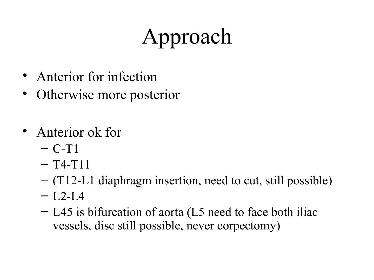
Q5.Which spinal levels can be approached anteriorly, and what are the level-specific caveats?
- Anterior approach for infection; otherwise more posterior
- Anterior OK for C-T1 and T4-T11
- T12-L1: at the diaphragm insertion --> diaphragm needs to be cut, approach still possible
- L2-L4 anterior OK
- L4-5: level of the bifurcation of the aorta
- L5: must face both iliac vessels --> disc still possible, never corpectomy
▸ Slide 423 · Left sided thoracolumbar junction approach (T10-L2)Spine · 3 questions expand
Q1-Q33 questions — tap to reveal all answerslist
- How is the skin incision planned for the left sided thoracolumbar junction approach?
- Describe the initial exposure for the left thoracolumbar junction approach.
- How are the diaphragm and spine exposed in the thoracolumbar approach?
Q1.How is the skin incision planned for the left sided thoracolumbar junction approach?
- Centred over fracture vertebra over a rib, from posterior proximal 2 levels above
- Rib selected should be 2 levels above the fractured vertebra (rib curves down)
Q2.Describe the initial exposure for the left thoracolumbar junction approach.
- Chest muscles overlying rib and abdominal muscle distal to costal cartilage incised with cautery; retract lat dorsi posteriorly
- Subperiosteal dissection of rib then rib resection (beware NV bundle at caudal edge)
- Incise parietal pleura
- Split the three muscle layers of the abdominal wall anteriorly and expose retroperitoneal cavity
Q3.How are the diaphragm and spine exposed in the thoracolumbar approach?
- Sweep peritoneum off inferior surface of diaphragm; take down to spine leaving 1cm margin for later attachment (access T12/L1)
- Cut diaphragm at the periphery to avoid denervation
- Lung retracted anteriorly (one lung ventilation) to expose spine
- Open parietal pleura longitudinally over fracture level: begin dissection at the hills (IVD) to avoid segmental vessels at the valleys
- Ligate segmental vessels; subperiosteal dissection to ALL with protection of great vessels
▸ Slide 424 · StabilitySpine · 4 questions expand

Q1-Q44 questions — tap to reveal all answerslist
- What is the White and Panjabi model of spinal stability?
- What is the White and Panjabi (1987) definition of spinal stability?
- What are the neutral zone and elastic zone of the spine?
- What is a functional spinal unit?
Q1.What is the White and Panjabi model of spinal stability?
- Active - muscle
- Passive - osteoligamentous
- Neural; the three subsystems interact to maintain stability
- Described by White and Panjabi 1987
Q2.What is the White and Panjabi (1987) definition of spinal stability?
- The spine's ability under physiologic loads to maintain its patterns of displacement
- So as to avoid initial or additional neurologic deficits
- Incapacitating deformity
- And intractable pain
Q3.What are the neutral zone and elastic zone of the spine?
- Load-displacement is nonlinear due to viscoelastic properties; two zones
- Neutral zone: maintained by active and neural subsystems; flexible with greater displacement under low loads
- Elastic zone: maintained by passive subsystem; stiffer with decreased slope of curve
- After injury (e.g. fracture), neutral zone decreases and passive zone increases, but passive zone exhausts quickly
Q4.What is a functional spinal unit?
- Small physiological unit of the spine
- Exhibits biomechanical properties similar to the entire spine excluding muscle
▸ Slide 425 · What to do if intraop durotomy?Spine · 5 questions 1 check expand

Q1-Q55 questions — tap to reveal all answerslist
- What is the incidence and what are the sequelae of an intraoperative durotomy?
- How is an intraoperative durotomy repaired?
- How is the repair checked and how is the patient managed postoperatively?
- What raises suspicion of a dural tear postoperatively?
- What is the management of a CSF leak discovered postoperatively?
Q1.What is the incidence and what are the sequelae of an intraoperative durotomy?
- Incidence 4-17%
- Pseudomeningocele - postural headache, back pain, neurocompression
- CSF leak
- Meningitis
Q2.How is an intraoperative durotomy repaired?
- Inform anaesthetist
- Trendelenberg position to decrease flow
- 5-0 prolene for watertight repair (continuous running)
- +/- fat graft / dura patch / fibrin glue / blood patch
- Subfascial drain with no suction
Q3.How is the repair checked and how is the patient managed postoperatively?
- Check with Valsalva to 40mmHg
- Bed rest 5-7 days - no benefit of prolonged bed rest (ESJ 2020)
- Prophylactic Rocephin for 5 days
Q4.What raises suspicion of a dural tear postoperatively?
- Headache on erect, vomiting, meningism
- Glucose on multistix in drain fluid
- Clear fluid in drain or from wound
- Beta-transferrin in fluid
Q5.What is the management of a CSF leak discovered postoperatively?
- Subfascial drain to prevent fistula
- 3rd generation cephalosporin (crosses BBB)
- Bed rest 3-5 days, gradual tilt up then mobilisation
- Observe 3 days; if persistent symptoms/leakage re-explore (Khan 2006 Spine)
- No long-term sequelae if suspected early
Prophylactic Rocephin (ceftriaxone) for 5 days after intraoperative durotomy — contested; routine prolonged postoperative antibiotics are not evidence-based — Meta-analyses show postoperative prophylactic antibiotics (including courses >24-48h) do not reduce surgical site infection after spine surgery; extended courses are not supported for uncomplicated durotomy repair — medium confidence — source
▸ Slide 426 · Functional spinal unit/ motion segmentSpine · 8 questions expand

Q1-Q88 questions — tap to reveal all answerslist
- What is the functional spinal unit (motion segment)?
- List the ligaments of the functional spinal unit and their cranial continuations.
- What forms the cruciform ligament and what is its function?
- What is the function of the alar ligament?
- What is the role of muscles in Panjabi's theory of stability?
- What are the ligaments specific to the C0-2 region?
- Describe the posterior spinal muscles by layer.
- What muscle plane does the Wiltze approach use?
Q1.What is the functional spinal unit (motion segment)?
- Smallest physiological motion unit of the spine
- Two adjacent vertebrae + intervertebral disc + all ligaments between them
- Excludes muscles
- The three joint complex is called the articular triad
Q2.List the ligaments of the functional spinal unit and their cranial continuations.
- ALL - continuation of anterior atlanto-occipital membrane
- PLL - continuation of tectorial membrane
- Ligamentum flavum - continuation of posterior atlanto-occipital membrane
- Facet capsules, inter- and supraspinous, intertransverse ligaments
Q3.What forms the cruciform ligament and what is its function?
- Transverse atlantoaxial ligament + superior and inferior fibres
- Limits flexion and extension while allowing rotation
Q4.What is the function of the alar ligament?
- Runs from dens tip to occipital condyles
- Limits flexion, rotation and lateral flexion
Q5.What is the role of muscles in Panjabi's theory of stability?
- Confer stability in the elastic zone
- Anterior C spine: SCM, scalene, longus coli
- Anterior TLS: abdominal muscles, psoas, quadratus lumborum
- Deep intrinsic: semispinalis (not lumbar), multifidus, rotators, short rotators
Q6.What are the ligaments specific to the C0-2 region?
- Anterior atlantoaxial membrane
- Tectorial membrane
- Posterior atlantoaxial membrane
Q7.Describe the posterior spinal muscles by layer.
- Extrinsic (thoracic): trapezius, serratus posterior superior and inferior (ICN), latissimus dorsi
- Intrinsic superficial (cervical): splenius capitis/cervicis, semispinalis capitis
- Intermediate (whole spine): spinalis, erector spinae = longissimus + iliocostalis
- Deep (whole spine): semispinalis (not in lumbar), multifidus, rotators, short rotators
- Deepest: interspinalis, intertransversalis
Q8.What muscle plane does the Wiltze approach use?
- The plane between multifidus and longissimus
▸ Slide 427 · Pedicles and screwsSpine · 6 questions 1 check expand

Q1-Q66 questions — tap to reveal all answerslist
- What is the facet orientation in each region of the spine?
- How does vertebral body shape differ between cervical, thoracic and lumbar vertebrae?
- Describe the spinous processes and transverse processes by region.
- What is the joint of Luschka and why is it surgically important?
- Describe pedicle dimensions and orientation by region.
- What are the free-hand entry points for thoracic and lumbar pedicle screws?
Q1.What is the facet orientation in each region of the spine?
- Cervical: coronal 0, sagittal 45
- Thoracic: coronal 20, sagittal 60
- Lumbar: coronal 45, sagittal 90
Q2.How does vertebral body shape differ between cervical, thoracic and lumbar vertebrae?
- Cervical: superior concave, inferior convex
- Thoracic: heart shape, with inferior/superior costal (demi) facets
- Lumbar: kidney shape
Q3.Describe the spinous processes and transverse processes by region.
- Cervical: short bifid SP; TP has anterior and posterior tubercle + transverse foramen
- Thoracic: long slender SP pointing down; TP has transverse costal facet
- Lumbar: broad and tall SP
- C7: SP not bifid, no prominent tubercle
- T12: no inferior demifacet / transverse costal facet
Q4.What is the joint of Luschka and why is it surgically important?
- Formed by the uncinate process in the cervical spine
- It is the lateral margin of dissection
- Damage risks the sympathetic trunk / vertebral artery
Q5.Describe pedicle dimensions and orientation by region.
- Cervical: mostly <3.5mm
- Thoracic: medial wall 2x thicker than lateral; smallest diameter at T4; convergence decreases caudally to T12 (20 deg at T1 to 0 deg at T12); 15 deg cephalad tilt
- Lumbar: diameter increases from L1; convergence increases caudally to S1; neural CC tilt
- mamillary process (mammillary process) = junction of ossification centres
Q6.What are the free-hand entry points for thoracic and lumbar pedicle screws?
- Thoracic: intersection of lateral margin of facet joint and midline of transverse process; entry more cephalad proximally
- Lumbar: midline of TP + SAP lateral border
- Lumbar: pars border + midline of TP
- Lumbar: mamillary process + midline of TP
Cervical spine pedicles are mostly <3.5mm in diameter — imprecise/misleading — Mean outer cervical pedicle width is about 4.8-6.6mm from C3 to C7; a 3.5mm screw generally needs an outer width >=4.5mm, which the narrowest upper subaxial pedicles (C3/C4) may not provide — medium confidence — source
▸ Slide 428 · Dual core diameter + dual pitch for bone specific anchorageSpine · 5 questions expand
Q1-Q55 questions — tap to reveal all answerslist
- What is the design of a dual core diameter, dual pitch pedicle screw?
- What are the overall advantages of this screw design?
- Why is the old conical core design less favourable?
- What are the head options and their advantages?
- What are the features of the screw tip and the double lead?
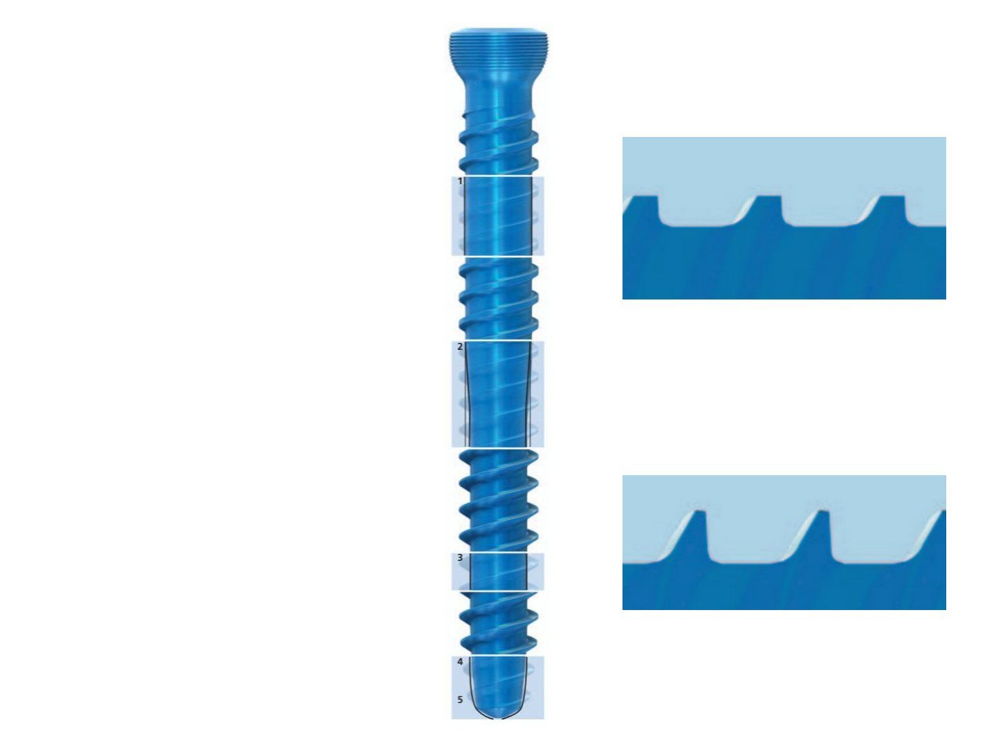
Q1.What is the design of a dual core diameter, dual pitch pedicle screw?
- Cylindrical large core + low broad threads - strength in the dense cortical bone of the pedicle
- Short smooth transition zone to decrease stress concentration
- Cylindrical small core + high thin threads - purchase in the cancellous bone of the vertebral body; increases area for load sharing
Q2.What are the overall advantages of this screw design?
- Increase pull-out resistance
- Length adjustment by backing out without abandoning safe anchorage
Q3.Why is the old conical core design less favourable?
- Decreases pull-out strength once backed out
- A loosened screw cannot be relied on for safe anchorage
Q4.What are the head options and their advantages?
- Low profile, threaded for the set screw
- Polyaxial: increases contact area between rod and locking mechanism (locking strength)
- Monoaxial: allows reduction and lordolisation (now overcome by new polyaxial reduction systems)
Q5.What are the features of the screw tip and the double lead?
- Tip: Self tap and self centering (self-tapping and self-centering)
- +/- round tip for bicortical purchase
- Double lead - faster insertion without compromising purchase
▸ Slide 429 · Prone positionSpine · 5 questions expand

Q1-Q55 questions — tap to reveal all answerslist
- Compare pin fixation and the horseshoe headrest for prone positioning.
- What equipment is used for prone positioning and what deformity does each produce?
- Why is reverse Trendelenburg used in prone spine surgery?
- How should the trunk and limbs be positioned?
- How is a Mayfield clamp applied and removed?
Q1.Compare pin fixation and the horseshoe headrest for prone positioning.
- Pin fixation device: rigid, no facial pressure; but pin wound complications and intraop loosening
- Horseshoe head set (headrest): non-invasive, easy adjustment; but less rigid, intraop displacement and facial pressure (sore, glaucoma, nasal cartilage necrosis)
Q2.What equipment is used for prone positioning and what deformity does each produce?
- OSI spine table
- Four poster (iliac and thigh support) - lordosis
- Wilson frame (chest to pelvis pad support) - kyphosis
Q3.Why is reverse Trendelenburg used in prone spine surgery?
- Reduces facial and periorbital oedema
- Reduces venous bleeding
Q4.How should the trunk and limbs be positioned?
- Free abdomen; free nipples and genitalia
- +/- adhesive strap to pull shoulders down for CS imaging
- Arms positioned, knee flexion, pad bony prominences
Q5.How is a Mayfield clamp applied and removed?
- Sweat band line; centreline through the EAM
- Just above pinna and slightly below equator
- Load 60lb adult, 40lb paediatric
- Removal: waterproof film + antibiotic ointment
▸ Slide 430 · SSEPSpine · 8 questions expand
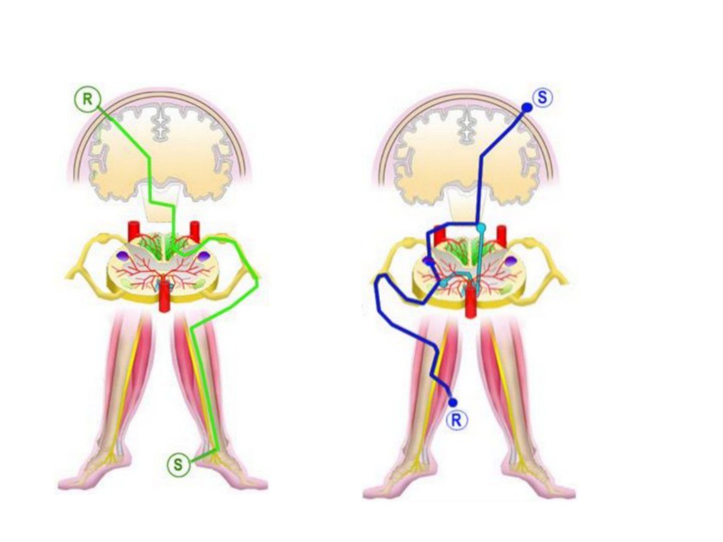
Q1-Q88 questions — tap to reveal all answerslist
- Describe SSEP monitoring in spine surgery.
- Describe MEP monitoring.
- What precautions are needed with intraoperative neuromonitoring?
- What defines a loss of signal and what should the surgeon do?
- What should anaesthesia and the neurophysiologist do after a loss of signal?
- If there is no improvement, what are the next steps and what is a Stagnara wake-up test?
- What other intraoperative neurological monitoring methods are available besides SSEP and MEP?
- What are the potential sources of false negatives and false positives in neuromonitoring?
Q1.Describe SSEP monitoring in spine surgery.
- Monitors the dorsal column
- Stimulation: UL ulnar nerve, LL posterior tibial nerve
- Recording: cortical, subcortical (C5), peripheral (Erb's point UL, popliteal fossa LL)
- Advantage: not affected by muscle relaxant
- Disadvantage: unremarkable in anterior cord syndrome
Q2.Describe MEP monitoring.
- Monitors the anterior and lateral corticospinal tracts
- Input: transcranial (motor cortex); output: gastrocnemius, EHL
- Advantage: detects ischaemic injury (anterior spinal artery)
- Disadvantage: altered by muscle relaxant
Q3.What precautions are needed with intraoperative neuromonitoring?
- Mouth guard so the patient does not bite their tongue
- Needles - shaving; risk of retained needles
- Seizures
- Burns under stimulating electrodes
- Movement-induced injury
- Transient arrhythmia
Q4.What defines a loss of signal and what should the surgeon do?
- SSEP amplitude reduced >50% or latency increased >10%; MEP reduced >50%; EMG firing
- Stop current manipulation; release traction
- Check dura for compression
- Reverse corrective manoeuvre and rod removal
- Confirm stability of the spine; look for implant malposition
- Assess blood loss; warm saline wound wash, less correction
Q5.What should anaesthesia and the neurophysiologist do after a loss of signal?
- Check if a bolus of anaesthetic drug or muscle relaxant was given
- Lighten depth of anaesthesia
- Increase BP (MAP >90mmHg)
- Optimise oxygenation, Hb, pH, temperature
- Neurophysiologist: repeat MEP trials, increase stimulus strength, assess pattern of changes
- OT technician: reduce noise and electrical interference
Q6.If there is no improvement, what are the next steps and what is a Stagnara wake-up test?
- Increase MAP >100mmHg
- Consider steroid administration
- Consider wake-up test
- Consider aborting surgery
- Stagnara test: test voluntary feet movement; can miss early ischaemia and is not continuous
Q7.What other intraoperative neurological monitoring methods are available besides SSEP and MEP?
- Intraoperative NCT
- Free-running EMG - real-time monitoring of motor nerve root function
- Neurotonic changes are a sensitive indicator of nerve root injury
- Stimulus-triggered EMG
Q8.What are the potential sources of false negatives and false positives in neuromonitoring?
- False negative: a transected distal part still has electrical activity before Wallerian degeneration
- False negative: higher stimulation threshold in pre-existing axonal damage; abnormal roots may not evoke neurotonic discharge
- False negative: screws may enlarge the holes on insertion
- False positive: electrocautery and irrigation cause interference and artefacts
▸ Slide 431 · How do you insert the halo ring?Spine · 4 questions expand
Q1-Q44 questions — tap to reveal all answerslist
- How do you prepare and position the patient for halo ring insertion?
- Where are the anterior pins placed and what are the dangers of malposition?
- How are the halo pins tightened?
- What are the limitations of halo immobilisation?
Q1.How do you prepare and position the patient for halo ring insertion?
- Prepare E trolley, assistant and ring of appropriate size
- Rule out local C/I: skull fracture, infection, type IIA hangman fracture
- Systemic C/I if using halo jacket: old age (20% mortality), obesity, barrel chest
- Patient supine with head on head spoon at end of bed; temporary fixation of ring using positional pins; ring 1cm above skin
- LA to pin site
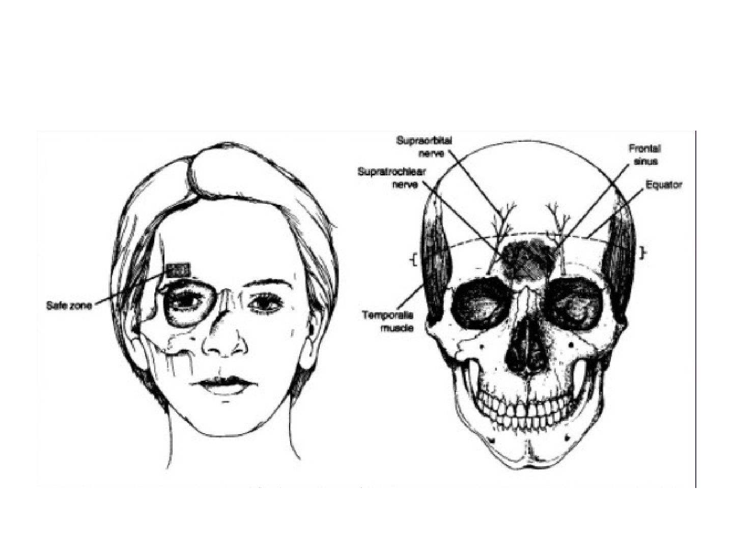
Q2.Where are the anterior pins placed and what are the dangers of malposition?
- 2 pins in front at lateral 2/3, 1cm above eyebrow, below equator; insert with eye closed
- Too medial: supraorbital nerve, frontal sinus
- Too lateral: thin temporal bone, temporal artery
- Too superior: easy slip out
Q3.How are the halo pins tightened?
- 2 posterior pins opposite the front pins, at the mastoid process
- Tighten opposite pins together with 8 pound-inch pressure
- Paediatric 4 pound; more pins 6-8
- Retighten 48hr later (48 hours), then connect to traction
Q4.What are the limitations of halo immobilisation?
- Good for the upper cervical spine
- Lower cervical spine: no good due to the snaking phenomenon
▸ Slide 432 · Anterior approach to C spineSpine · 5 questions expand
Q1-Q55 questions — tap to reveal all answerslist
- Describe the position for an anterior approach to the cervical spine.
- Describe the incision and dissection of the anterior cervical approach.
- Which structures may be ligated and which planes are incised?
- What are the dangers during subperiosteal dissection?
- What are the complications of the anterior cervical approach?
Q1.Describe the position for an anterior approach to the cervical spine.
- GA + reverse Trendelenburg
- Radiolucent table + head ring
- Sandbag between scapulae, slight neck extension to evade the jaw
Q2.Describe the incision and dissection of the anterior cervical approach.
- Left side incision (predictable recurrent laryngeal nerve course)
- Insert RT for easy identification of oesophagus; LA + adrenaline along planned incision
- Split platysma; deep investing cervical fascia just anterior to SCM; retract strap muscle medially
- Blunt dissection then incise pretracheal fascia medial to the carotid sheath
- Retract carotid sheath laterally, larynx and oesophagus medially
Q3.Which structures may be ligated and which planes are incised?
- +/- superior thyroid vessels (superior thyroid artery close to SLN at C3/4, superior thyroid vein at C6/7)
- +/- omohyoid
- Incise the prevertebral fascia
Q4.What are the dangers during subperiosteal dissection?
- Subperiosteal dissection of longus colli, retracted laterally
- Protect the sympathetic trunk and recurrent laryngeal nerve below C6
- Spine level time out; mount self-retaining retractor to bed
Q5.What are the complications of the anterior cervical approach?
- Vertebral artery injury: single = 3-5% stroke, bilateral = potentially fatal
- Haematoma
- Oesophageal injury 0.2-0.4%
- Dysphagia
- Vocal cord palsy (usually lower level C6-7)
▸ Slide 433 · C spine approach comparisonSpine · 4 questions expand

Q1-Q44 questions — tap to reveal all answerslist
- What are the advantages of an anterior approach to the cervical spine?
- What are the disadvantages of an anterior approach?
- What are the advantages of a posterior approach?
- What are the disadvantages of a posterior approach?
Q1.What are the advantages of an anterior approach to the cervical spine?
- Shorter fusion segment
- More deformity correction
- Directly addresses pathology (most pathology is anterior)
- Addresses axial neck pain with fusion
- Avoids violation of the posterior tension band
Q2.What are the disadvantages of an anterior approach?
- Approach complications (nerve, vessel, oesophagus, trachea)
- Fusion complications (pseudarthrosis, hardware failure)
- Graft complications (migration, donor site)
- Not for congenital / multilevel compression
- ASD in ACDF (adjacent segment disease in anterior cervical discectomy and fusion)
Q3.What are the advantages of a posterior approach?
- Deals with posterior pathology
- Addresses multilevel pathology
- More familiar to most surgeons
- Enlarge canal/ volume expansion (enlarges the canal)
Q4.What are the disadvantages of a posterior approach?
- Unable to deal with kyphosis >10 deg
- C5 palsy
- Instability (post-laminectomy kyphosis)
- Hinge fracture in laminoplasty
▸ Slide 434 · Posterior approach to C spineSpine · 4 questions expand
Q1-Q44 questions — tap to reveal all answerslist
- How is the patient positioned for a posterior approach to the cervical spine?
- How are levels checked before surgery?
- How is the dissection performed?
- Describe the laminoplasty technique.
Q1.How is the patient positioned for a posterior approach to the cervical spine?
- GA, prone reverse trendelenberg tilt (reverse Trendelenburg)
- OSI table, hands by side of body
- Mayfield skull clamp
- Neck slight flexion to open interlaminar space; strap to pull shoulders inferiorly
Q2.How are levels checked before surgery?
- By surface landmark
- By X-ray
Q3.How is the dissection performed?
- Posterior midline incision
- Cervical fascia along the nuchal ligament
- Subperiosteal dissection laterally to the facet
- Avoid dissecting muscle attachments at C2 and C7 to prevent kyphosis
Q4.Describe the laminoplasty technique.
- Locate the groove between lamina and facet
- Create a trough just medial to the groove
- Split the spinous process with a T saw / 1mm burr
- Plastic deformity of the anterior cortex
▸ Slide 435 · TL trauma burst fractureSpine · 12 questions expand

Q1-Q1212 questions — tap to reveal all answerslist
- Describe the X-ray findings and classification of this L1 burst fracture.
- What is the initial management of a thoracolumbar burst fracture?
- What is the TLICS and how is it scored?
- What are the indications for surgery in a thoracolumbar burst fracture?
- What is the timing and approach, and why is posterior chosen?
- What is the McCormack and Gaines load sharing classification?
- What is the aim of management in a thoracolumbar burst fracture?
- When is conservative management appropriate for a thoracolumbar burst fracture?
- What are the indications for decompression in a thoracolumbar burst fracture?
- How is reduction achieved and what counts as good reduction?
- What instrumentation is used and what determines the number of levels?
- When is fusion performed and why must implant removal be planned?
Q1.Describe the X-ray findings and classification of this L1 burst fracture.
- Anterior wedging of L1, some retropulsion, widening of interpedicular space
- Assess for posterior element widening/fracture on CT and MRI
- AO A3 or A4; Denis 2 column fracture
Q2.What is the initial management of a thoracolumbar burst fracture?
- High energy trauma - manage according to ATLS
- C spine immobilisation + pelvic binder; primary and secondary survey
- Assess neurology
- CT and MRI (whole spine) - no posterior element involvement, no nerve compression
Q3.What is the TLICS and how is it scored?
- Thoracolumbar injury classification system
- Calculate the TLICS for further mx
- Injury morphology (1-4), neurology (0-3), PLC injury (0-3)
- Provide scoring (1-10, </=3 conservative, >/= 5 operative )
- Total score 1-10; 3 or less conservative, 5 or more operative
Q4.What are the indications for surgery in a thoracolumbar burst fracture?
- TLICS score 5 or higher
- Neurological deficit needing decompression
- Unstable pattern: injury to the posterior ligament complex (PLC), progressive kyphosis
- 30 deg kyphosis (controversial)
- 50% loss of vertebral body height (controversial)
- 50% canal compromise (controversial)
Q5.What is the timing and approach, and why is posterior chosen?
- Early surgery (within 48hrs) if neurocompromise - Fehlings 2021 Lancet
- Posterior decompression + instrumentation from T10 to L4 +/- fusion
- Posterior: easier/familiar, shorter OT, less blood loss; If PLC disrupted, I can keep the intact ALL, avoiding further destabilisation of the spine; ligamentotaxis; three-column purchase; later implant removal avoids fusion
- Anterior if McCormack and Gaines >6, disc pathology, poor bone, chronic fracture, pincer fracture
Q6.What is the McCormack and Gaines load sharing classification?
- Comminution (<30, 30-60, >60%)
- Apposition of fragments (minimal, <50%, >50%)
- Reducibility of sagittal deformity (<=3, 4-9, >=10 deg)
- >6/9 = add anterior approach
Q7.What is the aim of management in a thoracolumbar burst fracture?
- Relieve pain
- Prevent neurological deficit and deformity
- Allow early mobilisation
Q8.When is conservative management appropriate for a thoracolumbar burst fracture?
- No neurology, relatively stable (PLC intact), acceptable deformity (size of anterior collapse)
- Protect with a brace
Q9.What are the indications for decompression in a thoracolumbar burst fracture?
- Neurological deterioration
- When deformity correction is needed (e.g. shortening osteotomy)
- Indirect decompression by ligamentotaxis, or facetectomy for the retropulsed fragment +/- expandable cage; directly deal with an entrapped nerve root
Q10.How is reduction achieved and what counts as good reduction?
- Distraction restores vertebral height and tenses the PLL to reduce the retropulsed fragment
- Lordolisation by cantilever rod reduction corrects deformity
- Good reduction: vertebral height restored, lumbar lordosis restored
Q11.What instrumentation is used and what determines the number of levels?
- Posterior pedicle-rod system able to engage all three columns
- Levels depend on fracture site, fracture age, deformity to correct and bone quality
- USS (universal spine system) reduction tools help lordolisation
Q12.When is fusion performed and why must implant removal be planned?
- Not fusing preserves the mobile segment
- Fuse if decompression destabilises the spine
- Plan removal of instrumentation, otherwise fatigue fracture or loosening
- Rehab: TLSO, FWB, DVT prophylaxis
▸ Slide 436 · TLJ prone to injury:Spine · 1 question expand

Q1-Q11 questions — tap to reveal all answerslist
- Why is the thoracolumbar junction prone to injury?
Q1.Why is the thoracolumbar junction prone to injury?
- Transition from kyphosis to lordosis
- Transition from rigid to mobile - more shearing
- Smaller vertebral body compared with the lower lumbar spine
- Increase bending moment
- Facet orientation changes from coronal to sagittal
▸ Slide 437 · TL trauma chance fractureSpine · 8 questions expand
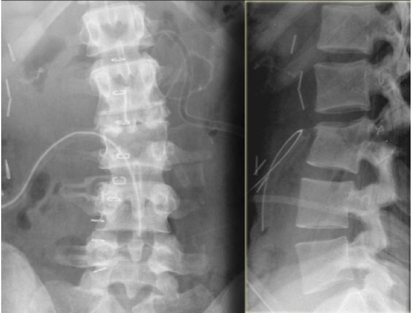
Q1-Q88 questions — tap to reveal all answerslist
- Describe the X-ray findings and diagnosis of this Chance fracture.
- What associated injuries must be assessed in a Chance fracture?
- What is the principle and timing of surgery?
- What determines the approach in a Chance fracture?
- How is reduction achieved and what is good reduction?
- What are the implications of a lamina fracture?
- What are the decompression and instrumentation options in a Chance fracture?
- What rehabilitation and implant planning follow surgery for a Chance fracture?
Q1.Describe the X-ray findings and diagnosis of this Chance fracture.
- Lateral: L1 fracture involving all three columns, vertebral height loss >50%, kyphosis >30 deg
- Mechanism RTA - Chance fracture: fulcrum anterior, distraction from posterior to anterior column, tensile failure of all three columns
- AP: widening of interpedicular distance
- AO B1, Denis three column fracture
Q2.What associated injuries must be assessed in a Chance fracture?
- ATLS, prevent secondary cord injury; spine precaution, bed rest
- PR + neuro examination
- Palpate abdomen (intraabdominal injury 40%) + assess LL pulses; bloods, urine for blood
- XR whole spine (non-contiguous fracture in 20%)
- CT for canal compromise by retropulsed fragment
- MRI not absolutely indicated: neuro compromise, suspected soft tissue Chance, high risk epidural haematoma (e.g. AS)
Q3.What is the principle and timing of surgery?
- Principle: pain relief, stabilise the spine for early mobilisation, prevent neurological deficits and future deformity
- Timing: early (within 24-48hrs) if neurocompromise - Fehlings 2021 Lancet
Q4.What determines the approach in a Chance fracture?
- Consider neurology and PLC
- Posterior: easier/familiar, shorter OT, less blood loss; keeps intact ALL if PLC disrupted; later implant removal avoids fusion - but decompression may be inadequate
- Anterior indications: severe collapse and kyphosis needing height restoration (McCormack and Gaines), disc pathology, poor bone quality, chronic fracture, failure of posterior surgery
Q5.How is reduction achieved and what is good reduction?
- Reduction by ligamentotaxis - needs to be done within 5-6 days
- Distraction restores vertebral height and tense up PLL to reduce the retropulsed fragment; lordolisation by cantilever rod reduction
- Good reduction: vertebral height restored, lumbar lordosis restored
- Fusion: not fuse, preserve mobile segment; fuse if decompression destabilises the spine; tend to fuse in soft tissue Chance as ligamentous healing is unpredictable
Q6.What are the implications of a lamina fracture?
- Dural tear
- Nerve root injury
- Need posterior decompression
Q7.What are the decompression and instrumentation options in a Chance fracture?
- Decompression for neurological deterioration or when deformity correction (shortening osteotomy) is needed
- Indirect decompression by ligamentotaxis, or facetectomy +/- expandable cage for the retropulsed fragment; Directly deal with entrapped nerve root if present
- Posterior pedicle screw-rod engages all three columns; levels depend on fracture site, age, deformity, bone quality
- Anterior: bone graft, cement or expandable cage (transpedicular from posterior)
Q8.What rehabilitation and implant planning follow surgery for a Chance fracture?
- Plan removal of instrumentation, otherwise fatigue fracture or loosening
- Rehab: TLSO, FWB
- Fusion not routine - preserve the mobile segment; fuse if decompression destabilises, and soft tissue Chance tends to be fused
▸ Slide 438 · Describe Xray:Spine · 8 questions 1 check expand

Q1-Q88 questions — tap to reveal all answerslist
- Describe the X-ray findings in this subaxial cervical injury.
- What is the initial management of a perched facet injury after high energy trauma?
- Will you perform urgent closed reduction and what are the contraindications?
- How do you perform closed reduction?
- After successful closed reduction, what are your management options?
- What is the Allen and Ferguson classification of subaxial cervical injuries?
- What are the pros and cons of anterior versus posterior subaxial cervical surgery?
- What is the halo pin retightening schedule after insertion?
Q1.Describe the X-ray findings in this subaxial cervical injury.
- Lateral C spine with exposure adequate C1-T1
- Kyphotic alignment at C5/6; C5/6 anterior translation <25% with angulation and disruption of 4 lines
- Loss of facet stacked parallelogram; C5 perched on C6; +/- bow tie sign
- Prevertebral soft tissue swelling; no obvious OC dissociation
- AP: upper spinous processes displaced towards the side of dislocation
- 10% of patients have a fracture elsewhere
Q2.What is the initial management of a perched facet injury after high energy trauma?
- ATLS - ABC, primary and secondary survey
- Protect whole spine, neck collar, spinal board; rule out other spine fractures; watch for neurogenic shock
- Assess GCS and neurology
- CT to look for fractures, may need to direct OR
- Pre-CR MRI if obtunded, or no neuro deficit + conscious
- MRI looks for large PID, transdiscal injury, epidural haematoma, PLC, adjacent level injury
Q3.Will you perform urgent closed reduction and what are the contraindications?
- Prerequisite of safe CR is a fully conscious and cooperative patient
- Order urgent MRI before reduction to r/o PID (bilateral facet dislocation: PID incidence 10-40%)
- CR in the presence of PID causes further neurological deterioration that may be irreversible
- Contraindications: PID, OC dissociation, fracture dislocation, skull fracture
- PID -> straight to OT for anterior discectomy; no PID -> closed reduction under X-ray guidance
Q4.How do you perform closed reduction?
- Controlled sequential traction with monitoring of X-ray and neurology; fluoroscopy suite, reverse trendelenberg position (reverse Trendelenburg) with head spoon
- Insert halo ring (3 ppl minimum, i.e. 3 people minimum, 1-2cm clearance); anterior pins at lateral 1/3 and 1cm above eyebrow (insert with eye closed), posterior pins above mastoid process; tighten 8 pound-inch
- Traction in flexion using a towel under the occiput
- Start 5kg (~weight of head), then add 5-10lb every 30min; min weight = 10lb (5kg) for head + 5lb (~2kg) per level (Crutchfield 1954); max = min x 2
- Monitor X-ray for disc space widening/OC dissociation and check neurology each time; additional manoeuvres: unilateral - rotate 30 deg to side; bilateral - anterior directed force
- Once facet out of perched position, extend neck and decrease to minimal maintaining weight; end points: neuro deficit/pain/decrease GCS, excessive distraction, max weight, successful reduction
Q5.After successful closed reduction, what are your management options?
- No neurology + unilateral facet dislocation: can treat conservatively
- Persistent neurological deficit after reduction: posterior decompression + instrumentation
- Bilateral facet dislocation: gross posterior ligament destruction - circumferential fixation
- PID: anterior discectomy first, indirect reduction with Caspar pins (max 1cm); if fails, posterior reduction then ASF; low threshold for 360 deg fusion
- Postop: neck collar, sit out and walk as tolerated, DVT and pressure ulcer prophylaxis
Q6.What is the Allen and Ferguson classification of subaxial cervical injuries?
- Flexion-distraction - facet dislocation
- Flexion-compression - large anterior (flexion type) teardrop; unstable, needs corpectomy + ASF
- Extension-distraction - small anterior (extension type) teardrop
- Extension-compression - facet fracture
- Vertical compression - burst fracture
- Lateral flexion
Q7.What are the pros and cons of anterior versus posterior subaxial cervical surgery?
- Anterior adv: fusion in compression; saves fusion levels; direct decompression for burst fracture or PID
- Anterior disadv: posterior tension band not restored; difficult to reduce complex facet dislocation
- Posterior adv: preserves mobility (implant removal after healing); restores tension band; easier exposure; stronger construct (pedicle screw > lateral mass screw)
- Posterior disadv: VA injury (pedicle/lateral mass); cord injury (pedicle screw); difficult to correct deformity; indirect decompression
Q8.What is the halo pin retightening schedule after insertion?
- Retighten pins after 24hrs, 3 days then weekly (AO guideline)
Crutchfield rule: minimum weight = 10lb (5kg) for the head plus 5lb (~2kg) per level, and maximum weight = minimum x 2 — misleading/inverted — Crutchfield's 'rule of fives' is usually cited as a MAXIMUM: 10lb for the head + 5lb per level (e.g. C4/5 = 30lb). Traction is typically started at 5-10lb and increased in 5-10lb increments with serial X-ray and neurological checks — medium confidence — source
▸ Slide 439 · Bilateral reversed hamburger signSpine · 2 questions expand
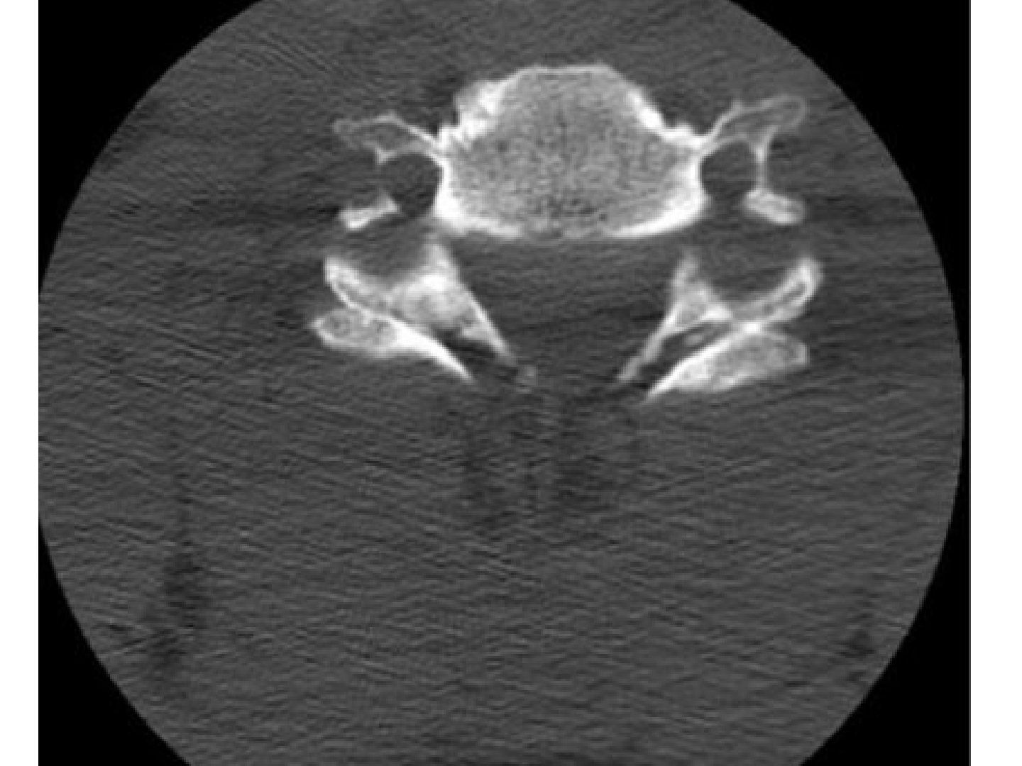
Q1-Q22 questions — tap to reveal all answerslist
- What does the bilateral reversed hamburger sign describe on imaging?
- What is the clinical significance of the bilateral reversed hamburger sign?
Q1.What does the bilateral reversed hamburger sign describe on imaging?
- Not covered in the speaker notes
Q2.What is the clinical significance of the bilateral reversed hamburger sign?
- Not covered in the speaker notes
▸ Slide 440 · C spine trauma dens fractureSpine · 9 questions expand

Q1-Q99 questions — tap to reveal all answerslist
- Describe the X-ray findings in this dens fracture.
- With intact neurology, how is a dens fracture managed by type?
- What are the risk factors for non-union of a type 2 dens fracture?
- How does age influence the management of a type 2 dens fracture?
- What are the operative choices for a dens fracture?
- Why is a type 2 dens fracture prone to non-union and what are the fusion rates?
- What history, examination and initial steps are needed in a dens fracture?
- What is the aim of treatment for a dens fracture?
- What are the C1/2 fusion options for a dens fracture?
Q1.Describe the X-ray findings in this dens fracture.
- Inadequate exposure
- Anderson and D'Alonzo type 2, Gruer A dens fracture with minimal displacement
- Spinal lines intact; prevertebral soft tissue swelling
- Sum of lateral mass normal; lateral ADI normal
Q2.With intact neurology, how is a dens fracture managed by type?
- Aim: stabilise the spine to promote healing, prevent neurological deficit, enhance early mobilisation
- Type 1: rigid neck collar
- Type 3: young - halo jacket; old - rigid neck collar (halo jacket in the elderly has higher mortality); non-union risk 7%
- Type 2: depends on the risk of non-union
Q3.What are the risk factors for non-union of a type 2 dens fracture?
- Patient factors: old age (>50), smoker, delay presentation >4 days
- Fracture factors: angulation >10 deg, displacement >5mm (strongest predictor), fracture gap >1mm, comminution
- Non-union rate up to 20-40%
Q4.How does age influence the management of a type 2 dens fracture?
- Young + no risk factors: can try halo jacket with close monitoring; surgery if risk factors
- Elderly: OT as age is already a risk factor and to avoid the jacket; OT if fit, neck collar if not
- Stable fibrous union in an asymptomatic patient >65yo may be acceptable (J Neurosurg 2020 Wilson)
Q5.What are the operative choices for a dens fracture?
- Osteosynthesis of the odontoid or C1/2 fusion
- Osteosynthesis retains rotation but depends on fracture configuration; may cause airway or swallowing problems
- C1/2 fusion does not rely on fracture configuration and is biomechanically more secure
- Osteosynthesis needs good reduction, fracture perpendicular to screw trajectory (Grauer II); C/I barrel chest, large BMI, fracture >3 weeks, poor bone quality
Q6.Why is a type 2 dens fracture prone to non-union and what are the fusion rates?
- Mechanical: fracture distal to the transverse ligament, so not stable
- Biological: watershed blood supply (superior internal carotid, inferior vertebral artery); small contact surface with high cortical:cancellous ratio
- C1/2 is a synovial joint - no fibrin clot; lack of cambium layer, no osteogenic potential
- Fusion rate: halo 70%, wiring 80%, Magerl/Harms/transarticular >95%
Q7.What history, examination and initial steps are needed in a dens fracture?
- ATLS and rule out other injury, especially head injury
- Temporary stabilisation of the C spine
- AMPLE history + chronicity, smoking, dysphagia (retropharyngeal haematoma)
- Assess neurology and body build
Q8.What is the aim of treatment for a dens fracture?
- Restore spinal stability and prevent late myelopathy
- Stabilise the spine to promote healing, prevent neurological deficit, enhance early mobilisation
Q9.What are the C1/2 fusion options for a dens fracture?
- Harms technique: C1 lateral mass screws + C2 pedicle screws
- C1/2 transarticular screws +/- wire augmentation - needs good reduction, pre-op CTA for vertebral artery anatomy
- Gallie or Brooks wiring with bone block + halo - sub-optimal rotational control, Brooks better
- Give neck collar x 3/12
▸ Slide 441 · Describe Xray:Spine · 4 questions expand
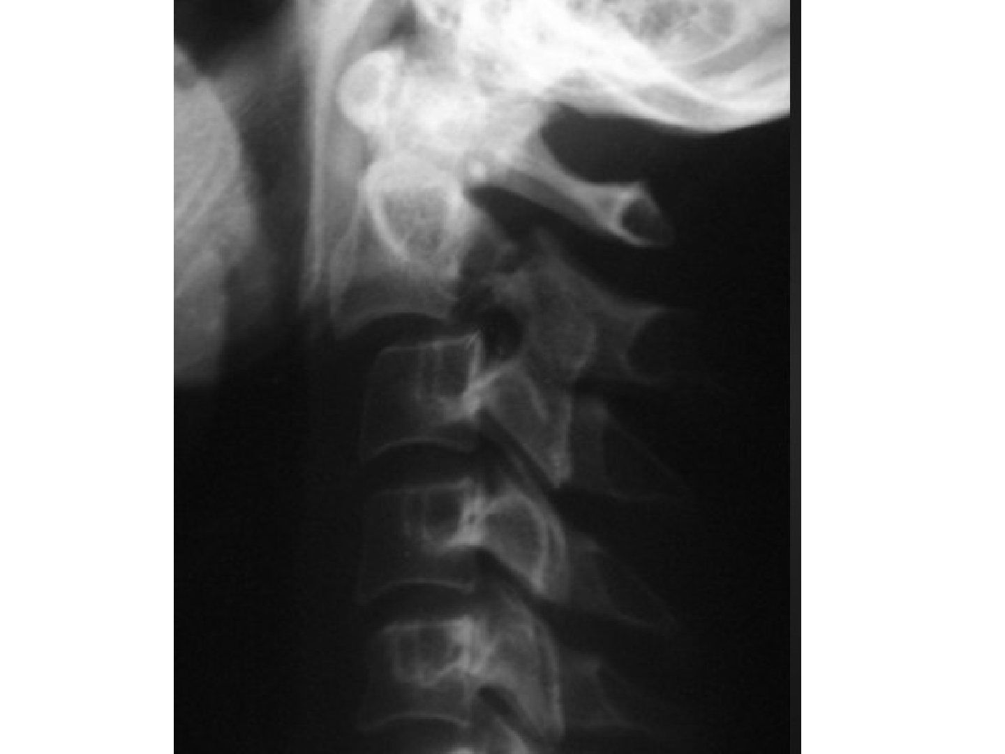
Q1-Q44 questions — tap to reveal all answerslist
- Describe the X-ray findings in this hangman's fracture.
- How do you classify a hangman's fracture?
- How is a hangman's fracture managed if there is no neurological deficit?
- What are the surgical options for C2/3 fixation?
Q1.Describe the X-ray findings in this hangman's fracture.
- C2/3 spondylolisthesis with disruption of anterior and posterior vertebral body lines
- spinolaminar and spinous process lines are intact
- Fracture seen at the pars, displacement and angulation (>11 deg = type 2A)
- Prevertebral soft tissue swelling; look for other subaxial C spine injury
- Hangman's fracture (traumatic spondylolisthesis of C2/3)
Q2.How do you classify a hangman's fracture?
- Levine/Effendi classification
- Type 1: anterior translation <3mm, minimal angulation (axial load + hyperextension)
- Type 2: translation >3mm, angulation <11 deg (axial load + hyperextension then hyperflexion); C2/3 disc and PLL disrupted
- Type 2A: minimal translation, horizontal fracture line, angulation >11 deg (flexion distraction; ALL intact as the hinge)
- Type 3: with facet dislocation (flexion distraction + hyperextension)
Q3.How is a hangman's fracture managed if there is no neurological deficit?
- Type I and II: non-op with halo/rigid collar; type IIa and III: C2/3 fusion (Li et al Eur Spine J 2006 - 50% pseudarthrosis with non-op)
- Type 1: rigid neck collar 4-6 weeks (stable, disc usually intact)
- Type 2: CR with halo traction then halo jacket 6-12 weeks; surgery if failed or displacement >5mm
- Type 2A: traction contraindicated - halo reduce by extension + axial load, then C2/3 fusion
- Type 3: more unstable, may need operation - C2/3 fusion
Q4.What are the surgical options for C2/3 fixation?
- Anterior plate and C2/3 interbody fusion
- Posterior C2/3 fusion (C2 pedicle screw + C3 lateral mass screw)
- Posterior C1-3 fusion; posterior C1-2-3 fusion
- Posterior Judet screw
- Non-union: C1-3 fusion
▸ Slide 442 · Describe Xray:Spine · 5 questions expand
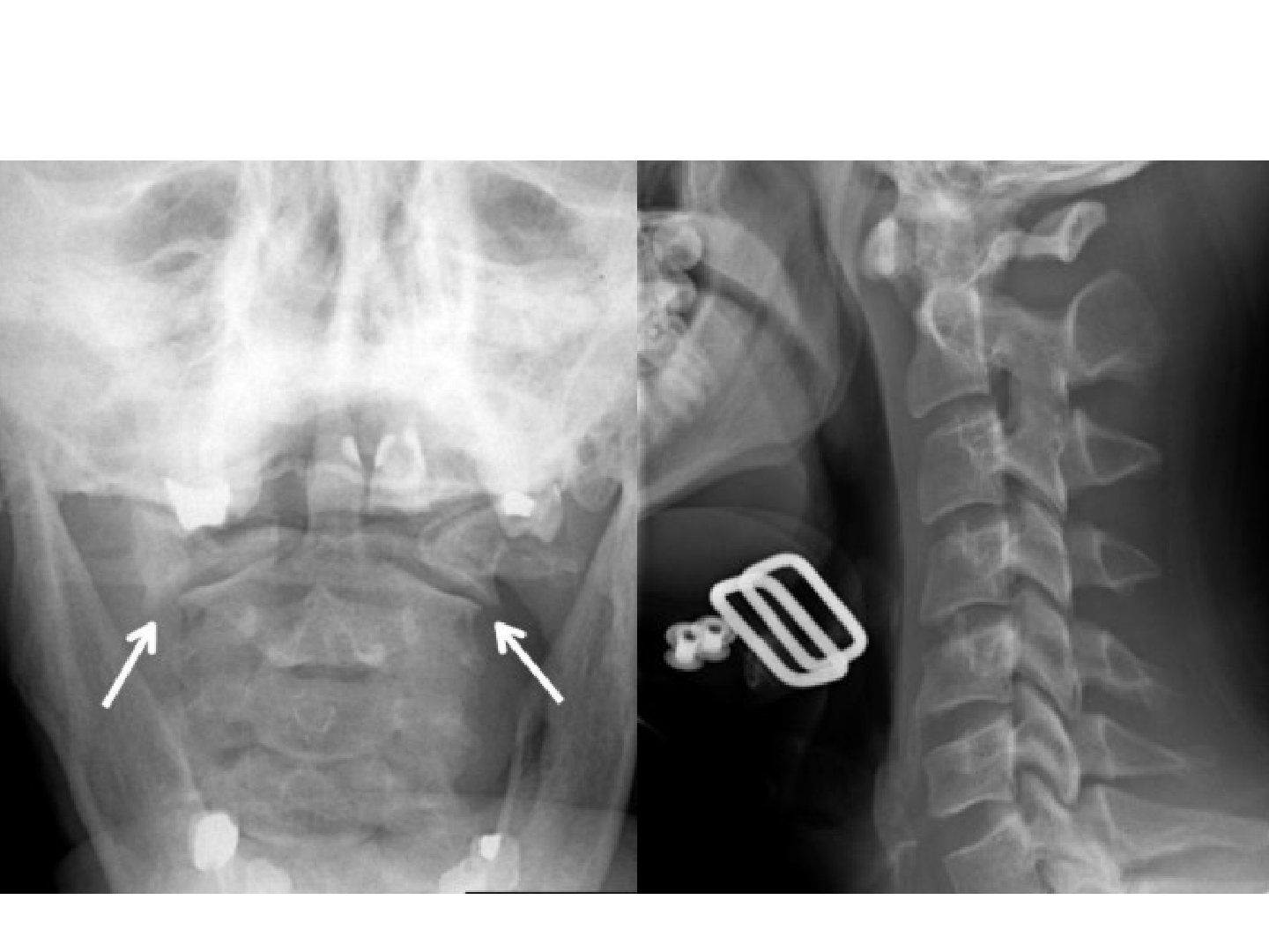
Q1-Q55 questions — tap to reveal all answerslist
- Describe the X-ray findings in this C1 injury.
- What is a Jefferson fracture and how is it classified?
- How is transverse ligament stability assessed in a Jefferson fracture?
- What is the Dickman classification and its treatment?
- What is the management of a Jefferson fracture?
Q1.Describe the X-ray findings in this C1 injury.
- Increased AADI on the lateral view
- Overhanging of C1 on C2 on the open mouth view (>7mm)
- Likely Jefferson fracture
Q2.What is a Jefferson fracture and how is it classified?
- Burst fracture of C1
- Landells: I isolated anterior or posterior arch fracture, II Jefferson burst fracture, III unilateral lateral mass fracture
- Gehweiler: I isolated anterior arch; II bilateral posterior arch; III classic Jefferson (anterior and posterior arch) A TAL intact / B TAL rupture; IV lateral mass; V transverse process
Q3.How is transverse ligament stability assessed in a Jefferson fracture?
- Combined overhang >7mm (rule of Spence)
- AADI >5mm (both alar and TL); if 3-5mm = TL only
- PADI <14mm
- Avulsion fracture of the transverse ligament
- MRI showed discontinuity
Q4.What is the Dickman classification and its treatment?
- Type I: intrasubstance tear - treat with C1/2 fusion
- Type II: bony avulsion - treat with halo vest
Q5.What is the management of a Jefferson fracture?
- Assess neurology - usually intact as the SAC is widened
- Gehweiler I, II, IIIA, V: SOMI brace / hard collar
- IIIB: halo +/- occiput to C2 or C1/2 fusion (now trend towards surgery)
- IV: halo majority of the time; O-C2 stabilisation if incongruence of atlanto-occipital or atlantoaxial joint, or sagittal split of lateral mass
▸ Slide 443 · Bilateral facet dislocation with neurologySpine · 6 questions expand

Q1-Q66 questions — tap to reveal all answerslist
- What is the initial management principle in bilateral facet dislocation with neurology?
- How do you assess the neurology in acute spinal cord injury?
- What is spinal shock?
- What is neurogenic shock?
- What is autonomic dysreflexia?
- What is the timing of surgery and the postoperative care?
Q1.What is the initial management principle in bilateral facet dislocation with neurology?
- ATLS, rule out other injury, temporary stabilisation with neck collar
- Minimise secondary injury, hypoxia and ischaemia at the cord injury site by ensuring adequate haemodynamics and oxygenation
- Immobilise the spine
- Shock can be neurogenic shock but need to r/o hypovolemic shock
Q2.How do you assess the neurology in acute spinal cord injury?
- Assess whether complete or incomplete cord injury (voluntary anal grip, deep anal sensation)
- If complete, determine neurological level (most caudal level with power >3/5 and normal sensation) by ASIA classification
- If incomplete, determine Frankel grading (A worst, E intact)
- Rule out spinal shock (return of bulbocavernosus reflex)
Q3.What is spinal shock?
- Temporary physiological response of the cord against trauma
- Complete paralysis, diaphragmatic breathing, paraesthesia, areflexia including bulbocavernosus reflex
- Only when spinal shock is over can the true neurological deficit be evaluated
Q4.What is neurogenic shock?
- Temporary generalised sympathectomy due to cord injury
- Triad of hypotension, bradycardia, peripheral vasodilatation
- SCI above C6
- Management: fluid support, vasopressor, atropine
Q5.What is autonomic dysreflexia?
- Occurs 3 weeks - 9 months (up to 12yrs); massive disordered reflex sympathetic surge to stimulation below the lesion, particularly T6 or above
- Emergency - can cause seizure, retinal haemorrhage, pulmonary oedema, MI
- Visceral stimuli (e.g. distended bladder); imbalanced reflex sympathetic discharge from the TL cord
- Uncontrolled hypertension, bradycardia, sweating, agitation
- Why above T6: below T6 there is greater vasodilatory reserve (splanchnic circulation) to offset vasoconstriction
Q6.What is the timing of surgery and the postoperative care?
- Rationale for early surgery: salvage neurology by decreasing secondary injury from swelling, ischaemia and free radicals
- STASCIS 2012 (Fehlings): operate within 24hrs - safe, better neurological recovery (increase 2 ASIA grades), less complications, similar mortality
- Confirmed by Fehlings 2021 Lancet meta-analysis: within 24hr for acute SCI
- Postop: avoid pneumonia/UTI, bedsore, DVT, paralytic ileus, gastric ulcer
- Rehab: reintegrate into community; key functions - transfer, mobility, hand skills
▸ Slide 444 · Steroid for Acute SCI?Spine · 5 questions expand

Q1-Q55 questions — tap to reveal all answerslist
- What is the rationale for methylprednisolone in acute spinal cord injury?
- What did the NASCIS I-III studies show?
- What are the criticisms of the NASCIS evidence?
- What is the dosage of methylprednisolone if used?
- What do guidelines recommend regarding steroids for acute SCI?
Q1.What is the rationale for methylprednisolone in acute spinal cord injury?
- Prevent secondary injury to the cord due to ischaemia, lipid peroxidation and free radicals
- Increase microcirculation, decrease inflammation and cell death
Q2.What did the NASCIS I-III studies show?
- NASCIS I: high vs low dose MP - no difference (dose)
- NASCIS II: MP vs naloxone vs placebo - no difference; posthoc high dose MP within 8hr improved 5 motor points
- NASCIS III: examined timing
Q3.What are the criticisms of the NASCIS evidence?
- Posthoc analysis is not level 1 evidence
- Not clinically significant
- Multiplicity of subgroup analysis increases type 1 error
- Complications: peptic ulcer and wound healing problems
Q4.What is the dosage of methylprednisolone if used?
- <3hr: 30mg/kg bolus, then 5.4mg/kg/hr drip for 23hr
- 3-8hr: same bolus until 48hr
- Indication: <8hr from injury, no wound
- Need gastric protection
- Usage controversial with significant SE (gastric ulcer, infection)
Q5.What do guidelines recommend regarding steroids for acute SCI?
- NICE - do not recommend it
- 2013 AANS - methylprednisolone should not be used for acute SCI
- AO Spine 2017: suggest not offering 24hr high dose MPSS if presenting >8hrs
- Suggest 24hr infusion MPSS be offered within 8 hours (30mg/kg loading then 5.4mg/kg/hr)
▸ Slide 445 · Central cord syndromeSpine · 5 questions expand
Q1-Q55 questions — tap to reveal all answerslist
- What is the pathoanatomy behind central cord syndrome?
- Why is the deficit UL >> LL and what is dissociative anaesthesia?
- What is the typical recovery pattern in central cord syndrome?
- When is surgery indicated in central cord syndrome?
- What did Anderson Neurosurgery 2015 conclude about the timing of surgery in central cord syndrome?
Q1.What is the pathoanatomy behind central cord syndrome?
- Pre-existing narrowing of the spinal canal (congenital or secondary to OA), usually after a flexion/extension neck injury
- A/P force distributes the greatest damaging effect on the central cord (anterior osteophytes, posterior thickened ligamentum flavum)
- Concentrated stress in the middle of the cord + the water-shed circulation between anterior and posterior horns -> ischemic insult to the central part of the SC
- Stasis of axoplasmic flow and/or Wallerian degeneration of the corticospinal tracts
Q2.Why is the deficit UL >> LL and what is dissociative anaesthesia?
- Lamination of the lateral corticospinal (motor) and spinothalamic (sensory) tracts: sacral segments are the most lateral, with lumbar, thoracic and cervical components arranged somatotopically, proceeding medially toward the central canal
- UL >> LL is much more pronounced in motor than sensory
- Dissociative anaesthesia: pain and light touch affected, deep touch and proprioception not affected
Q3.What is the typical recovery pattern in central cord syndrome?
- Lower limbs recover first
- Then bowel and bladder function
- Then proximal upper limb
- Hand function last to recover
Q4.When is surgery indicated in central cord syndrome?
- Early surgery if associated with a large disc protrusion, fracture or instability
- STASCIS 2012 and 2021 (Lancet) suggest surgical decompression within 24hrs
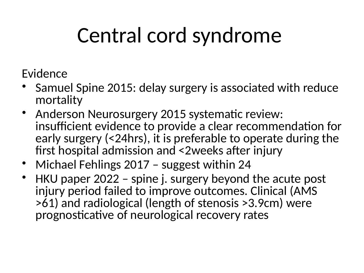
- Samuel Spine 2015: delay surgery is associated with reduced mortality
- Michael Fehlings 2017 – suggest within 24hrs
- HKU paper 2022 (Spine J): surgery beyond the acute post-injury period failed to improve outcomes; clinical (AMS >61) and radiological (length of stenosis >3.9cm) factors were prognosticative of neurological recovery rates
Q5.What did Anderson Neurosurgery 2015 conclude about the timing of surgery in central cord syndrome?
- Systematic review: insufficient evidence to give a clear recommendation for early surgery (<24hrs)
- It is preferable to operate during the first hospital admission and <2 weeks after injury
▸ Slide 446 · Clinical C spine clearanceSpine · 4 questions expand
Q1-Q44 questions — tap to reveal all answerslist
- What are the NEXUS criteria for cervical spine imaging?
- What do the Canadian C spine rules add?
- What are the different types of incomplete cord injury?
- What structures should be shown in a cross-section of the cervical spine?
Q1.What are the NEXUS criteria for cervical spine imaging?
- Any one criterion warrants imaging
- Decreased alertness
- Intoxication
- Distracting pain
- Neurological deficit
- Posterior midline C spine tenderness
Q2.What do the Canadian C spine rules add?
- Age 65
- Mechanism of injury
- Voluntary rotational range of motion >45 deg each side
Q3.What are the different types of incomplete cord injury?
- Central cord syndrome
- Anterior cord syndrome
- Posterior cord syndrome
- Brown-Sequard syndrome
- Conus medullaris and cauda equina syndromes
Q4.What structures should be shown in a cross-section of the cervical spine?
- Vertebral body and uncinate processes
- Transverse foramen with vertebral artery and vein
- Pedicles, laminae, spinous process, facet joints
- Spinal canal with the spinal cord
▸ Slide 447 · Cauda equina syndromeSpine · 5 questions expand

Q1-Q55 questions — tap to reveal all answerslist
- What is cauda equina syndrome (CES)?
- What is the pattern of bladder dysfunction in cord compression versus cauda equina syndrome?
- What is the timing of surgery in acute cauda equina syndrome?
- How would you perform a decompression for cauda equina syndrome?
- What is the clinical vignette of cauda equina syndrome shown?
Q1.What is cauda equina syndrome (CES)?
- Clinical syndrome from compression of the terminal spinal nerve roots in the lumbosacral spine
- Features: 1. bowel and bladder dysfunction, 2. saddle anaesthesia, 3. bilateral leg pain, 4. lower extremity sensorimotor changes
- Presentation can be acute or insidious
- Acute CES is an orthopaedic emergency warranting urgent MRI and surgery
Q2.What is the pattern of bladder dysfunction in cord compression versus cauda equina syndrome?
- Cord compression: reflex arc intact, lack of voluntary control -> bladder spasm, high pressure, incontinence
- Cauda equina: impaired reflex arc -> flaccid bladder, urinary retention, sphincter leakage, overflow incontinence
Q3.What is the timing of surgery in acute cauda equina syndrome?
- Controversial - studies quote <24h, 24-48h or within 48h
- Todd et al 2016 (Br J Neurosurg): CESS bilateral radiculopathy, CESI urinary difficulties, CESR retention with overflow, CESC absent perianal sensation/lax anus
- Urgency: S admit/observe, I emergency, R - <12hr emergency, >12hr next acute list, C next acute list
- Kumar et al 2021 (Eur Spine J): within 48h
- BASS: decompressive surgery should be undertaken at the earliest opportunity, balancing duration of pre-existing symptoms and night-time morbidity
- Do not wait for CESR or CESC - outcomes are poor despite surgery; if safe, operate as an emergency
Q4.How would you perform a decompression for cauda equina syndrome?
- Prone patient; II to identify the level of decompression
- Posterior midline approach through the inter-nervous plane between erector spinae; subperiosteal dissection to lamina
- Laminotomy of the cranial lamina above the ligamentum flavum; resect ligamentum flavum proximal to distal, then undercut the distal lamina
- Decompress laterally until the lateral edge of the dura is identified and the nerve root can be retracted to access the disc
Q5.What is the clinical vignette of cauda equina syndrome shown?
- Back pain after massage with radiating pain over bilateral lower limbs
- AROU x 1 day
- MRI showed L5/S1 PID with compression on the cauda equina
▸ Slide 448 · Interspinous distraction devices (IDDs)/Interspinous spacerSpine · 2 questions expand

Q1-Q22 questions — tap to reveal all answerslist
- What is the principle of interspinous distraction devices (IDDs)?
- What is the evidence for IDDs in lumbar spinal stenosis?
Q1.What is the principle of interspinous distraction devices (IDDs)?
- Create local flexion to stretch the posterior structures and increase space for the nerve roots
- Rationale: flexion makes back pain better
Q2.What is the evidence for IDDs in lumbar spinal stenosis?
- Borg 2021 (Neurosurgery): IDD cannot replace laminectomy
- Not as cost effective as laminectomy in lumbar spinal stenosis