18 slides
▸ Slide 401 · P&OP and O · 2 questions expand
Slide render
Question list
Q1-Q22 questions — tap to reveal all answerslist
- What does P&O stand for in this slide's title?
- What detailed content is provided for this P&O slide?
Answers · Q & A
Q1.What does P&O stand for in this slide's title?
- Prosthetics and Orthotics
- Not covered in the speaker notes
Q2.What detailed content is provided for this P&O slide?
- Slide image only - no speaker notes
- Content not in the speaker notes
▸ Slide 402 · Gait cycleP and O · 11 questions expand
Slide render

Question list
Q1-Q1111 questions — tap to reveal all answerslist
- What are the phases of the gait cycle and their proportions?
- Describe the loading response (1st rocker) of the gait cycle.
- Describe midstance (2nd rocker) of the gait cycle.
- Describe terminal stance (3rd rocker) and preswing.
- Describe the swing phase of the gait cycle.
- How does running differ from walking in the gait cycle?
- Define stride length and step length.
- What are Gage's prerequisites for efficient gait and give an example of each?
- Differentiate antalgic, short limb, Trendelenburg and gluteus maximus lurch gaits.
- How does antalgic gait differ for hip versus knee pain?
- What movements of the pelvis, knee, ankle and centre of gravity occur during the gait cycle?
Answers · Q & A
Q1.What are the phases of the gait cycle and their proportions?
- Cycle = heel strike to heel strike
- Stance 60% (closed chain), swing 40% (open chain)
- Most unstable part: initial contact and preswing
- Initial contact: hip flexed, knee nearly extended
Q2.Describe the loading response (1st rocker) of the gait cycle.
- From initial contact until elevation of the opposite limb; 0-10%
- Hip abductors eccentric contraction prevents pelvic tilt
- Knee flexes 15-20 degrees to minimize upward movement of CoG
- Quadriceps and tibialis anterior eccentric contraction; double limb stance
Q3.Describe midstance (2nd rocker) of the gait cycle.
- Elevation of opposite limb until both ankles aligned in coronal plane; 10-30%
- Single limb support; CoG progresses from posterior to anterior
- Eccentric gastrocnemius balances tibial progression (plantarflexion-knee extension couple)
- Hip extensors concentric contraction
Q4.Describe terminal stance (3rd rocker) and preswing.
- Supporting heel rises until the opposite heel touches the ground; 30-50%
- Concentric gastrosoleus contraction
- Tibialis posterior inverts the subtalar joint and locks the transverse tarsal joints - rigid lever arm
- Preswing: acceleration, CoG in front of hip, knee flexes for clearance/propulsion
Q5.Describe the swing phase of the gait cycle.
- Initial swing: elevation of limb to point of maximal knee flexion - active concentric hip flexor contraction
- Mid swing: from knee flexion to point where tibia is vertical - momentum of advancing tibia; HF, KE, ADF
- Terminal swing: tibia vertical to just prior to initial contact - deceleration by hamstrings; HF, KE, ADF
- Forward momentum provided by the hip flexors in initial swing
Q6.How does running differ from walking in the gait cycle?
- Stance phase decreases
- Double stance disappears
- Float phase appears
Q7.Define stride length and step length.
- Stride length: distance travelled between consecutive initial contacts of the same foot
- Step length: distance between initial contacts of alternating feet
Q8.What are Gage's prerequisites for efficient gait and give an example of each?
- Stability in stance (e.g. Trendelenburg)
- Foot clearance in swing (e.g. foot drop)
- Appropriate prepositioning of the swing phase foot (e.g. equinus)
- Adequate step length (e.g. antalgic limp)
- Energy conservation
Q9.Differentiate antalgic, short limb, Trendelenburg and gluteus maximus lurch gaits.
- Antalgic: trunk leans away from the painful side (opposite for hip), decreased stance on affected limb, longer stance on unaffected limb
- Short limb: pelvis tilts to the affected side (hip hiking), Foot may supinate to compensate, normal side hip/knee flex; equal proportion of stance and swing
- Trendelenburg: pelvis drops to the normal side from abductor weakness, trunk shift to painful side brings CG back to middle
- Glut max lurch: trunk leans backwards as the hip cannot be locked in extension
Q10.How does antalgic gait differ for hip versus knee pain?
- Knee/ankle pain: trunk leans away from the painful side in the coronal plane
- Hip pain: patient leans towards the side of pain to reduce the joint reaction force
- Knee pain: quadriceps avoidance mechanism - toe walking and decreased knee flexion to reduce tension on the knee capsule
Q11.What movements of the pelvis, knee, ankle and centre of gravity occur during the gait cycle?
- Pelvis: 4 degrees medial rotation and 4 degrees drop on the swing side to lengthen the limb; lateral displacement shifts COG over the stance limb
- Knee: flexion 15 degrees at heel strike lowers COG and absorbs shock; extends as the ankle plantarflexes and foot supinates
- Ankle: plantarflexion at heel strike and first part of stance
- COG: 5cm anterior to S2 in standing; highest at midstance, lowest at double limb support; 5cm horizontal displacement in an adult male step
▸ Slide 403 · How to ensureP and O · 6 questions expand
Slide render

Question list
Q1-Q66 questions — tap to reveal all answerslist
- What three factors determine good function after lower limb amputation?
- How is the patient optimized pre-operatively and intra-operatively?
- What post-operative stump care is required after amputation?
- When does pre-prosthetic training start and what is a preparatory prosthesis for?
- What factors determine the choice of socket and suspension at prosthetic fitting?
- What are the final stages of prosthetic rehabilitation?
Answers · Q & A
Q1.What three factors determine good function after lower limb amputation?
- Patient
- Amputated stump
- Prosthesis
Q2.How is the patient optimized pre-operatively and intra-operatively?
- Pre-op: optimize patient physically and psychologically
- Intra-op: optimal length
- Intra-op: optimal bulk
- Intra-op: soft tissue to prevent neuroma and maintain muscle balance
Q3.What post-operative stump care is required after amputation?
- Swelling control: differential crepe, elastic stocking
- Prevent contracture
- Skin desensitization
Q4.When does pre-prosthetic training start and what is a preparatory prosthesis for?
- When stump condition is stable, usually around 4 weeks
- Preparatory prosthesis allows stump maturation (till few months post-op)
Q5.What factors determine the choice of socket and suspension at prosthetic fitting?
- Patient factor
- Finance/cost
- Surgical factor
- P&O factor
Q6.What are the final stages of prosthetic rehabilitation?
- Proper walking mechanism taught by the P&O
- Good care of the stump
- Change to definitive prosthesis
▸ Slide 404 · Preparation for prosthesisP and O · 6 questions 1 check expand
Slide render
Question list
Q1-Q66 questions — tap to reveal all answerslist
- What is the timeline from amputation to normal gait?
- What is the Amputee Mobility Predictor (AMP)?
- What is assessed in the K level (Medicare functional classification) assessment?
- Define K0 and K1 functional levels and their prosthetic implications.
- What defines K2 and what prosthetic components are used?
- What defines K3 and K4 functional levels?
Answers · Q & A
Q1.What is the timeline from amputation to normal gait?
- Off stitches at 2 weeks
- Stump bandages from 2-4 weeks
- Temp prosthesis 4-6 weeks
- Stand with prosthesis 6-10 weeks
- Normal gait at 12 weeks
Q2.What is the Amputee Mobility Predictor (AMP)?
- Gailey et al 1999
- Predicts patient mobility with and without prosthesis by demo + PE - 21 tasks
- Used to predict K level
Q3.What is assessed in the K level (Medicare functional classification) assessment?
- Actual assessment of patient (CAT)
- Transfer
- Ambulation - level ground, low level barriers
- Cadence = steps per min
Q4.Define K0 and K1 functional levels and their prosthetic implications.
- K0 - fail to transfer/ambulate even with assistance; prosthesis does not improve QoL
- K1 - fixed cadence on level ground (household ambulator)
- K1 components: manual lock knee, SACH foot
Q5.What defines K2 and what prosthetic components are used?
- overcome low lever barrier (low level barrier) (limited community ambulator)
- Components: four bar polycentric/stance phase control, single axis foot
Q6.What defines K3 and K4 functional levels?
- K3 - variable cadence over most barriers (community ambulator) --> hydraulic, energy storage foot
- K4 - active adults/athlete
Fact check
Amputee Mobility Predictor (AMP) is attributed to Gailey et al 1999 and has 21 tasks — citation year — The 21-item AMP (with and without prosthesis) was published by Gailey et al in Arch Phys Med Rehabil 2002;83:613-627, developed from earlier work; the K-level system was adopted by Medicare in 1995 — medium confidence — source
▸ Slide 405 · Indication x amputation:P and O · 17 questions expand
Slide render
Question list
Q1-Q1717 questions — tap to reveal all answerslist
- What are the indications for lower limb amputation?
- How is the level of amputation chosen?
- What is the increase in energy consumption at each amputation level?
- What makes a good amputation stump?
- How do you perform a below-knee amputation (BKA)?
- What are the complications of amputation and the differential for pain in the missing limb?
- What are the key steps after the skin incision in a below-knee amputation?
- Compare the long posterior flap and sagittal flap for BKA.
- What are the length guidelines for transtibial and transfemoral amputation?
- How is an above-knee amputation (AKA) performed?
- What are the important considerations in the length of an AKA?
- What are the indications and technique of through-knee amputation?
- What are the advantages of through-knee amputation?
- What are the disadvantages of through-knee amputation?
- What did the LEAP study show for through-knee amputation?
- What is direct versus indirect load transfer in an amputation stump?
- What is the pathophysiology and treatment of phantom limb pain?
Answers · Q & A
Q1.What are the indications for lower limb amputation?
- DM/PVD
- Trauma
- Tumour
- Paediatric malformation/infection
Q2.How is the level of amputation chosen?
- Balance between pathology, wound healing and energy expenditure
- Ideal level: best for prosthesis fitting while minimising energy expenditure
Q3.What is the increase in energy consumption at each amputation level?
- Generally inversely proportional to remaining length
- Symes 15; BKA traumatic 25, vascular 40; through knee 50; AKA traumatic 70, vascular 100
- Bilateral BKA 40; BKA + AKA 120; bilateral AKA >200
Q4.What makes a good amputation stump?
- Ideal stump: mobile nonadherent muscle mass covered by full thickness skin tolerating weightbearing and shear
- Skin: scar mobile and away from bony edges, as sensate as possible
- Soft tissue: cylindrical or conical stump without excessive tissue
- Muscle: myodesis (muscle to bone) versus myoplasty (muscle to fascia)
- Nerve: clean section and retraction into soft tissue; bone ends bevelled and contoured
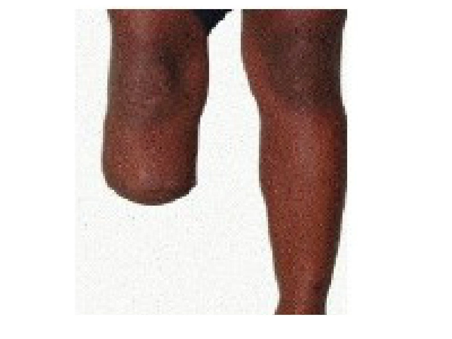
Q5.How do you perform a below-knee amputation (BKA)?
- Decide level (15cm below medial joint line); check pulse/healing and no contracture
- Bone cut 10-15cm from medial joint line; skin incision 15cm distal to tibial tubercle, posterior flap ~1.5x anterior flap (anterior 2/3, posterior 1/3)
- Section tibia, fibula 1cm above; bevel cut; Ertle osteomyoplasty fibula strut (paediatric)
- Ligate anterior and posterior tibial vessels and peroneal nerves; meticulous soft tissue balance; U slab to avoid knee contracture
Q6.What are the complications of amputation and the differential for pain in the missing limb?
- Unfit prosthesis - sore; neuroma; bone impingement; phantom limb
- Phantom limb risk factors: traumatic, young patients, upper limb, bilateral
- Ddx primary: neuroma, phantom limb pain, HO, bone impingement
- Ddx secondary: recurrence of primary pathology (tumour), CRPS, radiation pain from elsewhere
Q7.What are the key steps after the skin incision in a below-knee amputation?
- Position supine with tourniquet and hip support
- Divide anterolateral muscle down to intermuscular septum; ligate anterior tibial vessels and peroneal nerves
- Section tibia, and fibula 1cm above
- Dissect posterior soft tissues distally to the posterior transverse incision
- Ligate posterior tibial and peroneal vessels and the posterior tibial nerve
- Bevel cut; meticulous soft tissue balancing; do not trim dog-ear; U slab to avoid knee contracture
Q8.Compare the long posterior flap and sagittal flap for BKA.
- LPF: better flap blood supply from gastrocnemius; limited by proximal extent of infection/tumour
- SF: versatile, less infection in DM (Christensen JBJS 1989)
- SF disadvantage: tibial bone close to wound
- Ruckley J Vascular Surgery 1991: no difference
Q9.What are the length guidelines for transtibial and transfemoral amputation?
- Transtibial optimal 8cm for every 1m of height
- Transtibial shortest 7.5cm below medial joint line
- Transfemoral shortest 8cm below pubic rami
- Transfemoral longest 15cm above medial joint line
Q10.How is an above-knee amputation (AKA) performed?
- Bone: level of cut 8cm below pubic ramus to 15cm above joint line, smoothened and bevelled end
- Skin: AP flap
- Soft tissue: myodesis to counterbalance muscle pull and prevent contracture
- Myodesis of adductor and hamstring to prevent flexion and abduction deformity
- Not to do myodesis in vascular compromise
- Nerve: clear cut under gentle tension and allow retraction to prevent neuroma
Q11.What are the important considerations in the length of an AKA?
- Longer gives a better lever arm for control of the prosthesis
- Balance accommodation of the prosthesis
- Myoplasty/myodesis of adductor preserves 70% adductor power --> decreases AL drift
Q12.What are the indications and technique of through-knee amputation?
- Life saving
- Paediatric: preserve distal femur physis for longer lever arm
- Non-ambulatory patients
- Technique: suture patellar tendon to cruciate ligament
- Gastroc muscle (gastrocnemius) used as soft tissue envelope
Q13.What are the advantages of through-knee amputation?
- Less bleeding, faster OT
- Most thigh muscle preserved
- Long lever arm
- Broad end bearing surface
- Stable suspension
- No bony outgrowth
Q14.What are the disadvantages of through-knee amputation?
- Different levels of knee joint, less modularity
- Bulbous shape, less cosmetic
- Less room for error for flap (cannot cut more bone)
Q15.What did the LEAP study show for through-knee amputation?
- Similar pain to AKA/BKA
- Reduces walking speed and return to work
- Asymmetric knee + limited knee component choices
- Reduces satisfaction
Q16.What is direct versus indirect load transfer in an amputation stump?
- Direct - through knee/through ankle: wide metaphysis distributes the force of weight bearing
- Direct: less stiff bone at metaphysis dampens GRF at heel strike
- Indirect - BKA/AKA: terminal surface incapable of weight bearing; stiff bone
Q17.What is the pathophysiology and treatment of phantom limb pain?
- Pathophysiology: corresponding primary somatosensory cortex deprived of sensory input --> reorganization via increased expression of previously marked inputs - maladaptive plasticity model
- Physio: mirroring, USG, TENS, massage, immersive virtual reality
- Pharmacological: opioids, anticonvulsants, antidepressants, botulinum agents
- Surgical: stump revision, intrathecal implants, trigger point injections, dorsal column tractotomy
▸ Slide 406 · Tell me the different types of foot amputation and pros and cons of eachP and O · 6 questions 1 check expand
Slide render

Question list
Q1-Q66 questions — tap to reveal all answerslist
- What are the types of foot amputation?
- Describe transmetatarsal amputation and its problems.
- How are Lisfranc and Chopart amputations balanced?
- What are the prerequisites and indications for Syme amputation?
- Describe the Syme amputation technique.
- What are the advantages and disadvantages of Syme amputation, and what is Boyd amputation?
Answers · Q & A
Q1.What are the types of foot amputation?
- Transmetatarsal
- Lisfranc (TMTJ disarticulation)
- Chopart
- Syme
- Boyd
Q2.Describe transmetatarsal amputation and its problems.
- Lowest energy expenditure; no tendon transfer
- Bone cut >=3cm to MTB; preserve MT cascade; 5MTB
- Problem: equinus from loss of extensor lever arm; solution TA lengthening
Q3.How are Lisfranc and Chopart amputations balanced?
- Lisfranc: PB to cuboid transfer to reduce varus / shell out 5MTB to preserve PB insertion
- Lisfranc: +/- TA lengthening to reduce equinus
- Chopart: tib ant to talus transfer to reduce equinus and cavus; +/- TA lengthening
- Chopart problem: unstable hindfoot
Q4.What are the prerequisites and indications for Syme amputation?
- Patent posterior tibial artery (ischemic index >0.5)
- Healthy heel pad
- Indication: forefoot pathology - tumour/trauma/congenital; DM/infection
- Contraindicated in PVD
Q5.Describe the Syme amputation technique.
- Fishmouth flap 1cm distal + anterior to intermalleolar line; anterior through ankle joint, posterior through CCJ
- Ligate PTA as distal as possible
- Malleoli cut at 6cm proximal to joint line (see factcheck)
- Posterior flap anchored to distal tibia through drill holes to prevent heel pad posterior migration
- Preserve tibia articular surface for a resilient limb; avoid vigorous trimming of dogears
Q6.What are the advantages and disadvantages of Syme amputation, and what is Boyd amputation?
- Adv: end bearing stump (walk on unsupported stump); Self regulated walking speed, more natural gait pattern
- Adv: longer lever arm --> reduce energy expenditure
- Disadv: technically difficult; limited indication; fat pad migration; socket fabrication challenging
- Boyd = modification of Syme: talectomy + calcaneal tibial fusion; no heel pad migration
- Boyd disadv: bulky stump increases rehab time and nonunion
Fact check
In Syme amputation the malleoli are cut at 6cm proximal to the joint line — incorrect level — Classic Syme divides the tibia and fibula ~1-1.5cm above the ankle joint (or flush with the plafond) so the distal articular surface is preserved; 6cm would be a transtibial level — source
▸ Slide 407 · Hip disarticulationP and O · 4 questions expand

Question list
Q1-Q44 questions — tap to reveal all answerslist
- Describe the racquet-shaped incision for hip disarticulation.
- What are the anterior dissection steps in hip disarticulation?
- What are the posterior dissection steps in hip disarticulation?
- How is the stump covered after hip disarticulation?
Answers · Q & A
Q1.Describe the racquet-shaped incision for hip disarticulation.
- Anterior: ASIS to hip
- Medial: parallel to inguinal ligament to inferior pubic rami (adductor origin)
- Posterior: transverse 5cm distal to ischial tuberosity to 8cm distal to GT
- Lateral: curve up to anterior incision
Q2.What are the anterior dissection steps in hip disarticulation?
- ASIS: detach sartorius; AIIS: detach rectus femoris
- Divide femoral NAV at femoral triangle
- LT: detach iliopsoas (hip ER) and obturator externus
- Superior PR: detach pectineus (0.6cm residual cuff), divide obturator AVN underneath
- Inferior PR: detach adductors + gracilis; ischial tuberosity: detach hamstring
Q3.What are the posterior dissection steps in hip disarticulation?
- Divide fascia lata and glut max along posterior incision
- GT: detach glut med and min
- Trochanteric ridge: detach short ER
- Divide sciatic nerve
Q4.How is the stump covered after hip disarticulation?
- Deep myoplasty: obturator externus to glut med at acetabulum (GMOE)
- Superficial myoplasty: iliopsoas to quadratus femoris (IQ)
- Fascia: glut fascia --> inguinal ligament
▸ Slide 408 · What is an prosthesis?P and O · 14 questions expand
Slide render
Question list
Q1-Q1414 questions — tap to reveal all answerslist
- What is a prosthesis and how is it classified by structure?
- Compare quadrilateral and ischial containing (CAT-CAM) sockets.
- What is the modified design (NSNA) / Marlo anatomical socket?
- What suspension systems are available for a prosthesis?
- What is choke syndrome and how is phantom limb pain managed?
- How is an amputee assessed in clinic?
- How are prosthetic knee joints classified?
- What are the primitive knee control mechanisms and their trade-offs?
- What are the advanced knee control mechanisms and their disadvantages?
- What is the pilon and how do exoskeleton and endoskeleton pilons differ?
- What is the terminal device and how is it classified?
- What stump-related complications occur with prostheses?
- What dynamic movement complications occur with a prosthesis?
- What are the features of a transknee amputation relevant to prosthesis use?
Answers · Q & A
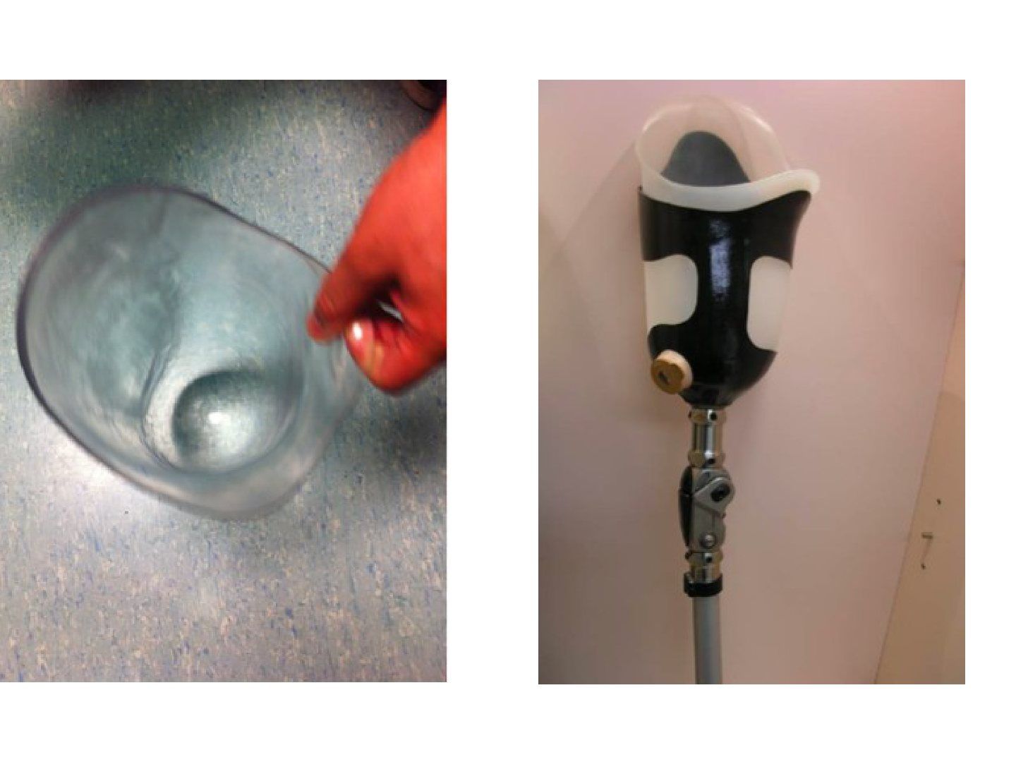
Q1.What is a prosthesis and how is it classified by structure?
- Artificial device externally applied to replace function or appearance of part of the body
- Exoskeleton: soft contoured foam + hard outer shell - durable, hard shell, heat resistant
- Endoskeleton: metal frame + soft covering - light, can exchange socket, adjustable
Q2.Compare quadrilateral and ischial containing (CAT-CAM) sockets.
- Quadrilateral: intentionally nonanatomical shape for soft tissue locking; narrow AP, wide ML; posteromedial platform for ischial bearing
- Quadrilateral disadv: cannot control abduction (wide ML), compensatory trendelenberg lurch; adv: comfortable sitting
- CAT-CAM: narrow ML, no posterior platform; groove contains ischium/rami --> bony lock (ramus bearing), rotational stability
- CAT-CAM: more anatomic - Distributes the proximal and medial concentration of forces more evenly
- CAT-CAM: allows 10 degrees adduction (stretch gluteus medius); Allows 5degs of flexion to stretch the gluteus maximus, increase hip extension strength for propulsion; more stable, less energy consuming
- CAT-CAM cons: bony lock discomfort/ulcers at the adductor region
Q3.What is the modified design (NSNA) / Marlo anatomical socket?
- Normal shape, normal alignment socket combining quadrilateral and ischial containing concepts
- Narrow ML to control abduction; high lateral to contain GT; posterior platform for ischium like quadrilateral
- Better ROM; smaller bony lock, less discomfort
Q4.What suspension systems are available for a prosthesis?
- Differential pressure (suction) - best fit/proprioceptive feedback, for mature limb; wrap or pull bag, seal-in liners, one-way valve
- Mechanical: liners with Velcro strap or pin-lock design
- Belt: total elastic suspension (neoprene), Silesian belt, pelvic belt
Q5.What is choke syndrome and how is phantom limb pain managed?
- Choke syndrome: venous outflow obstruction of residuum from narrow proximal socket + empty distal space; prevent with total contact sockets
- Phantom limb pain: pain felt in the amputated limb; treat with anti-depressants, NSAIDs, increased prosthesis use, TENS, sympathetic nerve block
Q6.How is an amputee assessed in clinic?
- Amputation related: wound condition, bone impingement, contracture, neuroma, phantom limb sensation/pain
- Prosthesis related function: can patient activate movement, walking aids, distance, stairs, ADL
- Prosthesis related complication: impingement, abrasion; assess wear
Q7.How are prosthetic knee joints classified?
- Knee joint mechanism: stable in stance, smooth swing, allow unrestricted motion for sitting and kneeling
- Axis: monocentric (single axis) or polycentric (multiple axis)
- Primitive control (all constant friction): constant friction, manual locking, stance control, single cadence 4-bar polycentric
- Advanced control (all variable friction): fluid (pneumatic/hydraulic), microprocessor
Q8.What are the primitive knee control mechanisms and their trade-offs?
- Constant friction (paediatric): durable, reliable; fixed cadence, stability in stance depends on alignment
- Manual locking (geriatric): locked, stable knee; affects gait efficiency, may need circumduction/ hip hiking
- Stance control (weight activated clutch-lock): easy swing, stable stance; difficulty shifting weight to sound limb when sitting
- Single cadence 4-bar polycentric: flex when loaded on forefoot, lock when loaded on heel; inherently stable; reduce energy dissipation by decreasing the magnitude of muscle activity; shortens for toe clearance; better sitting cosmesis
Q9.What are the advanced knee control mechanisms and their disadvantages?
- Fluid (pneumatic or hydraulic) control: cadence response changes knee flexion resistance via a piston mechanism
- Disadvantages: heavy, more expensive, need servicing
- Microprocessor control
Q10.What is the pilon and how do exoskeleton and endoskeleton pilons differ?
- Pilon connects the socket to the terminal device
- Exoskeleton pilon: transmits load via a rigid external structure; more durable; heavier; cannot adjust length
- Endoskeleton pilon: transmits load via an internal strut covered with foam; can adjust length
Q11.What is the terminal device and how is it classified?
- Most distal part of the prosthesis; provides shock absorption, adapts to uneven surfaces, stablise knee, reduce limb length during swing
- Classified by energy storing and articulating
- Includes SACH, single axis/poly axis, dynamic response foot
Q12.What stump-related complications occur with prostheses?
- Dermatological: contact dermatitis, blister, callosity
- Fitting problem: too tight/too loose
- Phantom limb sensation
Q13.What dynamic movement complications occur with a prosthesis?
- Gait cycle problems
- Pistoning
- Malalignment
Q14.What are the features of a transknee amputation relevant to prosthesis use?
- End bearing surface
- Long lever arm
- Stable suspension by metaphyseal flare
- Use a 4 bar knee joint to proximalise the COR of the knee
▸ Slide 409 · This is a LL prosthesis (device to replace missing part of body, in this case toP and O · 7 questions expand
Question list
Q1-Q77 questions — tap to reveal all answerslist
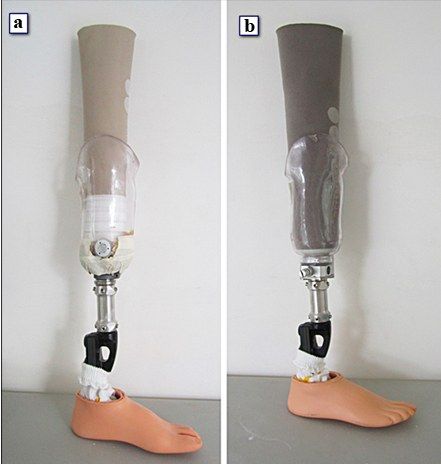
- What are the components of a below-knee prosthesis?
- Compare patellar tendon bearing and total contact sockets.
- What are the pressure tolerant and pressure sensitive areas in a BKA socket?
- What are the suspension options for a BKA prosthesis?
- What are the pros and cons of supracondylar/suprapatellar suspension?
- How does suction suspension compare with other suspension systems?
- What terminal devices can be used in a BKA prosthesis?
Answers · Q & A
Q1.What are the components of a below-knee prosthesis?
- Socket - interface between stump and prosthesis; transfers load and protects stump (inner soft liner + outer hard shell)
- Suspension - attachment mechanism for energy transfer and control
- Pilon - restores length (endoskeleton or exoskeleton)
- Terminal device - most distal part; energy storing or non-energy storing
Q2.Compare patellar tendon bearing and total contact sockets.
- Patellar bearing (PE foam, pelite): residual limb not in contact with bottom; problem of movement within distal socket
- Total contact (urethane/silicone gel): uniform distribution of pressure; disadvantage - volume control challenging
Q3.What are the pressure tolerant and pressure sensitive areas in a BKA socket?
- Tolerant: patella tendon, medial tibial flare, anterior compartment, gastrocnemius, fibular shaft --> add build-ups/convexities
- Sensitive: tibial crest and tubercle, distal fibula and fibular head, peroneal nerve, hamstring tendons --> reliefs/concavities
Q4.What are the suspension options for a BKA prosthesis?
- Differential pressure (negative atmospheric pressure, surface tension, muscle contraction) for total surface bearing; secure, best proprioception and ROM
- Differential pressure prerequisite: stable stump volume, good skin, precise fit, good cognition
- Mechanical: sleeve; shutter lock (pinlock) - needs daily washing and good hand function; strap for hyperextension (e.g. polio) - pistoning
- Hinge (thigh corset) - max stability and shared weight bearing but bulky, pistoning
- Anatomic supracondylar wedge +/- suprapatellar extension - for short stump <5cm
Q5.What are the pros and cons of supracondylar/suprapatellar suspension?
- Adv: short stump <5cm; increased contact area (supracondylar ML, suprapatellar AP); durable, economical, less maintenance
- Disadv: decreased knee ROM; local pressure at femoral condyles; requires good hand dexterity
Q6.How does suction suspension compare with other suspension systems?
- Relies on negative atmospheric pressure, surface tension and muscle contraction
- Usually for total surface bearing sockets; secure, best proprioception, best ROM
- Suction slightly > pin/shuttle lock, and 3 times supracondylar/straps
Q7.What terminal devices can be used in a BKA prosthesis?
- Divided into energy storing and non energy storing
- SACH
- Single axis
- Multiaxis
- Elastic keel (Safe heel)
- Dynamic response
▸ Slide 410 · Different knee jointsP and O · 5 questions expand
Slide render
Question list
Q1-Q55 questions — tap to reveal all answerslist
- Compare single axis constant friction and manual locking knee joints.
- What is the weight activated stance control (safety) knee?
- What are the pros and cons of a polycentric (4 bar linkage) knee?
- What are the pros and cons of fluid control/hydraulic knee joints?
- What is the knee extension-ankle plantarflexion coupling?
Answers · Q & A
Q1.Compare single axis constant friction and manual locking knee joints.
- Constant friction: simple, durable, light, cheap; less control, decreased stability, limited flexion
- Manual locking: more stability in standing; need circumduction and hip hitch for foot clearance; must unlock to sit
Q2.What is the weight activated stance control (safety) knee?
- Force sensitive drum brake/clutch lock
- Pros: versatile stability (adjustable housing); prosthetic length shortens for swing clearance
- Cons: more parts needing servicing; slower walking and smaller steps due to friction; difficult on stairs
Q3.What are the pros and cons of a polycentric (4 bar linkage) knee?
- Instant centre moves anterior and posterior to GRF, changing alignment stability and producing flexion or extension moment
- Pros: stability at heel strike/early stance; Reduce energy dissipation by decreasing the magnitude of muscle activity; prosthesis shortens in swing; better voluntary knee flexion control; better sitting cosmesis
- Cons: heavier than single axis; needs active knee extension moment in stance to prevent buckling; poor durability
Q4.What are the pros and cons of fluid control/hydraulic knee joints?
- Pros: variable cadence
- Cons: weight, cost
Q5.What is the knee extension-ankle plantarflexion coupling?
- Occurs in midstance/ankle rocker as CoG progresses posterior to anterior
- Purpose: decrease quadriceps work to maintain knee extension for energy conserving gait
- Soleus (biarticular) contracts, slows tibial forward progression and keeps CG anterior to knee --> extension moment
- Rigid lever then propulsed forward by concentric hamstrings, glut max and gastrosoleus
▸ Slide 411 · Terminal DeviceP and O · 7 questions expand
Slide render
Question list
Q1-Q77 questions — tap to reveal all answerslist
- What are the components and indication of a SACH foot?
- What are the pros and cons of the SACH foot?
- What is the elastic keel (SAFE) foot?
- What are the features of a single axis articulated foot and who is it used in?
- How do multi-axis and energy storing feet differ?
- Compare the Flex and Seattle prosthetic feet.
- What are the design features of a dynamic response (energy storing) foot?
Answers · Q & A
Q1.What are the components and indication of a SACH foot?
- For low demand patients
- Compressible cushioned foam heel simulating plantarflexion (3 densities: soft, median, hard)
- Solid ankle block for stability
- Wooden rigid keel for midstance stability
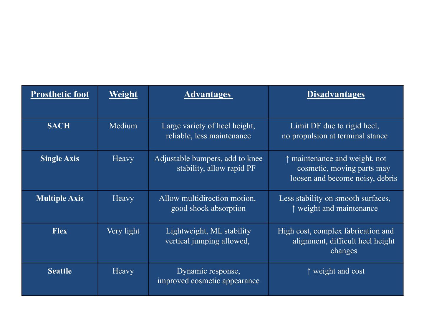
- Weight: medium
Q2.What are the pros and cons of the SACH foot?
- Pros: cheap and durable, large variety of heel height, reliable, less maintenance
- Cons: bad for uneven ground; limited DF due to rigid heel; no propulsion/push off at terminal stance
- Cons: fixed cadence; overloads the non-amputated foot
Q3.What is the elastic keel (SAFE) foot?
- Elastic keel = stationary attachment flexible endoskeleton (SAFE), a.k.a flexible keel foot - non-articulated
- Similar to SACH with a polyurethane section 45 degrees at sagittal plane to mimic the subtalar joint
- Internal keels permit triplanar movement and easy rollover, yet stiff and stable for stance and propulsion
- Disadvantage: ML instability
Q4.What are the features of a single axis articulated foot and who is it used in?
- Two rubber bumpers allow some passive DF/PF; adjustable bumpers add to knee stability and allow rapid PF
- Used in AKA only (not BKA as too heavy)
- DF bumper assists push off; weight: heavy
- Cons: not cosmetic; moving parts may loosen and become noisy; debris; increased maintenance and weight; less durable; does not adapt to uneven ground
Q5.How do multi-axis and energy storing feet differ?
- Multiple axis (heavy) adds inversion/eversion: good on uneven ground and shock absorption; less stability on smooth surfaces, increased weight and maintenance, poor durability
- Dynamic response: flexible heel, sagittal split, posterior projection of heel; allows variable cadence
- Mechanism: energy stored during heel strike re-used to spring back during push off; reduces energy expenditure; expensive
- Microprocessor foot: sensors (angle, accelerometers, gyroscopes, torque) adjust to various needs
Q6.Compare the Flex and Seattle prosthetic feet.
- Flex: very light; lightweight, ML stability, vertical jumping allowed
- Flex cons: high cost, complex fabrication and alignment, difficult heel height changes
- Seattle: heavy; dynamic response, improved cosmetic appearance
- Seattle cons: increased weight and cost
Q7.What are the design features of a dynamic response (energy storing) foot?
- Flexible heel for push off
- Sagittal split for inversion and eversion
- Posterior projection of the heel for heel strike
- Articulated vs non-articulated
- Allows variable cadence walking
▸ Slide 412 · What is orthosis?P and O · 10 questions expand
Slide render

Question list
Q1-Q1010 questions — tap to reveal all answerslist
- What is an orthosis and what are the ideal characteristics?
- How is an orthosis classified and described?
- What are the functions of an orthosis and the mechanism of an AFO?
- Compare thermosetting and thermoforming plastics.
- What are the types of AFO and their indications?
- Compare GRAFO and three point pressure AFO.
- What are the parts of an ankle foot orthosis (AFO)?
- What are the uses of a solid AFO?
- How does the GRAFO (posterior entry) work?
- What are the principles of a tone reducing AFO?
Answers · Q & A
Q1.What is an orthosis and what are the ideal characteristics?
- Device externally applied or attached to a body segment that facilitates or improves function by supporting, stabilizing, correcting or compensating for deformity or weakness
- Ideal: biomechanically effective, free of pressure, light weight, adjustable
- Ideal: cosmetically acceptable, easy to don and doff, cheap and durable
Q2.How is an orthosis classified and described?
- Corrective (tend to be hard): limit joint motion, stabilize flexible deformities
- Accommodative (tend to be soft): shock absorption, accommodate fixed deformities
- Describe by joint/region it encompasses, corrective or accommodative, static or dynamic, material
Q3.What are the functions of an orthosis and the mechanism of an AFO?
- Control angulation (hinged knee brace); control translation (PCL brace)
- Control axial force (weight relieving caliper); control line of GRF (lateral wedged shoes for cavus, GRFAFO)
- AFO mechanism: 3 point fixation (control moments about a joint, e.g. MCL); 4 point fixation (control translation, e.g. PCL); GRF
Q4.Compare thermosetting and thermoforming plastics.
- Thermosetting: Usually laminated using layers of fabric and monomer resins with catalyst, heat, vacuum/pressure; cannot reheat and remould; durable; usually used in prosthesis
- Thermosetting fabrics: fiberglass, nylon, carbon fibre, Kevlar; monomers: epoxy, polyester, polyurethane
- Thermoforming: can be reshaped after reheating; high temp softens at 120-190 degrees (polypropylene, copolymer, polyethylene)
- Medium temp plastozote; low temp orthoplast soft at <80 degrees, directly moulded on patient (hand splints)
Q5.What are the types of AFO and their indications?
- Solid: protection of ankle; anterior entry GRFAFO for knee recurvatum; prevent equinus in flaccid knee/ankle; suppress spasticity; trim line anterior to malleolus
- Posterior spring leaf for footdrop; trim line posterior to malleolus; prevents plantarflexion in swing/heel strike, allows dorsiflexion in stance
- GRAFO (posterior entry) for crouch gait with weak gastrocnemius/quadriceps and failure of knee extension-ankle plantarflexion couple; mid patella to MT; contraindicated in high tone
- Hinge for medial-lateral unstable ankle; trim anterior to malleolus; still allows dorsiflexion
- Tone reducing AFO for spastic tone; wraps foot, extends under toes; spastic inhibition bar + ML foot support
Q6.Compare GRAFO and three point pressure AFO.
- GRAFO: stance only (needs ground contact); related to shoe design/interface; joint control proximal to AFO
- GRAFO: decreases safety (uneven ground, malalignment); Increase efficiency (increase ROM)
- 3 point: swing and stance; independent of shoe design/interface; joint control within AFO
- 3 point: increases safety (stability in all planes); decreases efficiency (less ROM)
Q7.What are the parts of an ankle foot orthosis (AFO)?
- Calf shell: proximal anterior support, size of posterior support, trim line relative to malleolus
- Strap (calf/heel-retaining)
- Hinge
- Shoe insert: degree of coverage at foot
Q8.What are the uses of a solid AFO?
- Protection of the ankle (ML, prevent plantarflexion)
- Anterior entry GRFAFO for knee recurvatum
- Standing: prevents excessive plantarflexion; at heel strike GRF posterior to ankle COR produces a flexion moment at the knee
- Prevent equinus in flaccid knee and ankle
- Suppress spasticity (prevent sudden ankle dorsiflexion during terminal stance with clonus --> better toe off)
- Trim line anterior to malleolus; no hinge
Q9.How does the GRAFO (posterior entry) work?
- Indication: crouch gait with weak gastrocnemius and quadriceps with failure of the knee extension-ankle plantarflexion couple
- Static accommodative orthosis of high temp thermoforming plastic from mid patella to MT
- Uses Newton's third law to alter the GRF by controlling distal joints to alter the proximal joint
- Prevents excessive tibial anterior translation during ankle rocker with a rigid anterior tibial shell
- Prevents excessive ankle DF with a toe plate and riding ankle in neutral
- Keeps GRF anterior to the knee, creating an extension moment; stabilises knee and decreased quad workload
- Contraindicated in crouch gait due to high tone
Q10.What are the principles of a tone reducing AFO?
- For patients with spastic tone
- Well protection of whole foot and ankle; wraps around medial and lateral side and extends distally under the toes
- Prevent stimulation that will trigger increase tone over lower limb
- Spastic inhibition bar + ML foot support --> maintain STJ at stance phase --> no extensor activation
- Rigid shell --> proprioceptive feedback --> stimulate ankle dorsiflexion at swing phase
▸ Slide 413 · Upper foot dorsum coverageP and O · 4 questions expand
Slide render
Question list
Q1-Q44 questions — tap to reveal all answerslist
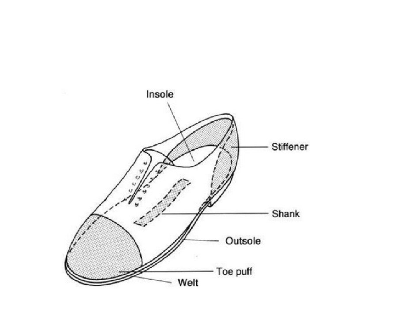
- What are the features of the shoe upper?
- What are the features of the shoe sole?
- What are the effects of a high heel and what is a heel counter for?
- What are the three point pressure principles?
Answers · Q & A
Q1.What are the features of the shoe upper?
- Height - high for ankle protection
- Closure
- Throat (= opening)
- Toe box (reinforcement) - stiffens anterior part of upper
Q2.What are the features of the shoe sole?
- Additional inner sole --> deformity adaptation
- Shank (reinforcement) - longitudinal support between in and out sole; decreases load to climber's feet in an ascent
- Heel under anatomical heel
Q3.What are the effects of a high heel and what is a heel counter for?
- High heel --> TA contract --> increase hallux valgus, hammer toes, metatarsalgia
- Heel counter stiffens posterior part of upper at anatomical heel
- Heel spur: snugly fit to heel to decrease heel abrasion
Q4.What are the three point pressure principles?
- Pressure = total force/area
- Sum of forces and bending moment = 0
- Lever arm: increase distance of load from joint --> increase moment arm --> decrease load needed for a given torque
▸ Slide 414 · This is an orthosis (which is a external device to improve function by supportinP and O · 5 questions expand

Question list
Q1-Q55 questions — tap to reveal all answerslist
- Describe the weight relieving caliper (KAFO).
- What are the components of a hinged KAFO?
- How do ring lock and bar lock knee joints work?
- What is the posterior offset knee joint and what does it require?
- What are B Joseph's principles of orthosis?
Answers · Q & A
Q1.Describe the weight relieving caliper (KAFO).
- Proximal circumferential brim moulded like a femoral prosthesis socket
- Medial and lateral uprights shaped to the patient's LL: it allows the weight to bypass the LL to be transferred to the shoe
- Bar lock at knee; adjustable bars for length; leather straps at mid thigh and mid leg
- Aims: Providing stability; Correcting/ preventing deformity; relieve weight bearing, relieve pain
- Mainly for NM weakness (polio, CP)
Q2.What are the components of a hinged KAFO?
- Thigh shell - ischial bearing, anchors into thigh for security/rotational control; cannot control pistoning
- Knee axis single or multi; flexion control - lock (drop lock safest, most durable; barlock auto locks in extension but poor valgus and varus control) or free (posterior offset)
- Deformity control (knee cap): sagittal pretibia/supracondylar pad; frontal calf shell extension
- Medial + lateral uprights; solid AFO +/- custom moulded insole
Q3.How do ring lock and bar lock knee joints work?
- Ring lock (drop lock): actively push down to lock; requires sufficient finger power
- Barlock (Swiss lock): spring loaded release bar; semi auto, auto locks at knee extension
- Sitting/manual lifting up of bail to unlock uprights --> knee flexion
Q4.What is the posterior offset knee joint and what does it require?
- No locking
- Requires quad power grade 3 or above
- Orthosis COR posterior to knee COR
- Knee flexion at swing/sitting; active knee extension locks orthosis
Q5.What are B Joseph's principles of orthosis?
- Leave as many joints as possible free
- As light as possible
- Contact areas between orthosis and limb should be as large as possible
- Attempt to discard the orthosis by the time child is skeletally mature
▸ Slide 415 · LeftP and O · 4 questions expand

Question list
Q1-Q44 questions — tap to reveal all answerslist
- What are the components of a reciprocal gait orthosis (RGO)?
- How does the RGO produce gait?
- What keeps the torso erect in an RGO and why is it needed?
- What is an HKAFO and how does the patient mobilise?
Answers · Q & A
Q1.What are the components of a reciprocal gait orthosis (RGO)?
- Pelvic and thorax straps
- Isocentric bar/cables connect 2 hips - lock limb at stance, allow limb flexion at swing
- Hip and knees manual locking
- Solid AFO
Q2.How does the RGO produce gait?
- Limb advancement by torso rotation
- Cable tightened at swing
- 3 way linkage (torso-hip-knee): movement at one side --> contralateral side locks at extension, preventing torso collapse
- Weight shift at double support --> cable loose --> allows contralateral advancement
- Manual cable loosening for sitting
Q3.What keeps the torso erect in an RGO and why is it needed?
- Pelvic and thorax straps
- Compensates for weak hip extensors
Q4.What is an HKAFO and how does the patient mobilise?
- Components: pelvic +/- thoracic band, hip joints, lateral uprights, KAFO
- Mostly standing
- Locomotion: swing to/swing through gait
- Needs hand support
- Very energy consuming
▸ Slide 416 · This is a UCBL insertP and O · 3 questions expand

Question list
Q1-Q33 questions — tap to reveal all answerslist
- What is a UCBL insert?
- What are the design characteristics of a UCBL insert?
- What force system does a UCBL insert use?
Answers · Q & A
Q1.What is a UCBL insert?
- Type of corrective, static orthosis
- Rigid plastic insert with total contact design to stabilize flexible foot deformity
Q2.What are the design characteristics of a UCBL insert?
- Heel cup encompasses the heel and midfoot
- Medial calcaneal grip merges with contour of the foot arch
- Fabricated over a cast of the foot held in maximal manual correction
Q3.What force system does a UCBL insert use?
- 3 point force system to control forefoot abduction
- Grip on sustentaculum tali, lateral forefoot control and medial arch support
▸ Slide 417 · This is a spinal orthosisP and O · 7 questions expand

Question list
Q1-Q77 questions — tap to reveal all answerslist
- What is the Milwaukee brace and when is it indicated?
- What are the components of the Milwaukee brace?
- What is the principle of correction of the Milwaukee brace?
- What is the dose-dependent effect and success rate of the Milwaukee brace?
- What are the contraindications to Milwaukee bracing?
- What are the weaning indications for a Milwaukee brace?
- What are the complications of the Milwaukee brace?
Answers · Q & A
Q1.What is the Milwaukee brace and when is it indicated?
- Spinal orthosis
- For scoliosis with apex above T7
Q2.What are the components of the Milwaukee brace?
- Thermoplastic pelvic girdle - most important, custom made, stable base; avoids iliac crest, allows hip flexion, clears seat when sitting
- Two posterior and one anterior upright
- Outrigger corrective pads (lumbar/thoracic/trapezius/kyphosis); axillary ring NOT for correction
- Neck ring with throat and occipital padding pieces
Q3.What is the principle of correction of the Milwaukee brace?
- 3 point fixation
- At lumbar apical vertebrae
- At rib of thoracic apex
- Plus relief of pressure on the opposite side
Q4.What is the dose-dependent effect and success rate of the Milwaukee brace?
- Dose dependent effect >18 hours
- Success 90%
Q5.What are the contraindications to Milwaukee bracing?
- Disease: curves <20 degrees (not indicated); >50 degrees (not effective); short sharp curve
- Disease: loss of thoracic kyphosis (hypokyphosis or lordosis)
- Patient: skeletal maturity (Risser >4); predisposing emotional problems; sensory impairment; OI
Q6.What are the weaning indications for a Milwaukee brace?
- Menarche >2 years
- Height plateau x 1.5 years
- Risser >4
- TWIII RUS to DR and DU
Q7.What are the complications of the Milwaukee brace?
- Psychological and social
- Physical: pressure sore, pain, irritation of the anterior cutaneous nerve of thigh
- Physical: dental problem and under development of mandible
- Progression of the curve is a concern
▸ Slide 418 · TLSO with asymmetry: Boston braceP and O · 4 questions expand

Question list
Q1-Q44 questions — tap to reveal all answerslist
- What is a Boston brace and when is it used?
- How does the Boston brace correct deformity?
- How is the Boston brace made?
- How do you prevent complications of orthotics at the interface?
Answers · Q & A
Q1.What is a Boston brace and when is it used?
- TLSO with asymmetry; external spine orthosis
- Used in flexible scoliosis with apex at T8 or below
- Controls curve by 3 point fixation and HV principle
Q2.How does the Boston brace correct deformity?
- Sagittal: control lumbar lordosis to control forward pelvic tilting
- Coronal: padding applies pressure on paravertebral muscle or articulating rib to control the curve
- 3. longitudinal: intra abdominal pressure to aim for correction and distraction
Q3.How is the Boston brace made?
- Made from high temperature thermoplastic (NOT thermosetting), from cast of patient in corrected position
- Apply dynacast on trunk of patient (= negative mould); apply thermoplastics on plaster cast
- Liner (soft, semi-rigid) - urethane, silicone, pelite; padding over pressure points
- Underarm brace: for double curve
Q4.How do you prevent complications of orthotics at the interface?
- Maximise lever arm
- Maximise surface contact area and conformity
- Protect bony prominence
- Moist absorbant lining