9 slides
▸ Slide 392 · OthersOthers · 2 questions expand
Slide render

Question list
Q1-Q22 questions — tap to reveal all answerslist
- Which section of the course does this slide belong to?
- What source material is available for this slide?
Answers · Q & A
Q1.Which section of the course does this slide belong to?
- Others - miscellaneous topics
- Not covered in the speaker notes
Q2.What source material is available for this slide?
- Slide image only - no speaker notes
- Not in the speaker notes
▸ Slide 393 · Longitudinal(incision(preferred,(avoid(damage(Gluteal(nerve(and(skin(necrosis(Others · 3 questions expand
Slide render
Question list
Q1-Q33 questions — tap to reveal all answerslist
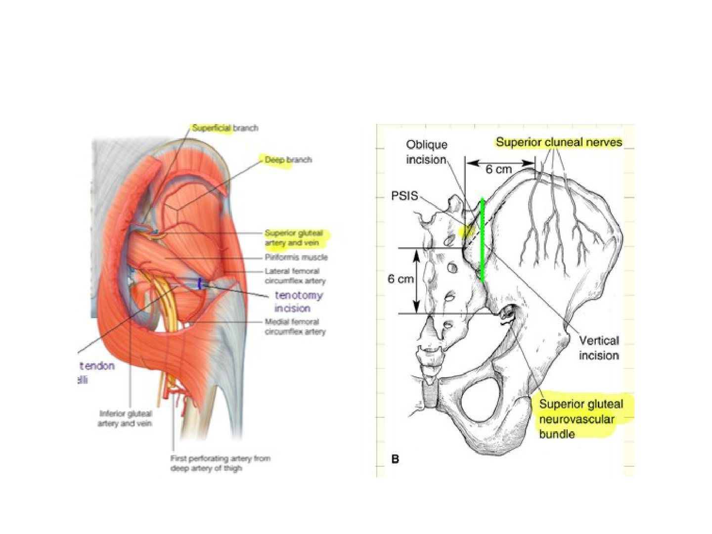
- Why is a longitudinal incision preferred in this approach?
- Describe the incision used.
- What structures are at risk of damage in this approach?
Answers · Q & A
Q1.Why is a longitudinal incision preferred in this approach?
- Avoids damage to the gluteal nerve
- Avoids skin necrosis
Q2.Describe the incision used.
- Extends from 1cm lateral to the PSIS
- Upwards to 2cm above the crest
- Elevate gluteus medius/periosteum
Q3.What structures are at risk of damage in this approach?
- Cluneal nerve
- Superior gluteal vessels
▸ Slide 394 · Lateral cutaneous nerve of thighOthers · 6 questions expand
Slide render
Question list
Q1-Q66 questions — tap to reveal all answerslist
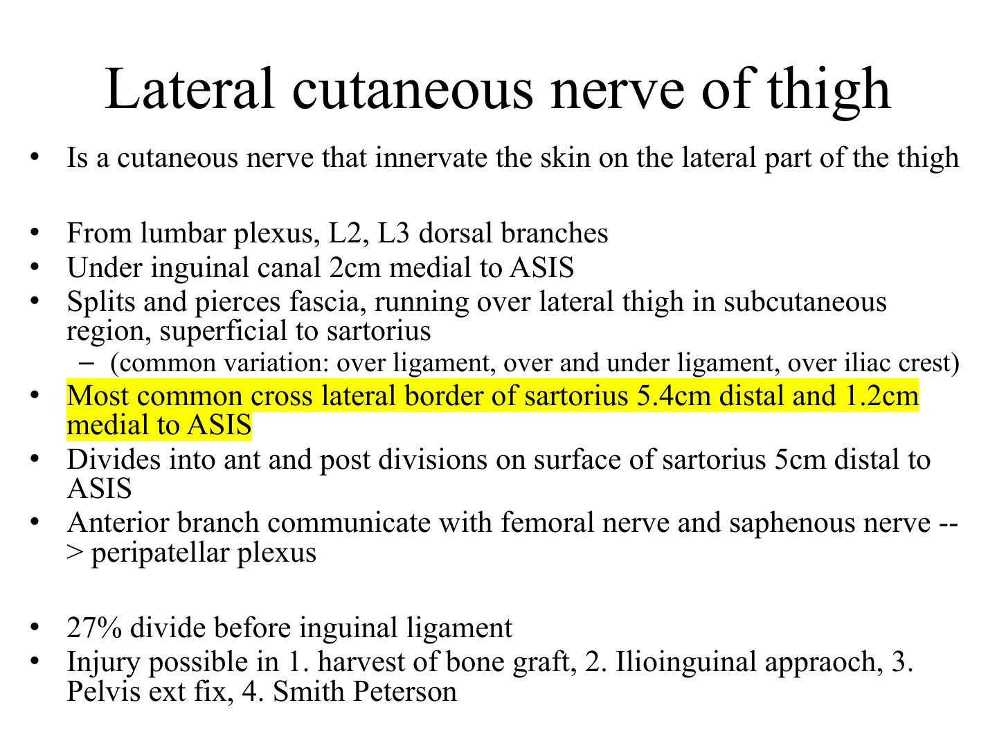
- What is the lateral cutaneous nerve of the thigh and from where does it arise?
- Describe the course of the lateral cutaneous nerve of the thigh into the thigh.
- Where does the nerve cross and divide?
- What are the common anatomical variations of the nerve?
- What does the anterior branch communicate with?
- Where can the lateral cutaneous nerve of the thigh be injured, and which condition does it cause?
Answers · Q & A
Q1.What is the lateral cutaneous nerve of the thigh and from where does it arise?
- Is a cutaneous nerve that innervates the skin on the lateral part of the thigh
- From the lumbar plexus, L2, L3 dorsal branches
Q2.Describe the course of the lateral cutaneous nerve of the thigh into the thigh.
- Passes under the inguinal canal, 2 cm medial to the ASIS
- Splits and pierces the fascia, running over the lateral thigh in the subcutaneous region, superficial to sartorius
Q3.Where does the nerve cross and divide?
- Most commonly crosses the lateral border of sartorius 5.4 cm distal and 1.2 cm medial to ASIS
- Divides into anterior and posterior divisions on the surface of sartorius 5 cm distal to ASIS
- 27% divide before the inguinal ligament
Q4.What are the common anatomical variations of the nerve?
- Over the ligament
- Over and under the ligament
- Over the iliac crest
Q5.What does the anterior branch communicate with?
- Communicates with the femoral nerve and saphenous nerve --> peripatellar plexus
Q6.Where can the lateral cutaneous nerve of the thigh be injured, and which condition does it cause?
- 1. Harvest of bone graft
- 2. Ilioinguinal approach
- 3. Pelvis external fixation
- 4. Smith-Peterson approach
- Injury causes meralgia paresthetica
▸ Slide 395 · Cast wedgingOthers · 3 questions expand
Slide render

Question list
Q1-Q33 questions — tap to reveal all answerslist
- What is the indication and timing for cast wedging?
- Describe the open wedging technique.
- Where is the wedge placed relative to the deformity?
Answers · Q & A
Q1.What is the indication and timing for cast wedging?
- To correct small angular malalignments
- Delayed 3-4 days until the cast is fully hardened
Q2.Describe the open wedging technique.
- Half-circumferential cut with a 1cm bridge
- Wedge applied on the concave side
- Cuts perpendicular to the plane of angulation; bridge lies on the axis of rotation
- Cork keeps the wedge open; fresh POP bandaging ~5cm distal and proximal
Q3.Where is the wedge placed relative to the deformity?
- On the concave side of the angulation
- The 1cm bridge lies on the axis of rotation
▸ Slide 396 · 50% - die from non-survivable injuries immediately, or within minutesOthers · 4 questions expand
Question list
Q1-Q44 questions — tap to reveal all answerslist
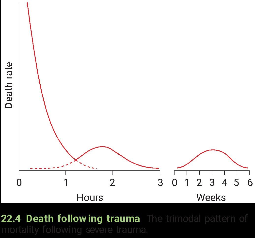
- How are trauma deaths distributed over time according to the slide?
- What characterises the first peak of trauma deaths?
- What characterises the second peak of trauma deaths?
- What characterises the third peak of trauma deaths?
Answers · Q & A
Q1.How are trauma deaths distributed over time according to the slide?
- 50% - die from non-survivable injuries immediately, or within minutes
- 30% - survive the initial trauma, but die within 1-3 hours
- 20% - die from complications at a late stage during the 6 weeks after injury
Q2.What characterises the first peak of trauma deaths?
- 50% of deaths
- Due to non-survivable injuries, immediately or within minutes
Q3.What characterises the second peak of trauma deaths?
- 30% of deaths
- Survive the initial trauma but die within 1-3 hours
Q4.What characterises the third peak of trauma deaths?
- 20% of deaths
- Die from complications at a late stage, during the 6 weeks after injury
▸ Slide 397 · Non unionOthers · 12 questions expand
Slide render
Question list
Q1-Q1212 questions — tap to reveal all answerslist
- What is the FDA definition of non union and the definition of delayed union?
- How is delayed union treated?
- Describe the Paley and descriptive classifications of non union.
- What are the causes of non union according to the diamond concept?
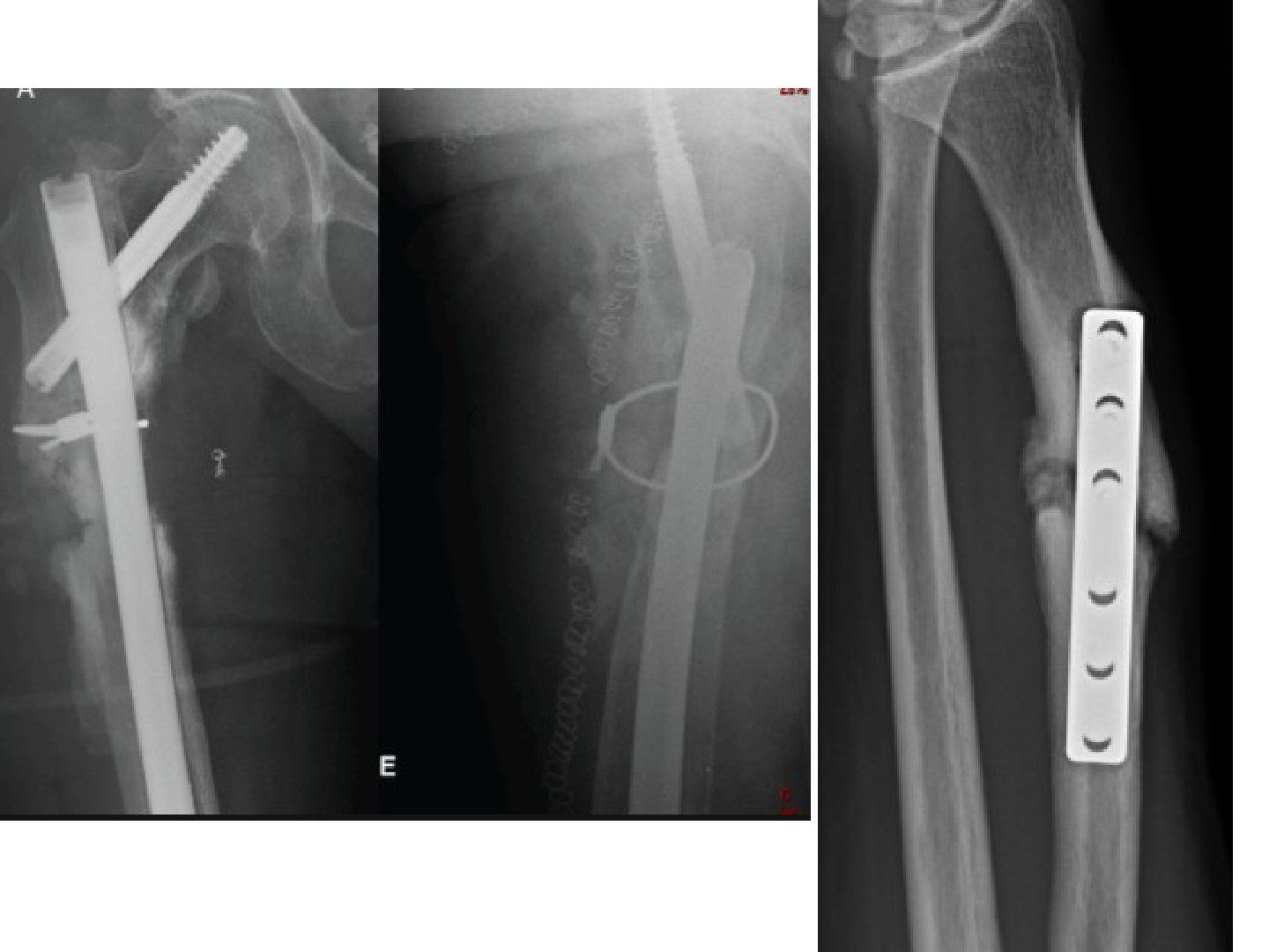
- Describe the X-ray findings and postulated causes in this subtrochanteric non union.
- How is this subtrochanteric non union managed?
- How is non union assessed?
- What are the management principles in non union?
- What are the local causes of atrophic versus hypertrophic non union?
- Describe the X-ray findings and management in the distal radius non union.
- How are bone defects managed?
- Why is exchange nailing used for non union and what are the results?
Answers · Q & A
Q1.What is the FDA definition of non union and the definition of delayed union?
- FDA: fracture at least 9 months old with no signs of healing for 3 consecutive months
- Delayed union: bone unable to achieve union in the expected time frame (6 months)
Q2.How is delayed union treated?
- Conservative: shock wave USG (early, 2-3 months), vibration (train muscle mass, proprioception, promote union)
- Electrical stimulation - increased expression of BMP2,7 and osteoblast proliferation (direct current, pulsed electromagnetic field)
- Operative: dynamization, bone graft, bone marrow
Q3.Describe the Paley and descriptive classifications of non union.
- Paley type 1: <1cm defect (then lax/stiff/deformity)
- Paley type 2: >1cm defect (shortening/gap/both)
- Descriptive: atrophic, oligotrophic, hypertrophic (horse hoof/elephant foot), pseudarthrosis
- Also infective non union
Q4.What are the causes of non union according to the diamond concept?
- Diamond concept: inductive, conductive, osteogenic, host and vascular factors
- Mechanical instability: inadequate fixation, bone loss, poor bone quality
- Inadequate vascularity: severe soft tissue injury with periosteal stripping, malalignment, bone loss, distraction
- Poor bone contact: soft tissue interposition, malalignment, bone loss, distraction
Q5.Describe the X-ray findings and postulated causes in this subtrochanteric non union.
- Previous subtrochanteric fracture fixed with a long cephalomedullary device + 1 cerclage wire
- No healing, bone defect, varus alignment, no broken implant
- Atrophic non union
- Biology: watershed area; mechanical: wide medullary canal, large deforming force; varus increases the bending moment, leading to increase in fracture strain per unit length; the region experiences the highest tensile stress according to Koch's diagram
Q6.How is this subtrochanteric non union managed?
- Deal with both the biological and mechanical problems
- Exchange nail: larger nail provides more stability
- Reaming provides bone graft
Q7.How is non union assessed?
- Rule out infection (initial open injury, wound infection, erythema/sinus, blood tests)
- Delineate the cause: atrophic vs hypertrophic non union
- Atrophic: biological +/- mechanical causes; consider periosteal/endosteal blood supply
- r/o complication due to non union (broken implant, AVN, arthritis)
Q8.What are the management principles in non union?
- Alignment, stability, bone potential to heal, +/- adjacent joint stiffness
- Rule out infection
- Address patient/systemic factors: DM, nutrition, smoking, compliance
- Formulate a plan: bring in blood supply, optimal reduction, stable fixation, +/- manage bone defect
Q9.What are the local causes of atrophic versus hypertrophic non union?
- Atrophic - bone factors: vulnerable blood supply, minimal bone contact, high stress, no periosteum
- Atrophic - fracture factors: open injury, butterfly fragment with no blood supply
- Atrophic - surgeon factors: diminished blood supply (distruption of endosteum + periosteum blood supply) - disruption of endosteal/periosteal supply, non-optimal fixation method
- Hypertrophic - bone factors: minimal bone contact, high stress, no periosteum
- Hypertrophic - fracture factors: degree of initial comminution --> poor initial stability if fixed with load sharing device
- Hypertrophic - surgeon factors: wrong implant, poor reduction, poor fixation skill
Q10.Describe the X-ray findings and management in the distal radius non union.
- Fracture shaft of distal radius fixed with 6-hole DCP with compression
- Positive callus, fracture gap still seen; need proper AP + lateral X-ray to delineate alignment; no ulnar fracture
- One distal screw very near the fracture gap
- Hypertrophic/oligotrophic non union
- Management: rule out infection, delineate cause (initial malreduction, unstable fixation), whole forearm X-ray, revision plating + bone graft
Q11.How are bone defects managed?
- Further management depends on size of defect and integrity of soft tissue coverage, vascularity of tissue bed
- Small <6cm: acute shortening, non-vascularised bone graft, Papineau technique
- Intermediate up to 12cm: vascularised bone graft (e.g. fibular), can also provide soft tissue coverage in same flap
- Large >6cm: acute shortening + distraction osteogenesis, bone transport, Masquelet technique
- Amputation for significant bone/soft tissue/NV defect or patients unfit for multiple surgery
Q12.Why is exchange nailing used for non union and what are the results?
- Largest series by group in Edinburgh, published in 2016 BJJ: union 75% after 1st exchange, rising to 95% after 2nd exchange
- In infected non union: 35% after 1st, 65% after second
- Works by 3 mechanisms: larger nail improves mechanical stability; Reaming increases periosteal blood flow – stimulate the formation of periosteal new bone; reaming products are osteoinductive
- Simple JBJS 2016: the strongest predictor of failure of exchange nailing was infection and suggested other treatment options such as ilizarov treatment should be preferred
▸ Slide 398 · 1/12 post opOthers · 11 questions expand
Slide render

Question list
Q1-Q1111 questions — tap to reveal all answerslist
- Describe the X-ray findings and clinical presentation in this infected non union.
- What causes of non union are listed (diamond concept)?
- What are the risk factors for infected non union?
- What investigations are used in suspected infected non union?
- What are the general principles for managing an infected non union with an implant?
- What is the role of ESR/CRP and imaging in acute infected non union?
- How is an infected non union with a stable implant managed?
- How is an infected non union with an unstable implant managed and how is infection timing classified?
- How does infected non union differ from prosthetic joint infection in terms of implant retention?
- What are the principles of management in infected non union?
- What is the Masquelet technique?
Answers · Q & A
Q1.Describe the X-ray findings and clinical presentation in this infected non union.
- Infected nonunion: previous fracture of the tibia, fibula and syndesmosis, fixed with plates + syndesmosis screw
- Tibia reduction acceptable; syndesmosis widening (reduced tibiofibular overlap, increased clear space)
- Periosteal elevation over the lateral tibia, no lucency around implant, fracture not healed
- Suspected infected implant
- At 1/12 post op: pain, redness
Q2.What causes of non union are listed (diamond concept)?
- Mechanical instability: inadequate fixation, Bone loss, Poor bone quality
- Inadequate vascularity: severe soft tissue injury with periosteal stripping, malposition or malalignment, bone loss, distraction
- Poor bone contact: soft tissue interposition, malposition or malalignment, bone loss, distraction
Q3.What are the risk factors for infected non union?
- Patient: old, smoking, DM, PVD, previous injury
- Injury: open fracture, wound comminution
- Surgeon: multiple operations, stripped periosteum, bone necrosis during drilling
Q4.What investigations are used in suspected infected non union?
- Blood: ESR, CRP, albumin
- CT and USG for collection
- Gallium scan
- Intraoperative deep cultures are most reliable
Q5.What are the general principles for managing an infected non union with an implant?
- Confirm infection, assess implant stability and bone union
- Healed -> remove implant
- Not healed + unstable -> remove and keep stable
- Not healed + stable -> antibiotics
Q6.What is the role of ESR/CRP and imaging in acute infected non union?
- ESR, CRP: 100% PPV, 80% NPV
- CT and USG for subperiosteal and intramedullary collection
- Debridement and intraoperative culture, start antibiotics afterwards
Q7.How is an infected non union with a stable implant managed?
- Retain the implant + debridement
- PMMA antibiotic to fill defects and coat the implant
- Antibiotics x 3 months (BJJ 2021 Shen) + soft tissue coverage
- Second stage after 6-8 weeks: remove cement/implant, revision fixation + autograft
Q8.How is an infected non union with an unstable implant managed and how is infection timing classified?
- Remove implant + debridement + external fixation
- Gentamicin beads/cement
- Acute <2 weeks, delayed 2-10 weeks, chronic >10 weeks
- Low grade infection can coexist with a healing fracture
Q9.How does infected non union differ from prosthetic joint infection in terms of implant retention?
- Aim in fracture-related infection: stability until union
- Retention is successful in 50-90% of infected fracture implants
- Joint replacement: only 15-50% successful (aimed at long-term function)
- The concept is different from joint replacement infection
Q10.What are the principles of management in infected non union?
- Optimise host factors
- Identify the microorganism involved
- Debridement + antibiotics
- Reconstruction: soft tissue coverage + bone defect management (antibiotic beads for small defects, Masquelet for larger)
Q11.What is the Masquelet technique?
- Antibiotic-impregnated cement beads/spacer deliver local high-dose antibiotics
- Reaming also debrides; circular external fixator allows shortening of any defect
- Soft tissue envelope reconstructed with a vascularised flap
- Second-stage bone grafting at ~8 weeks to the pseudomembranous tube (prevents graft resorption)
▸ Slide 399 · D1 post opOthers · 5 questions 1 check expand

Question list
Q1-Q55 questions — tap to reveal all answerslist
- What is the likely timing and reported incidence of this periprosthetic fracture?
- Describe the Vancouver classification as given for periprosthetic fractures.
- How is a periprosthetic femoral fracture managed according to stem stability?
- What are the risk factors and management of acetabular-side periprosthetic fractures?
- How can periprosthetic fractures be prevented?
Answers · Q & A
Q1.What is the likely timing and reported incidence of this periprosthetic fracture?
- Day 1 post op; likely an intra-operative fracture
- Incidence 4% cementless, 0.4% cemented, 0.1% post op
- Stem appears stable, bone stock adequate
Q2.Describe the Vancouver classification as given for periprosthetic fractures.
- Type A diaphyseal
- Type B metaphyseal
- Type C distal to stem (not amendable to longest stem)
- Subtypes: cortical perforation, undisplaced, displaced
Q3.How is a periprosthetic femoral fracture managed according to stem stability?
- Stable stem -> fix
- Unstable stem -> revise with a long stem
- Lateral plating: at least 3 screws and 3 cables (cables resist bending; screws resist rotation and control length)
Q4.What are the risk factors and management of acetabular-side periprosthetic fractures?
- Patient: osteoporosis, radiotherapy, dysplasia/fusion, revision surgery
- Surgeon: underream
- Implant: cementless, elliptical cup
- Stable -> protected WB 12 weeks; unstable -> add screw/jumbo cup/fix + protected WB 12 weeks
Q5.How can periprosthetic fractures be prevented?
- Pre-op planning
- Careful exposure
Fact check
Vancouver classification types are A diaphyseal, B metaphyseal, C distal to stem — non-standard mapping — Standard (postoperative) Vancouver classification: A = trochanteric region (AG/AL), B = around/just below the stem (B1 well-fixed, B2 loose, B3 loose + poor bone), C = well below the stem — medium confidence — source
▸ Slide 400 · FlapOthers · 7 questions expand
Question list
Q1-Q77 questions — tap to reveal all answerslist
- Describe the Mathes classification of muscle flaps with examples.
- How are flaps classified by location, blood supply and composition?
- Which systemic and design factors are checked before raising a flap?
- Describe the intra-operative principles of flap surgery.
- How is flap circulation monitored after surgery and what is done if it becomes impaired?
- Which pedicled flaps are used for proximal, middle and distal leg defects?
- List common free flaps and their pedicles.
Answers · Q & A
Q1.Describe the Mathes classification of muscle flaps with examples.
- I: 1 dominant (tensor fascia lata)
- II: 1 dominant + 1 minor (gracilis)
- III: 2 dominant (gluteus maximus)
- IV: multiple minor (sartorius)
- V: 1 dominant + multiple minor (latissimus dorsi)
Q2.How are flaps classified by location, blood supply and composition?
- Location: local (transposition, rotational, advancement, axial), regional (same limb), distant, free
- Blood supply: random or axial
- Composite: cutaneous, fasciocutaneous, musculocutaneous, osseocutaneous
Q3.Which systemic and design factors are checked before raising a flap?
- Systemic: smoking, obesity, hypertension, PVD, immunosuppression
- Flap design: type, size, pivot point, axis, arc of rotation
- Doppler +/- to identify the pedicle; know pedicle type (Mathes I-V), with I, III, V more reliable and preferred with a broad base
Q4.Describe the intra-operative principles of flap surgery.
- Elevation: preserve minor pedicles; complete pedicle mobilisation not required unless free flap; avoid tension during rotation and inset
- Insetting: tunnel size = flap base x2, avoid tension, haemostasis
Q5.How is flap circulation monitored after surgery and what is done if it becomes impaired?
- Monitor flap circulation initially
- If impaired: consider systemic haemodynamic problem or local arterial/venous obstruction
- Can try to release stitches and reposition the flap
- Avoid pressure, constrictive bandage and motion (splint)
Q6.Which pedicled flaps are used for proximal, middle and distal leg defects?
- Proximal: medial gastrocnemius (medial sural artery)
- Middle: soleus (branches of posterior tibial and peroneal arteries)
- Distal: distant/reverse sural flap (superficial sural artery via peroneal perforator)
Q7.List common free flaps and their pedicles.
- LD flap (thoracodorsal artery), gracilis (medial circumflex femoral), serratus anterior (subscapular)
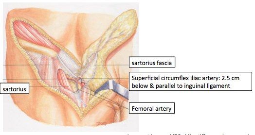
- Groin flap (superficial circumflex iliac, 2.5cm inferior to inguinal ligament)
- ALT flap (descending branch of lateral circumflex femoral artery)
- Radial forearm flap (radial artery; skin +/- muscle +/- bone)