▸ Slide 296 · TraumaTrauma · 3 questions expand
Q1-Q33 questions — tap to reveal all answerslist
- What is the definition of polytrauma?
- What are the priorities of the primary survey in a trauma patient?
- What are the common life-threatening injuries in major trauma?
Q1.What is the definition of polytrauma?
- Not covered in the speaker notes
Q2.What are the priorities of the primary survey in a trauma patient?
- Not covered in the speaker notes
Q3.What are the common life-threatening injuries in major trauma?
- Not covered in the speaker notes
▸ Slide 297 · DCO vs ETC ISSTrauma · 19 questions expand

Q1-Q1919 questions — tap to reveal all answerslist
- What do DCO, ETC and ISS stand for in this slide title?
- Which classification is listed for open fractures?
- Which scores are listed for a mangled limb?
- Which classifications are listed for pelvic fractures?
- Which classification is listed for acetabular fractures?
- Which classifications are listed for hip dislocation?
- Which classifications are listed for femoral neck fractures (NOF)?
- Which classification is listed for trochanteric fractures (TOF)?
- Which classification is listed for subtrochanteric fractures?
- Which body defines an atypical femoral fracture (AFF)?
- Which classification is listed for femoral shaft fractures?
- Which classification is listed for a floating knee?
- Which classifications are listed for distal femur fractures?
- Which classification is listed for tibial plateau fractures?
- Which classification is listed for pilon fractures?
- Which classification is listed for ankle fractures?
- Which classifications are listed for calcaneal fractures?
- Which classifications are listed for talar neck fractures?
- Which classification is listed for Lisfranc injuries?
Q1.What do DCO, ETC and ISS stand for in this slide title?
- The slide is titled DCO vs ETC ISS
- Not covered in the speaker notes - the slide only lists classifications
Q2.Which classification is listed for open fractures?
- Gustilo classification
Q3.Which scores are listed for a mangled limb?
- MESS
- LEAP
Q4.Which classifications are listed for pelvic fractures?
- Young & Burgess
- Tile
Q5.Which classification is listed for acetabular fractures?
- Judet & Letournel (Latournal) classification
Q6.Which classifications are listed for hip dislocation?
- Epstein
- Pipkin
Q7.Which classifications are listed for femoral neck fractures (NOF)?
- Garden
- Pauwel
- Leighton
Q8.Which classification is listed for trochanteric fractures (TOF)?
- Kyle classification
Q9.Which classification is listed for subtrochanteric fractures?
- Russell Taylor classification
Q10.Which body defines an atypical femoral fracture (AFF)?
- American Society for Bone & Mineral Research
Q11.Which classification is listed for femoral shaft fractures?
- Winquist & Hansen
- The list also includes NOF + shaft
Q12.Which classification is listed for a floating knee?
- Fraser classification
Q13.Which classifications are listed for distal femur fractures?
- AO
- Hoffa
Q14.Which classification is listed for tibial plateau fractures?
- Schatzker classification
Q15.Which classification is listed for pilon fractures?
- Reudi & Allgowa classification
Q16.Which classification is listed for ankle fractures?
- Lauge Hansen classification
Q17.Which classifications are listed for calcaneal fractures?
- Essex Lopresti
- Sander
- Bohler
- Gissane
- Broden
Q18.Which classifications are listed for talar neck fractures?
- Hawkin
- Canale
Q19.Which classification is listed for Lisfranc injuries?
- Myerson classification
▸ Slide 298 · fatigue fracture and fragility fractureTrauma · 19 questions expand
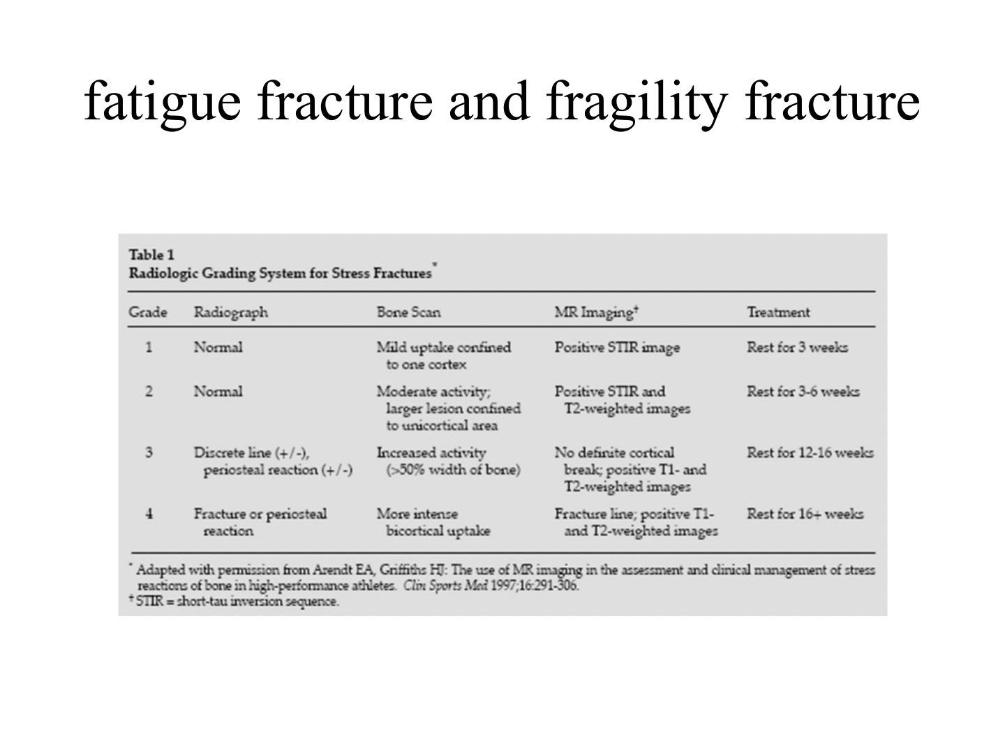
Q1-Q1919 questions — tap to reveal all answerslist
- What is a stress fracture?
- Differentiate an insufficiency fracture from a fatigue fracture.
- Describe the pathogenesis of stress fractures.
- Which femoral neck stress fractures are more serious and how are they treated?
- What is the concern with a femoral shaft stress fracture?
- What is notable about pelvic stress fractures?
- What is the presentation of a stress fracture?
- What is the fulcrum test used for?
- What are the X-ray findings and timing in a stress fracture?
- What is the role of MRI in stress fractures?
- What are the bone scan findings in an acute stress fracture, a healing fracture and a soft tissue injury?
- What is the general management of a stress fracture?
- What is risk stratification in stress fracture management?
- Which is the most common stress fracture and where does the compression type begin?
- Describe the tibial tension stress fracture.
- Why do tibial tension fractures predispose to complete fracture, delayed union or nonunion?
- How are tibial tension stress fractures treated?
- How is a femoral neck stress fracture managed?
- Where do 5th metatarsal stress fractures occur and how are they managed?
Q1.What is a stress fracture?
- A fracture of bone at stresses below the ultimate tensile strength
Q2.Differentiate an insufficiency fracture from a fatigue fracture.
- Insufficiency fracture: physiological stress on a weakened bone (normal stress, abnormal bone)
- Fatigue fracture: repetitive loading below the ultimate tensile stress on normal bone (abnormal stress, normal bone)
Q3.Describe the pathogenesis of stress fractures.
- Bone formation lags behind bone resorption
- Ultimate tensile strength reduced, rendering the bone susceptible to microfractures
- This propagates to a stress fracture
Q4.Which femoral neck stress fractures are more serious and how are they treated?
- Tension fractures are more serious
- They may require operation
Q5.What is the concern with a femoral shaft stress fracture?
- It responds to protected weight bearing
- It can progress to a complete fracture if unrecognised
Q6.What is notable about pelvic stress fractures?
- They are rare
- They involve the sacrum and pelvis
Q7.What is the presentation of a stress fracture?
- History of overuse
- Insidious onset of pain
- Localised swelling and tenderness
Q8.What is the fulcrum test used for?
- It is used for a femoral shaft stress fracture
Q9.What are the X-ray findings and timing in a stress fracture?
- XR is normal in the first 2-3 weeks
- Later: periosteal reaction, thin fracture line, cortical lucency
Q10.What is the role of MRI in stress fractures?
- MRI (STIR sequence) is the most specific test but is not specific
Q11.What are the bone scan findings in an acute stress fracture, a healing fracture and a soft tissue injury?
- Bone scan is highly sensitive but non-specific
- Acute stress fracture: linear area of increased uptake at all 3 phases
- Healing fracture: back to normal at phase I, II and III sequentially
- Soft tissue injury: uptake in the first 2 phases
Q12.What is the general management of a stress fracture?
- Rule out anorexia nervosa or metabolic disease
- Nonoperative: protected weight bearing, rest, cross training, analgesics, therapeutic modalities
Q13.What is risk stratification in stress fracture management?
- Low-risk fracture
- High-risk fracture
Q14.Which is the most common stress fracture and where does the compression type begin?
- Tibial stress fracture - the most common
- Compression fracture begins at the posteromedial (PM) cortex
- Transverse in orientation, common in distance runners
- Treated nonoperatively with pneumatic bracing
Q15.Describe the tibial tension stress fracture.
- Anterior cortex of the middle 1/3 of the tibia
- XR features are subtle but better seen on the lateral view - a V-shaped defect at the anterior cortex
- Common in repetitive jumping and heaping activities
Q16.Why do tibial tension fractures predispose to complete fracture, delayed union or nonunion?
- The tension side effect (Wolff's law)
- Hypovascularity of the anterior cortex
Q17.How are tibial tension stress fractures treated?
- Initial: trial of rest +/- immobilisation
- Once a wide fissure develops, operative treatment is needed
- Delayed/nonunion: excision and bone grafting, or IMN
Q18.How is a femoral neck stress fracture managed?
- Compression type -> nonoperative
- Tension type -> operative
Q19.Where do 5th metatarsal stress fractures occur and how are they managed?
- At the metadiaphyseal junction
- Sx+ / XR- -> NWB + brace
- Sx+ / XR+ -> low demand: NWB; high demand: IM screw
- Delayed union -> intramedullary screw fixation
▸ Slide 299 · Osteopetrosis with pathological subtrochanteric fractureTrauma · 11 questions expand

Q1-Q1111 questions — tap to reveal all answerslist
- Describe the X-ray findings in this osteopetrotic subtrochanteric fracture.
- What is the differential diagnosis of this sclerotic fracture?
- What other X-rays should be requested and what do they show in osteopetrosis?
- What is pyknodysostosis?
- What is the pathophysiology of osteopetrosis?
- How do patients with osteopetrosis present?
- What are the types of osteopetrosis?
- What investigations are performed in osteopetrosis?
- What medical treatment is used in osteopetrosis?
- What are the special considerations when fixing a fracture in osteopetrosis?
- What are the special considerations for replacement arthroplasty in osteopetrosis?
Q1.Describe the X-ray findings in this osteopetrotic subtrochanteric fracture.
- Polyostotic sclerotic bone
- Bone in bone pelvis
- Pathological fracture with lateral beaking and medial spike, non-comminuted, subtrochanteric, short oblique
- Suspect underlying osteopetrosis
Q2.What is the differential diagnosis of this sclerotic fracture?
- Metastasis
- Hypoparathyroidism
- Paget's disease
- Pyknodysostosis
Q3.What other X-rays should be requested and what do they show in osteopetrosis?
- Spine: rugger jersey appearance
- Hand: bone in bone
- Lower limb: Erlenmeyer flask appearance
Q4.What is pyknodysostosis?
- Autosomal recessive trait with mutation in cathepsin K
- Generalized osteosclerosis like osteopetrosis but with preservation of the medullary canal of long bones
Q5.What is the pathophysiology of osteopetrosis?
- Defect in osteoclastic resorption due to defective carbonic anhydrase / chloride channel
- Osteoclasts also lack a normal ruffled border and clear zone
Q6.How do patients with osteopetrosis present?
- Fracture (cartilage remains in the diaphysis -> inferior mechanical properties)
- Deformity from repeated fractures
- Infection (osteomyelitis)
- Pancytopenia
- Cranial nerve palsy from overgrowth of skull foramina
- Osteoarthritis
Q7.What are the types of osteopetrosis?
- AD tarda = Albers-Schonberg disease: mild anaemia, pathological fractures, premature OA, increase serum acid phosphatase
- AR: patients die in infancy
- Intermediate type
Q8.What investigations are performed in osteopetrosis?
- Biopsy: empty lacunae and plugged-up haversian system with necrotic cartilage
- Enzyme studies: TRAP and Creatinine kinase BB isoenzyme
Q9.What medical treatment is used in osteopetrosis?
- Bone marrow transplant
- High-dose vitamin D with a low-calcium diet
- Inferon 1 gamma beta
Q10.What are the special considerations when fixing a fracture in osteopetrosis?
- Freshen the fracture edges
- Fix with plating (tension band +/- medial plating)
- Sharp reaming with irrigation and a prepared burr
- Rigid fixation and bone graft
- Warn of a high failure rate
Q11.What are the special considerations for replacement arthroplasty in osteopetrosis?
- Use cementless implants - there is no interdigitation for cement
- Use a short stemmed implant
- The Acetabulum need multiple screws
▸ Slide 300 · Pelvic #:Trauma · 22 questions expand
Q1-Q2222 questions — tap to reveal all answerslist
- Describe the AP pelvis X-ray findings of gross pelvic disruption.
- How is this injury classified?
- What are the three Tile categories of pelvic fracture?
- What are the three Young-Burgess mechanisms?
- Outline the immediate ATLS management of a high-energy pelvic fracture.
- How is circulation managed in the acute pelvic fracture?
- Describe the pelvic binder application and its goal.
- What is the tranexamic acid regimen in pelvic trauma?
- What scans are ordered after the primary survey?
- What local associated injuries are sought in the second survey?
- What is a Morel-Lavallee lesion and how is it monitored?
- What is the operative management if the patient remains unstable?
- What are the three sites of retroperitoneal bleeding in pelvic fracture?
- What are the definitive management principles for a pelvic ring injury?
- What is the New Berlin definition of a polytrauma patient?
- What are the three peaks of mortality in trauma?
- What are the ISS body regions, AIS grades and ISS threshold?
- What is DCO and what are its five components?
- Describe the stages of DCO.
- What key physiologic values did Heather Vallier publish for DCO?
- How are trauma patients classified, and when is DCO considered in a borderline patient?
- What is the more recent evidence about early definitive fracture fixation?
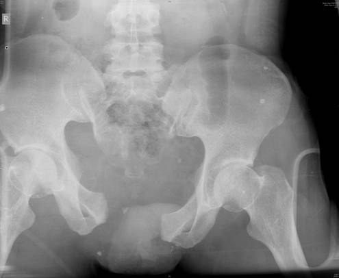
Q1.Describe the AP pelvis X-ray findings of gross pelvic disruption.
- Widening of the pubic symphysis
- Suspected widening of the right SI joint
- Vertical translation of the hemipelvis
- No fracture over the femoral neck/head
- Remember: assess anterior then posterior; the lines are not as important
Q2.How is this injury classified?
- Young and Burgess - vertical shear
- Tile type C
Q3.What are the three Tile categories of pelvic fracture?
- Stable
- Rotationally unstable
- Vertically unstable
Q4.What are the three Young-Burgess mechanisms?
- Lateral compression
- AP compression
- Vertical shear
Q5.Outline the immediate ATLS management of a high-energy pelvic fracture.
- High energy -> ATLS protocol + AMPLE -> primary survery ABCDE
- Secure airway and breathing
- C-spine triple immobilisation
- Disability: examine lower limb neurology
- Exposure
Q6.How is circulation managed in the acute pelvic fracture?
- Pelvic binder application
- Fluid challenge with warmed 2L crystalloid
- Early prescription of unmatched whole blood
- If not responding, activate the massive transfusion protocol
- ROTEM-based substitution of single components
- Time limited permissive hypotension
Q7.Describe the pelvic binder application and its goal.
- Ask assistance to hold the legs in internal rotation (IR)
- Apply at the level of the greater trochanter (GT)
- Goal: protect the first clot with a tamponade effect
Q8.What is the tranexamic acid regimen in pelvic trauma?
- IV tranexamic acid 1g over 10 minutes, then 1g over 8 hours
- Given within 3 hours
Q9.What scans are ordered after the primary survey?
- FAST scan
- XR (trauma series) and CT
- Second survey for life-/limb-threatening injuries
Q10.What local associated injuries are sought in the second survey?
- Open wound
- Soft tissue: morel Lavelle (15% infection rate)
- Urinary: blood from urethral meatus, distended bladder, floating prostate -> urinary diversion
- GI: blood from rectum -> treat as open fracture + faecal diversion
- GU: blood from vagina
Q11.What is a Morel-Lavallee lesion and how is it monitored?
- Soft tissue degloving injury in the subcutaneous plane/fascia
- 15% infection rate
- Monitor +/- contrast CT - collection with contrast enhancement at the subcutaneous plane
Q12.What is the operative management if the patient remains unstable?
- Rule out other sources of bleeding
- DCO operative management (3 in 1) - a local study from a tertiary trauma centre in HK showed 3 in 1 reduces mortality
- Aim: skeletal stabilisation and haemorrhage control -> stabilise and nurture the first clot
- External fixator
- Angiogram + embolization
Q13.What are the three sites of retroperitoneal bleeding in pelvic fracture?
- One in front of the SIJ
- One in front of the quadrilateral plate
- One behind the pubic rami
Q14.What are the definitive management principles for a pelvic ring injury?
- AIM: restore integrity of the pelvic ring
- Anterior: plating
- Posterior: SI screw or plating
- If fixation is still tenuous, add an exfix to augment the fixation
Q15.What is the New Berlin definition of a polytrauma patient?
- AIS >=3 for 2 or more different body regions
- Plus >=1 of: age >=70, GCS <=8, hypotension sBP <90, acidosis BE <=-6, coagulopathy
Q16.What are the three peaks of mortality in trauma?
- On scene
- Golden hours (bleeding)
- 2-3 weeks (infection, MODS, ARDS)
Q17.What are the ISS body regions, AIS grades and ISS threshold?
- ISS regions: 1 head and neck, 2 face, 3 chest, 4 abdomen/pelvis, 5 extremities, 6 external
- AIS: mild, moderate, severe non-life-threatening, severe life-threatening, critical, fatal
- ISS >15 = mortality 10%
Q18.What is DCO and what are its five components?
- Damage control orthopaedics - a staged approach for the polytraumatised patient
- Limits CARS and SIRS by minimising the impact of surgery (the 2nd hit)
- Components: haemorrhage control, decontamination, decompression, temporary skeletal stabilisation, revascularisation
Q19.Describe the stages of DCO.
- It involves a 3 stage approach
- Stage I: initial life-saving procedure (day 1, +/- day 2-3 for second look)
- Stage II: ICU optimisation to avoid the lethal triad (acidosis, hypothermia, coagulopathy)
- Stage III: definitive treatment once stabilised (day 5-10; stable BP, no inotropes, no hypothermia, normal urine output, no acidosis/coagulopathy)
- Stage IV: immunosuppressed D7-21 - avoid OT
Q20.What key physiologic values did Heather Vallier publish for DCO?
- pH >7.25
- BE >=-5.5
- Lactate <4
- (J Orthop Surg Res 2015)
Q21.How are trauma patients classified, and when is DCO considered in a borderline patient?
- Classified stable, borderline, unstable, extremes
- Borderline: ISS >40 or ISS >20 + chest injury
- GCS <=8
- Bilateral femoral fracture, pulmonary contusion, pelvic/abdominal injury
- Hypothermia; head injury with AIS >=3; IL6 above 500pg/dL
Q22.What is the more recent evidence about early definitive fracture fixation?
- Nahm J Trauma 2011: most polytrauma patients benefit from early definitive fracture fixation
- New concept of early appropriate care (within 36hrs of injury)
- Key is individualised, based on intraoperative response to resuscitation
- Continued reassessment and ability to change from ETO to DCO
▸ Slide 301 · IOOTrauma · 12 questions 1 check expand
Q1-Q1212 questions — tap to reveal all answerslist
- Which radiographic lines and walls are assessed on an AP pelvis X-ray?
- What displacement findings indicate vertical/rotational instability?
- What posterior injuries should be looked for on a pelvis X-ray?
- What are the indications and contraindications for a pelvic external fixator?
- What is the principle of pelvic external fixation?
- What is the preoperative setup for pelvic external fixation?
- Describe the ASIS frame for pelvic external fixation: advantages and disadvantages.
- Describe the technique of ASIS frame pin placement.
- Describe the AIIS frame for pelvic external fixation: advantages and disadvantages.
- Describe the technique of AIIS frame placement.
- What are the indications and contraindications for a C clamp?
- How is a C clamp applied?
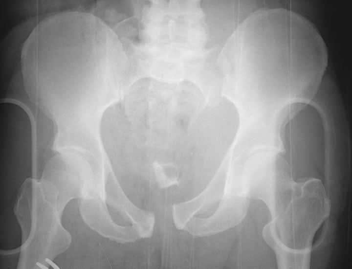
Q1.Which radiographic lines and walls are assessed on an AP pelvis X-ray?
- Ilioischial line
- Iliopecteneal line
- Post/ ant wall
- +/- Shenton's line
Q2.What displacement findings indicate vertical/rotational instability?
- Obvious displacement
- Vertical/rotational instability
- Widening of the pubic symphysis 2.5mm and SIJ 5mm
Q3.What posterior injuries should be looked for on a pelvis X-ray?
- Avulsion fracture of L5 transverse process, ischial tuberosity, ischial spine
- Sarcal foramen + fracture
Q4.What are the indications and contraindications for a pelvic external fixator?
- Indications: mechanically unstable ring (VS, APC2-3, LC2-3) and haemodynamically unstable patient
- Contraindications: iliac fracture involving tracking of pins; acetabular fracture
Q5.What is the principle of pelvic external fixation?
- Decrease pelvic volume
- Stabilise the bleeding bone surface + venous plexus bleeding (tamponade effect) -> form the first clot
Q6.What is the preoperative setup for pelvic external fixation?
- 2 surgeons
- Iliac crest draped with the groin well covered
- Remove the pelvic binder at the last minute
- Radiolucent table + X-ray
Q7.Describe the ASIS frame for pelvic external fixation: advantages and disadvantages.
- Advantages: suitable for most patterns, fast and relatively easy, minimum X-ray
- Disadvantage: not for AP dissociation
Q8.Describe the technique of ASIS frame pin placement.
- Start 2cm posterior to ASIS (thickest gluteal pillar, avoid LFCN)
- Skin incision more medial than anticipated as the pelvis will close up
- Stab wound, then 3 x 5.0mm pins
- Entry point on the iliac crest: more medial for older, middle for young
- Drill trajectory 30-45 degrees towards the ipsilateral hip
- Confirm with the OOO view (45/45)
Q9.Describe the AIIS frame for pelvic external fixation: advantages and disadvantages.
- Advantages: no limitations (usable in AP dissociation), less bulky construct, facilitates nursing care
- Disadvantages: needs XR and a good OOO view; increased time and technique
Q10.Describe the technique of AIIS frame placement.
- Use a good OOO view (tear drop sign at the acetabular roof) for the entry site
- Stab wound 2-3cm medial and below ASIS
- Blunt dissection (watch out for the LFCN)
- Trajectory AIIS to PSIS
- Confirm trajectory with the IOO view (20/35)
- Rod connection to form L and R modules -> reduction by using the modules as handles
Q11.What are the indications and contraindications for a C clamp?
- Indications: posterior instability not controlled by an anterior frame; fracture of ilium and acetabulum; refractory haemodynamic instability
- Contraindications: fracture within the ilium (transiliac fracture dislocation); comminuted sacral fracture with risk of compression of the sacral nerve plexus (relative)
Q12.How is a C clamp applied?
- 3 surgeons: hold device, reduction, tightening; preassemble at the side; involved limb draped free for manipulation
- Landmarks: line joining ASIS and PSIS, and another along the femoral shaft
- Push clamps along the transverse rail until bilateral bone contact, while applying traction and IR to the affected leg
- Bony landmark: intersection between anterior and posterior iliac wing
- Hammer the nail 1cm into bone, then push the side arms towards each other to tighten
Pelvic ring instability is indicated by widening of the pubic symphysis of 2.5mm and SIJ of 5mm — wrong unit — Classic teaching defines anterior instability by symphysis diastasis >2.5 cm (not mm); SI joint widening >5 mm is a sign of posterior injury, though recent literature questions the exact 2.5 cm cut-off — source
▸ Slide 302 · Ilioinguinal approachTrauma · 10 questions expand

Q1-Q1010 questions — tap to reveal all answerslist
- What are the indications, positioning and incision for the ilioinguinal approach?
- How is the internal iliac fossa exposed in the ilioinguinal approach?
- How is the inguinal canal unroofed in the ilioinguinal approach?
- Describe the medial deep dissection of the ilioinguinal approach.
- How is the iliopectineal fascia delineated in the ilioinguinal approach?
- What are the three slings used in the ilioinguinal approach and what do they contain?
- What are the three windows of the ilioinguinal approach and what does each expose?
- How can visualisation through the medial window be improved?
- What is the corona mortis and how is it managed?
- What are the dangers and complications of the ilioinguinal approach?
Q1.What are the indications, positioning and incision for the ilioinguinal approach?
- Anterior approach to the pelvis and acetabulum
- Supine on a radiolucent table, GA, Foley
- Flex the hip to relax the psoas and NV bundle
- Incision from 5cm above ASIS to 1cm above pubic symphysis
Q2.How is the internal iliac fossa exposed in the ilioinguinal approach?
- Skin + fascia from lateral to medial
- Release the external oblique insertion from the iliac crest
- Subperiosteal dissection to mobilise the iliacus muscle
- Pack the iliac fossa with gauze -> lateral window
Q3.How is the inguinal canal unroofed in the ilioinguinal approach?
- Incise the external oblique aponeurosis from ASIS to the external inguinal ring (beware of the LFCN)
- This unroofs the inguinal canal, exposing the inguinal ligament
- Mobilise the spermatic cord/round ligament medially and sling with a Penrose drain
- Sharply incise the inguinal ligament, leaving a cuff of IO and transversus abdominis for later repair (beware of the ilioinguinal nerve)
Q4.Describe the medial deep dissection of the ilioinguinal approach.
- Divide the rectus sheath and rectus 1cm proximal to its insertion
- Develop the plane between the symphysis pubis and bladder (Cave of Retzius)
Q5.How is the iliopectineal fascia delineated in the ilioinguinal approach?
- Retract the femoral vessels (lacuna vasorum) medially
- Retract the femoral nerve and iliopsoas (lacuna musculorum) laterally
- Divide distally down to the pectineal eminence
Q6.What are the three slings used in the ilioinguinal approach and what do they contain?
- Lateral sling: iliopsoas and femoral nerve
- Middle sling: femoral vessels
- Medial sling: spermatic cord
Q7.What are the three windows of the ilioinguinal approach and what does each expose?
- Lateral window (lateral to the lateral sling): entire iliac fossa, SIJ, sacral ala, superior iliopectineal eminence
- Middle window (between middle and lateral slings): pelvic brim, quadrilateral surface
- Medial window (between medial and middle slings): pubic ramus, pubic symphysis, retropubic space of Retzius
Q8.How can visualisation through the medial window be improved?
- The surgeon stands on the opposite side of the table
- With tilting of the table
Q9.What is the corona mortis and how is it managed?
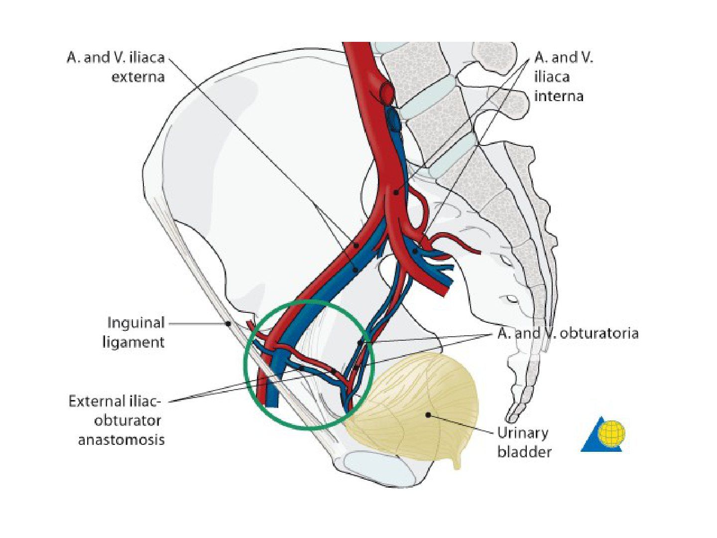
- Ligate retropubic anastomosis - obturator artery from the external iliac (inferior epigastric branch)
- Ligated during the ilioinguinal approach while developing the space of Retzius
Q10.What are the dangers and complications of the ilioinguinal approach?
- Femoral nerve; femoral and external iliac arteries
- Lymphatics; LCFN; inferior epigastric artery; spermatic cord
- Heterotopic ossification; obturator nerve; ilioinguinal nerve
▸ Slide 303 · Modified Stoppa approachTrauma · 5 questions expand
Q1-Q55 questions — tap to reveal all answerslist
- What are the indications and access of the modified Stoppa approach?
- What is the setup and incision for the modified Stoppa approach?
- Describe the superficial dissection of the modified Stoppa approach.
- Describe the deep dissection of the modified Stoppa approach.
- What is the corona mortis and how is it managed in the modified Stoppa approach?
Q1.What are the indications and access of the modified Stoppa approach?
- Indication: acetabular and pelvic ring fractures
- Access: pubic body, superior pubic ramus, pubic root, anterior column, mid posterior column, quadrilateral plate, anterior SIJ
Q2.What is the setup and incision for the modified Stoppa approach?
- Radiolucent table, supine, Foley, flex hips and knees
- Operating surgeon on the opposite side
- Midline transverse incision 2cm above the pubic symphysis (laterally beware the external inguinal ring)
Q3.Describe the superficial dissection of the modified Stoppa approach.
- Incision of subcutaneous tissue and rectus fascia in line
- Rectus abdominis fascia split along the linea alba
- Pyramidalis muscle released
- Retract the rectus abdominis muscle laterally
Q4.Describe the deep dissection of the modified Stoppa approach.
- Transversalis fascia opened sup to PS
- Enter the space of Retzius (between pubic symphysis and bladder)
- Hohman retraction over the pubic tubercle to retract the rectus abdominis
- Subperiosteal dissection along the pubic ramus, then continue retroramus dissection to the quadrilateral surface
- Release the iliopectineal fascia from the pelvic brim
Q5.What is the corona mortis and how is it managed in the modified Stoppa approach?
- Identified during subperiosteal dissection along the pubic ramus
- Ligated: 2 veins and 1 artery
▸ Slide 304 · Disruption of pelvisTrauma · 2 questions expand

Q1-Q22 questions — tap to reveal all answerslist
- Describe the X-ray findings in this pelvic disruption.
- How would you classify this fracture?
Q1.Describe the X-ray findings in this pelvic disruption.
- Widening of the pubic symphysis ~2cm
- Fracture of the right inferior pubic ramus
- Widening of the left SIJ with vertical displacement of the left hemipelvis
- Fracture of the left L5 transverse process
Q2.How would you classify this fracture?
- Vertically and rotationally unstable fracture
▸ Slide 305 · Pelvic fracture acute MxTrauma · 11 questions expand

Q1-Q1111 questions — tap to reveal all answerslist
- Which ligaments provide vertical and rotational stability of the pelvic ring?
- Describe Xray findings in a suspected pelvic fracture.
- What are the ATLS priorities in the acute management of a pelvic fracture?
- What is the role of the pelvic binder and what are its indications?
- If haemodynamically unstable, how is the pelvic fracture resuscitated?
- How does FAST alter the acute management sequence?
- What is the role of embolization in pelvic trauma?
- What anterior frame is used acutely and how are pins placed?
- What is pelvic packing and how is it performed?
- How are PR and GU bleeding managed in pelvic trauma?
- What trauma patterns must be bewared of in pelvic fractures?
Q1.Which ligaments provide vertical and rotational stability of the pelvic ring?
- Vertical stability: iliolumbar, sacrotuberous, posterior SI ligaments
- Rotational stability: pubic symphysis, sacrospinous (suspect pelvic floor injury), anterior SI ligaments
Q2.Describe Xray findings in a suspected pelvic fracture.
- Pubic symphysis, rami (transverse -> LC; vertical -> open book), iliac wing, SIJ, sacrum
- Avulsion fracture: L5 transverse process, STL, SSL
- Look for an associated hip fracture
- This is a rotational/vertical unstable fracture
Q3.What are the ATLS priorities in the acute management of a pelvic fracture?
- ATLS - resuscitate according to ABC
- Rule out other life-threatening chest/abdomen trauma by clinical examination, Xray and FAST
Q4.What is the role of the pelvic binder and what are its indications?
- Pelvic binder centre at the GT with LL IR
- Indication: blunt trauma, high energy, shock
- Protect the first clot by tamponade effect
Q5.If haemodynamically unstable, how is the pelvic fracture resuscitated?
- Give 2L crystalloid and see response
- Take blood for cross-match
- If still unstable give O-negative blood and start massive transfusion protocol -> DCO
Q6.How does FAST alter the acute management sequence?
- FAST +ve (intraperitoneal bleeding): laparotomy first
- FAST equivocal (60% of bleeding due to pelvis) or -ve: ex fix first
Q7.What is the role of embolization in pelvic trauma?
- Considered after pelvic packing if still not stable
- Stable patient -> highly selective embolization
- Unstable patient -> non-selective embolization of the internal iliac
- If no extravasation, bleeding may be masked by shock/vasospasm
- Material: gel foam / metallic coil
Q8.What anterior frame is used acutely and how are pins placed?
- Anterior frame iliac crest construct if there is no iliac fracture
- +/- C clamp
- Pins start 2cm posterior to ASIS
- Use the OOO Xray view and orient to ASIS-PSIS and the femoral shaft
Q9.What is pelvic packing and how is it performed?
- Performed via the Stoppa approach (2cm above PS, between transversus abdominis and bladder)
- Pack anterior to the SIJ, the quadrilateral plate, and posterior to the PS
- Performed if the patient is still not stable before embolization
Q10.How are PR and GU bleeding managed in pelvic trauma?
- PR/GU bleeding: +/- diversion + antibiotic
- Morel-Lavallee: subcutaneous/fascia plane with contrast enhancement; drainage in 2nd look OT - there is a risk of injury to the inferior epigastric artery/obturator artery; no drainage in D0
Q11.What trauma patterns must be bewared of in pelvic fractures?
- Tilt fracture (lateral compression): starts benign-looking, then severe displacement, LLD, pain in sexual intercourse, difficult childbirth
- Pelvic acetabular fracture
▸ Slide 306 · Pelvic fracture definitive MxTrauma · 3 questions expand
Q1-Q33 questions — tap to reveal all answerslist
- What is the load transmission of the posterior and anterior pelvic complexes?
- Why should both the anterior and posterior complexes be fixed?
- What must be considered when there is both column involvement?
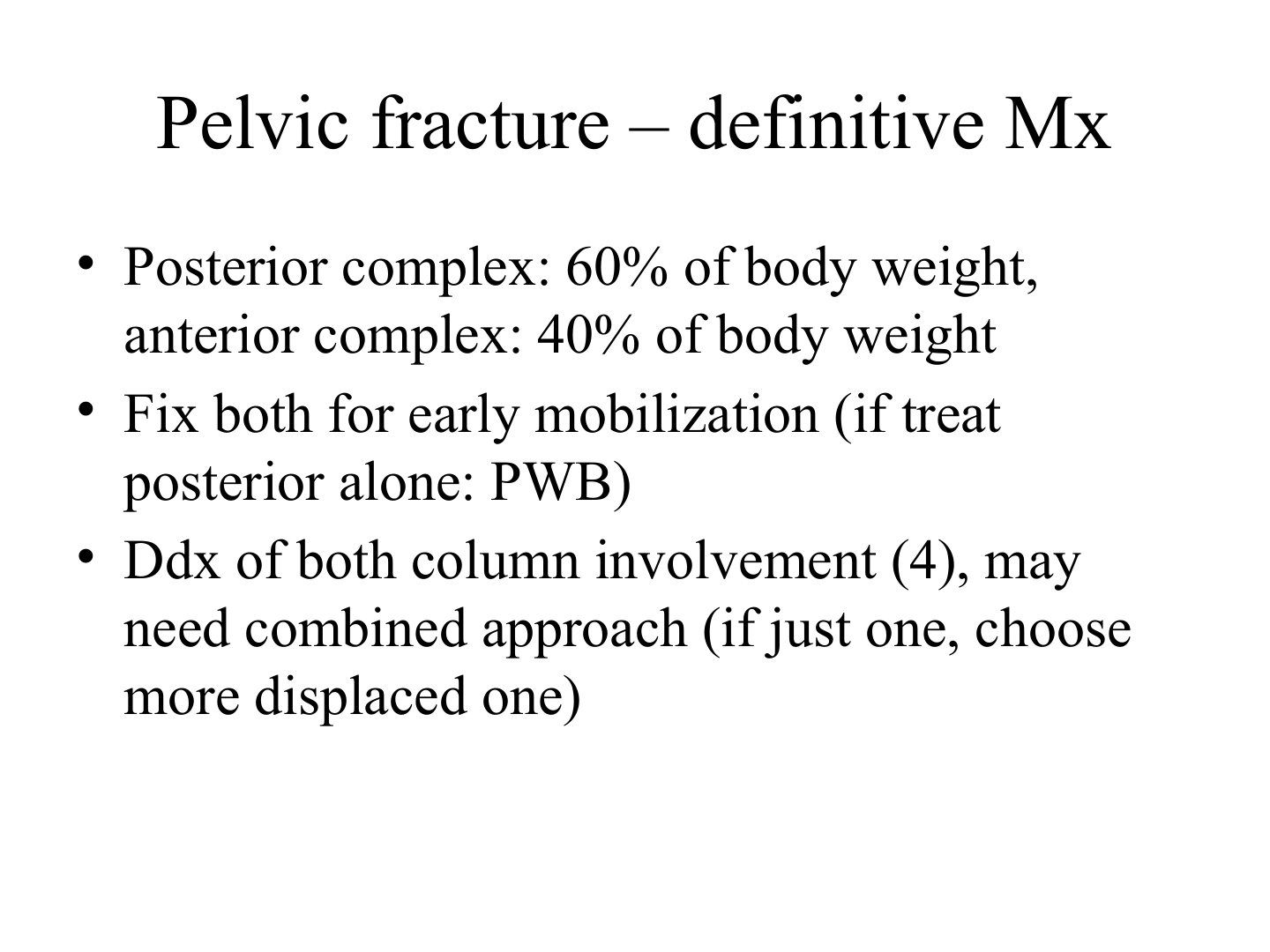
Q1.What is the load transmission of the posterior and anterior pelvic complexes?
- Posterior complex: 60% of body weight
- Anterior complex: 40% of body weight
Q2.Why should both the anterior and posterior complexes be fixed?
- Fix both for early mobilization
- If treating the posterior alone: PWB
Q3.What must be considered when there is both column involvement?
- Ddx of both column involvement (4) - the four differentials are listed on the slide but not expanded in the notes
- May need a combined approach
- If just one column, choose the more displaced one
▸ Slide 307 · DCO:Trauma · 4 questions expand

Q1-Q44 questions — tap to reveal all answerslist
- What is damage control orthopaedics (DCO)?
- What are the components of damage control surgery?
- Who should undergo DCO rather than early total care?
- What are the peaks of mortality after major trauma?
Q1.What is damage control orthopaedics (DCO)?
- Staged operation in the poly-trauma patient to minimize the second hit
- Aim to avoid the terrible triad: coagulopathy, acidosis, hypothermia
- Minimal surgical intervention to achieve hemodynamic & skeletal stabilisation and nursing care
Q2.What are the components of damage control surgery?
- Haemostasis
- Decontamination
- Decompression
- Stabilization
- Revascularize
Q3.Who should undergo DCO rather than early total care?
- Decide according to hemodynamic status
- Unstable/Borderline patient
- Severe chest injury, ISS >40
- Multiple fracture, shock
Q4.What are the peaks of mortality after major trauma?
- 1st peak: major bleeding, brain injury
- 1 hr (golden hour): haemorrhage control
- 2-3 week: sepsis, multi-organ failure
▸ Slide 308 · Acetabular fractureTrauma · 13 questions 1 check expand
Q1-Q1313 questions — tap to reveal all answerslist
- Describe the X-ray assessment of an acetabular fracture.
- Outline the immediate management and further imaging of an acetabular fracture.
- What are the indications for operative fixation of an acetabular fracture?
- What are the aims and timing of definitive acetabular surgery?
- What are the operative details and postoperative care for acetabular fracture fixation?
- What are the pros and cons of the lateral versus prone position for acetabular surgery?
- Which approach (ilioinguinal or Kocher-Langenbeck) is chosen for an acetabular fracture?
- What are the poor prognostic factors in an acetabular fracture?
- Why may a both-column fracture have a better prognosis?
- How is an elderly acetabular fracture managed with respect to acute THR?
- What is the evidence for combined hip procedures in acetabular fractures?
- What is Olson's subchondral arc measurement?
- What is the nerve supply of the gluteal muscles and what lies between them?
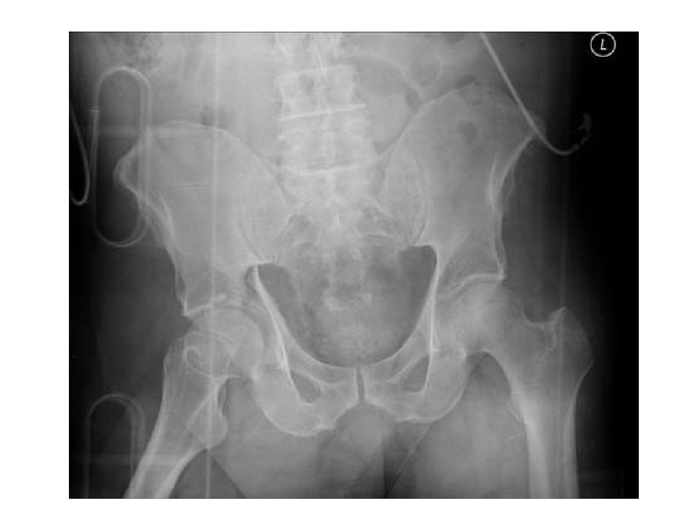
Q1.Describe the X-ray assessment of an acetabular fracture.
- Assess IP and II lines, sourcil, teardrop
- Posterior and anterior wall
- Fractures in iliac crest and inferior pubic rami
- Joint subluxation/dislocation
- Associated fracture (femoral neck and head, pelvis)
Q2.Outline the immediate management and further imaging of an acetabular fracture.
- ATLS primary and secondary survey; rule out other injury, in particular knee, Morel-Lavallee, sciatic and femoral nerve
- +/- Skeletal traction via a distal femur traction pin
- Judet (OO) view - look for the spur sign -> both column
- Fine-cut CT for fracture pattern, loose bodies, marginal impaction, articular step off or gap
- Judet-Letournel classification: no prognostic value, used for surgical exposure
Q3.What are the indications for operative fixation of an acetabular fracture?
- Location of fracture line - whether it is in the weight-bearing zone (roof arc <45 of Matta on AP, IO and OO views, or subchondral arc measurement by Olson)
- Incongruent joint (>2mm step, marginal impaction, loose body)
- Instability (Moed's method: 20% - posterior wall fragment size % + intraop screening after dislocation)
- Associated fractures (femoral head/neck)
Q4.What are the aims and timing of definitive acetabular surgery?
- Aim: anatomical reduction of the articular surface, congruent joint, stability for mobilization
- Timing 2-5 days post injury (according to BOAST guidelines); earlier OT gives a higher chance of anatomical reduction
- <2 days - risk of bleeding
- Letournel: >3 weeks - only 50% anatomical reduction
Q5.What are the operative details and postoperative care for acetabular fracture fixation?
- Single approach to the most displaced fracture +/- an additional approach
- Reduction with joystick, colinear clamp, Farabeuf clamp
- ORIF with 3.5mm reconstruction plate / 3.5mm lag screw / spring plate
- Assess fixation with OOO (joint penetration), IOO (supraacetabular screw within the iliac table), IIO (AP position in the pubic rami)
- Postop HO prophylaxis, NWB for 3 months until healing consolidated, XR + MRI to look for AVN
Q6.What are the pros and cons of the lateral versus prone position for acetabular surgery?
- Lateral pros: less problematic for the chest, lower infection and revision rates, easier limb manoeuvrability
- Lateral cons: sciatic nerve, difficult manual traction, difficult reduction of the posterior column, impaired access through the greater sciatic notch
- Prone position is better for T-type or transverse fractures
Q7.Which approach (ilioinguinal or Kocher-Langenbeck) is chosen for an acetabular fracture?
- If choosing one -> depends on which side is more displaced/more unstable
- Both column fracture / anterior column + posterior hemi-transverse -> usually anterior
- T / transverse -> usually posterior approach
- May need a combined approach / staged operation
Q8.What are the poor prognostic factors in an acetabular fracture?
- Posterosuperior wall (very important for stability/joint congruency)
- Marginal impaction
- Roof arc angle <45 degrees
- Hip dislocation
- Femoral head fracture or impaction
Q9.Why may a both-column fracture have a better prognosis?
- The whole acetabulum is separated from the axial skeleton
- Secondary congruity: a floating acetabulum - the joint is congruent but its orientation differs from the axial skeleton
Q10.How is an elderly acetabular fracture managed with respect to acute THR?
- Primary THR - advantages: earlier weight bearing, shorter OT time
- Primary THR disadvantages: risk of aseptic loosening due to poor fixation, risk of dislocation, risk of HO, need for special implants
- ORIF then THR advantage: defined bone stock
- ORIF then THR disadvantages: higher failure rate due to abnormal anatomy, other implants, more scar tissue, HO, soft tissue insufficiency, infection
Q11.What is the evidence for combined hip procedures in acetabular fractures?
- SICOT 2017: 20-25% of acetabular fractures need revision ORIF or THR, so each case needs an individualised treatment plan
- BJJ 2019 Borg prospective study (minimum 2-year FU): combined hip procedure reduces the need for further surgery compared with ORIF
- Outcome: 78% survival at 10 years
- Poor prognostic factors: male, <50 years old, >80kg, significant acetabular defect
Q12.What is Olson's subchondral arc measurement?
- Measures the superior 10mm of the acetabulum
- Any fracture line within it will be in the weight-bearing zone
Q13.What is the nerve supply of the gluteal muscles and what lies between them?
- Gluteus maximus: inferior gluteal nerve
- Gluteus medius and minimus: superior gluteal nerve
- Between maximus and medius lie the superior gluteal artery and nerve
Acetabular fracture surgery should be performed 2-5 days post injury according to BOAST guidelines — misattributed — The BOAST pelvic fracture standard recommends definitive fixation within 72 hours of stabilisation; a 2-5 day window is not a BOAST recommendation — medium confidence — source
▸ Slide 309 · Xray showing dislocation of hip joint, associated # of post wall of acetabulumTrauma · 19 questions expand

Q1-Q1919 questions — tap to reveal all answerslist
- What radiographic features suggest a posterior rather than anterior hip dislocation, and how is the diagnosis confirmed?
- How are hip dislocations classified?
- Describe the initial ATLS assessment of a hip dislocation.
- What is the timing and technique of closed reduction for a hip dislocation?
- What must be checked after reduction of a hip dislocation?
- What is the definitive management of a reduced hip dislocation with a posterior wall fracture?
- What are the aims, timing and approach of surgery for a dislocated hip with posterior wall fracture?
- Describe the position, incision and superficial dissection of the Kocher-Langenbeck approach.
- How are the short rotators managed in the Kocher-Langenbeck approach?
- Which nerves must be identified and protected in the Kocher-Langenbeck approach?
- Describe the capsulotomy and exposure adjuncts of the Kocher-Langenbeck approach.
- Describe the trochanteric flip osteotomy used in the Kocher-Langenbeck approach.
- What fixation and postoperative care follow the Kocher-Langenbeck approach?
- What are the early complications of the Kocher-Langenbeck approach and hip dislocation?
- What is the prognosis and management of sciatic nerve injury after hip dislocation?
- What are the late complications of hip dislocation and acetabular fracture?
- What obstacles may prevent closed reduction of a hip dislocation?
- What are the contents of the greater sciatic notch?
- What are the contents of the lesser sciatic notch?
Q1.What radiographic features suggest a posterior rather than anterior hip dislocation, and how is the diagnosis confirmed?
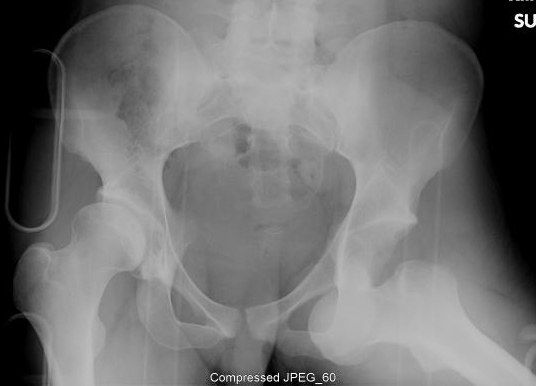
- Femur appears adducted, with the lesser trochanter less prominent
- No fracture over the femoral head and neck
- Likely posterior dislocation, but there is no lateral XR in the trauma setting - confirm with clinical examination
Q2.How are hip dislocations classified?
- Thompson and Epstein (posterior): 1 small post fragment, 2 large post fragment, 3 comminuted post fragment, 4 acetabular floor, 5 femoral head
- Anterior (Epstein): 1 superior, 2 inferior; A no fracture, B femoral head fracture, C acetabular fracture
Q3.Describe the initial ATLS assessment of a hip dislocation.
- ATLS, AMPLE history, primary and secondary survey
- Local assessment: wounds, Morelle Lavalle, knee dislocation, sciatic/femoral nerve
- Systemic survey to rule out other injuries
Q4.What is the timing and technique of closed reduction for a hip dislocation?
- CT if available; if time is needed, proceed with CR and screen for neck fracture under II before reduction
- CR under GA with muscle relaxant within 6 hours
- AVN risk: >6 hours, repeated attempts >2, neck fracture
- Apply traction in line with the deformity regardless of the direction of dislocation
Q5.What must be checked after reduction of a hip dislocation?
- Stability: hip flex, IR, axial load
- NV status
- +/- skeletal traction if needed
- Post-reduction CT for congruency, iatrogenic fracture, marginal impaction, loose bodies
Q6.What is the definitive management of a reduced hip dislocation with a posterior wall fracture?
- Traction x 4/52 if unstable but concentrically reduced; PWB for 4 weeks if stable and concentric
- Surgery if instability: posterior wall fragment >20% (dynamic screening is more reliable - Moed, J Orthop Trauma 2009)
- Surgery if incongruent: marginal impaction, articular step >2mm, loose bodies
- Surgery based on fracture location (roof arc angle of Matta or subchondral arc of Olson) and associated fractures
Q7.What are the aims, timing and approach of surgery for a dislocated hip with posterior wall fracture?
- Aim: anatomical reduction, congruent joint, stability for mobilisation
- Timing 2-5 days post injury (according to BOAST guidelines); earlier OT higher chance of anatomical reduction; <2 days - risk of bleeding
- Letournel: >3 weeks - only 50% anatomical reduction
- Approach: Kocher-Langenbeck
Q8.Describe the position, incision and superficial dissection of the Kocher-Langenbeck approach.
- Lateral position, hip extended and knee flexed
- Incision from PSIS along the femoral shaft, centred on the GT
- Split fascia lata
- Split gluteus maximus along its avascular plane
Q9.How are the short rotators managed in the Kocher-Langenbeck approach?
- Cut the short rotators 1.5cm from their GT insertion
- Beware the ascending branch of the medial circumflex artery, which runs anterior to quadratus femoris
- Therefore do NOT incise the quadratus femoris
Q10.Which nerves must be identified and protected in the Kocher-Langenbeck approach?
- Trace the piriformis to the greater sciatic notch (GSN)
- Trace the internal oblique (internal obturator) to the lesser sciatic notch (LSN)
- Hoffman on the LSN to protect the sciatic nerve
Q11.Describe the capsulotomy and exposure adjuncts of the Kocher-Langenbeck approach.
- Marginal capsulotomy (acetabular-based), keeping the blood supply to the posterior wall fragment
- Subperiosteal dissection of the greater and lesser sciatic notches to expose the retroacetabular surface
- May insert a Shanz pin into the femoral neck to allow traction and assess for loose bodies
- +/- release the gluteus maximus insertion into the femur to aid retraction and reduce stretch on the sciatic nerve
Q12.Describe the trochanteric flip osteotomy used in the Kocher-Langenbeck approach.
- Indicated when better visualisation of the joint is needed
- Predrill the GT
- Posterior to anterior oscillating saw digastric cut
- Detach the vastus lateralis and gluteus medius from the underlying attachment
Q13.What fixation and postoperative care follow the Kocher-Langenbeck approach?
- Reduction with joystick, colinear clamp
- ORIF with 3.5mm reconstruction plate / 3.5mm lag screw / spring plate
- Assess with OOO (joint penetration), IOO (supraacetabular screw within the iliac table), IIO (AP position in the pubic rami)
- Postop HO prophylaxis, NWB for 3 months until healing consolidated, XR + MRI to look for AVN
Q14.What are the early complications of the Kocher-Langenbeck approach and hip dislocation?
- <2% recurrent dislocation
- 20% sciatic nerve injury
- Femoral nerve injury
- Inferior gluteal artery and nerve injury (from vigorous piriformis retraction)
- Superior gluteal artery and nerve injury (superior retraction of glut med/gluteus medius)
- 1st perforator of the profunda femoris (during gluteus maximus release)
Q15.What is the prognosis and management of sciatic nerve injury after hip dislocation?
- 50% recovery
- EMG post injury at 3-4 months
- OT if suspected entrapment or no recovery at 1 year
Q16.What are the late complications of hip dislocation and acetabular fracture?
- Post-traumatic OA (20%)
- AVN (up to 40%)
- Chondrolysis
- Heterotopic ossification
Q17.What obstacles may prevent closed reduction of a hip dislocation?
- Intracapsular: bony fragment, labrum
- Extracapsular: psoas and rectus femoris in front; gluteus maximus and piriformis at the back
- Open reduction if failed 2 times - via the KL approach
Q18.What are the contents of the greater sciatic notch?
- Superior and inferior gluteal nerve and vessels
- Piriformis
- Sciatic nerve and posterior cutaneous nerve of thigh
- Pudendal nerve
- Nerves to obturator internus and quadratus femoris
Q19.What are the contents of the lesser sciatic notch?
- Pudendal nerve
- Internal pudendal artery and vein
- Nerve to obturator internus
- Obturator internus tendon
▸ Slide 310 · Pipkin IIITrauma · 5 questions expand
Q1-Q55 questions — tap to reveal all answerslist
- How is a Pipkin III fracture managed?
- Which approaches are used for Pipkin I, II and III fractures?
- How does fragment size guide treatment of a Pipkin I fracture?
- Which approach is used for a Pipkin IV fracture?
- What is the long-term outcome of Pipkin fractures, and how do they relate to the Thompson-Epstein classification?
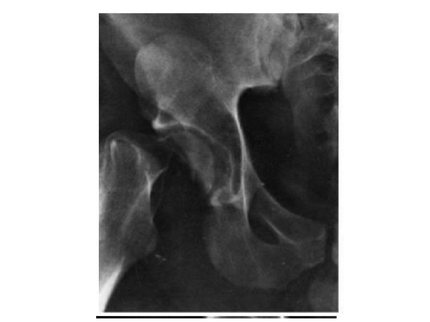
Q1.How is a Pipkin III fracture managed?
- No closed reduction (NO CR)
- Young: fix the neck before the head
- Old: hemiarthroplasty or THR
Q2.Which approaches are used for Pipkin I, II and III fractures?
- Anterior or anterolateral approach
Q3.How does fragment size guide treatment of a Pipkin I fracture?
- <1cm2: excise the fragment
- >1cm2: fix the fragment
Q4.Which approach is used for a Pipkin IV fracture?
- Kocher-Langenbeck approach with trochanteric flip osteotomy
- Then fix the fracture
Q5.What is the long-term outcome of Pipkin fractures, and how do they relate to the Thompson-Epstein classification?
- Around 20% of patients require THA in the first 6 months
- Dislocation of the femur + fracture head = TE5, then use Pipkin
- Both fracture + dislocation = Pipkin 4 + TE5
▸ Slide 311 · Fixation of femoral head with headless screwsTrauma · 5 questions expand

Q1-Q55 questions — tap to reveal all answerslist
- What approaches are used for open reduction and fixation of femoral head fractures?
- What implant is used for fixation of a femoral head fracture?
- Describe the Kocher-Langenbeck approach with trochanteric flip.
- Describe the Z capsulotomy used in surgical hip dislocation.
- How is the hip dislocated anteriorly in the surgical hip dislocation approach?
Q1.What approaches are used for open reduction and fixation of femoral head fractures?
- Depends on the direction of dislocation
- Anterior: Smith-Petersen; posterior; trans-trochanteric
- Surgical hip dislocation - allows 360 degrees direct visualisation of the head
Q2.What implant is used for fixation of a femoral head fracture?
- The slide title states fixation of the femoral head with headless screws
- The approach depends on the direction of dislocation
Q3.Describe the Kocher-Langenbeck approach with trochanteric flip.
- Subcutaneous fat, TFL and gluteus maximus split
- Trochanteric osteotomy: from the posterosuperior edge of the GT extending distally to the posterior border of the VL ridge; mobilise anteriorly
Q4.Describe the Z capsulotomy used in surgical hip dislocation.
- First limb along the anterolateral axis of the femoral neck
- Second limb along the distal anterior edge of the capsule around the calcar
- Third limb parallel to the edge of the acetabulum
Q5.How is the hip dislocated anteriorly in the surgical hip dislocation approach?
- Flexion, adduction and external rotation of the hip
▸ Slide 312 · Hip flexed, abducted and ERTrauma · 3 questions expand
Q1-Q33 questions — tap to reveal all answerslist
- What is the clinical significance of a hip held in flexion, abduction and external rotation?
- Which Epstein classification applies to this injury?
- Describe the Walker modification of Allis reduction for anterior hip dislocation.
Q1.What is the clinical significance of a hip held in flexion, abduction and external rotation?
- Clinically this indicates an anterior dislocation
Q2.Which Epstein classification applies to this injury?
- Epstein II inferior - as the hip is flexed
Q3.Describe the Walker modification of Allis reduction for anterior hip dislocation.
- Supine; stabilise the pelvis; in-line traction; knee flexed
- Hip external rotation + flexion
- Laterally directed force + abduction
- Stabilise in internal rotation
▸ Slide 313 · Pelviacetabular fracture + bilateral hip dislocationTrauma · 3 questions expand
Q1-Q33 questions — tap to reveal all answerslist
- Describe the injury pattern shown.
- How is anterior hip fracture dislocation classified?
- In the Thompson-Epstein classification, what do grades IV and V represent?
Q1.Describe the injury pattern shown.
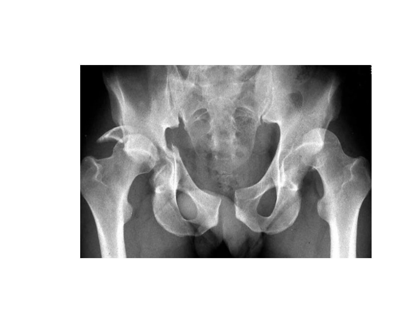
- Pelviacetabular fracture + bilateral hip dislocation
- Right Thompson-Epstein IV (medial wall)
- Left Thompson-Epstein V
Q2.How is anterior hip fracture dislocation classified?
- 1 Superior
- 2 Inferior
- Modifiers: A no fracture, B femoral head fracture, C acetabular fracture
Q3.In the Thompson-Epstein classification, what do grades IV and V represent?
- Type IV: acetabular floor (medial wall)
- Type V: femoral head
▸ Slide 314 · Young NOF#Trauma · 16 questions 1 check expand
Q1-Q1616 questions — tap to reveal all answerslist
- Describe the X-ray findings in this young femoral neck fracture.
- What does Garden III mean in this fracture?
- What is the initial management of a young femoral neck fracture?
- What is the aim of definitive management in a young femoral neck fracture?
- What is the timing of surgery and the evidence in a young femoral neck fracture?
- What fixation options are used in a young femoral neck fracture?
- Describe the Leadbetter technique for reducing a femoral neck fracture.
- How is reduction assessed clinically and radiographically?
- What is done if closed reduction fails?
- Describe the principle of screw fixation for a femoral neck fracture.
- Describe the screw insertion sequence and why the anterosuperior screw is tightened first.
- What is the postoperative care after fixation of a young femoral neck fracture?
- What factors predict AVN after a femoral neck fracture?
- Describe the blood supply of the femoral head.
- What are the AVN and nonunion rates by Garden grade?
- What is the controversial evidence regarding timing, capsulotomy and implant choice?
Q1.Describe the X-ray findings in this young femoral neck fracture.
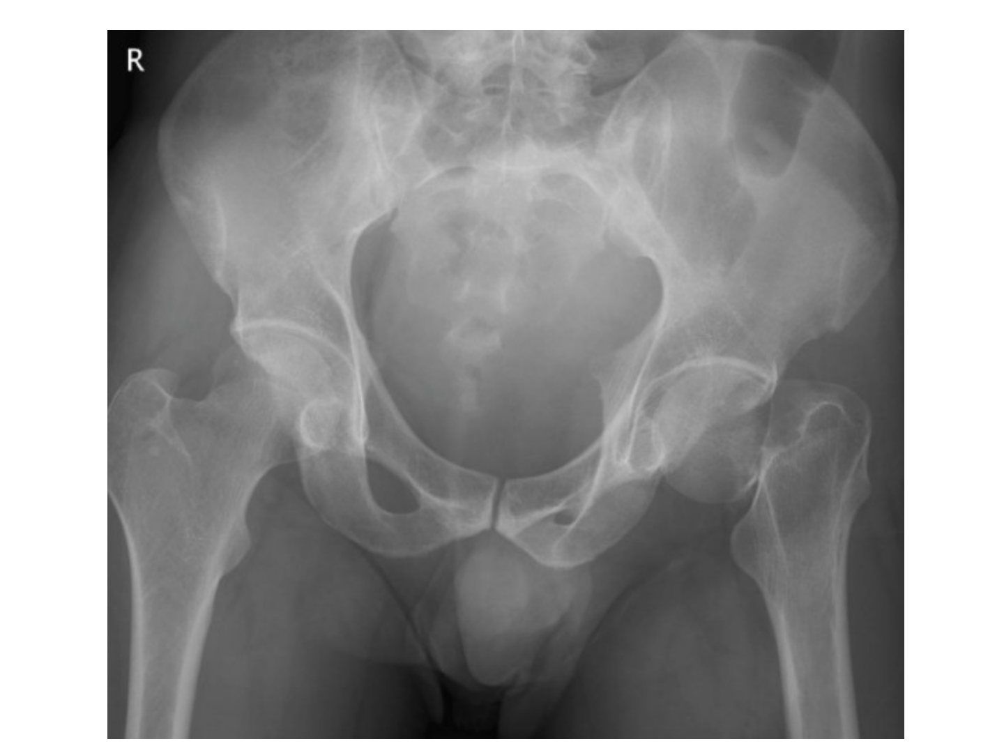
- AP X-ray of the pelvis and bilateral proximal femur
- Over the left side there is a transcervical displaced fracture, with no underlying lytic lesion, +/- medial comminution
- Shenton line broken; bone quality okay
- Garden III, Pauwel III
- Fracture orientation and Pauwel grading are difficult to appreciate
Q2.What does Garden III mean in this fracture?
- Acetabular and femoral head trabeculae are not aligned
- The capsule is likely intact
Q3.What is the initial management of a young femoral neck fracture?
- Rule out life-threatening injury; make sure this is an isolated, closed fracture with intact NV
- Targeted history and PE: premorbid status, mechanism and chronicity of the fracture
Q4.What is the aim of definitive management in a young femoral neck fracture?
- Achieve anatomical reduction and stable fixation to preserve the femoral head and avoid AVN
Q5.What is the timing of surgery and the evidence in a young femoral neck fracture?
- Papakostidis (Giannoudis group) 2015 meta-analysis: no association between AVN and timing of surgery
- >24hr could increase substantially the odds of nonunion
- Go for reduction and fixation at the next available trauma list by a senior trauma surgeon, under GA
Q6.What fixation options are used in a young femoral neck fracture?
- DHS + one anti-rotational screw - angular stability + tension band plating for a Pauwel 3 fracture
- Or 3 cannulated 6.5mm hip screws
Q7.Describe the Leadbetter technique for reducing a femoral neck fracture.
- Hip flexion, adduction and internal rotation to relax the psoas, hamstring and Y ligament
- In-line traction -> book open the fracture
- While maintaining traction and IR: circumduct, abduct and extend the hip
Q8.How is reduction assessed clinically and radiographically?
- Clinical: heel-palm test - put both legs in IR; good if they stay in IR
- X-ray: Garden's alignment index and Lowell's S lines
- AP view: medial cortex of femoral shaft and central axis of medial trabeculae of the capital fragment should be 160 degrees, accept valgus to 180 degrees
- Lateral neutral (axis of neck and centre of capital fragment), accept 155-180 degrees in both views
Q9.What is done if closed reduction fails?
- Go for open reduction and internal fixation
- Approach: Watson's Jones
- DHS + anti-rotational screw + capsulotomy
Q10.Describe the principle of screw fixation for a femoral neck fracture.
- Insert three screws in an inverted triangle configuration
- Avoid multiple drill holes and keep all screws proximal to the lesser trochanter
- Screws perpendicular to the fracture site to subchondral bone
- Parallel screw placement
- All threads pass into the proximal fragment
Q11.Describe the screw insertion sequence and why the anterosuperior screw is tightened first.
- Use a guide pin to identify neck anteversion
- Insert the anterosuperior pin first, then posterosuperior and inferior
- 6.5mm partial-threaded cannulated screws in the same sequence
- Comminution is usually posteromedial, so tightening the AS screw first gives less varus and retroversion
Q12.What is the postoperative care after fixation of a young femoral neck fracture?
- TTWB/NWB for 6 weeks
- Treat osteoporosis if present
- Monitor for AVN with XR/MRI
Q13.What factors predict AVN after a femoral neck fracture?
- Initial fracture displacement
- Quality of fracture reduction
- Loss of reduction post surgery
- Fracture nonunion
Q14.Describe the blood supply of the femoral head.
- Extracapsular arterial ring: MCFA (posterior), LCFA (anterior)
- Ascending cervical branch (major: posterosuperior)
- Subsynovial intracapsular ring at the subcapital region (major: lateral epiphyseal artery)
- Artery to ligamentum teres
- Intraosseous supply
Q15.What are the AVN and nonunion rates by Garden grade?
- Garden I/II: AVN <10%, nonunion 5%
- Garden III/IV: AVN >80%, nonunion 30%
Q16.What is the controversial evidence regarding timing, capsulotomy and implant choice?
- Time to surgery: within or beyond 12 hours (Papakostidis 2015 - no AVN association; >24hr increases nonunion)
- Capsulotomy: theoretically reduces intra-articular pressure
- DHS vs cannulated screws (JBJS 2008): DHS has lower short-term failure rates; backed by Boston group Gardnet 2015 and Singh 2017 for Pauwel II and III
- FEA (Jiang, Injury 2022): supports DHS + BS or FNS
Garden III/IV femoral neck fractures have an AVN rate >80% — overstated — Pooled meta-analysis data show AVN after displaced (Garden III-IV) fractures of about 20-33% (Garden IV ~33%, Garden III ~17%); a rate >80% is not supported — source
▸ Slide 315 · Watsons Jones approachTrauma · 5 questions expand
Q1-Q55 questions — tap to reveal all answerslist
- What are the key steps of the Watson-Jones approach to the hip?
- Why is the Watson-Jones approach described as having no true internervous plane?
- How is exposure improved proximally and distally in the Watson-Jones approach?
- How do you open reduce a femoral neck fracture through the Watson-Jones approach?
- What are the advantages and disadvantages of the Watson-Jones approach compared with the Smith-Petersen approach?
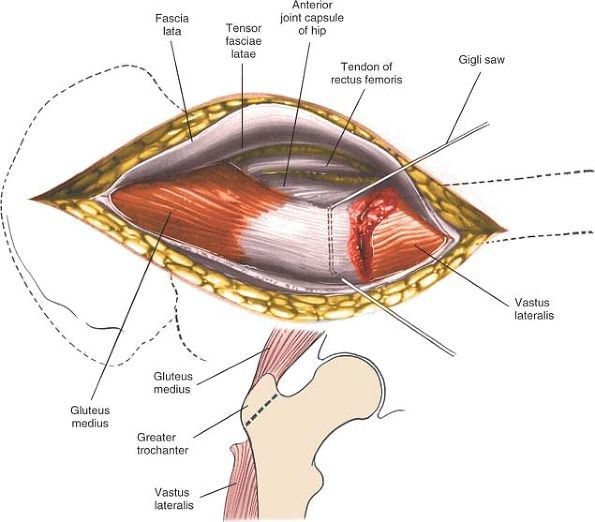
Q1.What are the key steps of the Watson-Jones approach to the hip?
- Anterolateral approach to the hip, no internervous plane
- GA, supine, radiolucent table
- Incision from 2cm inferior and posterior to ASIS to posterior 1/3 of GT down the shaft
- Plane between TF and GM (both supplied by superior gluteal nerve)
- Ligate perforating vessels (superior gluteal arteries) before retraction
- Acetabular-based T capsulotomy
Q2.Why is the Watson-Jones approach described as having no true internervous plane?
- The interval is between tensor fascia lata (TF) and gluteus medius (GM)
- Both muscles are supplied by the superior gluteal nerve
- Hence it is not a true internervous plane
Q3.How is exposure improved proximally and distally in the Watson-Jones approach?
- Improve exposure by: proximal - detach the reflected head of rectus femoris
- Distal: release VL anterior fibres or perform trochanteric osteotomy
Q4.How do you open reduce a femoral neck fracture through the Watson-Jones approach?
- Bone hook at GT + disimpact fracture by traction and ER
- Lever at fracture site (neck always anterior to head)
- LL max IR (maximal internal rotation) of the limb to maintain reduction
Q5.What are the advantages and disadvantages of the Watson-Jones approach compared with the Smith-Petersen approach?
- Advantages: same wound for fixation; better visualisation of the base of neck/trochanteric region
- Disadvantages: poor visualisation of the head; perforator bleeding
▸ Slide 316 · oldTrauma · 11 questions expand

Q1-Q1111 questions — tap to reveal all answerslist
- Describe the X-ray findings in this elderly patient with a transcervical femoral neck fracture.
- What does the focused history and examination include in an elderly hip fracture patient?
- List the BOA six standards of hip fracture care.
- What is the definitive management of a displaced osteoporotic NOF fracture in a geriatric patient?
- What is the evidence for THR vs hemiarthroplasty, and cemented vs cementless hemiarthroplasty?
- What are the risk factors for hip instability after hemiarthroplasty?
- What is the evidence for bipolar vs unipolar hemiarthroplasty?
- What is the 1-year mortality after hip fracture and its risk factors?
- What is the evidence for DHS vs cannulated screw in patients over 50 years old?
- What are the minor complications of hemiarthroplasty?
- What are the major complications of hemiarthroplasty?
Q1.Describe the X-ray findings in this elderly patient with a transcervical femoral neck fracture.
- Transcervical displaced fracture of the right femur on AP pelvis
- Osteoporotic bone, no lytic lesion; broken Shenton's line
- No other fractures in the pelvic ring
- Garden IV osteoporotic fracture
Q2.What does the focused history and examination include in an elderly hip fracture patient?
- Premorbid mental and functional status, PMH, mechanism of injury: 70y fell on level ground, osteoporosis and medications
- Rule out life-threatening conditions, especially HI; make sure it is an isolated injury
- Look for open wound and distal NV deficit
Q3.List the BOA six standards of hip fracture care.
- Admit to ortho ward within 4hrs
- Surgery within 48hrs if medically fit, during normal working hours
- Pressure sore precautions
- MDT assessment and intervention
- Orthogeriatric combined care within 72hrs
- Secondary prevention of osteoporotic fractures
Q4.What is the definitive management of a displaced osteoporotic NOF fracture in a geriatric patient?
- Aim: allow weight bearing and mobilisation ASAP and repatriation to place of usual abode
- Surgery: cemented bipolar hemiarthroplasty
- Long run: osteoporosis and fall prevention by the fracture liason service according to the IOF guidelines
- +/- DVT prophylaxis, +/- fascia iliaca block
Q5.What is the evidence for THR vs hemiarthroplasty, and cemented vs cementless hemiarthroplasty?
- Bhandari 2019 NEJM (HEALTH): THR gave a clinically unimportant improvement in function/QoL over 24months, with more dislocation
- Fernandez 2022 NEJM: cemented hemi = significantly better QoL and lower periprosthetic fracture risk in NOF patients >60yo
- Cemented Exeter stem: 100% survival rate in 17years according to Carrington’s series in JBBS Br 2009; Norwegian registry: lower fracture, infection and revision
- Cemented gives immediate stability, less anterior thigh pain and fewer periprosthetic fractures
Q6.What are the risk factors for hip instability after hemiarthroplasty?
- Surgeon: posterior approach, unaddressed acetabular defect/GT fracture/glut med tear, failure to restore soft tissue tension, Cement or bone fragment impingement, incorrect stem version
- Improper posterior capsular repair (Kwon meta-analysis: dislocation reduced 10 times if repaired)
- Patient: delirium, Parkinson's, stroke, dementia
Q7.What is the evidence for bipolar vs unipolar hemiarthroplasty?
- Theoretically bipolar has two bearing surfaces: less dislocation, acetabular erosion and femoral head protrusio
- Australian joint replacement registry 2021: less risk of revision, at least in the young (9.9% vs 6.9%)
- Injury 2019 meta-analysis: greater ROM, less acetabular erosion, lower reoperation; longer operative time; no difference in HHS/mortality
- Cochrane review 2022 Lewis et al: insufficient evidence to determine whether bipolar is superior to unipolar
Q8.What is the 1-year mortality after hip fracture and its risk factors?
- 25% mortality at 1 year
- Risk factors: male, TOF, ASA 3 or above, OT >48 hrs, age 85 or above
Q9.What is the evidence for DHS vs cannulated screw in patients over 50 years old?
- FAITH trial: no difference in reoperation rate
- Smokers, displaced or base of neck fractures might do better with a sliding hip screw
Q10.What are the minor complications of hemiarthroplasty?
- Persistent hip pain
- Poor mobility
- LL muscle wasting
- Superficial infection
Q11.What are the major complications of hemiarthroplasty?
- Periprosthetic fracture
- Deep infection
- Dislocation
- Aseptic loosening
- Acetabular erosion
▸ Slide 317 · 60yTrauma · 10 questions expand
Q1-Q1010 questions — tap to reveal all answerslist
- How do you comment on screw fixation of a femoral neck fracture on X-ray?
- What does this X-ray show and what is the summary?
- List the mechanical and biological reasons for fracture fixation failure.
- Describe the Leighton classification of femoral neck nonunion and its treatment.
- If joint salvage is chosen, what is the revision fixation technique?
- What investigations are needed in suspected femoral neck nonunion?
- What history and examination points are important in failed femoral neck fixation?
- What factors determine the choice of operation for femoral neck nonunion?
- What are the surgical options: joint salvage versus joint sacrifice?
- Why are femoral neck fractures difficult to heal?
Q1.How do you comment on screw fixation of a femoral neck fracture on X-ray?
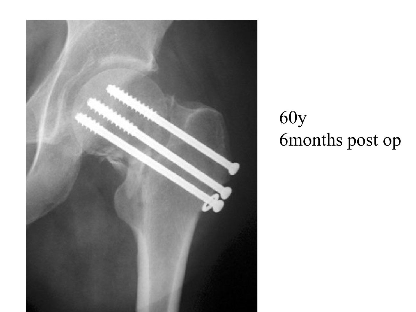
- All screw threads passed the fracture site
- All screws reached subchondral bone
- Check the entry site of the screws
- In inverted triangle configuration, screws should be parallel
Q2.What does this X-ray show and what is the summary?
- Fracture pattern: medial comminuted with a relatively vertical fracture line
- Varus malreduction
- No signs of union; no signs of infection
- Femoral head spherical (signs of AVN); Acetabular side intact
- Summary: NOF fracture with nonunion and varus collapse
Q3.List the mechanical and biological reasons for fracture fixation failure.
- Mechanical (5): fracture pattern, location, reduction, fixation method, fixation quality
- Biological: patient, fracture, infection
Q4.Describe the Leighton classification of femoral neck nonunion and its treatment.
- I (<6 weeks): inadequate fixation/malreduction to revision ORIF
- II (3 months): loss of fixation with good initial alignment to valgus osteotomy
- III (fibrous nonunion): free vascularised bone graft (Meyers BG, fibular graft, iliac crest bone graft)
Q5.If joint salvage is chosen, what is the revision fixation technique?
- Remove implant + valgus osteotomy + revision fixation by angle blade plate + bone graft
- Aim to convert shearing force to compressive force
Q6.What investigations are needed in suspected femoral neck nonunion?
- WBC, ESR, CRP; +/- hip aspirate if suspicious
- MRI/bone scan to assess AVN
- CT for bone stock
- Compare previous Xray see if there was any mal-reduction or displacement of fracture
Q7.What history and examination points are important in failed femoral neck fixation?
- Hx: premorbid status, smoker, pre/intra/post-op problems, recurrent injury, current symptoms, limitations, expectations
- PE local: soft tissue, scar, infection signs
- PE systemic: LLD, muscle power, walking status, general fitness for GA
Q8.What factors determine the choice of operation for femoral neck nonunion?
- Factors to consider: age and premorbid status
- Bone quality and bone stock
- AVN
- Acetabular integrity
- Infection
Q9.What are the surgical options: joint salvage versus joint sacrifice?
- Salvage: revision fixation - unreliable fixation due to osteoporosis, defeats the goal of early weight bearing
- Joint sacrifice: arthroplasty (hemi vs THR) or Girdlestone
Q10.Why are femoral neck fractures difficult to heal?
- Biological: synovial fluid, haematoma difficult to form, no cambium layer of periosteum
- Mechanical: high stress, little bone contact
▸ Slide 318 · Non union of femoral neck #Trauma · 4 questions expand
Q1-Q44 questions — tap to reveal all answerslist
- What are the nonunion rates and time definitions after a displaced femoral neck fracture?
- What is Leighton's classification of femoral neck nonunion?
- How is each Leighton type managed?
- What is Meyer's bone graft and what does it provide?
Q1.What are the nonunion rates and time definitions after a displaced femoral neck fracture?
- 20% nonunion after displaced NOF fracture
- Delayed union: 6 weeks
- Nonunion: 6 months
Q2.What is Leighton's classification of femoral neck nonunion?
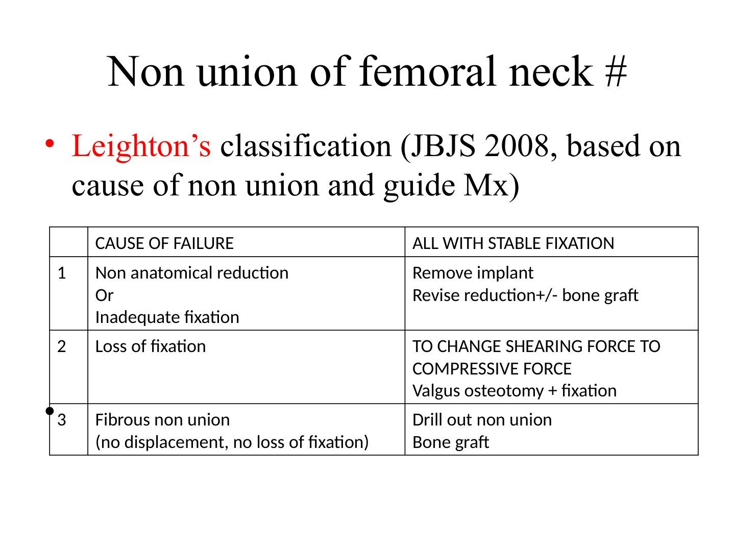
- Leighton's classification (JBJS 2008), based on the cause of nonunion, guides management
- Applies to fractures all with stable fixation
- Type 1: non-anatomical reduction or inadequate fixation
- Type 2: loss of fixation
- Type 3: fibrous nonunion (no displacement, no loss of fixation)
Q3.How is each Leighton type managed?
- Type 1: remove implant, revise reduction +/- bone graft
- Type 2: change shearing force to compressive force with valgus osteotomy + fixation
- Type 3: drill out nonunion, bone graft
Q4.What is Meyer's bone graft and what does it provide?
- Quadratus femoris muscle pedicle graft
- Provides mechanical support and brings in blood supply
▸ Slide 319 · 60y, 6 months post opTrauma · 7 questions expand

Q1-Q77 questions — tap to reveal all answerslist
- Describe the X-ray findings at 6 months post fixation.
- List the reasons for failure of this fixation.
- What workup is required?
- What factors guide the choice of operation?
- What reconstruction is chosen and what is the supporting evidence?
- What history and examination are needed in this failed fixation case?
- What are the aims and options of the operation?
Q1.Describe the X-ray findings at 6 months post fixation.
- Failure of hip screw fixation with back out and protrusion of screws
- No healing over the transcervical fracture
- Femoral head collapse suggesting AVN; acetabulum shows no sourcil sign
- Suspected lucency around screw
- Suspected nonunion and AVN
Q2.List the reasons for failure of this fixation.
- Mechanical: fracture pattern, location, reduction, fixation method, fixation quality
- Biological: patient, fracture, infection
Q3.What workup is required?
- WBC, ESR, CRP; +/- hip aspirate if suspicious
- MRI/bone scan to see any AVN
- CT for bone stock
- Compare previous X-ray for malreduction or displacement
Q4.What factors guide the choice of operation?
- Age and premorbid status
- AVN
- Acetabular integrity
- Infection
- Bone quality and bone stock
Q5.What reconstruction is chosen and what is the supporting evidence?
- Uncemented acetabular cup with cemented PE cup and cemented Exeter stem
- Carrington JBJS Br 2009: 90% 17-year survival for this combination
- Average male life expectancy in HK is 80 so THR is the best option
Q6.What history and examination are needed in this failed fixation case?
- Hx: premorbid status, smoker, perioperative problems, recurrent injury, current symptoms, limitations, expectations
- PE local: soft tissue, scar, infection signs
- PE systemic: LLD, muscle power, walking status, general fitness for GA
Q7.What are the aims and options of the operation?
- Aim of operation to restore a functional hip
- Joint salvage vs joint sacrificing
- Salvage: revision fixation - unreliable due to osteoporosis, defeats early weight bearing
- Joint sacrifice: arthroplasty vs Girdlestone vs arthrodesis; hemi vs THR
▸ Slide 320 · Describe Xray (right)Trauma · 13 questions expand
Q1-Q1313 questions — tap to reveal all answerslist
- Describe the right and left hip X-rays.
- What makes an intertrochanteric fracture unstable?
- Outline the BOA guideline for initial management of hip fractures.
- What are the pre-requisites and technique for a good DHS?
- How does a cephalomedullary (PFNA) device stably fix an unstable fracture?
- What are Kaufer's variables affecting construct strength?
- What focused history and examination are needed in a trochanteric fracture?
- How does barrel choice affect DHS sliding and failure?
- How is a pre-existing OA with a trochanteric fracture managed?
- What is the epidemiology and mortality of trochanteric fractures?
- Describe the Kyle classification of trochanteric fractures.
- Describe the AO classification of trochanteric fractures.
- What is the evidence for cephalomedullary nailing versus DHS?
Q1.Describe the right and left hip X-rays.
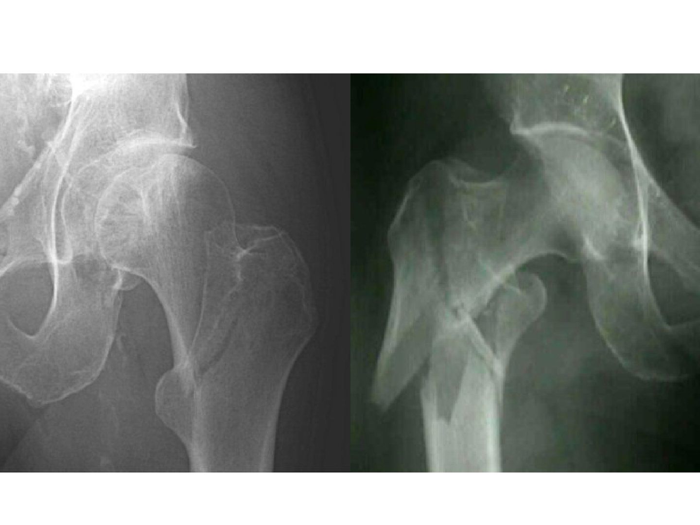
- Right: intertrochanteric fracture, minimally displaced, no features of instability, Kyle 1
- Left: intertrochanteric fracture, unstable - large posteromedial fragment, fracture GT, reversed oblique configuration, Kyle 4
Q2.What makes an intertrochanteric fracture unstable?
- No medial calcar support
- No lateral buttressing effect (fracture greater trochanter, transverse/reverse oblique fracture)
- Kyle 3 (large posteromedial fragment + GT fracture) and Kyle 4 (extension to subtrochanteric) are unstable
Q3.Outline the BOA guideline for initial management of hip fractures.
- Admitted to acute ortho ward within 4 hours
- Surgery within 48 hours of admission if medically fit, during normal working hours
- Minimise risk of pressure ulcer
- Routine orthogeriatric medical support from admission
- Bone-protective therapy to prevent further osteoporotic fractures
- Offered MDT assessment and intervention
Q4.What are the pre-requisites and technique for a good DHS?
- Pre-requisites: no medial calcar comminution, intact lateral buttressing
- Principle: allow controlled collapse across the fracture to enhance healing
- Good reduction to prevent varus; lag screw avoiding superior/posterior cortex; TAD < 25mm; antirotation guide pin
Q5.How does a cephalomedullary (PFNA) device stably fix an unstable fracture?
- Stability does not rely on lateral buttress or medial calcar
- Efficient load transfer from blade to nail to locking bolt and shaft, not through the medial calcar
- Decreased bending moment as the nail is more medialised than the DHS plate
- Nail can control impaction of fracture by physically blocking the sliding
Q6.What are Kaufer's variables affecting construct strength?
- Uncontrolled: bone quality, fracture geometry (stable vs unstable pattern)
- Controllable: reduction, choice of implant and mode of application
Q7.What focused history and examination are needed in a trochanteric fracture?
- Premorbid mental and functional status, PMH, mechanism of injury, osteoporosis and medications
- Rule out life-threatening conditions, especially HI; ensure it is an isolated injury
- Look for open wound and distal NV deficit
Q8.How does barrel choice affect DHS sliding and failure?
- Short barrel (25mm): longer sliding distance but higher bending moment (increased moment arm)
- Long barrel (38mm): less bending moment, less screw breakage, less friction and better sliding
- Gundle: sliding <1cm gives worse outcome; short barrel if lag screw <85; recommended sliding 25mm
- DHS thread 22mm
Q9.How is a pre-existing OA with a trochanteric fracture managed?
- CRIF is difficult: the proximal part is stiff
- Fixation +/- second-stage THR if symptomatic
- Or primary THR with fixation of GT/calcar-replacing prosthesis
Q10.What is the epidemiology and mortality of trochanteric fractures?
- Nonunion/malunion rare <2%
- 20-30% mortality risk in the 1st year (TOF worse than NOF)
- Increased mortality: male, operation >48 hours, age >85, ASA III or above
Q11.Describe the Kyle classification of trochanteric fractures.
- I - undisplaced
- II - displaced, minimal comminution
- III - greater trochanter fragment
- IV - subtrochanteric extension
Q12.Describe the AO classification of trochanteric fractures.
- 31A-A1: peritrochanteric simple two part (lateral cortex intact)
- 31A-A2: pertrochanteric with a separate PM fragment (lateral cortex intact)
- 31A-A3: fracture extends through lateral and medial cortices
Q13.What is the evidence for cephalomedullary nailing versus DHS?
- Nail only shown to be superior to DHS in unstable fractures (Cochrane review 2014)
▸ Slide 321 · (describe fracture and fixation) Fracture TOF with fracture GT fixed with DHS + Trauma · 11 questions expand
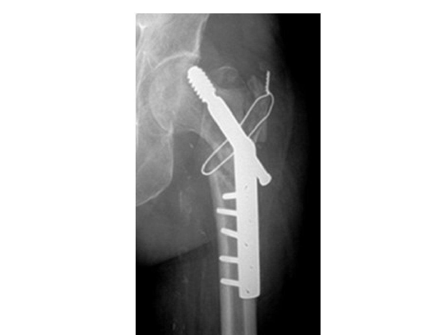
Q1-Q1111 questions — tap to reveal all answerslist
- Describe the fracture, fixation and current status on X-ray.
- What are the reasons for failure in this case?
- What investigations are required before revision surgery for this nonunion?
- How would you perform revision fixation?
- What history and examination are needed for this failed fixation?
- What management factors and options apply in this failed TOF fixation?
- What are the approach and exposure considerations for this revision?
- How are the hip replacement options chosen in this failed TOF fixation?
- How are the acetabulum and femur reconstructed?
- What is done at reduction if the hip is unstable?
- What is the rehabilitation plan after revision?
Q1.Describe the fracture, fixation and current status on X-ray.
- Fracture TOF with fracture GT fixed with DHS + 1 cerclage wire
- No union, varus deformity
- Cut out of lag screw superiorly, complete sliding of DHS, no implant fracture
- Acetabulum intact, femoral head spherical, no halo/lucency around screws
- Summary: TOF nonunion with implant cut out
Q2.What are the reasons for failure in this case?
- Mechanical: unstable fracture pattern and location, varus reduction, wrong implant (should have used short barrel), lag screw not in centre-centre position, tip-to-apex
- Biological: infection, osteoporotic patient, fracture
Q3.What investigations are required before revision surgery for this nonunion?
- Rule out infection: bloods, gallium scan, +/- hip aspirate if suspicious
- Serial XR to see progress of alignment
- CT for acetabular defect and healing status
- MRI for head viability
Q4.How would you perform revision fixation?
- Remove implant, then revision fixation
- Dimon-Hughston osteotomy: medialized shaft (medialised shaft), reconstruct medial calcar, more valgus configuration
- Converts shearing to compression
Q5.What history and examination are needed for this failed fixation?
- Hx: premorbid status, smoker, pre/intra/post-op problems, recurrent injury, current symptoms, limitations, expectations
- PE local: soft tissue, scar, infection signs
- PE systemic: LLD, muscle power, walking status, general fitness for GA
Q6.What management factors and options apply in this failed TOF fixation?
- Factors: age/premorbid, bone quality and bone stock, AVN, acetabulum status, infection
- Joint salvage: revision fixation - unreliable due to osteoporosis, defeats early weight bearing
- Joint sacrifice: arthroplasty vs Girdlestone vs arthrodesis; hemi vs THR
Q7.What are the approach and exposure considerations for this revision?
- Use the old/incorporate the old incision
- Identify and protect the sciatic nerve; soft tissue release
- Dislocate before removal of the implant
- Careful manipulation of the osteoporotic leg
- Implant removal: correct screwdriver, removal of nail, prepare a broken screw set
Q8.How are the hip replacement options chosen in this failed TOF fixation?
- Hemi vs THR depending on acetabulum status
- Hybrid/cementless THR with metal-on-PE (joint registry data)
Q9.How are the acetabulum and femur reconstructed?
- Acetabulum: Acetabular defect (paprovsky) (Paprosky); cementless +/- screw augment, standby cemented
- Femur: Defect management (paprovsky) (Paprosky); cementless calcar replacement vs long diaphyseal stem to bypass screw holes by 2 cortical diameter
- Cemented femur: cement leakage problem; GT reattachment
Q10.What is done at reduction if the hip is unstable?
- Aim combined anteversion 35
- Femur: increase size of head, increase offset, GT advancement
- Acetabulum: anteversion and opening angle, lipped or constrained liner, MDM
Q11.What is the rehabilitation plan after revision?
- Rehab plan: Weight bear as tolerated
- Hip precautions
- HO prophylaxis
- Osteoporosis treatment
▸ Slide 322 · Pelvic fragility fractureTrauma · 5 questions expand

Q1-Q55 questions — tap to reveal all answerslist
- Describe the Rommens classification of pelvic fragility fractures.
- What is the management principle and recommendation by Rommens type?
- What are the problems of non-operative management, and those specific to pelvic fragility fractures?
- What is the evidence on fracture progression and anterior fixation in FFP?
- When should pelvic fragility fractures be operated on?
Q1.Describe the Rommens classification of pelvic fragility fractures.
- 1: anterior only
- 2: undisplaced + posterior
- 3: displaced posterior
- 4: bilateral posterior
Q2.What is the management principle and recommendation by Rommens type?
- Regain walking ability ASAP while balancing the high operation risk
- Type 1: non-operative
- Type 2: early weight bear; if OK non-op, if not tolerated then operation
- Types 3/4: operative
Q3.What are the problems of non-operative management, and those specific to pelvic fragility fractures?
- Non-operative: non/malunion, fracture progression
- PFF-specific: poor bone, higher rate of loss of fixation
- Measures: cemented screw, interdigitation of fixation, robust fixation
Q4.What is the evidence on fracture progression and anterior fixation in FFP?
- Rommens JBJS 2019: fracture progression rate 14%
- Herteleer Eur J Trauma Emerg Surg 2022: no correlation between anterior fixation and overall fixation failure (no consensus)
Q5.When should pelvic fragility fractures be operated on?
- SR by Daniel BMC MSK Disord 2021: fixation considered for patients failing a brief period of non-operative management
- Fixation should be robust enough to allow immediate weight-bearing but minimise operative morbidity and post-op complications
▸ Slide 323 · Xray right hip showing subtrochanteric fracture with displacementTrauma · 12 questions expand

Q1-Q1212 questions — tap to reveal all answerslist
- Describe the displacement and location of this subtrochanteric fracture on X-ray.
- What is a subtrochanteric fracture and what are the deforming forces?
- Why is a subtrochanteric fracture difficult to heal?
- What is the management of choice and why?
- What difficulties are expected with reduction and fixation?
- Describe the classification systems for subtrochanteric fractures.
- What history and examination findings are important in a subtrochanteric fracture?
- What imaging is required for a subtrochanteric fracture?
- What is the aim of surgery for a subtrochanteric fracture?
- If fixed with an IM nail, what is the common malreduced position?
- How do you perform safe reaming for a subtrochanteric nail?
- When is a plate/angle device used instead of a nail?
Q1.Describe the displacement and location of this subtrochanteric fracture on X-ray.
- Subtrochanteric fracture with displacement, extending into the piriformis fossa and to the lesser trochanter
- Proximal fragment in varus and abducted
- Main distal fragment shortened and adducted
- Lesser trochanter fragment migrated proximally
Q2.What is a subtrochanteric fracture and what are the deforming forces?
- Area from the lesser trochanter to 5cm distal
- Abduction by gluteus medius and minimus
- Flexion by iliopsoas
- ER by short external rotators
- Adduction and proximal migration of the distal fragment by adductors
Q3.Why is a subtrochanteric fracture difficult to heal?
- Biologically a watershed area
- Mechanical high stress according to Koch diagram, multiple muscle pull, high cortical-to-cancellous ratio
- Fielding classification: the more distal the fracture, the higher the nonunion rate
Q4.What is the management of choice and why?
- Long cephalomedullary nail: load-sharing with efficient load transfer
- Good torsional and bending rigidity with high tensile and fatigue strength
- Medialised lever arm decreases bending stress; biologically friendly
- Most recent NICE guidelines recommend IM nail fixation
Q5.What difficulties are expected with reduction and fixation?
- Difficult reduction from strong muscle pull; reduce on traction table with not too much traction (align distal to proximal with flexion, abduction, ER)
- Assist with Shanz screw (Schanz screw), cerclage wires, colinear clamp; lateral nailing if all fails (especially obese patients)
- More body lateral flexion (entry point); entry point more MEDIAL + POST (medial and posterior) to avoid flexion and coxa vara
Q6.Describe the classification systems for subtrochanteric fractures.
- Russell-Taylor: 1/2 piriformis fossa intact; A/B loss of medial calcar/lesser trochanter; 1A can use centromedullary nail, others cephalomedullary
- Fielding: type 1 at, type 2 <2.5cm, type 3 >2.5cm from LT (more distal = more nonunion)
- Seinsheimer: 1 undisplaced two-part to 5 subtrochanteric-intertrochanteric extending to GT
Q7.What history and examination findings are important in a subtrochanteric fracture?
- Premorbid status; history of bisphosphonate/malignancy
- Mechanism of injury, preceding symptoms or prodromal pain
- Rule out HI
- PE: open wound or skin impingement; document distal NV conduction
Q8.What imaging is required for a subtrochanteric fracture?
- X-ray lateral view, whole femur and one joint below
- Offer operative management for fixation
Q9.What is the aim of surgery for a subtrochanteric fracture?
- Secondary bone healing with relative stability
- Correct alignment, rotation and length
- Adequate bone contact
Q10.If fixed with an IM nail, what is the common malreduced position?
- Varus and procurvatum
Q11.How do you perform safe reaming for a subtrochanteric nail?
- Technique: high speed, slow advancement, serial reaming, irrigation, clear bone debris and suction, vent hole, no tourniquet
- Reamer design: Bixcut with side and forward cutting flutes, sharp flutes
- Anaesthesia: avoid dehydration, maintain BP
Q12.When is a plate/angle device used instead of a nail?
- For pre-existing deformity or a narrow IM canal
▸ Slide 324 · XR showing comminuted femoral shaft fracture with large butterfly fragment with Trauma · 5 questions expand
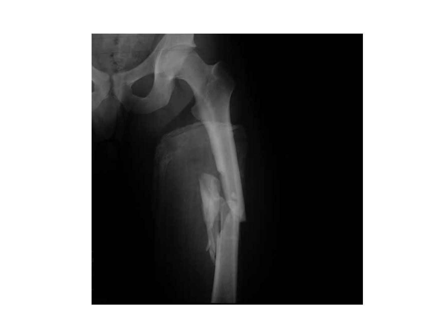
Q1-Q55 questions — tap to reveal all answerslist
- Describe the femoral shaft fracture and degree of comminution on X-ray.
- Describe the Winquist and Hansen classification.
- How is bilateral femur fracture managed?
- How do you determine rotation during femoral nailing?
- Describe the AO classification of femoral shaft fractures.
Q1.Describe the femoral shaft fracture and degree of comminution on X-ray.
- Comminuted femoral shaft fracture with a large butterfly fragment and <50% cortical contact
- No associated femoral neck fracture
- Proximal fragment is abduction and ER (abducted and externally rotated)
- Winquist-Hansen type 3
- XR views required: entire bone + one joint above and below
Q2.Describe the Winquist and Hansen classification.
- 0: no comminution
- 1: small butterfly fragment
- 2: large butterfly fragment, cortical contact >50%
- 3: large butterfly fragment, cortical contact <50%
- 4: gross comminution
Q3.How is bilateral femur fracture managed?
- DCO in unstable/selected borderline patients to reduce the second hit (inflammatory response against surgery)
- Hemostasis, revascularisation, decompression, decontamination, stabilisation
- Definitive surgery: IM nail with distal and proximal locking
Q4.How do you determine rotation during femoral nailing?
- Cortical thickness
- Lesser trochanter and knee X-ray, compare both sides
- Replicate the anteversion of the contralateral side
- Anterior bowing compared with the contralateral side
Q5.Describe the AO classification of femoral shaft fractures.
- A simple
- B wedge
- C comminuted
▸ Slide 325 · Fracture femur shaft + neckTrauma · 9 questions expand
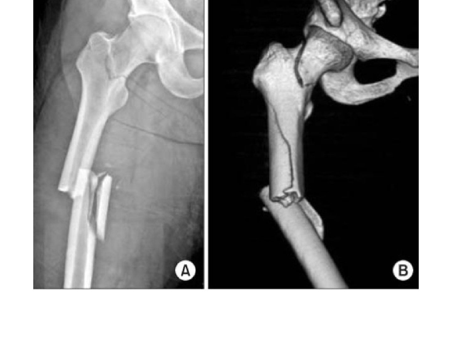
Q1-Q99 questions — tap to reveal all answerslist
- How common is a concomitant femoral neck fracture with a femoral shaft fracture, and what is the prognosis?
- What is the aim and sequence of fixation for ipsilateral femur shaft and neck fractures?
- How is an undisplaced neck fracture managed?
- How is a displaced neck fracture managed?
- What are the indications for retrograde nailing?
- What are the contraindications to retrograde nailing?
- What factors decide between retrograde and antegrade nailing?
- What is the risk of a cephalomedullary nail for combined shaft and neck fractures?
- What injuries are associated with femoral shaft fracture?
Q1.How common is a concomitant femoral neck fracture with a femoral shaft fracture, and what is the prognosis?
- High energy trauma; 5-10% of femoral shaft fractures have a concomitant femoral neck fracture
- Prognosis of the neck fracture is usually better than isolated NOF (usually undisplaced)
Q2.What is the aim and sequence of fixation for ipsilateral femur shaft and neck fractures?
- Aim: anatomical reduction + stable fixation, prevent complications
- Dual construct preferred (some studies show high failure rates with a single construct)
- Priority is the neck to prevent AVN; fix the neck first
Q3.How is an undisplaced neck fracture managed?
- Recognised before nailing: A+P hip screws + centre nail, or hip screw + DHS + long plate, or hip screw x3 + retrograde nail/lateral plate
- Recognised after nailing: miss-a-nail technique
Q4.How is a displaced neck fracture managed?
- Recognised before nailing: ORIF of NOF with cannulated screw/DHS, then retrograde nailing
- Recognised after nailing: remove sliding hip screw then reduction then fix +/- augment
Q5.What are the indications for retrograde nailing?
- Reduce time for positioning: bilateral femur fracture
- Reduce further insult to concomitant fractures: acetabular fracture, NOF, spine fracture
- Same incision: floating knee, fracture patella, periprosthetic TKR fracture
Q6.What are the contraindications to retrograde nailing?
- Open fracture
- Stiff knee
- Patella baja
Q7.What factors decide between retrograde and antegrade nailing?
- Injury factors: fracture location, entry site condition, associated injury (e.g. concomitant tibial fracture - cannot use traction table), pre-existing proximal implant
- Patient factors: knee ROM, (obesity)
- Cons of retrograde nail: infection will cause septic arthritis
Q8.What is the risk of a cephalomedullary nail for combined shaft and neck fractures?
- Compared with two separate devices for each fracture, the cephalomedullary nail risks displacing either fracture while introducing the nail
Q9.What injuries are associated with femoral shaft fracture?
- Hip dislocation, femoral neck fracture, knee fracture
▸ Slide 326 · AP XR of right hip and proximal femur showing fracture shaft of femur and right Trauma · 3 questions expand
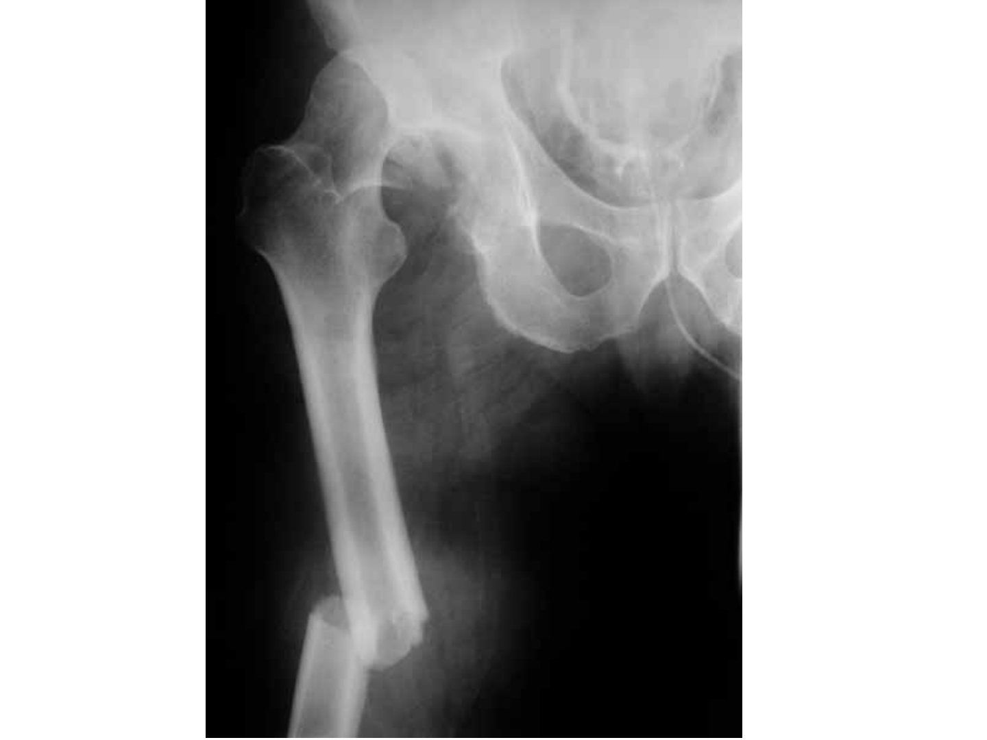
Q1-Q33 questions — tap to reveal all answerslist
- What injuries are shown on this AP X-ray of the right hip and proximal femur?
- What is the initial management and what takes priority?
- Describe damage control orthopaedics in this situation.
Q1.What injuries are shown on this AP X-ray of the right hip and proximal femur?
- Femoral shaft fracture: short oblique, medially angulated, laterally translated, no bone contact
- Right hip dislocation (AP view cannot tell anterior vs posterior)
- Small bony fragment inferior to the femoral head, could be femoral head or acetabular fracture
- No femoral neck fracture
Q2.What is the initial management and what takes priority?
- MOI: RTA/FFH; high energy trauma then ATLS; check open wound and NV status (sciatic nerve)
- If stable: stabilise with Thomas splint, then go for CT scan to see where the bony fragment come from
- Hip dislocation takes priority
- Unstable: DCO; stable: ETC (anatomical reduction, stable fixation, early mobilisation)
Q3.Describe damage control orthopaedics in this situation.
- Attempt CR +/- OR; insert Schanz screws into proximal femur avoiding the canal, connect to rod
- OR approach depends on the direction of dislocation
- External fixation of the femoral shaft
- Definitive surgery: plate/nail
▸ Slide 327 · AP Xray of prxoimal thigh showing fracture over proximal shaftTrauma · 8 questions 1 check expand
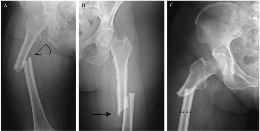
Q1-Q88 questions — tap to reveal all answerslist
- What are the fracture features and diagnosis on this X-ray of the proximal thigh?
- Describe the 2013 ASBMR case definition of AFF.
- What are the minor features of AFF?
- What history and management apply to AFF?
- What is the association between AFF and bisphosphonates?
- How is the contralateral limb surveyed?
- What is the QEH experience with AFF?
- What did Schilcher 2014 NEJM show about bisphosphonates and AFF?
Q1.What are the fracture features and diagnosis on this X-ray of the proximal thigh?
- Transverse fracture over the lateral cortex of the proximal shaft with medial spike, no comminution
- Evidence of beaking over the lateral cortex
- Diagnosis: atypical femoral fracture (AFF)
Q2.Describe the 2013 ASBMR case definition of AFF.
- MUST be along the femoral diaphysis from distal to the lesser trochanter to proximal to the supracondylar flare
- Plus at least 4 of 5 major features; no minor features required
- Major: minimal/no trauma; origin at lateral cortex, transverse (may become oblique medially)
- Complete fracture may have a medial spike; incomplete involves only the lateral cortex
- Major: beaking (periosteal/endosteal thickening of lateral cortex); not or minimally comminuted
Q3.What are the minor features of AFF?
- Delayed healing
- Prodromal pain
- Generalised increase in cortical thickness
- Bilateral
Q4.What history and management apply to AFF?
- History: prodromal symptoms, symptoms on the other side, history of bisphosphonates
- Stop BP and switch to another agent
- Treat fracture side with a long cephalomedullary nail; surveillance of the contralateral side
- Warn about nonunion/malunion (8%)
Q5.What is the association between AFF and bisphosphonates?
- More common with long-term BP (median 7 years); 1.78/100,000/year with exposure <2 years, rising with 8-9 years exposure
- Risk of AFF declines when BP stopped (70%/yr since last use); RR 2-128, AR <50/100,000 person-years
- Asian 8x risk; oral BP stop after 5 years, IV after 3 years
Q6.How is the contralateral limb surveyed?
- Clinical + radiological; pain + normal X-ray then rule out referred pain, bone scan
- Pain + beaking then prophylactic nailing
- Beaking then MRI for bone marrow oedema; if MRI positive or stress osteolysis on X-ray then nail (protected weight bearing if patient declines operation)
Q7.What is the QEH experience with AFF?
- 20% bilateral (UK 7%)
- 30% prodromal pain (UK 40%)
- Nonunion 8%
Q8.What did Schilcher 2014 NEJM show about bisphosphonates and AFF?
- Risk of atypical femoral fracture during and after bisphosphonate use (2014, New England journal of medicine) by Schilcher on 5300 Swedish men and women
- Risk-benefit ratio for prolonged use >5 years would be inverted
- Preventive effect likely lasts several years after cessation, whereas AFF risk decreases rapidly (70%/yr since last use)
- Unclear benefit for age >80; 2020 NEJM: absolute risk of AFF remains very low vs fracture risk reduction
AFF incidence is 100 per 100,000/year with 8-9 years of bisphosphonate exposure — imprecise — The source (Dell 2012 JBMR) reports 113.1 per 100,000/year for 8-9.9 years exposure; the 1.78/100,000/year figure for <2 years matches — source
▸ Slide 328 · Fraser classification for floating kneeTrauma · 2 questions expand
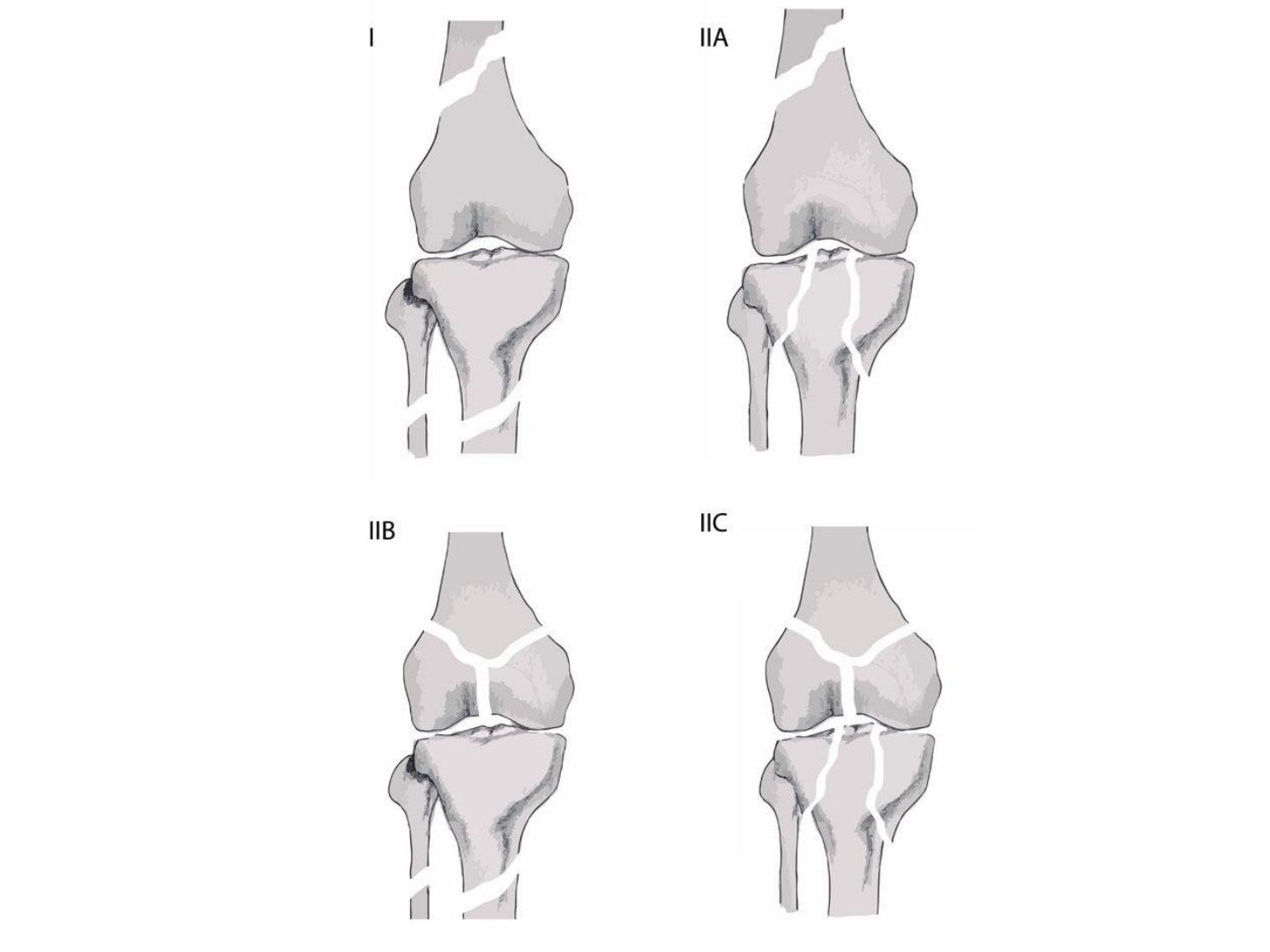
Q1-Q22 questions — tap to reveal all answerslist
- What is the Fraser classification for floating knee and what are the surgical considerations?
- What is the management if there is a vascular injury?
Q1.What is the Fraser classification for floating knee and what are the surgical considerations?
- Type 1 extraarticular
- Femur first; if it becomes unstable can slab the tibia (better than traction on the femur)
- Can immobilise tibia during femoral fixation but not vice versa (need knee flexion for tibial fixation)
- Type 1 can consider retrograde nail, same wound for both fractures
Q2.What is the management if there is a vascular injury?
- Reduce the fracture, reassess
- CTA +/- exploration
▸ Slide 329 · Describe Xray:Trauma · 14 questions expand

Q1-Q1414 questions — tap to reveal all answerslist
- Describe the distal femoral fracture configuration and articular involvement on X-ray.
- What is the initial assessment and local examination in this distal femoral fracture?
- What further imaging is required?
- What are the aims of management?
- Describe the surgical approaches.
- How is the articular block reduced and compressed?
- Describe the MIPO plating technique and parameters.
- What are the cons of non-operative management of a distal femoral fracture?
- What other fixation options exist for distal femoral fractures?
- How is reduction assessed intra-operatively?
- What is the rehabilitation protocol after distal femoral fixation?
- How do you manage an associated neurovascular injury?
- What is the evidence for MIPO plating and how does it work?
- How can an elderly patient be allowed to weight bear immediately?
Q1.Describe the distal femoral fracture configuration and articular involvement on X-ray.
- Displaced fracture over the metaphyseal region of the femur with a vertical split on AP extending to the articular surface
- Some comminution
- AO complete articular fracture type C
Q2.What is the initial assessment and local examination in this distal femoral fracture?
- ATLS, primary and secondary survey, rule out fracture in the same limb; AMPLE history
- Locally: skin (impingement, wound), compartment, NV injury (popliteal artery tethered in this region)
- Systemic: secondary survey
Q3.What further imaging is required?
- XR whole femur to make sure there is no synchronous fracture and to check for proximal implant
- CT scan to delineate fracture pattern, r/o Hoffa fracture (40%)
- XR contralateral side for preop templating
Q4.What are the aims of management?
- Anatomical reduction of the articular surface
- Stable fixation of the articular block and the shaft for secondary bone healing
- Restore rotation, coronal and sagittal alignment, and length
- Allow early mobilisation; timing ETC
Q5.Describe the surgical approaches.
- Lateral parapatellar approach with arthrotomy for joint visualisation
- Or lateral approach: Gerdy tubercle to midaxial line, incise ITB, elevate VL anteromedially from the intermuscular septum, ligate perforators (superolateral geniculate artery)
- +/- medial subvastus approach if double plating: plane between sartorius and VMO, then ligate the descending genicular artery
Q6.How is the articular block reduced and compressed?
- K-wire as joystick, pointed reduction, mini-pelvic clamp; adjuncts: co-linear clamp, cerclage wires
- Reference pins: pin 1 parallel to anterior femoral cortex (axial), pin 2 parallel to joint line (coronal)
- K-wire at junction of anterior 1/4 and posterior 3/4, 2cm from joint line; lag screws for interfragmentary compression
Q7.Describe the MIPO plating technique and parameters.
- Connect articular block to metaphysis; bolster under the knee to relax gastrocnemius
- Plate span ratio 1:3 comminuted, 1:8 not comminuted; plate screw ratio 1/2
- Working length of plate at least double the fracture length
- Bone graft the void; assess alignment (7.3mm screw parallel to joint line, cable method, Blumensaat line)
Q8.What are the cons of non-operative management of a distal femoral fracture?
- Stiffness
- Malunion
- Bedbound patient
Q9.What other fixation options exist for distal femoral fractures?
- LISS plate, angled blade plate, DCS
- Retrograde nail with at least 6cm distal fragment
Q10.How is reduction assessed intra-operatively?
- Varus/valgus: the 7.3mm screw in the plate parallel to the joint line
- Coronal alignment: cable method
- Rotation by checking lesser trochanter, or comparing cortical thickness
- Sagittal: Blumensaat line-shaft angle
Q11.What is the rehabilitation protocol after distal femoral fixation?
- Passive ROM
- NWB for 6 weeks
- FWB by 8-10 weeks
- Treat osteoporosis if needed
Q12.How do you manage an associated neurovascular injury?
- Primary aim is to revascularise the limb
- Early vascular surgeons, vascular shunt, followed by skeletal stabilisation
- +/- prophylactic fasciotomy if warm ischaemic time >6 hrs
Q13.What is the evidence for MIPO plating and how does it work?
- Baumgaertel Injury 1998 introduced biological plating: indirect reduction and bridge plating superior to direct anatomical fixation for bone healing
- Increase the working length of the construct - evenly distributed forces over a long segment, sharing strain between fragments
Q14.How can an elderly patient be allowed to weight bear immediately?
- Double plating
- Shortening of the fracture
- IM nail (prerequisite: fracture line >6cm from joint line)
- Megaprosthesis or nail plate construct
▸ Slide 330 · Hoffa fragment (coronal fracture of femoral condyle)Trauma · 2 questions expand
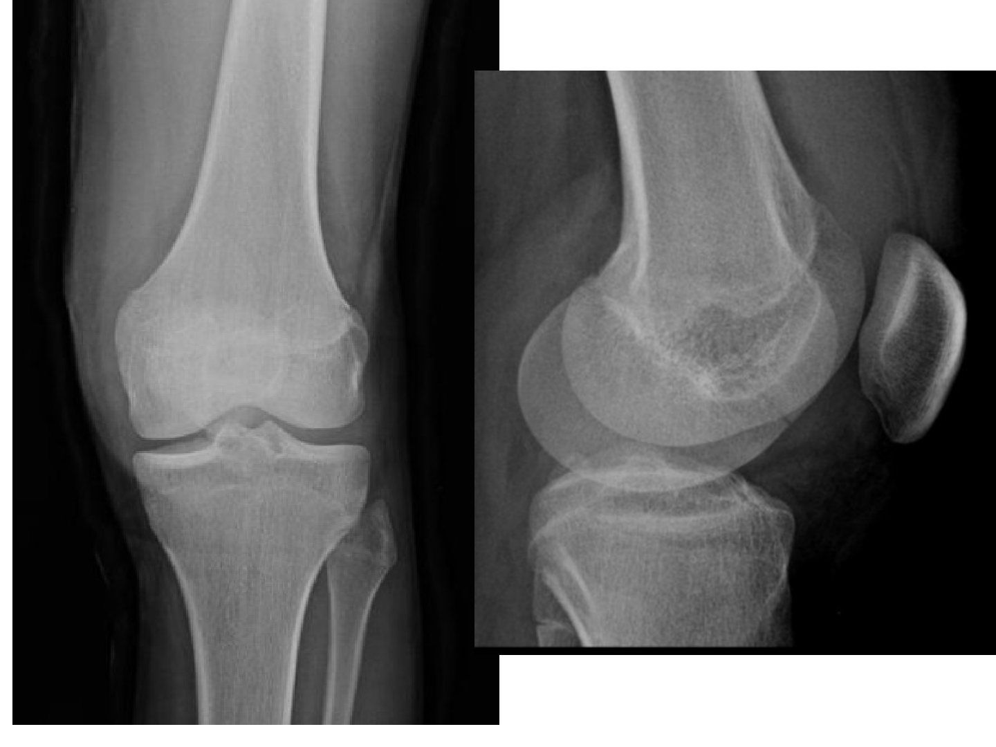
Q1-Q22 questions — tap to reveal all answerslist
- What is a Hoffa fragment and how is it detected?
- What are the considerations in managing a Hoffa fragment?
Q1.What is a Hoffa fragment and how is it detected?
- Coronal fracture of the femoral condyle
- Easily missed - request lateral X-ray/CT in any distal femur fracture
Q2.What are the considerations in managing a Hoffa fragment?
- Fragment may not be well fixed with a lateral locking plate; needs anatomical reduction and lag screw in AP manner before plating
- Use countersink or headless screw
- Isolated fragment may need posteromedial/posterolateral incision
▸ Slide 331 · XR:Trauma · 7 questions expand
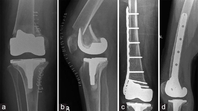
Q1-Q77 questions — tap to reveal all answerslist
- Describe the periprosthetic fracture in relation to the TKR components on X-ray.
- What TKR-specific history and examination findings are important?
- What imaging is required to assess the implant and distal bone stock?
- What are the aims of definitive management?
- How do you manage a loose implant?
- How do you manage a well-fixed implant?
- What is the immediate management of a periprosthetic distal femoral fracture?
Q1.Describe the periprosthetic fracture in relation to the TKR components on X-ray.
- Supracondylar femur fracture distal to the upper edge of the anterior flange of the femoral component
- Shortening and apex anterior angulation; transverse, not comminuted
- TKR posterior stabilised, no sign of loosening, patella not replaced, no notching
- Osteopenia
- Su type II; Rorabeck type II
Q2.What TKR-specific history and examination findings are important?
- Premorbid functional status; details of TKR (implant model, timing, posterior sacrificing or retaining)
- Ipsilateral THR; pre-existing pain, stiffness, infection; mechanism of injury
- PE: soft tissue wound/impingement, distal NV status, compartment syndrome
Q3.What imaging is required to assess the implant and distal bone stock?
- XR of the whole bone
- CT: assess loosening and how much bone stock is left distally
Q4.What are the aims of definitive management?
- Stable fixation of implant and fracture
- Restore mechanical axis to avoid abnormal stresses on the implant
- Allow early weight bearing
Q5.How do you manage a loose implant?
- Revision arthroplasty with a long stem to bypass at least 2 cortical diameters (prefer cementless)
- 2/3 zonal fixation
- Address massive bone loss when removing the old TKR; prepare allograft
- Always standby constrain or even megaprosthesis
Q6.How do you manage a well-fixed implant?
- Locking plate - better restoration of alignment; nail entry/alignment is restricted by the notch; more distal fixation screws
- +/- medial plate for poor bone
- Retrograde nail for posterior cruciate retaining TKR
- Not recommended in ipsilateral THR as a stress riser in the unprotected femoral shaft
Q7.What is the immediate management of a periprosthetic distal femoral fracture?
- Back slab and analgesics
- Ex fix if the initial soft tissue is compromised
▸ Slide 332Trauma · 2 questions expand
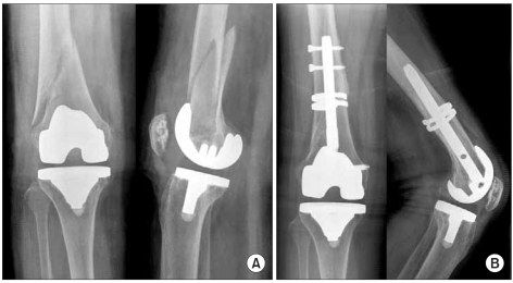
Q1-Q22 questions — tap to reveal all answerslist
- What trauma subject does this slide cover?
- What are the key teaching points on this slide?
Q1.What trauma subject does this slide cover?
- Not covered in the speaker notes - the slide image is the only source
- Topic listed: Trauma
Q2.What are the key teaching points on this slide?
- Not covered in the speaker notes - the slide image is the only source
- No speaker notes were provided for this slide
▸ Slide 333 · Describe Xray:Trauma · 14 questions expand
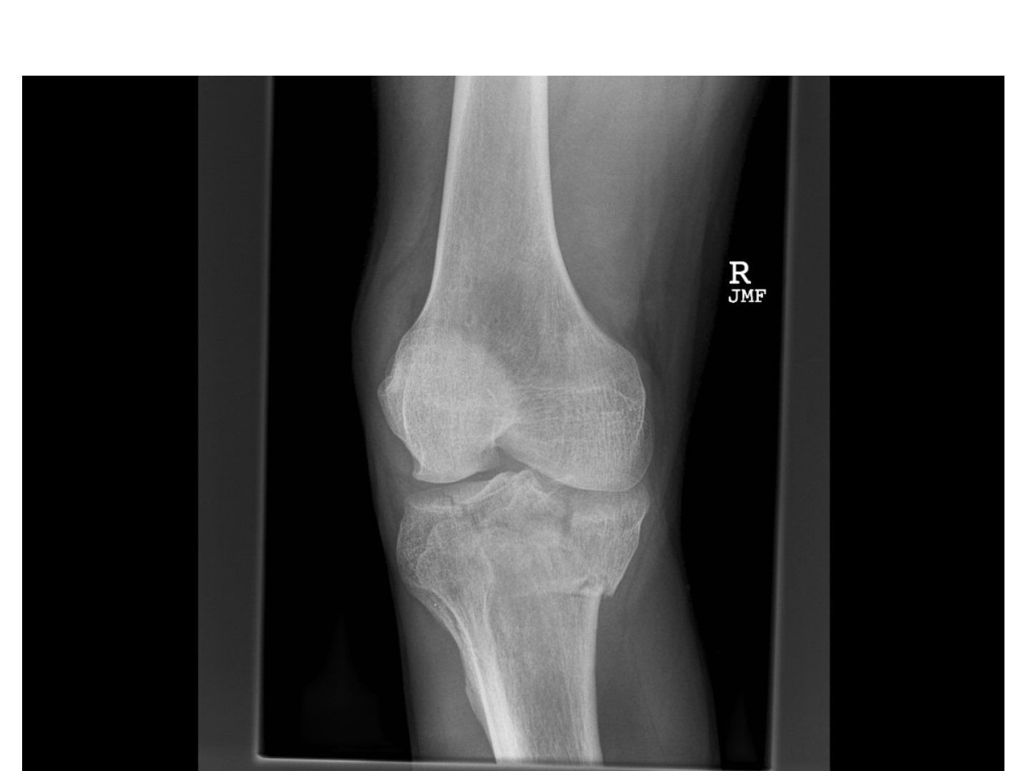
Q1-Q1414 questions — tap to reveal all answerslist
- Describe the tibial plateau fracture pattern and metaphyseal extension on X-ray.
- What is the acute assessment and soft tissue management?
- What are the aims of definitive fixation?
- Describe the approaches.
- How is reduction and fixation performed?
- What is the prognosis?
- What is the incidence of compartment syndrome in tibial plateau fractures?
- What is the evidence comparing ORIF with circular external fixation?
- Describe the anterolateral approach to the tibial plateau.
- Describe the lateral approach to the tibial plateau.
- Describe the posteromedial approach to the tibial plateau.
- What are the criteria for non-operative management?
- What is the rehabilitation after tibial plateau fixation?
- What is the evidence for primary arthroplasty in elderly patients?
Q1.Describe the tibial plateau fracture pattern and metaphyseal extension on X-ray.
- Tibial plateau fracture involving both medial and lateral condyles, articular stepping, metaphyseal involvement, metaphyseal-diaphyseal dissociation
- +/- fibular fracture, joint subluxation
- Schatzker type VI
Q2.What is the acute assessment and soft tissue management?
- ATLS, primary survey, AMPLE history; full circumferential soft tissue examination, compartments
- NV status: popliteal artery tethered at adductor hiatus and soleus arch; common peroneal nerve
- Soft tissue compromise: Span Scan Plan
- CT to assess the three columns and plan approach
Q3.What are the aims of definitive fixation?
- Anatomical reduction of the articular surface; primary bone healing with absolute rigidity
- Correct coronal and sagittal alignment
- Buttress plating for the metaphysis
Q4.Describe the approaches.
- Anterolateral (TA/bone) and posteromedial (medial hamstring/medial gastrocnemius; risk saphaneus nerve (saphenous), MCL)
- Assess articular reduction through submeniscal arthrotomy/scope for meniscus and cruciate status
Q5.How is reduction and fixation performed?
- Split: periarticular clamp/femoral distractor; depression: bone punch from the split
- Fill void with bone graft (autograft, allograft, synthetic bone graft, injectable bone cement)
- Support articular fragment with 3.5mm raft screw, followed by an angular stable device such as a 4.5mm periarticular locking plate in buttressing mode
- Assess overall alignment with the cable method; too comminuted then ring/hybrid external fixator
Q6.What is the prognosis?
- Key to good outcome: restoration of joint line and mechanical axis (mechanical axis > articular surface)
- ~7% had TKR 10 years after index operation (Wasserstein JBJS 2014)
- OA incidence up to 30% if axis deviation more than 5 degrees, increasing TKA likelihood 5 times
- Complications ~10%; 1-year mortality 14%
Q7.What is the incidence of compartment syndrome in tibial plateau fractures?
- 0.7-12%
Q8.What is the evidence comparing ORIF with circular external fixation?
- Canadian OTS multicentre RCT 2006: functional results similar
- ORIF group had more deep infections and unplanned returns to theatre
Q9.Describe the anterolateral approach to the tibial plateau.
- Longitudinal incision lateral to patella starting 2cm below Gerdy tubercle
- Incise deep fascia anterior to ITB
- Subperiosteal dissection of the proximal attachment of tibialis anterior
- Submeniscal arthrotomy; +/- lateral epicondyle osteotomy (extended AL approach)
Q10.Describe the lateral approach to the tibial plateau.
- Skin incision from lateral epicondyle to posterior fibular head
- Protect the CPN
- Interval: ITB and biceps femoris; enter the joint posteriorly between LCL and gastroc LH (gastrocnemius lateral head), or anterior to the LCL
- +/- fibular osteotomy (Lobenhoffer) for better exposure
Q11.Describe the posteromedial approach to the tibial plateau.
- Skin incision from medial epicondyle to posterior edge of tibia
- Release pes anserinus, retract it anteriorly; medial head of gastrocnemius posteriorly
- May release part of the popliteus for exposure; submeniscal arthrotomy
Q12.What are the criteria for non-operative management?
- <2mm articular step
- Stable in knee extension
- No varus/valgus malalignment
Q13.What is the rehabilitation after tibial plateau fixation?
- Protected weight bearing
- Controlled ROM exercises
- Regime tailor made to fixation type, comminution at surgery and associated injuries
Q14.What is the evidence for primary arthroplasty in elderly patients?
- For elderly patients with tibial plateau fracture and pre-existing knee OA
- EFFORT group 2016; also Injury 2021 Sabaitini
▸ Slide 334 · Tibial Plateau FractureTrauma · 4 questions expand

Q1-Q44 questions — tap to reveal all answerslist
- What is the initial assessment and acute management of a tibial plateau fracture?
- What is the aim of operation for a tibial plateau fracture?
- What approaches and fixation are used for a tibial plateau fracture?
- What is the post-operative regimen after tibial plateau fracture fixation?
Q1.What is the initial assessment and acute management of a tibial plateau fracture?
- High energy mechanism: follow ATLS and rule out hip and ankle pathology
- R/O open fracture, compartment Sx and NV injury
- Think possible knee dislocation
- Ext fix +/- compartmental release
- CT to delineate fracture pattern and comminution
- Expect 1-2/52 for soft tissue to settle
Q2.What is the aim of operation for a tibial plateau fracture?
- Restore articular surface
- Stable fixation
Q3.What approaches and fixation are used for a tibial plateau fracture?
- AL (anterolateral) and medial approach
- Buttress plate + raft screw +/- bone graft
- +/- submeniscal dissection x articular reduction
- EUA x ligamentous injury
Q4.What is the post-operative regimen after tibial plateau fracture fixation?
- Post-op ROM 0-90
- NWB
▸ Slide 335 · AP lat XR left tibia and fibula including knee and part of ankle jointTrauma · 7 questions expand
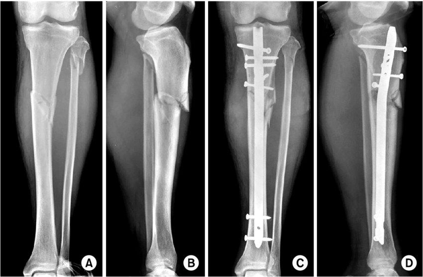
Q1-Q77 questions — tap to reveal all answerslist
- Describe the tibial shaft fracture and associated fibular injury on X-ray.
- What is the initial assessment and management?
- What are the principles of definitive management and why use an antegrade IM nail?
- What is the classic deformity and how is it corrected?
- Describe the entry point and nailing steps.
- What are the non-operative regime and alignment targets?
- What is the post-operative rehabilitation?
Q1.Describe the tibial shaft fracture and associated fibular injury on X-ray.
- Fracture of the proximal 1/3 of the tibial shaft with valgus and apex anterior displacement
- Fibula neck fracture
- AO B2
Q2.What is the initial assessment and management?
- ATLS, primary survey, AMPLE history
- Local: soft tissue, skin impingement, NV status (CPN), compartment syndrome; systemic secondary survey
- XR one joint above and below
- Long leg slab + analgesics; close monitoring for compartment syndrome
Q3.What are the principles of definitive management and why use an antegrade IM nail?
- Restore tibial length, correct angulation and rotation; achieve secondary bone healing; prevent complications
- Load sharing device that is biomechanically more rigid than a plate
- Preserves biology (not disrupting fracture haematoma); minimally invasive; Have the option of secondary dynamization if delay union
Q4.What is the classic deformity and how is it corrected?
- Proximal: procurvatum (patellar tendon) + valgus (by ATT - anterior tibial tendon)
- Distal: varus + IR (PTT, FHL, FDL)
- Reduction: traction, manipulation, reduction forceps, unicortical Shanz pins
- Poller screw on concave side in proximal fragment (lateral, posterior), convex side distally; +/- anterior unicortical plate
Q5.Describe the entry point and nailing steps.
- Lateral to midline, medial to lateral tibial spine (more lateral prevents valgus); just anterior to articular surface, as proximal/posterior as possible without breaking it
- Knee in slightly extended position; awl, guidewire to centre of tibial plafond at physeal scar; serial reaming; measure length; static locked nail
- Choose a reamed nail: bone graft, bigger nail more rigid, better endosteal fit, reduced working length
- SPRINT trial: reaming decreased future bone grafting and nail exchange
Q6.What are the non-operative regime and alignment targets?
- Long leg cast 4 wk then patellar bearing brace/ Sarmiento brace for 12 week; union rate 90%
- Varus/valgus <5 degrees, AP angulation <10 degrees, rotation 10 degrees, shortening <1cm, joint line parallel
Q7.What is the post-operative rehabilitation?
- FWB (full weight bearing)
▸ Slide 336 · Gustilo I fracture tibial shaft with proximal fragment protruding out of skin inTrauma · 7 questions 1 check expand

Q1-Q77 questions — tap to reveal all answerslist
- Describe the injury.
- What is the acute management of an open fracture per BOA guidelines?
- What is the timing of early total care?
- Describe the procedure.
- What is the evidence for nailing in open fractures?
- How do you avoid compartment syndrome and when should soft tissue coverage be performed?
- Would you ream in this isolated open tibial fracture and why?
Q1.Describe the injury.
- Gustilo I fracture of the tibial shaft with the proximal fragment protruding out of skin in an inside-out manner
- Leg pink
Q2.What is the acute management of an open fracture per BOA guidelines?
- Antibiotic within 1 hour (1st gen cephalosporin + aminoglycoside) + anti-tetanus
- Remove gross contamination, take a picture, dressing with NS gauze + impermeable film
- Direct pressure for bleeding control; temporary reduction + slab
- Next step is XR
Q3.What is the timing of early total care?
- Within 12 hours for solitary high energy open fractures
- Within 24 hours for all other low energy open fractures
- If there is no vascular compromise
Q4.Describe the procedure.
- GA, supine with triangular support; irrigation with 3L NS under low pressure; remove non-viable bone/soft tissue; assess wound coverage after debridement
- If soft tissue acceptable, go for definitive nailing via a split patellar approach
- Entry site: coronal plane parallel to the canal; sagittal plane just distal to the angle between tibial plateau and anterior tibial metaphysis
- 2 static locking screws proximal and distal; continue antibiotics until wound ok; Closely monitor for compartment syndrome; WBAT
Q5.What is the evidence for nailing in open fractures?
- Unreamed IMN interlocking showed superior results for Gustilo II/IIIA/B: 1less malunion, 2lower infection rate, 3↓no. of OT needed (Bhandari 2001 JBJS Br)
- Foote from canada published in CORR 2015 network meta-analysis: definitive IM nail once soft tissue settled may be superior independent of Gustilo grade; Use of unreamed nails over reamed nails also may be advantageous in the setting of open fractures. LESS REOPERATION
- In multitrauma open fracture, no evidence for reaming (SPRINT trial)
Q6.How do you avoid compartment syndrome and when should soft tissue coverage be performed?
- High index of suspicion; avoid regional anaesthetic blocks
- Definitive soft tissue coverage within 5 days
Q7.Would you ream in this isolated open tibial fracture and why?
- Isolated trauma with risk of nonunion - ream to allow a larger nail
- Biological advantage of bone graft deposition
- In multitrauma open fracture there is no evidence for reaming (SPRINT trial)
Definitive soft tissue coverage should be within 5 days — outdated — BOA/BAPRAS (BOAST 4) standard is definitive closure or coverage within 72 hours of injury if it cannot be performed at the time of debridement — source
▸ Slide 337 · Distal 1/3 extraarticular tibia fractureTrauma · 7 questions expand

Q1-Q77 questions — tap to reveal all answerslist
- What are the advantages of plating a distal 1/3 extraarticular tibia fracture?
- What are the disadvantages of plating a distal 1/3 tibia fracture?
- What are the mechanical and biological advantages of IM nailing?
- Which implant is load bearing and which is load sharing?
- What did the FixDT trial (Costa 2018) show?
- What did the Injury 2021 Bleeker meta-analysis show?
- Should the fibula be plated if the tibia is nailed?
Q1.What are the advantages of plating a distal 1/3 extraarticular tibia fracture?
- Anatomical reduction
- Primary bone healing
Q2.What are the disadvantages of plating a distal 1/3 tibia fracture?
- More dissection, especially at the site of anastomosis between PTA and ATA
- Load bearing construct
- Implant impingement
Q3.What are the mechanical and biological advantages of IM nailing?
- Mechanical: load sharing implant, more torsional and bending rigidity
- Biology: does not disturb the fracture hematoma; reaming deposits bone graft
- Cons: more anterior knee pain
Q4.Which implant is load bearing and which is load sharing?
- Plate = load bearing construct
- IM nail = load sharing implant
- Nail has more torsional and bending rigidity
Q5.What did the FixDT trial (Costa 2018) show?
- IM nail: greater recovery - less disability, better ankle function and health-related QoL at 3 months
- Cost lower with IM nail
- Longer term: similar disability ratings and infection
Q6.What did the Injury 2021 Bleeker meta-analysis show?
- Nail - more malunion
- Plate - more infection
- Management should be individualized
Q7.Should the fibula be plated if the tibia is nailed?
- Frodl EFFORT 2021 metaanalysis: no difference in nonunion
- Fixing fibula may reduce secondary valgus/varus malunion but higher risk of wound infection
- Individualized decision
▸ Slide 338 · Clinical photo: Open fracture with mangled left leg with vascular compromise, asTrauma · 11 questions 1 check expand
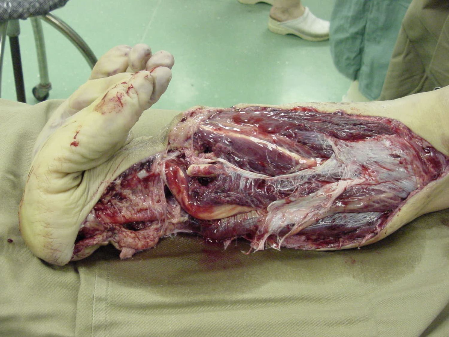
Q1-Q1111 questions — tap to reveal all answerslist
- Describe the injury and X-ray findings in this mangled leg.
- What is the initial ATLS/AED management of this open fracture?
- What is a mangled extremity and how is the amputation decision made?
- What is the MESS score and what are its components?
- Which factors determine the choice between amputation and reconstruction?
- What are the general indications for amputation?
- What did the LEAP study show?
- What are the common problems after limb salvage?
- What is the initial operative plan and early care?
- Which flaps are used for soft tissue coverage in limb salvage?
- How is conversion from external to internal fixation timed?
Q1.Describe the injury and X-ray findings in this mangled leg.
- Open fracture, mangled left leg with vascular compromise (pale looking sole)
- No gross contamination with soil/feces
- X-ray: comminuted, segmental tibial shaft fracture, extraarticular with gas shadow
- Gustilo determined intraop - at least IIIB, probably IIIC
Q2.What is the initial ATLS/AED management of this open fracture?
- ATLS ABCDs, tranexamic acid, massive transfusion protocol; primary and secondary survey
- Antibiotics within 1 hr (1st gen cephalosporin) + tetanus; BOAST 4: <3hr infection 4.7%, >3hr 7.4%
- Direct pressure to control bleeding; remove gross contamination; sterile saline-soaked gauze dressing
- Temporary stabilization with splintage; AMPLE history incl ischemic time
Q3.What is a mangled extremity and how is the amputation decision made?
- Consider 4 factors: soft tissue, bone, nerve, vessel; if 3/4 are disrupted it is a mangled extremity
- Consider patient/injury factors and scoring systems e.g. MESS
- Per LEAP, the single most important predictive factor is the patient's self efficacy
- If possible, will try to salvage the limb in acute setting if we are able to revascularize the limb; then buy time to have thorough discussion with patient and relative for delay primary amputation
- Patient need to understand limb salvage operation will likely involve multiple surgery and long rehab
Q4.What is the MESS score and what are its components?
- MESS 1990 original paper: score >7 had 100% predictive value of amputation in that series
- Patient factors: age, shock
- Disease/injury factors: energy, extremity perfusion
Q5.Which factors determine the choice between amputation and reconstruction?
- If possible keep the limb first; can do secondary amputation later
- Factors (referenced from MESS): shock, energy of trauma, distal pulse, age, time of injury
- Whether the ipsilateral foot is reconstructable
Q6.What are the general indications for amputation?
- Dead (non viable)
- Dangerous (life threatening)
- Damn nuisance (non functional)
Q7.What did the LEAP study show?
- Amputation vs reconstruction: no difference in outcome
- Self efficacy is the single determining factor for long-term outcome
- Satisfaction driven by function and pain; psychosocial factors more important
- Lifetime cost of salvage < amputation; no single score can be used for decision making
- Tibial nerve integrity not reliable/predictive - at 2 years almost 50% regained some plantar sensation
Q8.What are the common problems after limb salvage?
- Recurrent infection, nonunion, delayed union
- LLD, OA, pain and stiffness
- Second amputation
Q9.What is the initial operative plan and early care?
- DCO - damage control orthopaedics (staged management)
- Consent: external fixator + debridement + temporary wound coverage +/- vascular repair/shunting +/- amputation
- Early 2nd look in 48 hr; gentamicin beads (high local concentration, high MIC, fill dead space)
- If salvage succeeds: early fix and flap as early as D3, within 1 week, before infection sets in
Q10.Which flaps are used for soft tissue coverage in limb salvage?
- Medial gastrocnemius flap
- Medial soleus flap
- Reverse sural flap (fasciocutaneous)
Q11.How is conversion from external to internal fixation timed?
- <2/52: can be one stage
- >2/52: pin tract resting x 1 week (daily pin tract resting + antibiotics)
MESS score >7 has a 100% predictive value of amputation — misleading/outdated — True only in Johansen's original 1990 series; LEAP and later studies could not validate any trauma score, with MESS >7 sensitivity around 44% in recent cohorts — source
▸ Slide 339 · Same PatientTrauma · 8 questions expand
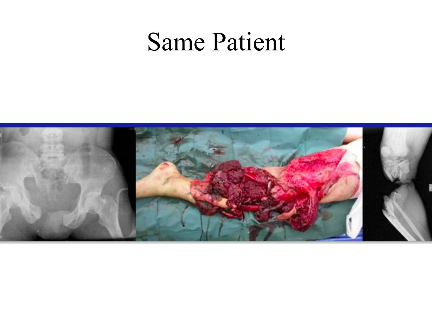
Q1-Q88 questions — tap to reveal all answerslist
- What injuries does this multiply injured patient have and what are the initial priorities?
- What is the 'second hit' phenomenon in damage control orthopaedics?
- What are the 3 stages of DCO?
- What is the DCO timeline?
- When is the patient stable for definitive surgery?
- What is early total care?
- Which patients are classified as borderline for DCO?
- Does damage control orthopaedics improve mortality?
Q1.What injuries does this multiply injured patient have and what are the initial priorities?
- Unstable pelvic fracture, open leg fracture and tibial plateau fracture
- ATLS protocol and pelvic binder
- Rule out life-threatening injury
- Manage according to damage control orthopaedics
Q2.What is the 'second hit' phenomenon in damage control orthopaedics?
- The initial injury is the first hit; surgery is a potential second hit
- Triggers inflammatory, immunological and coagulatory cascades
- May push the patient into an irreversible physiological response (lethal triad) with increased mortality
- Limiting the second hit is the rationale for a staged approach
Q3.What are the 3 stages of DCO?
- 1. Initial stabilization: life-saving surgery for skeletal and hemorrhagic stabilization, to facilitate nursing care (hemostasis, skeletal stabilization, decompression, decontaminate, revascularize)
- 2. ICU resuscitation and optimization
- 3. Definitive surgery
Q4.What is the DCO timeline?
- D1 DCO
- D2 second look
- D5-10 window of opportunity
- 3+ can go to OT
Q5.When is the patient stable for definitive surgery?
- When acidosis, hypothermia and coagulopathy are corrected
- Clinical markers: BP, urine output, inotrope use, temperature
- Biochemical markers: pH (lactate, base excess), coagulation
Q6.What is early total care?
- Immediate fixation of all fractures
- Aim: early OT, early mobilization, less recumbency, early discharge
- Increased ARDS and multi-organ failure in borderline patients
Q7.Which patients are classified as borderline for DCO?
- Patients classified: stable, borderline, unstable, extremities
- Borderline: ISS >40 or ISS >20 + chest injury
- Bilateral femoral fracture, chest contusion, pelvic/abdominal injury
Q8.Does damage control orthopaedics improve mortality?
- DCO is shown to improve mortality compared with immediate definitive care in the unstable patient
▸ Slide 340 · Topics to discuss:Trauma · 8 questions 1 check expand

Q1-Q88 questions — tap to reveal all answerslist
- What three topics are discussed in this case?
- What are the priorities in managing an open fracture?
- What is the acute AED management of an open fracture?
- What examination and imaging must be documented in open fracture assessment?
- What are the operative debridement and irrigation principles?
- What is the timing of second look, flap cover and antibiotics?
- What is the evidence for timing of surgery in open fractures?
- How is a bone defect managed?
Q1.What three topics are discussed in this case?
- Open fracture management
- Definitive management for bone (bone defect) and soft tissue (flap coverage)
- Mangled extremity management
Q2.What are the priorities in managing an open fracture?
- 1. NV status
- 2. Prevent infection
- 3. Soft tissue
- 4. Bone
Q3.What is the acute AED management of an open fracture?
- ATLS, BOA/BOAST guidelines, AMPLE history
- Antibiotic within 1 hr (1st gen cephalosporin; BOAST suggests 1.5g IV cefuroxime + aminoglycoside) + anti-tetanus
- Remove gross contamination, take a photo, dressing with NS gauze + impermeable film
- Direct pressure for bleeding control; temporary reduction + slab
Q4.What examination and imaging must be documented in open fracture assessment?
- Local: compartment syndrome and NV status
- Systemic: secondary survey to rule out other injuries
- XR of the injured part
Q5.What are the operative debridement and irrigation principles?
- Early OT within 24 hrs: ex fix + debridement (keep skin/bone, remove all doubtful muscle)
- Irrigation: 3, 6, 9 L at low pressure 8 psi; no evidence for which fluid type
- NPWT if primary closure not possible (WOLLF trial JAMA 2019 Costa: no improvement in self-rated disability at 12 months)
Q6.What is the timing of second look, flap cover and antibiotics?
- Early 2nd look at 48 hr with gentamicin beads
- Early fix and flap within 72hrs (should not exceed 7 days) per BOAST, before infection sets in
- Antibiotics continued for 72 hrs or until wound closure
Q7.What is the evidence for timing of surgery in open fractures?
- Within 24 hrs; older studies recommended 6 hrs but based on animal studies
- LEAP: no difference in infection rates when managed within 6 hr or 24 hrs
- Latest BOAST: urgent surgery if vascular injury or heavily contaminated with sewage, marine or agricultural matter
Q8.How is a bone defect managed?
- Eradicate infection first; depends on size of defect, soft tissue coverage and vascularity of the tissue bed
- Small <6cm: acute shortening, non-vascularized bone graft (Hammon PL bone grafting or Papineau)
- Large >6cm: Masquelet technique (cement spacer 4-5 weeks + staged bone graft), vascularized bone graft (fibula), bone transport
- Large defect + soft tissue/NV defect or unfit patient: consider amputation
BOAST guidelines suggest 1.5g IV cefuroxime + aminoglycoside for open fractures — imprecise attribution — 2017 BOAST-4 recommends IV antibiotics ideally within 1 hour of injury but defers the specific antibiotic choice to local network guidelines; it does not mandate this regimen — medium confidence — source
▸ Slide 341 · Soft tissue coverageTrauma · 2 questions expand
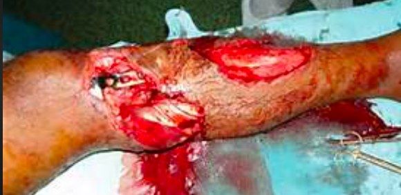
Q1-Q22 questions — tap to reveal all answerslist
- What flap options cover proximal, middle and distal third tibial defects?
- What is the BOA initial treatment guideline for adult open fractures?
Q1.What flap options cover proximal, middle and distal third tibial defects?
- Proximal 1/3: medial gastroc flap (medial sural artery) + PTSG
- Middle 1/3: soleal flap (posterior tibial artery)
- Distal 1/3: classic free flap; recent reverse hemisoleal (tibial perforators) or reverse sural (superficial sural artery)
Q2.What is the BOA initial treatment guideline for adult open fractures?
- IV antibiotics within 1 hr + tetanus
- Document distal NV status and compartments
- Re-align the fracture and apply a splint
- Remove gross contamination, dress with saline-soaked gauze + occlusive film, take a clinical photo
▸ Slide 342 · FlapTrauma · 10 questions expand

Q1-Q1010 questions — tap to reveal all answerslist
- How are flaps classified?
- What are the Mathes types of muscle flap pedicles?
- Which Mathes pedicle types are most reliable in muscle flaps?
- What systemic factors are assessed pre-operatively before a flap?
- What must be planned in flap design?
- How is a flap elevated and inset intra-operatively?
- What are the causes and management of impaired flap circulation post-operatively?
- What is the mechanism of VAC?
- What are the common pedicle flaps for the leg?
- List common free flap choices and their pedicles.
Q1.How are flaps classified?
- Location: local, regional, distant (pedicle/free)
- Pedicles: random, axial
- Composite/tissue: cutaneous, fasciocutaneous, musculocutaneous, osseocutaneous
Q2.What are the Mathes types of muscle flap pedicles?
- I: one dominant pedicle (e.g. gastrocnemius, TFL)
- II: one dominant + one minor pedicle (e.g. gracilis)
- III: two dominant pedicles (e.g. rectus abdominis)
- IV: segmental pedicles (e.g. sartorius)
- V: one dominant + secondary segmental pedicles (e.g. pectoralis major)
Q3.Which Mathes pedicle types are most reliable in muscle flaps?
- Types I, III and V are more reliable
- Preferred because they have a broad base
Q4.What systemic factors are assessed pre-operatively before a flap?
- Smoking, obesity, hypertension, PVD, immunosuppression
Q5.What must be planned in flap design?
- Type of flap, size
- Pivot point, axis, arc of rotation
- +/- Doppler to identify the pedicle
Q6.How is a flap elevated and inset intra-operatively?
- Elevation: preserve minor pedicles; complete pedicle mobilization not required unless free flap; avoid tension during rotation and inset
- Inset: tunnel size = flap base x 2, avoid tension, ensure haemostasis
Q7.What are the causes and management of impaired flap circulation post-operatively?
- Consider a systemic haemodynamic problem or local arterial/venous obstruction
- Try releasing stitches and repositioning the flap
- Avoid pressure, constrictive bandage and motion (splint)
Q8.What is the mechanism of VAC?
- Induces granulation tissue and epithelisation
- Decreases dead space and edema
- Down grade soft tissue reconstruction ladder
Q9.What are the common pedicle flaps for the leg?
- Proximal: medial gastrocnemius flap (muscle flap; medial sural artery from popliteal artery)
- Middle: soleus flap (muscle flap; branches from posterior tibial and peroneal arteries)
- Distal: distant flap/reverse sural (fasciocutaneous; superficial sural artery depending on perforating branch of peroneal artery)
Q10.List common free flap choices and their pedicles.
- LD flap: myocutaneous, thoracodorsal artery
- Groin flap: fasciocutaneous, superficial circumflex iliac artery 2.5cm inferior to inguinal ligament, very consistent
- ALT flap: fasciocutaneous, descending branch of lateral femoral circumflex artery
- Radial forearm flap: radial artery; skin +/- muscle +/- bone; can be retrograde/antegrade
▸ Slide 343 · Key points in gunshot woundsTrauma · 3 questions expand
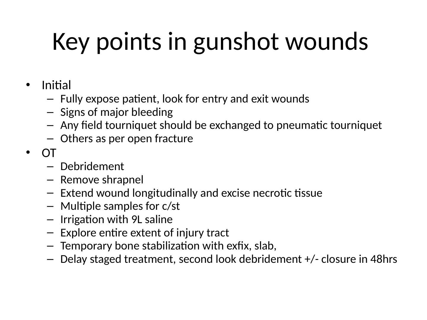
Q1-Q33 questions — tap to reveal all answerslist
- What are the key points in the initial assessment of a gunshot wound?
- What are the key points of OT debridement in a gunshot wound?
- What is the staged treatment plan after debridement of a gunshot wound?
Q1.What are the key points in the initial assessment of a gunshot wound?
- Fully expose the patient and look for entry and exit wounds
- Look for signs of major bleeding
- Any field tourniquet should be exchanged for a pneumatic tourniquet
- Otherwise manage as per open fracture
Q2.What are the key points of OT debridement in a gunshot wound?
- Remove shrapnel
- Extend the wound longitudinally and excise necrotic tissue
- Take multiple samples for c/st
- Irrigation with 9L saline
- Explore the entire extent of the injury tract
- Temporary bone stabilization with exfix, slab
Q3.What is the staged treatment plan after debridement of a gunshot wound?
- Delay staged treatment: second look debridement +/- closure at 48 hrs
▸ Slide 344 · Vacuum assisted closureTrauma · 3 questions expand
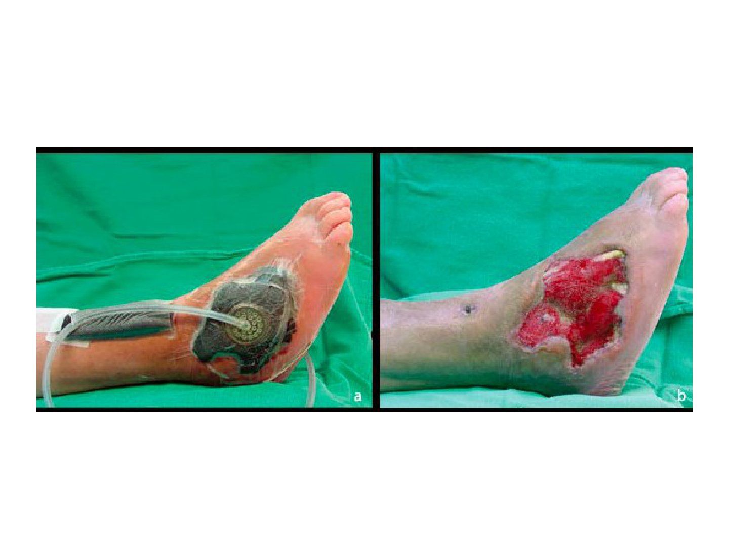
Q1-Q33 questions — tap to reveal all answerslist
- How does VAC affect wound size and the reconstruction ladder?
- How does VAC affect edema and dead space?
- What tissue processes does VAC promote?
Q1.How does VAC affect wound size and the reconstruction ladder?
- Reduces wound size
- Downgrades the soft tissue reconstruction ladder
Q2.How does VAC affect edema and dead space?
- Reduces edema
- Reduces dead space
Q3.What tissue processes does VAC promote?
- Promotes granulation tissue
- Promotes epithelialization
▸ Slide 345 · Tscherne classification of soft tissue injury in close fractureTrauma · 2 questions expand
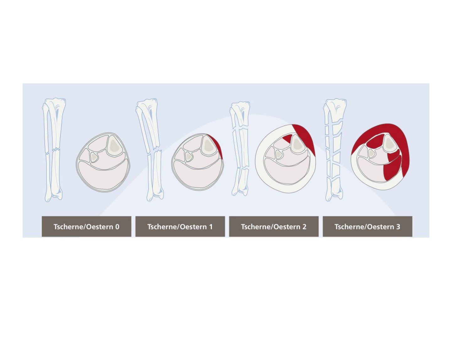
Q1-Q22 questions — tap to reveal all answerslist
- Describe the Tscherne classification of soft tissue injury in closed fractures.
- Describe the Tscherne classification of open fractures.
Q1.Describe the Tscherne classification of soft tissue injury in closed fractures.
- Grade 0: minimal, simple fracture, indirect force
- Grade 1: superficial abrasion, mild fracture pattern
- Grade 2: deep abrasion, skin or muscle contusion, comminuted fracture
- Grade 3: crush/degloving injury
Q2.Describe the Tscherne classification of open fractures.
- Grades 1-4
- Based on: wound size, degree of contamination, fracture pattern
- Grade 4 = incomplete/complete amputation
▸ Slide 346 · XR left ankle showing comminuted tibial metaphyseal fracture with intraarticularTrauma · 15 questions expand
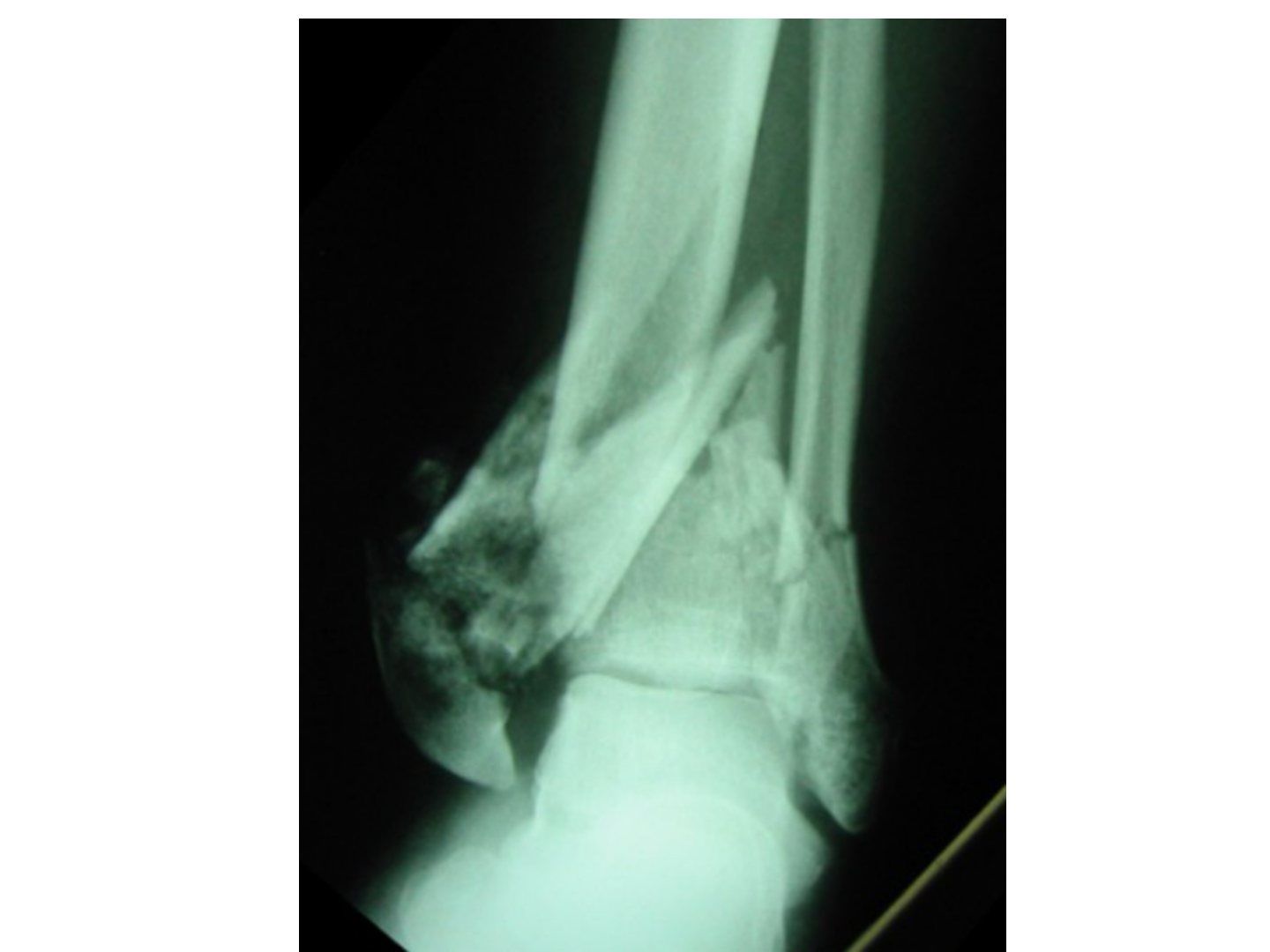
Q1-Q1515 questions — tap to reveal all answerslist
- Describe the X-ray findings in this ankle.
- What classifications are used for tibial plafond (pilon) fractures?
- What is the initial assessment for a high-energy pilon fracture?
- What local and systemic assessment is needed in a pilon fracture?
- What is the initial Span Scan Plan management?
- Why is the fibula plated alongside the spanning fixator?
- What are the typical CT fragments and their ligament attachments?
- What is the Topliss classification?
- What are the definitive management options and timing?
- What are the Ruedi-Allgower principles of fixation?
- What are the surgical approach and fixation details?
- Which approaches are used and what is the evidence for fragment-specific fixation?
- What adjuncts and checks are used during reduction?
- What is the evidence for ORIF versus external fixation in pilon fractures?
- Describe the anterolateral (AL) approach.
Q1.Describe the X-ray findings in this ankle.
- Comminuted tibial metaphyseal fracture with intraarticular extension, fracture distal fibula
- Articular impaction and comminution
- Any association joint subluxation or dislocation. The talar dome appears intact
Q2.What classifications are used for tibial plafond (pilon) fractures?
- Ruedi-Allgower: 1 undisplaced, 2 displaced with minimal comminution, 3 comminuted
- AO: A extraarticular, B partial articular, C complete articular
- CT fragment description: Topliss classification
Q3.What is the initial assessment for a high-energy pilon fracture?
- High-energy trauma - ATLS protocol
- AMPLE history
- Primary and secondary survey, looking for other axial loading type injuries
Q4.What local and systemic assessment is needed in a pilon fracture?
- Local: circumferential assessment of the limb - soft tissue, compartment syndrome, NV status
- Systemic: secondary survey for axial load injuries - calcaneus, knee, spine
Q5.What is the initial Span Scan Plan management?
- Analgesia, elevate, ice therapy
- Span: ankle spanning external fixator - delta frame construct; tibial pin >=3cm from fracture site to prevent fracture propagation; +/- plate fibula
- Scan: CT for planning - typically 3 fragments: medial, Chaput (AL), Volkman (PL), central, die punch
Q6.Why is the fibula plated alongside the spanning fixator?
- According to Robertson's paper
- Maintain length and better soft tissue resting
Q7.What are the typical CT fragments and their ligament attachments?
- Typically 3 fragments; ligaments usually intact
- Medial, Chaput (AL) - AITFL, Volkman (PL) - PITFL, central, die punch
- Descriptive Topliss classification
Q8.What is the Topliss classification?
- Coronal: CS, PS, AS, V, Y
- Sagittal: SS, IV, T
Q9.What are the definitive management options and timing?
- ORIF with plating; definitive external fixator (hybrid/ring); combined ex fix + ORIF
- Consider patient factors: premorbid state, DM, PVD, smoking
- Timing: when wrinkle sign positive and blisters epithelialized (13-28 days)
Q10.What are the Ruedi-Allgower principles of fixation?
- 1. Restore fibular length and rotation
- 2. Reconstruct the articular surface posterior to anterior (PL -> C -> AL -> M); lag screw A to P
- 3. Restoration of bone defect (bone graft)
- 4. Tibial buttressing (articular block to shaft); plate medial if varus, lateral if valgus
Q11.What are the surgical approach and fixation details?
- Classic PL + AM approach; beware 7cm skin bridge; fragment-specific (AM for plate, AL for Chaput) if <7cm
- Fix fibula: simple fracture - fix before tibia; complex - fix after
- Reduce articular block first, then connect to metaphysis + compression if possible
- Lag screw anterior to posterior; angular stable plate 2.7/3.5 in buttressing mode (medial), 3 screws above and below; +/- concept of bicolumn plating
- Rehab: NWB walking, resting AFO to prevent equinus
Q12.Which approaches are used and what is the evidence for fragment-specific fixation?
- Classic PL + AM (AL if small AL fragment, AM if large AL fragment); or anterior approach (EHL/EDC)
- Fragment specific (AM for plate, AL for Chaput): <7cm skin bridge acceptable with good timing and careful soft tissue dissection (Howard JOT 2008)
Q13.What adjuncts and checks are used during reduction?
- K wire joystick and reduction clamps; distractor as adjunct
- Reduce articular block first, then connect to metaphysis + compression if possible
- + Bone graft for bone defect
- Assess reduction of fibular length by dime sign and talocrural angle (83deg); mortise by Shenton line, overlap, clear space
Q14.What is the evidence for ORIF versus external fixation in pilon fractures?
- Wang et al J Foot Ankle Surg 2015 meta-analysis: equivalent results
- Equal incidence of complications including arthritis
- ORIF has more severe complications
Q15.Describe the anterolateral (AL) approach.
- Skin incision centred at the ankle joint proximally, parallel to the 4th MT distally
- Protect branches of the superficial peroneal nerve
- Raise full thickness flaps; incise extensor retinaculum and retract anterior compartment tendons medially
▸ Slide 347 · What is syndesmosis ligament complex? consist of ATFL, PTFL, ITL and interosseouTrauma · 7 questions 1 check expand

Q1-Q77 questions — tap to reveal all answerslist
- What is the syndesmosis ligament complex?
- How is syndesmosis injury assessed clinically and on X-ray?
- Where is the syndesmosis assessed and how is the TF clear space defined?
- What does arthroscopy add in syndesmosis injury?
- How do you insert a syndesmosis screw?
- What is the evidence for Tightrope versus syndesmosis screw fixation?
- When would you fix the posterior malleolus?
Q1.What is the syndesmosis ligament complex?
- Consists of AITFL, PITFL, ITL and interosseous ligament
- PITFL is the strongest
Q2.How is syndesmosis injury assessed clinically and on X-ray?
- Clinical: local tenderness at AITFL, squeeze test, ER stress test, cotton test
- AP: medial clear space <4mm, TF clear space <6mm, TF overlap >6mm
- Mortise: Shenton line, symmetrical joint space (<5 degrees talar tilt), talocrural angle 83 degrees, dime sign
- Lateral: ATFI/TW = 39 +/- 9%; stress views (abd + ER on DF), hook test, CT, arthroscopy
- Ramsey: 1mm lateral talar shift = 42% reduction in tibiotalar contact area
Q3.Where is the syndesmosis assessed and how is the TF clear space defined?
- Syndesmosis is assessed 1cm above the joint line
- TF clear space: from the medial border of the fibula to the incisural surface of the tibia
- Mortise view: TF clear space <6mm, TF overlap >1mm
Q4.What does arthroscopy add in syndesmosis injury?
- Detects multi-directional instability, especially sagittal and rotational
- Lui arthroscopy 2005
Q5.How do you insert a syndesmosis screw?
- X-ray control +/- CT; reduce with IR and anterior drawer, hold with periarticular clamp or K wires
- Keep ankle in neutral position (traditional teaching used DF; Gonzalez Injury 2017 - an anatomically reduced syndesmosis will not be over-tightened)
- 2cm above ankle joint, posterolateral to anteromedial (20 deg from horizontal), parallel to joint line
- One 3.5mm cortical positional screw through 3 cortices (2 for Maisonneuve)
- NWB for 8/52, then remove screw and gradually increase weight bearing
Q6.What is the evidence for Tightrope versus syndesmosis screw fixation?
- Injury 2015 RCT Kortekangas: similar postop malreduction rates
- Similar functional outcomes
- Similar OA incidence
Q7.When would you fix the posterior malleolus?
- Traditionally if >25% articular surface, >2mm step off, or unstable syndesmosis
- Recent studies suggest a lower threshold - it is the attachment of the PITFL (contributes 42% of syndesmosis stability)
- Isolated PM fixation restores 70% of syndesmosis stiffness vs 40% with isolated syndesmosis fixation
- 2017 JBJS Solan suggests much lower threshold; POSTFIX trial to be released in 2023
Posterior malleolus fixation threshold is to be confirmed by the POSTFIX trial (to be released in 2023) — outdated — POSTFIX one-year results (Bone Joint J 2025) found ORIF of medium-sized (5-25%) posterior fragments in AO type B fractures did not improve outcome, so size threshold alone is not validated — source
▸ Slide 348 · Lauge-Hansen: Foot Position and Force applied relative to tibiaTrauma · 5 questions expand
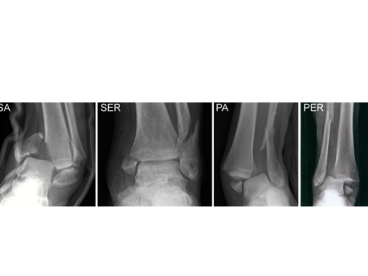
Q1-Q55 questions — tap to reveal all answerslist
- Describe the Lauge-Hansen classification.
- What is the general fixation for each malleolus?
- What is the classic SAD pattern and its fixation?
- How are PAB and PER patterns managed?
- What are the poor prognostic factors for ankle fractures?
Q1.Describe the Lauge-Hansen classification.
- Based on foot position and force applied relative to tibia
- SAD: transverse Weber A + vertical medial malleolus +/- impaction
- SER: Weber B + transverse medial malleolus + PM# (AITFL -> fibula -> PITFL/PM -> deltoid)
- PAB: comminuted impacted Weber C + transverse medial malleolus (Transverse MM --> PITFL/ PM --> Fibula)
- PER: spiral Weber C + transverse medial malleolus + PM# (Maisonneuve) (Deltoid/ avulsion from MM --> AITFL --> high fibula --> PITFL)
Q2.What is the general fixation for each malleolus?
- Lateral malleolus: posterolateral approach - lag screw + neutralization plate, locking plate, or antiglide plate/buttress
- Medial malleolus: anteromedial approach with screw/plate (plate if vertical)
- Posterior malleolus: percutaneous screw
Q3.What is the classic SAD pattern and its fixation?
- Marginal impaction - reduce and bone graft
- Fix with buttress screw/antiglide plate
Q4.How are PAB and PER patterns managed?
- PAB: comminuted impaction of Weber B distal fibula - plate; transverse medial malleolus - tension band fixation
- PER: lateral fibula plating to restore length; restore ankle mortise and syndesmosis stability
- PM fixation if >25% articular surface, 2mm step off
Q5.What are the poor prognostic factors for ankle fractures?
- Smoking/alcohol
- DM
- Osteoporosis
- IOD
▸ Slide 349 · Xray AP view of patients ankle with fractured tibia and fibulaTrauma · 3 questions expand
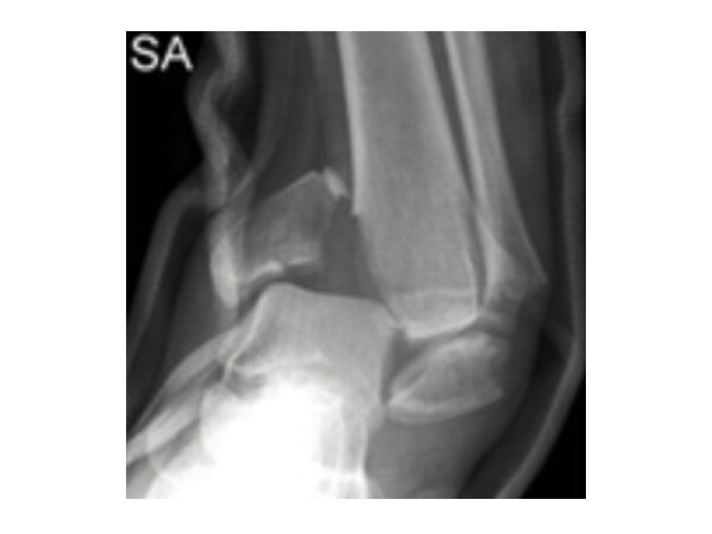
Q1-Q33 questions — tap to reveal all answerslist
- Describe the X-ray findings and injury pattern.
- What other views and relevant negatives do you need?
- What are the definitive management aims?
Q1.Describe the X-ray findings and injury pattern.
- AP view of ankle with fractured tibia and fibula
- Lauge-Hansen supination-adduction type injury
- Vertical fracture of medial malleolus + transverse infrasyndesmosis fibular fracture
Q2.What other views and relevant negatives do you need?
- Ask for lateral X-ray and X-ray one joint above
- Relevant negatives: no talus fracture
- No os calcis fracture
- No MT fracture
Q3.What are the definitive management aims?
- Anatomical reduction and stable fixation for primary healing
- Restore joint congruency of the tibial plafond
- Bone graft for defects
▸ Slide 350 · Maisonneuve --> prox fibula, compartmentTrauma · 2 questions expand
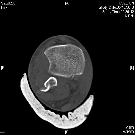
Q1-Q22 questions — tap to reveal all answerslist
- What is a Maisonneuve fracture and what must you check?
- What is a Bosworth fracture and how is it managed?
Q1.What is a Maisonneuve fracture and what must you check?
- Proximal fibula fracture
- Check the proximal fibula and compartments
Q2.What is a Bosworth fracture and how is it managed?
- Fibula trapped behind the incisura fibularis (SER pattern)
- Requires urgent ORIF
▸ Slide 351 · Aviator AstragalusTrauma · 14 questions expand

Q1-Q1414 questions — tap to reveal all answerslist
- Describe the X-ray findings in this talar neck fracture.
- What is the mechanism of a talar neck fracture?
- Describe the Hawkins classification and AVN risk.
- How is a talar neck fracture reduced acutely?
- How is the Canale view performed?
- What is the definitive management of a talar neck fracture?
- What are the reduction and fixation details of ORIF for talar neck fracture?
- What is the post-operative rehabilitation and what are the risk factors for AVN?
- What is Hawkins sign and its significance?
- What is the blood supply of the talus and the complications of talar neck fractures?
- What causes varus malunion after talar neck fracture?
- How is AVN of the talus managed?
- Describe the anatomy of the talus.
- How is a medial malleolar osteotomy performed?
Q1.Describe the X-ray findings in this talar neck fracture.
- Hawkin 2 fracture of talar neck with subtalar joint dislocation; tibiotalar and TNJ congruent
- No soft tissue gas (open fracture in 50% of type 3 and 4)
- Need other X-rays - ipsilateral foot fracture in 90%
Q2.What is the mechanism of a talar neck fracture?
- Dorsiflexion with axial load
- Impingement of the talar neck by the anterior lip of the tibia
Q3.Describe the Hawkins classification and AVN risk.
- 1: no dislocation, minimally displaced (0-13%)
- 2: subtalar joint dislocation (50%)
- 3: subtalar joint + ankle dislocation (90%)
- 4: subtalar + TN + ankle joint dislocation (100%)
Q4.How is a talar neck fracture reduced acutely?
- ATLS; assess skin condition, compartment syndrome and NV injury
- Sedation; flexed knee relaxes gastroc, ankle PF relaxes TA; stabilise malleoli, apply longitudinal traction +/- varus/valgus correction
- No multiple attempts; failed CR in 25% -> OR require MM osteotomy to preserve deltoid artery
- Analgesia and backslab after CR; Canale view and CT for fracture personality/medial comminution
Q5.How is the Canale view performed?
- Ankle in maximum plantarflexion
- Foot pronated 15 degrees
- XR beam 75deg from horizontal
Q6.What is the definitive management of a talar neck fracture?
- Aim: anatomical reduction, rigid fixation for primary bone healing
- Non-op if CT-confirmed Hawkin I with no articular step off: short leg cast 8-12 weeks
- ORIF: usually AL approach first (beware superficial peroneal nerve, full-thickness flaps, EDB retracted)
- Fix lateral first as foundation or cortical key for reduction, then medial; medial buttress plate to prevent varus
- Posterior: percutaneous headless cannulated screw posterior to anterior
Q7.What are the reduction and fixation details of ORIF for talar neck fracture?
- Reduction with forceps, direct manipulation, K wire as joystick +/- joint distractor; prelim K wire fixation assessed on Canale view
- Fix with lag screw/positional screw
- Medial approach: medial malleolus to base of 1st MT, plane between TA and TP, protect saphenous nerve and vein; +/- medial malleolar osteotomy + buttress plate/positional screw
- Posterior: percutaneous cannulated headless titanium screw (countersink), posterior to anterior, perpendicular to fracture line, maximal bone purchase (P to A mechanically more stable, does not disrupt TNJ)
Q8.What is the post-operative rehabilitation and what are the risk factors for AVN?
- NWB walking for 10-12 weeks
- AVN risk factors: Hawkins classification and timing of reduction
Q9.What is Hawkins sign and its significance?
- Subchondral radiolucency at the talar dome at 6-8 weeks on serial AP XR
- Reactive hyperaemia causing bone resorption - likely no AVN (but not r/o)
- Sclerosis indicates loss of blood supply
- Sensitivity 100%, specificity 58%; MRI if in doubt
Q10.What is the blood supply of the talus and the complications of talar neck fractures?
- Posterior tibial artery via artery of tarsal canal (inferior body) and deltoid branch (superior body)
- Perforating peroneal artery via artery of sinus tarsi (lateral); anterior tibial artery via dorsalis pedis (head/neck)
- Talus prone to AVN: 80% covered by articular surface, no muscular attachment, tenuous retrograde supply
- AVN overall 30%, subtalar arthritis 50%, ankle OA 33%
Q11.What causes varus malunion after talar neck fracture?
- Medial comminution -> varus malunion
- Decreased subtalar eversion and weight bearing on the lateral foot
Q12.How is AVN of the talus managed?
- Differentiate the cause of pain (talus vs ankle joint)
- NWB until creeping substitution
- Usually symptoms are not bad
Q13.Describe the anatomy of the talus.
- Head, neck and body
- Superior = trochlear surface (articulates tibia and fibula); inferior lateral process articulates with the posterior facet of the calcaneum
- Posterior process: medial and lateral tubercles separated by a groove for FHL; PTFL attaches to the lateral tubercle (Shepherd fracture); medial tubercle fracture = Cedell fracture
- Anterior surface articulates with navicular and sustentaculum tali
Q14.How is a medial malleolar osteotomy performed?
- AM approach; open capsule to visualise the apex between the medial malleolus and tibial plafond
- Predrill screw tract and pretap
- Osteotomy at 90 degrees to the screw tract (45 degrees from tibial plafond); finish cut with osteotome
▸ Slide 352Trauma · 2 questions expand
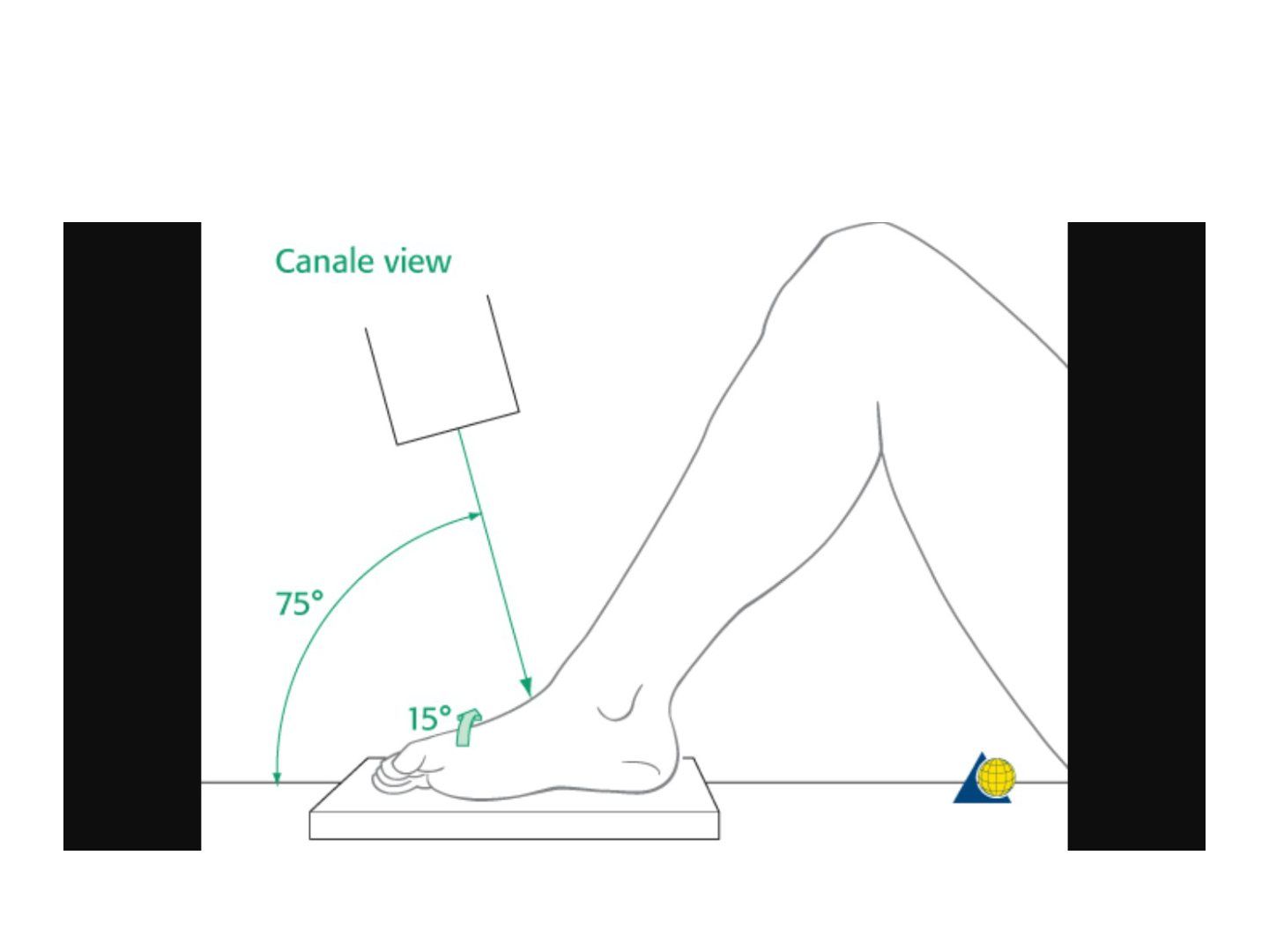
Q1-Q22 questions — tap to reveal all answerslist
- What topic does this slide cover?
- Summarise the key learning points from this slide.
Q1.What topic does this slide cover?
- Not covered in the speaker notes
Q2.Summarise the key learning points from this slide.
- Not covered in the speaker notes
▸ Slide 353 · Lateral and oblique Xray of patients left ankle and foot showing fracture over Trauma · 22 questions expand
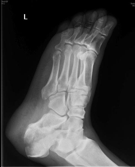
Q1-Q2222 questions — tap to reveal all answerslist
- Describe the X-ray findings and fracture type.
- What imaging views and classification are used?
- What is the initial assessment and management of a calcaneal fracture?
- What associated injuries must be ruled out?
- Describe the Sanders classification.
- Which patient and fracture factors affect definitive management?
- What is the timing of calcaneal surgery?
- What are the aims of operative treatment?
- Describe the sinus tarsi approach and reduction sequence.
- How are the Broden and Harris views taken?
- What is the rehabilitation after calcaneal fracture?
- What causes long-term chronic pain after calcaneal fracture?
- What are the epidemiological associations of calcaneal fractures?
- Describe the relevant calcaneal anatomy.
- How are Bohler and Gissane angles measured and what are the normal values?
- What are the emergency presentations of a calcaneal fracture?
- Describe the Essex-Lopresti classification.
- Describe the primary fracture line and the constant fragment.
- Describe the extensile L-shaped approach.
- What are the complications and their management?
- What is the evidence and prognosis?
- What are the details and criticisms of the key trials, and what predicts a favourable outcome?
Q1.Describe the X-ray findings and fracture type.
- Intraarticular calcaneal fracture extending into the posterior facet
- Reduced Bohler and increased Gissane angle; no obvious fracture to the CC joint
- Essex-Lopresti tongue type (extends to posterior tuberosity); otherwise joint depression type
Q2.What imaging views and classification are used?
- Lateral: Bohler, Gissane, calcaneal shortening
- Harris (axial): widening, varus
- Broden: posterior facet
- CT to plan; Sanders classification on Widest 30 deg semicoronal cut (1-4)
Q3.What is the initial assessment and management of a calcaneal fracture?
- High energy trauma: ATLS, AMPLE, primary survey
- Slab for rest + elevate and ice
- Frequent monitoring for compartment syndrome; unconscious patient needs continuous slit catheter monitoring
- CT to plan management
Q4.What associated injuries must be ruled out?
- Local: wound, skin impingement, compartment syndrome, NV status
- Systemic signpost injuries: TL/L spine compression fracture 10%, contralateral os calcis fracture 10%
Q5.Describe the Sanders classification.
- Based on the widest 30-degree semicoronal CT cut
- Grades 1-4 (1: no stepping; 2-4: 2-4 parts)
Q6.Which patient and fracture factors affect definitive management?
- Patient: DM, PVD, smoking - >90% wound complication rate and poor outcome
- Fracture: articular step >2mm, calcaneal height, width, valgus/varus alignment
- Soft tissue factors such as skin impingement
Q7.What is the timing of calcaneal surgery?
- Wait until the wrinkle sign is positive and blisters are epithelialized
Q8.What are the aims of operative treatment?
- OT aim: restore articular surface of posterior facet
- Restore calcaneal height and length (Bohler and Gissane angles) to restore triceps surae lever arm
- Correct varus to prevent late instability (hindfoot in varus locks up CC and TN joints, more lateral foot weight bear, more metatarsalgia)
- Restore width to prevent peroneal tendon impingement
Q9.Describe the sinus tarsi approach and reduction sequence.
- Incision 2cm distal to fibular tip to base of 4th MT; beware sural nerve, IDCN and peroneal tendons
- Use sustenaculum tali as the keystone for reduction (constant fragment held by surrounding TC ligament); reduce anterior process first, then jig up posterior facet with osteotome or lamina spreader, K wires to sustentaculum tali
- Steinman pin restores length and varus/valgus; Xray to assess reduction for posterior facet by broden's view, calcaneal alignment by Harris view
- Bone graft, subchondral raft screws with 3.5mm cortical screw, lateral wall, low-profile locking plate, Allgower technique closure
Q10.How are the Broden and Harris views taken?
- Broden: ankle neutral dorsiflexion, 45 IR (45 degrees internal rotation), XR with 40, 30, 20, 10 degrees cephalic tilt
- Harris: ankle maximum dorsiflexion, beam 45 degrees from horizontal caudal tilt
Q11.What is the rehabilitation after calcaneal fracture?
- NWB for 6-8 weeks
Q12.What causes long-term chronic pain after calcaneal fracture?
- 40% will have long term chronic pain
- Causes: subtalar incongruity, penetration of screws, lateral impingement, peroneal tendinitis, talar neck impingement
Q13.What are the epidemiological associations of calcaneal fractures?
- 17% of fractures are open
- 10% bilateral, 10% involve spine fracture
- 63% involve the CCJ
Q14.Describe the relevant calcaneal anatomy.
- Posterior, middle and anterior facets for talus; anterior facet for cuboid
- Sinus tarsi: groove dividing anterior and posterior subtalar joint
- Sustenaculum tali: medial projection supporting the talar neck, FHL runs below; deltoid and talocalcaneal ligament attachments -> constant AM fragment
- Bifurcate ligament: anterior process to CC and CN, Y shaped
Q15.How are Bohler and Gissane angles measured and what are the normal values?
- Bohler: angle from highest point of anterior process to highest point of posterior facet and highest point of superior tuberosity; normal 20-40 degrees; reduced = collapse of posterior facet
- Gissane: angle between posterior facet and line joining lowest point of posterior facet to highest point of anterior process; normal 130-145 degrees; increased = collapse
Q16.What are the emergency presentations of a calcaneal fracture?
- Skin impingement in tongue type/avulsion fracture
- Compartment syndrome (10%) and NV injury
- Open fracture (10%)
Q17.Describe the Essex-Lopresti classification.
- Tongue type: secondary fracture line exits at the posterior calcaneal surface
- Joint depression type: secondary fracture line exits at the superior calcaneal surface
Q18.Describe the primary fracture line and the constant fragment.
- Oblique fracture line from anterolateral to posteromedial
- Two fragments: AM & PL
- AM contains the sustentaculum tali, attached to the proximal fragment by strong ligaments (deltoid, medial talocalcaneal, spring) = constant fragment
Q19.Describe the extensile L-shaped approach.
- Vertical limb between lateral malleolus and Achilles; >100 degree turn; horizontal limb between glabrous and non-glabrous skin
- Protect sural nerve and lateral calcaneal branch of the peroneal artery
- Subperiosteal dissection, elevate full thickness periosteocutaneous flap; extend proximally to body of talus; excise CF ligament; bent K wires as retractors
Q20.What are the complications and their management?
- Wound complications 25%; FHL irritation from a long screw at the sustentaculum tali; compartment syndrome
- Malunion (Stephen and Sanders classification: talar declination angle <20, talocalcaneal angle)
- Lateral exostosis - resection; subtalar OA - fusion (distraction bone block); varus - valgus osteotomy
Q21.What is the evidence and prognosis?
- UK heel trial BMJ 2014 and extended Heft trial 2021 BJJ do not support surgery (methodological flaws)
- Buckley JBJS 2002: outcomes similar; surgery may be better when IOD group is excluded
- Overall poor prognosis: >40% long-term chronic pain
- Complications: wound complications 25%, FHL irritation, compartment syndrome, malunion
Q22.What are the details and criticisms of the key trials, and what predicts a favourable outcome?
- UK heel trial BMJ 2014: wound complication 19%, many excluded as 'obviously needed surgery', postop CT only looked at subtalar stepoff
- Buckley JBJS Am 2002: outcomes similar; when IOD excluded, surgery better
- Favourable prognosis: female, non-IOD, sedentary work, anatomical reduction, less initial disruption of Gissane/Bohler
- Later study: overall cost less with surgical management due to fusion surgery and disability costs in non-op patients
▸ Slide 354 · Lateral xray of the left ankle and foot showing fracture calcaneumTrauma · 2 questions expand

Q1-Q22 questions — tap to reveal all answerslist
- Describe the fracture seen on this lateral X-ray.
- What is the significance of the changed Bohler and Gissane angles?
Q1.Describe the fracture seen on this lateral X-ray.
- Intraarticular calcaneal fracture - joint depression / tongue / both JD&T type
- Fracture extends from the posterior facet and exits the inferior/posterior calcaneal surface
- Posterior facet collapsed: decreased Bohler angle, increased angle of Gissane
Q2.What is the significance of the changed Bohler and Gissane angles?
- Indicates collapse of the posterior facet
- Decreased Bohler angle
- Increased angle of Gissane
▸ Slide 355 · Broden viewTrauma · 2 questions expand

Q1-Q22 questions — tap to reveal all answerslist
- What is the Broden view and when is it used?
- What is the Harris view and what does it show?
Q1.What is the Broden view and when is it used?
- Used intraoperatively for facet joint reduction
- Ankle in neutral dorsiflexion
- X-ray taken at 45 degrees internal rotation
Q2.What is the Harris view and what does it show?
- Shows widening of the calcaneum and angulation of the tuberosity fragment
- Ankle in dorsiflexion
- X-ray beam at 45 degrees caudal tilt
▸ Slide 356 · Lateral XR of left foot and ankle region showing fracture calcaneal tuberosity wTrauma · 4 questions expand
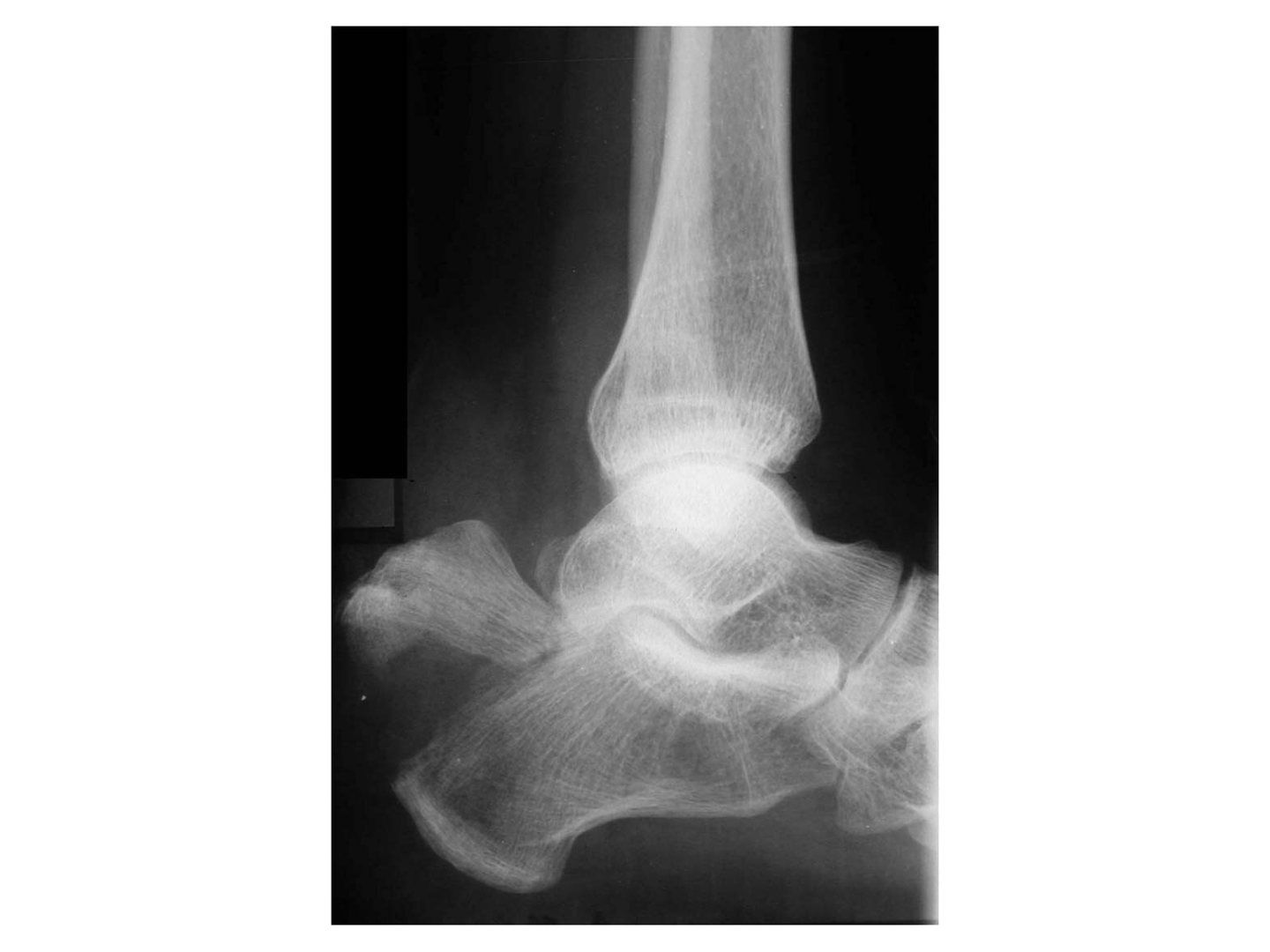
Q1-Q44 questions — tap to reveal all answerslist
- What is the mechanism and clinical concern in this fracture?
- What is examined in a calcaneal tuberosity fracture?
- What is the Beavis classification of calcaneal tuberosity fractures?
- What is the management and what are the complications?
Q1.What is the mechanism and clinical concern in this fracture?
- Avulsion fracture from eccentric contraction of gastrocnemius
- Displaced fracture -> skin tenting, skin necrosis
Q2.What is examined in a calcaneal tuberosity fracture?
- Soft tissue: skin impingement
- Distal NV status
Q3.What is the Beavis classification of calcaneal tuberosity fractures?
- Type 1: avulsion (usually osteoporotic insufficiency fracture)
- Type 2: beak (direct blow, EOT)
- Type 3: infrabursal avulsion (superficial fibres involved; suture anchor)
- Type 4 (modified by Lee): only deep fibres involved (conservative treatment)
Q4.What is the management and what are the complications?
- CR + percutaneous screw fixation with washers
- OR + tension band wire fixation
- Undisplaced: conservative - below knee cast with ankle in plantarflexion
- Complications: skin necrosis, fracture displacement, weak push-off strength
▸ Slide 357 · Xray left foot of DP viewTrauma · 2 questions expand
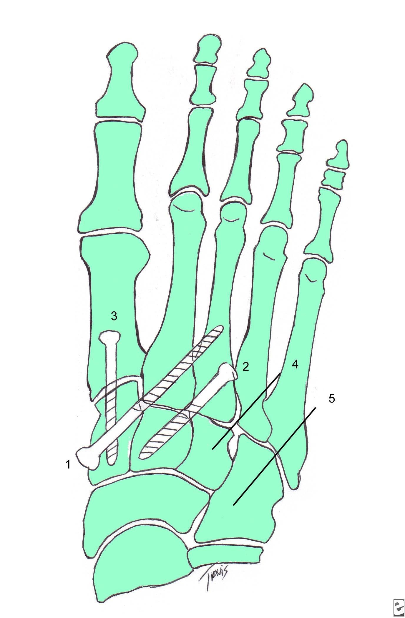
Q1-Q22 questions — tap to reveal all answerslist
- What injury is shown and which radiographic signs confirm it?
- What additional views are needed and what do they show?
Q1.What injury is shown and which radiographic signs confirm it?
- This is a bony Lisfranc injury
- DP view: widening of the space between the 1st and 2nd MT with a fleck sign
- The line along the medial border of the middle cuneiform and 2nd MT is disrupted
- No other cuboid or navicular fractures seen
Q2.What additional views are needed and what do they show?
- Lateral: dorsal subluxation of the 1st or 2nd MT base
- Oblique: 4th MT medial line with medial cuboid; 3rd MT lateral line with lateral border of lateral cuneiform
▸ Slide 358 · XrayTrauma · 11 questions expand
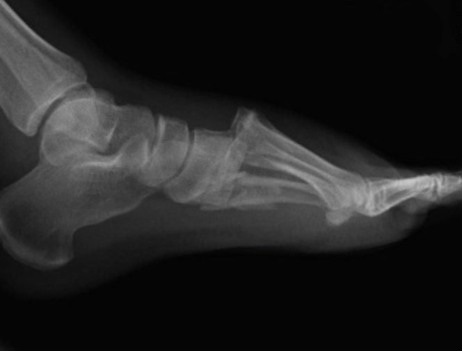
Q1-Q1111 questions — tap to reveal all answerslist
- What X-ray views are required in suspected Lisfranc injury?
- What is the spectrum of Lisfranc injury and the Nunley classification?
- What structures provide Lisfranc stability?
- What is the Lisfranc ligament?
- What is the Myerson classification and what initial management is required?
- What are the aims and timing of definitive management?
- What are the indications for primary fusion?
- How would you perform an EUA?
- Describe the operative details of ORIF.
- What is the evidence for ORIF versus primary arthrodesis?
- What is the prognosis of a Lisfranc injury?
Q1.What X-ray views are required in suspected Lisfranc injury?
- DP: line along medial 2nd MT and middle cuneiform; 1st-2nd MT widening >3mm; fleck sign
- Oblique: TMTJ overlap; medial 4th MT and cuboid
- Lateral: dorsal subluxation of 1st/2nd MT base
- Relevant negatives: cuboid and dorsal navicular avulsion fracture; abduction stress views if delayed presentation
Q2.What is the spectrum of Lisfranc injury and the Nunley classification?
- Spectrum from sprain to dislocation of the TMT joint complex
- High energy/athletic: axial load through hyperplantarflexed forefoot, associated MT/T fractures
- Nunley (low energy): Stage 1 = undisplaced; Stage 2 = widening >2mm on AP with no dorsal subluxation; Stage 3 = dorsal subluxation on lateral
Q3.What structures provide Lisfranc stability?
- Lisfranc joint complex = MT, intermetatarsal and intertarsal articulations
- Static bone: roman arch, keystone (mortise created by medial cuneiform and recessed middle cuneiform)
- Static ligaments: Lisfranc ligament, interMT, plantar (C1M2M3) and dorsal TMT ligaments
- Dynamic: plantar aponeurosis, intrinsics, PTT and ATT, peroneus longus
Q4.What is the Lisfranc ligament?
- Interosseous ligament from the medial cuneiform to the base of the 2nd MT on the plantar surface
- Tightens with pronation and abduction of the forefoot
Q5.What is the Myerson classification and what initial management is required?
- Myerson: total, partial incongruent, divergent
- Rule out compartment syndrome; if grossly displaced try CR then temporary immobilization + elevate and ice
- Assess soft tissue: plantar ecchymosis is the hallmark feature
- +/- CT (mainly preop planning) / MRI (pure ligamentous injury)
Q6.What are the aims and timing of definitive management?
- Anatomical reduction of the Lisfranc joint, prevent instability and early OA
- If missed -> rocker bottom deformity
- Emergent if open/compartment/irreducible; else wait 2-3/52
- Options: ORIF for fracture dislocation (medial screw, lateral K wire); primary medial column arthrodesis for purely ligamentous arch injuries
Q7.What are the indications for primary fusion?
- Ligamentous injury + multiplanar instability
- Delayed presentation
- Mid to advanced age
- Intraarticular comminution
Q8.How would you perform an EUA?
- Stabilise the midfoot then apply varus/valgus stress, supination/pronation, dorsoplantar force to the forefoot
- Widening with pronation and abduction of the forefoot
- Assess TMTJ dorsal subluxation
Q9.Describe the operative details of ORIF.
- GA, tourniquet; mark DP before tourniquet
- Dorsal incisions between 1st-2nd MT and over 4th MT (at risk: dorsalis pedis, superficial and deep peroneal nerves)
- Dorsomedial approach: plane between TA and EHL
- Reduce 2nd MT into the keystone, then reduce and fix 1st TMTJ; assess reduction on XR
- Medial column rigid fixation: retrograde 3.5mm cortical positional screw (pocket hole) or 2.4/2.7 VALCP in bridging mode; 2.0 K wire lateral column (4th MT to cuboid)
- Assess the foot for intercunieform instability and fix if needed; short leg cast/splint
- NWB 6 weeks; K wires out at 6 weeks then gradual weight bearing; screws out at 6 months; arch support 6 months
Q10.What is the evidence for ORIF versus primary arthrodesis?
- JBJS 2006 Ly: primary arthrodesis of 2-3 medial rays - better pain control and function, less hardware removal in pure ligamentous injury
- Henning 2009 / CORR 2016 Smith: ORIF more hardware removal, functional outcome no difference
- ORIF shifting towards bridging plates rather than screw fixation
Q11.What is the prognosis of a Lisfranc injury?
- 50% posttraumatic OA
- Poor prognosis: IOD, female and young, smoker
▸ Slide 359 · Cuboid fractureTrauma · 4 questions expand
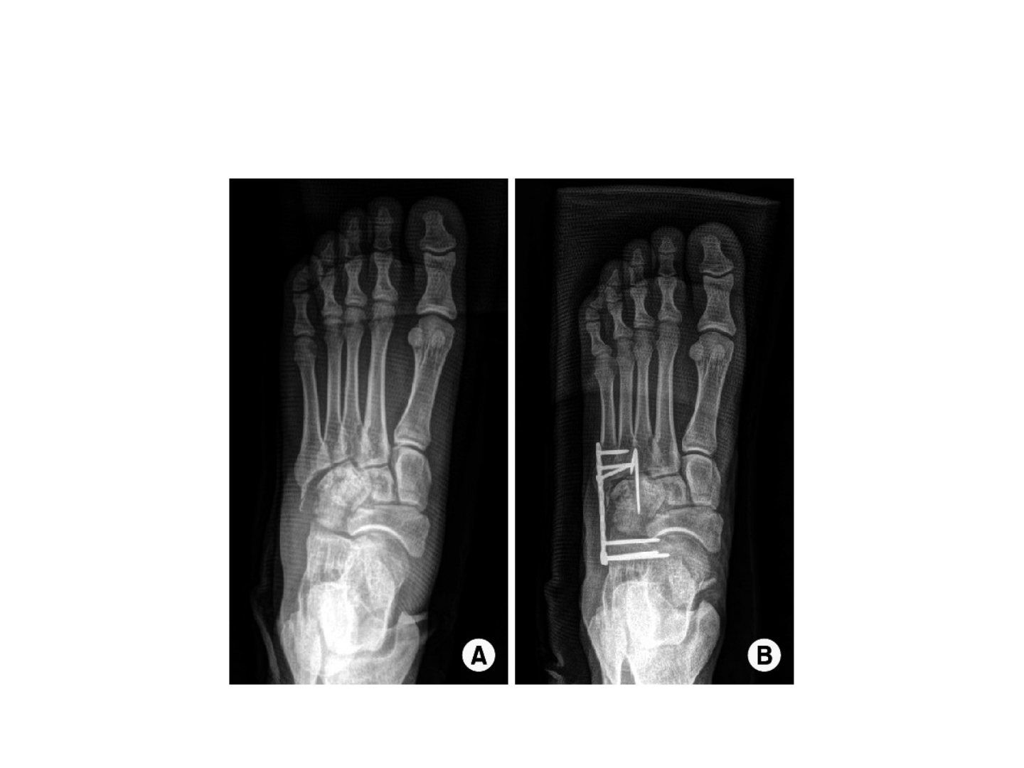
Q1-Q44 questions — tap to reveal all answerslist
- What force causes a cuboid fracture and what are the surgical indications?
- How is a cuboid fracture treated operatively?
- What are the poor prognostic factors for a cuboid fracture?
- What is the management of a failed ORIF of a cuboid fracture?
Q1.What force causes a cuboid fracture and what are the surgical indications?
- Abduction force
- Surgery if articular step >2mm or lateral column shortening
Q2.How is a cuboid fracture treated operatively?
- Simple - plating/screw
- Comminuted/shortening/severe/bone loss - bridge plate or ex fix
Q3.What are the poor prognostic factors for a cuboid fracture?
- Patient: young age, IOD, high energy/direct trauma
- Fracture: non-anatomical reduction
Q4.What is the management of a failed ORIF of a cuboid fracture?
- Salvage fusion
▸ Slide 360 · DP XR of left foot showing medial subtalar dislocationTrauma · 6 questions expand
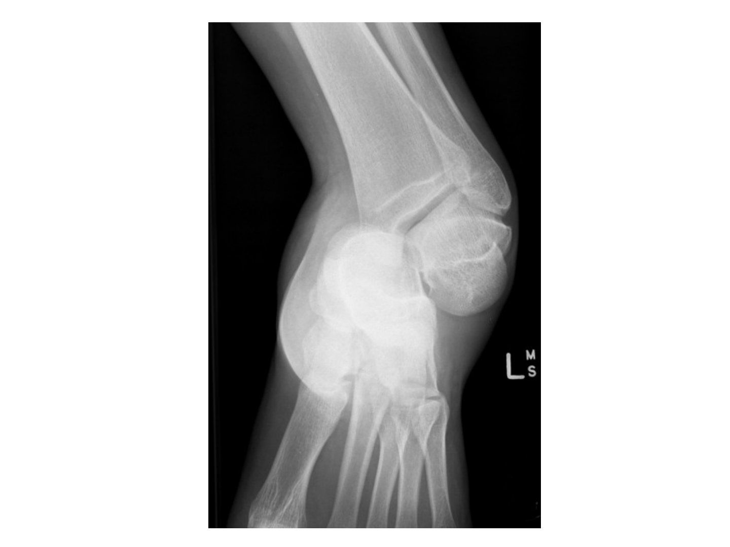
Q1-Q66 questions — tap to reveal all answerslist
- Which subtalar dislocation is more common and how does it lock?
- What structures can block reduction in lateral versus medial subtalar dislocation?
- How is closed reduction performed for a subtalar dislocation?
- What imaging is required in a subtalar dislocation?
- What is the definitive management of a subtalar dislocation?
- Which side is associated with a fracture in a subtalar dislocation?
Q1.Which subtalar dislocation is more common and how does it lock?
- Medial dislocation is more common - 70%
- Medial locks in supination; lateral locks in pronation
- Lateral - more high energy, often open
Q2.What structures can block reduction in lateral versus medial subtalar dislocation?
- Lateral dislocation - medial structures: PT, FDL, FHL
- Medial dislocation - lateral structures: EDB, peroneal tendon, TN dorsal capsule
Q3.How is closed reduction performed for a subtalar dislocation?
- Knee flexed
- Inversion for lateral dislocation
- Eversion for medial dislocation
Q4.What imaging is required in a subtalar dislocation?
- CT to rule out fracture
- 40% have an associated fracture
Q5.What is the definitive management of a subtalar dislocation?
- Most are stable after reduction: NWB with below knee cast
- Unstable with no fracture: ensure no soft tissue interposition, then transfixing K wire
- Failed CR - OR (medial dislocation via AL/sinus tarsi approach and vice versa)
Q6.Which side is associated with a fracture in a subtalar dislocation?
- Fracture is associated on the displaced side
▸ Slide 361 · Navicular fractureTrauma · 6 questions expand
Q1-Q66 questions — tap to reveal all answerslist
- Which classification is used for navicular fractures?
- Describe a Sangeorzan type I navicular fracture.
- Describe a Sangeorzan type II navicular fracture.
- Describe a Sangeorzan type III navicular fracture.
- What are the indications for surgery in a navicular fracture?
- What are the complications of a navicular fracture?
Q1.Which classification is used for navicular fractures?
- Sangeorzan classification
- Based on fracture plane, comminution and direction of forefoot displacement
Q2.Describe a Sangeorzan type I navicular fracture.
- Fracture in the coronal plane
- No angulation of the forefoot
Q3.Describe a Sangeorzan type II navicular fracture.
- Fracture line from dorsal-lateral to plantar-medial
- Forefoot displaced medially
Q4.Describe a Sangeorzan type III navicular fracture.
- Comminuted fracture in the sagittal plane
- Forefoot displaced laterally
Q5.What are the indications for surgery in a navicular fracture?
- >2 mm displacement
Q6.What are the complications of a navicular fracture?
- Nonunion
- Loss of medial longitudinal arch support
- Painful TNJ (talonavicular joint)
- Post-traumatic OA
- AVN and collapse
▸ Slide 362 · Foot CompartmentsTrauma · 7 questions expand

Q1-Q77 questions — tap to reveal all answerslist
- Why do you release the foot compartments?
- How many compartments does the foot have and what are they?
- What does the medial release of the foot decompress?
- What does the DL release decompress?
- What does the DM release decompress?
- What is the evidence for management of foot compartment syndrome?
- What surgical wounds are used to release the foot compartments?
Q1.Why do you release the foot compartments?
- Ischaemia of the intrinsic muscles leads to clawing
- Compartment release is performed to prevent this
Q2.How many compartments does the foot have and what are they?
- 9 compartments
- Medial and lateral compartments
- 3 central compartments - superficial, mid and deep
- 4 interosseous compartments
Q3.What does the medial release of the foot decompress?
- Medial foot border
- Central superficial and middle compartments
- Performed through a medial wound
Q4.What does the DL release decompress?
- Lateral 2 interossei
- Lateral compartment
- Performed through a dorsal wound over the 2nd and 4th MT
Q5.What does the DM release decompress?
- Medial 2 interossei
- Central deep compartment
Q6.What is the evidence for management of foot compartment syndrome?
- AAOS 2013 review article: no strong evidence exists to guide management
- Emergent fasciotomy is commonly recommended to prevent pain and deformity
Q7.What surgical wounds are used to release the foot compartments?
- Dorsal wound over the 2nd and 4th MT
- Medial wound
▸ Slide 363 · ULTrauma · 4 questions expand
Q1-Q44 questions — tap to reveal all answerslist
- Which classifications are used for ACJ and clavicle injuries?
- Which classifications are used for the proximal humerus?
- Which classifications are used for humeral shaft and distal humerus fractures?
- Which classifications are used around the elbow?
Q1.Which classifications are used for ACJ and clavicle injuries?
- ACJ: Rockwood
- Clavicle: Allman & Neer
Q2.Which classifications are used for the proximal humerus?
- Neer
- Hertel
Q3.Which classifications are used for humeral shaft and distal humerus fractures?
- Humeral shaft: by morphology
- Distal humerus: AO, Holstein Lewis
Q4.Which classifications are used around the elbow?
- Elbow dislocation: terrible triad, Cori cycle
- Olecranon: Mayo
- Coronoid process: Regand & Morray
- Radial head & neck: Mason
- Capitellum: Regand & Morray
▸ Slide 364 · AP radiograph of the left shoulder showing posterior dislocation of right shouldTrauma · 9 questions expand

Q1-Q99 questions — tap to reveal all answerslist
- Describe the radiographic signs of a posterior shoulder dislocation.
- What history and examination findings suggest a posterior shoulder dislocation?
- How do you confirm the diagnosis of posterior shoulder dislocation?
- How is an acute traumatic posterior shoulder dislocation reduced?
- What are the primary stabilisers against posterior shoulder dislocation?
- How is a chronic missed posterior dislocation (>6 weeks) managed?
- What history points are important in a suspected posterior shoulder dislocation?
- How are the axillary and Velpeau views taken?
- What is the difference between the classic and modified McLaughlin procedure?
Q1.Describe the radiographic signs of a posterior shoulder dislocation.
- Light bulb sign
- Trough line
- Positive rim sign (widening of GHJ)
- Absence of half-moon overlap
- Proximal humerus internally rotated and adducted
- No associated fracture in LT or surgical neck
Q2.What history and examination findings suggest a posterior shoulder dislocation?
- MOI: direct - axial load on arm in flexion, adduction, IR; indirect - electric shock/convulsion
- Loss of shoulder contour, asymmetrical axillary fold, prominent coracoid (not obvious)
- Arm held in IR and adduction; painful reduced ROM particularly ER and FF
- Check NV status esp axillary nerve, brachial plexus, axillary artery; associated trauma (high energy), SSSC
Q3.How do you confirm the diagnosis of posterior shoulder dislocation?
- Clinical examination
- Axillary view - most useful to assess direction of injury
- Velpeau view - most useful if patient is in pain
- CT - confirm direction of injury and associated fractures
Q4.How is an acute traumatic posterior shoulder dislocation reduced?
- Closed reduction under GA with an assistant
- Delpalma method: caudal traction in line of the adducted IR deformity (elbow flexed) with lateral lifting
- Recheck NV status post CR; immobilise in ER for 4-6 weeks
- Arrange CT to assess associated occult fracture/bone defect
Q5.What are the primary stabilisers against posterior shoulder dislocation?
- Posterior band of IGHL - static restraint in IR
- Subscapularis - dynamic restraint in ER
- CH ligament
Q6.How is a chronic missed posterior dislocation (>6 weeks) managed?
- Reverse HS lesion = loss of normal convexity in the anteromedial part, depression/defect of humeral head
- CR unlikely successful; complete workup with XR/CT to assess degree of reverse HS defect
- Open reduction with subscapularis transfer (McLaughlin) or LT transfer (modified McLaughlin)
- Castagna: modified McLaughlin - significant functional improvement, no intra/postop complications at mean FU 62.8 months
Q7.What history points are important in a suspected posterior shoulder dislocation?
- Mechanism of injury
- Patient's age and hand dominance
Q8.How are the axillary and Velpeau views taken?
- Axillary: arm in 90 degrees abduction, plate above the shoulder, beam into the axilla
- Velpeau: patient leans 45deg backward, plate on the floor, beam directed caudally
Q9.What is the difference between the classic and modified McLaughlin procedure?
- Classic: subscapularis transfer
- Modified: adds transfer of the lesser tuberosity for better bone filling of the defect and more secure subscapularis insertion
▸ Slide 365 · Anterior fracture dislocation of shoulderTrauma · 3 questions expand
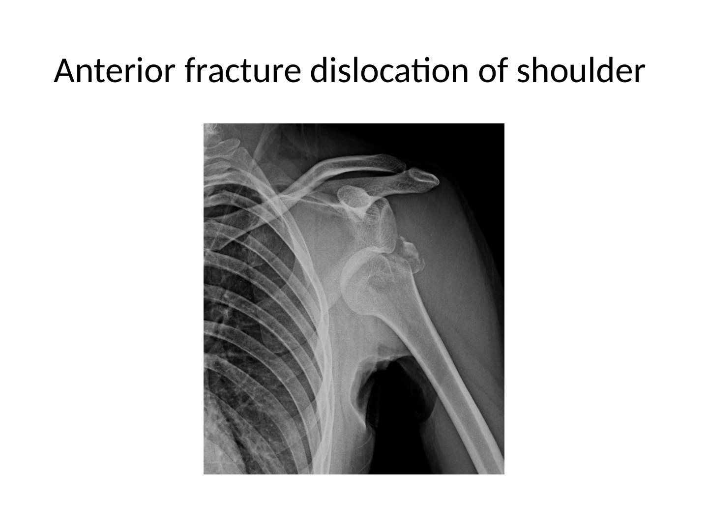
Q1-Q33 questions — tap to reveal all answerslist
- What is the terrible triad of shoulder dislocation?
- When should a greater tuberosity fracture be fixed after reduction?
- What is the evidence for arthroscopic fixation of the greater tuberosity?
Q1.What is the terrible triad of shoulder dislocation?
- Dislocation
- Cuff injury
- Brachial plexus injury
Q2.When should a greater tuberosity fracture be fixed after reduction?
- If GT displaced >5mm post reduction
- May be fixed open or arthroscopically
Q3.What is the evidence for arthroscopic fixation of the greater tuberosity?
- Fixation with a double row of anchors shown to have superior postoperative ROM
- CORR 2016
▸ Slide 366 · AP Xray showing ACJ dislocation with marked superior displacement of the distal Trauma · 11 questions expand
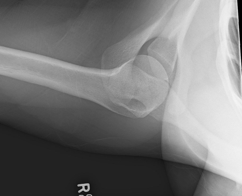
Q1-Q1111 questions — tap to reveal all answerslist
- How does the Rockwood classification guide management of ACJ dislocation?
- What imaging views are used for ACJ dislocation?
- What is the Cochrane 2019 evidence for ACJ dislocation?
- When is distal clavicle resection indicated in ACJ disease and how much bone is removed?
- What is the role of tendon reconstruction in chronic ACJ dislocation?
- Describe the X-ray findings in this ACJ dislocation.
- What history and examination points are important in ACJ dislocation?
- What are the options for CC fixation in acute ACJ dislocation?
- What is the direct ACJ fixation option and its drawback?
- How is chronic ACJ dislocation managed?
- What are the ACJ stabilisers?
Q1.How does the Rockwood classification guide management of ACJ dislocation?
- 1-3: static stabilisers disrupted (AC sprain, AC torn, CC torn) - conservative
- 4-6: dynamic (posterior through trapezius, D+T torn, inferior below coracoid) - surgical
- Type 3: +/- operation
- If untreated the shoulder becomes protracted
Q2.What imaging views are used for ACJ dislocation?
- Contralateral side for comparison
- Axillary view - AP translation
- Zanca view - 10 degree cephalic tilt, vertical translation
- +/- stress view
Q3.What is the Cochrane 2019 evidence for ACJ dislocation?
- Low evidence for surgery
- At 10 yr FU, minimal degenerative changes
- Conservative actually has better functional outcomes at 6 weeks
Q4.When is distal clavicle resection indicated in ACJ disease and how much bone is removed?
- Indicated for ACJ arthritis without instability
- Resect 7-8mm, <1.5cm of distal clavicle - too much jeopardises the ACJ capsule
- Can be performed arthroscopically
Q5.What is the role of tendon reconstruction in chronic ACJ dislocation?
- Tauber 2009 AJSM: semitendinosus tendon graft for coracoclavicular ligament reconstruction
- Gave superior clinical and radiologic outcomes compared with the modified Weaver-Dunn procedure
Q6.Describe the X-ray findings in this ACJ dislocation.
- AP: ACJ dislocation with marked superior displacement of the distal end of the clavicle
- No associated fractures; will need further Xray to classify
- Axillary view: posterior dislocation of the distal clavicle
- Summary: Rockwood 3 posterior dislocation of the ACJ
Q7.What history and examination points are important in ACJ dislocation?
- Mechanism of injury; acute vs chronic
- Associated injury around the shoulder girdle - SSSC, floating shoulder
- Neurovascular status
- Compare with the contralateral side
Q8.What are the options for CC fixation in acute ACJ dislocation?
- Bosworth screw, CC sling, fibre tape
- Tightrope loop suspensory fixation (A-assisted)
Q9.What is the direct ACJ fixation option and its drawback?
- Hook plate
- Provides anatomical reduction
- Needs a second operation for implant removal
Q10.How is chronic ACJ dislocation managed?
- Modified Weaver-Dunn procedure (classic without additional fixation)
- If just ACJ arthritis with no instability: resection of the distal clavicle
- Tendon reconstruction with semitendinosus graft (Tauber 2009 AJSM)
Q11.What are the ACJ stabilisers?
- Static: AC lig, CC lig, ACJ capsule
- Dynamic: Trapezius, deltoid muscle
▸ Slide 367 · Xray showing ACJ dislocation with marked superior displacement of the distal endTrauma · 9 questions expand
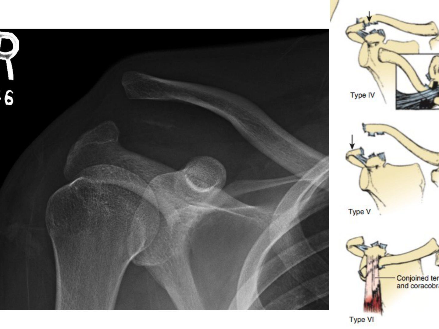
Q1-Q99 questions — tap to reveal all answerslist
- How do you assess a patient with a suspected ACJ dislocation?
- What is the evidence from the Cochrane 2019 review?
- What are the options for CC fixation in acute ACJ dislocation?
- What does the Arirachakaran 2016 meta-analysis show?
- Describe the modified Weaver-Dunn procedure.
- What are the static and dynamic stabilisers of the ACJ?
- Describe the X-ray findings in this ACJ dislocation.
- What is the direct fixation option for ACJ dislocation?
- When is resection of the distal clavicle performed and how much bone is removed?
Q1.How do you assess a patient with a suspected ACJ dislocation?
- Hx: mechanism of injury, acute vs chronic
- P/E: associated injury around the shoulder girdle - floating shoulder / SSSC
- Neurovascular status and contralateral side for comparison
Q2.What is the evidence from the Cochrane 2019 review?
- Low evidence for surgery; most studies used hook plate
- At 10 yr FU, minimal degenerative change; conservative better functional outcomes at 6 weeks
- Late reconstruction results comparable to early surgery; main problem is deformity
- One report quotes 15% reduced bench press strength
Q3.What are the options for CC fixation in acute ACJ dislocation?
- Loop suspensory fixation (tightrope, A-assisted)
- LARS ligament, Bosworth screw, CC sling, fibre tape
Q4.What does the Arirachakaran 2016 meta-analysis show?
- Suspensory device fixation gives higher shoulder function scores than metallic fixation (hook plate)
- Suspensory fixation also had lower postop pain
Q5.Describe the modified Weaver-Dunn procedure.
- Excision of distal clavicle
- Transfer acromial end of CA ligament to distal clavicle
- CC fixation
- Indicated for chronic ACJ dislocation with +ve arthritis
Q6.What are the static and dynamic stabilisers of the ACJ?
- Static: AC lig, CC lig, ACJ capsule
- Dynamic: trapezius, deltoid muscle
Q7.Describe the X-ray findings in this ACJ dislocation.
- ACJ dislocation with marked superior displacement of the distal end of the clavicle
- Look for associated fracture, especially floating shoulder or SSSC injury
- Further X-ray needed to classify
Q8.What is the direct fixation option for ACJ dislocation?
- Hook plate
- Provides anatomical reduction but needs a second operation for removal of the implant
Q9.When is resection of the distal clavicle performed and how much bone is removed?
- For chronic ACJ arthrosis without instability
- Remove 7-8mm, less than 1.5cm of distal clavicle
- Too much resection jeopardises the capsule
- Can be performed arthroscopically
▸ Slide 368 · Describe Xray:Trauma · 10 questions 1 check expand
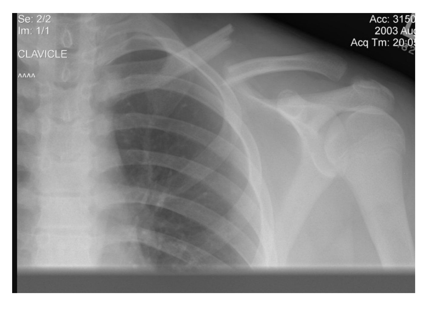
Q1-Q1010 questions — tap to reveal all answerslist
- Describe the X-ray features of this midshaft clavicle fracture.
- What are the deforming forces in a midshaft clavicle fracture?
- What patient and fracture factors favour surgery in a midshaft clavicle fracture?
- What are the absolute indications for surgery in a clavicle fracture?
- What is the evidence for and against surgery in midshaft clavicle fractures?
- Compare plate versus nail fixation for midshaft clavicle fracture.
- What is the initial assessment and classification of this clavicle fracture?
- Describe the surgical details of clavicle fixation.
- Compare superior versus anterior plating of the clavicle.
- What should a patient choosing nonoperative treatment be told?
Q1.Describe the X-ray features of this midshaft clavicle fracture.
- Fracture midshaft clavicle with superior displacement of the proximal fragment
- Shortening ~1cm
- No fracture of ribs/humerus
Q2.What are the deforming forces in a midshaft clavicle fracture?
- SCM pulls the proximal part superior
- Weight of the arm pulls the distal part inferior
- Pectoralis major adducts the arm causing shortening
Q3.What patient and fracture factors favour surgery in a midshaft clavicle fracture?
- >2cm shortening, complete displacement
- Smoking, comminution, old female
- Increased PROM at 6 weeks (Mckee JBJS 2012)
Q4.What are the absolute indications for surgery in a clavicle fracture?
- Open injury
- Vascular injury
- Scapulothoracic dissociation/floating shoulder
- Progressive neurological deficit
Q5.What is the evidence for and against surgery in midshaft clavicle fractures?
- For: 2012 McKee meta-analysis - earlier return of function; 2013 Robinson RCT - better functional score
- Against: 2020 CORR Bandari meta-analysis - more nonunion with conservative care but no functional difference
- Surgery increases likelihood of union (about 10 patients would need to undergo surgery to avoid one nonunion) but do not expect better function
- Weigh against complications and possible second procedure for hardware removal
Q6.Compare plate versus nail fixation for midshaft clavicle fracture.
- Plate: faster recovery, superior biomechanics esp comminution; NV injury, hardware prominence/irritation
- Nail: small wound, less soft tissue dissection, shorter op, less infection; rotationally unstable, implant migration, more nonunion
- BJJ 2017 Fuglesang RCT: nail for no comminution, plate for comminuted
Q7.What is the initial assessment and classification of this clavicle fracture?
- ATLS if high energy; rule out other injury
- Local: skin impingement wound, brachial plexus, SSSC, subclavian and lung injury
- Classification: Allman type 1
Q8.Describe the surgical details of clavicle fixation.
- GA, beach chair position
- Horizontal incision over the clavicle (superior or inferior)
- Platysma split, protect the supraclavicular nerve, open the deltotrapezial fascia
Q9.Compare superior versus anterior plating of the clavicle.
- Superior: biomechanically higher load to failure and bending, better for inferior comminution; risk of injury to subclavian artery/vein
- Anterior: quicker operation, less blood loss, plate significantly better tolerated
Q10.What should a patient choosing nonoperative treatment be told?
- Nonunion occurs in slightly more than 10% of patients
- Nonunions can be more difficult to manage than acute fractures
Bandari 2020 CORR meta-analysis: more nonunion with conservative (union rate 97% vs 89%) — union-rate figures appear assigned to the wrong groups — Contemporary meta-analyses show operative treatment has the higher union rate; the 97% vs 89% figures should read operative vs conservative respectively — medium confidence — source
▸ Slide 369 · Fracture of distal 1/3 clavicle with displacement, no comminution, mild shorteniTrauma · 12 questions 1 check expand

Q1-Q1212 questions — tap to reveal all answerslist
- Describe the X-ray features of this distal clavicle fracture.
- What is the classification and nonunion risk?
- What are the nonunion risk factors for distal clavicle fracture?
- What did Robinson 2004 JBJS show?
- What did the AJSM 2021 Uittenbogaard meta-analysis of 2282 patients show?
- What are the complications of surgery for distal clavicle fracture?
- What history and examination points are important in a distal clavicle fracture?
- What are the absolute indications for surgery in a distal clavicle fracture?
- What is the nonunion rate in Neer type II fractures and is it clinically significant?
- What are the weaknesses of the evidence supporting nonoperative management?
- What fixation is preferred for a young active patient with a distal clavicle fracture?
- What methods of fixation are available for distal clavicle fracture?
Q1.Describe the X-ray features of this distal clavicle fracture.
- Fracture of distal 1/3 clavicle with displacement, no comminution, mild shortening
- Acute fracture, no lytic lesion
- No fracture over scapula, humerus or ribs; no pneumothorax; no gas in soft tissue
Q2.What is the classification and nonunion risk?
- Allman II, Neer II, displaced
- Risk factor for nonunion - up to 60% in Neer IIA
Q3.What are the nonunion risk factors for distal clavicle fracture?
- Patient factors: age, smoking, female
- Disease factors: Allman II, Neers II, comminution, shortening 2cm, displacement
Q4.What did Robinson 2004 JBJS show?
- Nonop treatment given good medium term result
- Symptomatic nonunion that required late recon 14%
- Asymptomatic nonunion does not affect functional outcome
- Recommended 6 months as an acceptable cut off for nonoperative treatment, after which recon surgery may be required if patient has persistent sx
Q5.What did the AJSM 2021 Uittenbogaard meta-analysis of 2282 patients show?
- Hook plates had lower functional scores than CC fixation; no difference in union rates
- Nonoperative patients showed good functional outcomes despite a 31% nonunion rate
- With locking plate, additional CC fixation led to better function outcomes
Q6.What are the complications of surgery for distal clavicle fracture?
- General: infection (4%)
- Hardware failure (1%)
- Bone: nonunion (2%)
- Soft tissue: NV injury (2%), frozen shoulder (4%)
Q7.What history and examination points are important in a distal clavicle fracture?
- Hx: mechanism of injury, associated injury
- P/E: breathing, haemodynamic status, C spine, ipsilateral limb
Q8.What are the absolute indications for surgery in a distal clavicle fracture?
- Neurovascular compromise
- Open fracture
- Floating shoulder
Q9.What is the nonunion rate in Neer type II fractures and is it clinically significant?
- Nonunion rate is very high - about 1/3 to 2/3
- Incidence of symptomatic nonunion is very low (Neer, Nordqvist about 1/3)
Q10.What are the weaknesses of the evidence supporting nonoperative management?
- Relatively small sample sizes (type II error)
- Fixation was mainly by hook plate, which always needs a second operation for implant removal
Q11.What fixation is preferred for a young active patient with a distal clavicle fracture?
- Suture button construct - no need for removal of implant
- Healing rate is quite reliable
Q12.What methods of fixation are available for distal clavicle fracture?
- Superior vs anterior plate
- Hook plate
- CC screw
- Suture
Hook plates have lower functional scores and revision rates compared with CC fixation — revision-rate direction likely reversed — Uittenbogaard 2021 found hook plates had lower functional scores, no union-rate difference, but higher revision/complication rates (implant removal) than CC fixation — medium confidence — source
▸ Slide 370 · Describe Xray:Trauma · 9 questions expand

Q1-Q99 questions — tap to reveal all answerslist
- Describe the X-ray features of this scapular neck fracture.
- What classifications apply to scapular fractures?
- What are the indications for surgery in scapular fractures?
- What is Comoli sign?
- What is the SSSC and how does it affect management?
- Describe the posterior approach to the shoulder.
- What is the initial management and imaging workup of a scapular neck fracture?
- What is the glenopolar angle?
- What long-term complications occur if SSSC disruption is treated conservatively?
Q1.Describe the X-ray features of this scapular neck fracture.
- Displaced fracture of the glenoid neck with medial displacement and possible angulation
- Fracture extends to the medial scapular and supraspinatus fossa; articular surface not involved
- No fracture clavicle/acromion/proximal humerus; coracoid difficult to assess; no rib fracture or pneumothorax
Q2.What classifications apply to scapular fractures?
- Ideberg - glenoid fracture morphology and exit site
- Goss - scapular neck: A anatomical, B surgical, C inferior to scapula spine
- Ogawa - coracoid; Kuhn - acromion; clavicle by Allman/Neer
- Scapula = AO 14F
Q3.What are the indications for surgery in scapular fractures?
- Displaced intra-articular fracture >25% articular surface, step 5mm
- Scapular neck with >45 degree angulation or 2cm medial translation
- Glenopolar angle <20 (rotational malalignment of glenoid)
- Acromion fracture with subacromial impingement; coracoid fracture causing AC joint separation
- Relative: floating shoulder - scapular neck fracture with displaced clavicle/ACJ disruption
Q4.What is Comoli sign?
- Swelling outlining the scapula
- Suggests compartment syndrome of the infraspinatus
Q5.What is the SSSC and how does it affect management?
- Superior shoulder suspensory complex - bone/soft tissue ring at the end of superior and inferior struts
- Superior strut: acromion and clavicle; inferior strut: scapular spine and glenoid; connected by CA, CC, AC ligaments
- Disruption of 2 structures theoretically compromises stability = floating shoulder (>1cm displacement, >45 degrees angulation)
- Edwards JBJS 2000 (20 patients): nonoperative strength comparable to normal - no absolute indication for surgery
Q6.Describe the posterior approach to the shoulder.
- Modified Judet - do not detach deltoid from spine; Classic Judet - detach deltoid from spine; Brodsky - incision parallel to lateral border
- Lateral decubitus, GA, ipsilateral arm draped free; landmarks scapular spine and acromion
- Incision along scapular spine then medial border (reverse 7 on right, 7 on left)
- Internervous plane infraspinatus and teres minor (SSN and AN); at risk: suprascapular nerve superior, axillary nerve + posterior circumflex artery inferior to TM
Q7.What is the initial management and imaging workup of a scapular neck fracture?
- ATLS; rule out neck injury
- Rule out compartment syndrome (Comoli sign)
- CXR to look for scapulothoracic dissociation
- Axillary/Velpeau view for dislocation; CT for glenoid articular surface, glenoid position (translation, GPA) and scapular body angulation
Q8.What is the glenopolar angle?
- Angle between a line across the superior and inferior glenoid and a line between the superior glenoid and inferior scapular angle
- <20 degrees is an indication for surgery (rotational malalignment of the glenoid)
Q9.What long-term complications occur if SSSC disruption is treated conservatively?
- Nonunion, malunion
- Impingement and altered shoulder mechanics
- Osteoarthritis
▸ Slide 371 · Xray showingTrauma · 10 questions expand
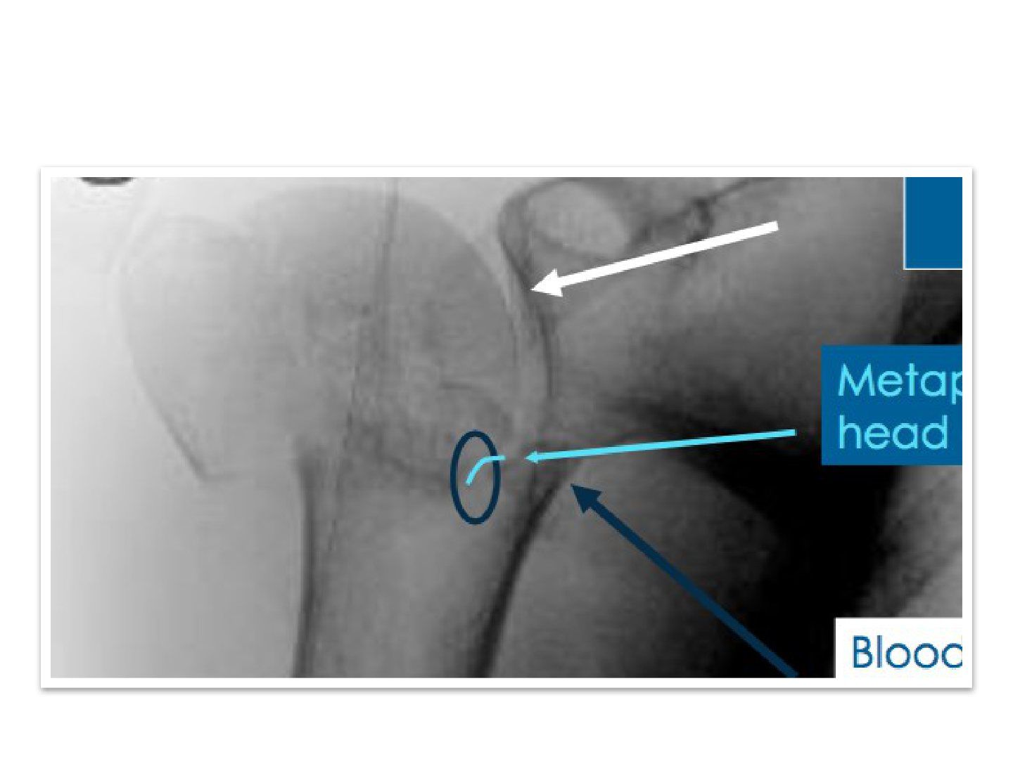
Q1-Q1010 questions — tap to reveal all answerslist
- Describe the X-ray features of this comminuted proximal humeral fracture.
- What are Hertel criteria and their significance?
- What did the ProFHer trial show?
- What is the evidence for fixation versus replacement?
- What is the evidence for implant choice?
- Describe the deltopectoral approach and fixation.
- What history and examination are required in a proximal humeral fracture?
- What is the acute management of a proximal humeral fracture?
- What patient and fracture factors determine whether to operate?
- Describe the rehabilitation phases after proximal humerus fixation.
Q1.Describe the X-ray features of this comminuted proximal humeral fracture.
- Comminuted fracture of the right proximal humerus with GT, head and neck fragments
- Fracture over the anatomical head with short metaphyseal extension <8mm
- Displaced with medial hinge disruption >=2mm
- No fracture scapula/ribs, no pneumothorax, no underlying lytic lesion
Q2.What are Hertel criteria and their significance?
- Hertel 2004 predicts AVN of the humeral head
- 97% chance of AVN if all three criteria met
- Minor criteria: head split, 3-4 part, dislocation
- Poor predictors of ischaemia: dislocation, tuberosity displacement, angular displacement of the head, 3-4 part fracture
Q3.What did the ProFHer trial show?
- JAMA 2015 - no significant benefit of surgery at 2-year FU
- Critiques: selection bias (clear indications for surgery excluded), low cases per surgeon
- Fracture patterns categorised by Neer classification rather than pathomorphology
Q4.What is the evidence for fixation versus replacement?
- Gupta 2015 JOT: higher reoperation in ORIF vs HA/RSA but significantly better outcomes in ORIF
- DelPhi trial JBJS 2020: for B2, C2 fractures in the elderly, RSA gives better functional outcomes
Q5.What is the evidence for implant choice?
- Fixation: nail or plate - Metanalysis 2018 Int Ortho Sun et al: no difference
- Arthroplasty: Cuff et al JBJS 2013 - RSA better clinical outcomes than hemi, similar complication rate
Q6.Describe the deltopectoral approach and fixation.
- Skin incision from coracoid along deltopectoral groove; retract cephalic vein laterally
- Plane between deltoid and pec major; stay lateral to the conjoint tendon; clavipectoral fascia
- Tag tuberosities; enter through fracture site; head to shaft fixation with K wire +/- bone graft
- Aim CCD angle >120, retroversion 20; tuberosities tied together restore metaphyseal torus
- 3.5mm locking plate in neutralization mode 5mm distal to GT tip, 2mm lateral to bicipital groove; 5 screws proximally, 3 distally
- Pin and make sure position good for calcar screw to prevent varus collapse
Q7.What history and examination are required in a proximal humeral fracture?
- Assess for associated injuries: C spine, scapula, clavicle, ribs
- NV condition esp brachial plexus, axillary nerve, axillary artery
Q8.What is the acute management of a proximal humeral fracture?
- Analgesics
- Immobilisation with a shoulder immobiliser
- Long-term management should address osteoporosis
Q9.What patient and fracture factors determine whether to operate?
- Patient: age, comorbidities, associated injuries, premorbid function
- Fracture: displacement, comminution, bone quality
Q10.Describe the rehabilitation phases after proximal humerus fixation.
- Phase 1 (first 3 weeks): pendular exercises, gentle assisted motion; avoid ER for the first 6 weeks
- Phase 2 (3-9 weeks): active assisted forward flexion and abduction; no abduction against resistance weeks 3-6; reduce assistance from week 6
- Phase 3 (after week 9): isotonic concentric and eccentric strengthening; add passive stretching if stiff
▸ Slide 372 · Describe Xray:Trauma · 8 questions 1 check expand
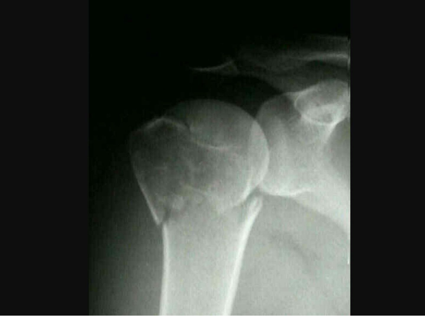
Q1-Q88 questions — tap to reveal all answerslist
- Describe this X-ray and classify the proximal humeral fracture using Neer's classification.
- What is the blood supply to the humeral head?
- What is Neer's classification threshold for a displaced part?
- How do age and AVN risk guide management?
- What are the principles and key parameters of hemiarthroplasty?
- What are the complications of hemiarthroplasty?
- What history, examination and imaging are needed in a proximal humeral fracture?
- Describe the technical steps of hemiarthroplasty for proximal humerus fracture.
Q1.Describe this X-ray and classify the proximal humeral fracture using Neer's classification.
- Fracture of the GT and surgical neck of the humerus with angular displacement
- Likely a 3-part fracture by Neer classification
- Short medial metaphyseal extension (<8mm) with disrupted medial hinge - 97% chance of AVN (Hertel 2004)
Q2.What is the blood supply to the humeral head?
- Traditional belief: mainly the arcuate branch of the ascending anterior circumflex
- Mainly from the posterior circumflex according to Henrich 2010 JBJS (64%)
Q3.What is Neer's classification threshold for a displaced part?
- 1cm displacement defines a displaced part
- 45 degree angulation also defines a displaced part
Q4.How do age and AVN risk guide management?
- Old patient, low functional demand and fit for surgery - advise hemiarthroplasty/RSA (Delphi trial)
- Young patient - still attempt ORIF with plating (long-term hemiarthroplasty outcome uncertain; Gupta JOT 2015)
Q5.What are the principles and key parameters of hemiarthroplasty?
- Needs an intact CA arch
- Aim: restore biomechanics of the shoulder and allow early mobilisation; principles include anatomical and stable fixation of hemiarthroplasty, correct tensioning and position of implant, secure reattachment of tuberosities
- Deltopectoral approach; identify GT/LT by position of biceps tendon and tag fragments
- Retroversion ~30 degrees; top of implant to GT 7-8mm; top of humeral head to upper border of pec major insertion = 5.6cm; should translate <50% of head
Q6.What are the complications of hemiarthroplasty?
- Approach related: axillary nerve injury, subscapularis rupture, traction injury to musculocutaneous nerve
- Implant related: poor tensioning/positioning, cuff retraction with weakness, infection, loosening, glenoid erosion, anterosuperior escape
Q7.What history, examination and imaging are needed in a proximal humeral fracture?
- Hx: age, function, injury mechanism; rule out associated fracture (distal radius, clavicle, chest, neck)
- PE: wound, axillary nerve function
- Obtain Y view or Velpeau axillary view to rule out dislocation
Q8.Describe the technical steps of hemiarthroplasty for proximal humerus fracture.
- Deltopectoral approach; identify GT/LT by the position of the biceps tendon and tag the fragments
- Dislocate/retrieve the humeral head, prepare the canal by reaming, trial then cement the implant
- Retroversion ~30 degrees - lateral fin slightly posterior to the bicipital groove
- Height: sit on medial calcar if present, top of implant to GT 7-8mm, head top to upper border of pec major insertion 5.6cm, translate <50% of head
- Head size templated from the contralateral shoulder; reattach GT/LT/SS under the collar by suture
Humeral head blood supply mainly from the posterior circumflex according to the study of Henrich 2010 JBJS (64%) — citation author name is incorrect — The study is Hettrich et al., JBJS 2010;92(4):943-8, which found the posterior humeral circumflex artery supplied 64% of the humeral head — source
▸ Slide 373 · Anterior approach to shoulder with internervous plane between axillary nerve andTrauma · 5 questions expand
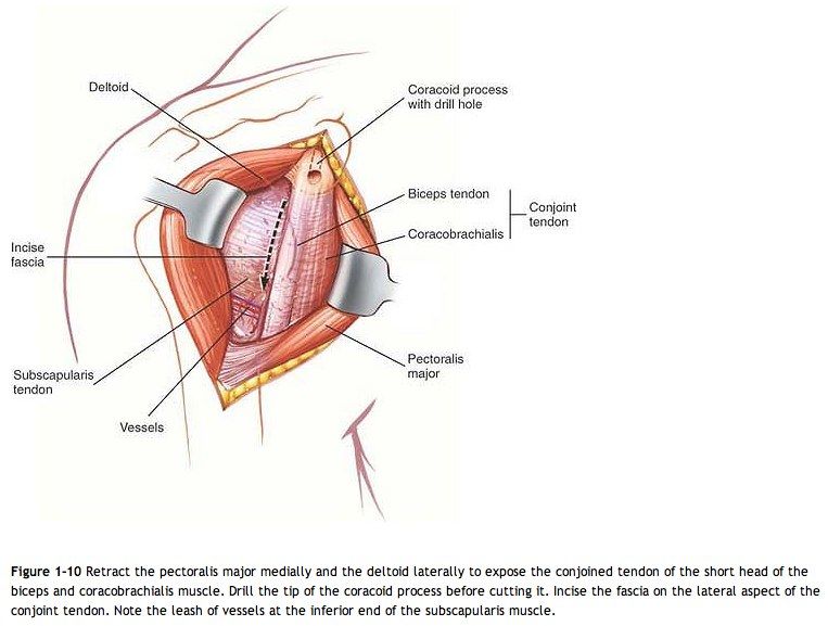
Q1-Q55 questions — tap to reveal all answerslist
- What are the indications for the anterior approach to the shoulder?
- How is the patient positioned for the anterior shoulder approach?
- What are the landmarks and key steps of the anterior approach?
- What is the internervous plane of the anterior shoulder approach?
- What structures are at risk in the anterior shoulder approach?
Q1.What are the indications for the anterior approach to the shoulder?
- Trauma
- Infection
- Tumour
Q2.How is the patient positioned for the anterior shoulder approach?
- GA, beach chair position, head turned to the other side
- Sandbag under the scapula
- Xray available
Q3.What are the landmarks and key steps of the anterior approach?
- Landmarks: coracoid, deltopectoral groove
- Find the cephalic vein and retract it laterally
- Plane between deltoid and pectoralis; clavipectoral fascia
- Stay lateral to the conjoint tendon, careful with medial retraction
- Subscapularis tendon - find NV beneath; externally rotate arm and incise longitudinally
Q4.What is the internervous plane of the anterior shoulder approach?
- Between the axillary nerve supplying deltoid
- And the pectoral nerve supplying pectoralis
Q5.What structures are at risk in the anterior shoulder approach?
- Cephalic vein
- Musculocutaneous nerve
- Brachial plexus
- Axillary nerve and vessels
▸ Slide 374 · Anterior appraoch to armTrauma · 5 questions expand
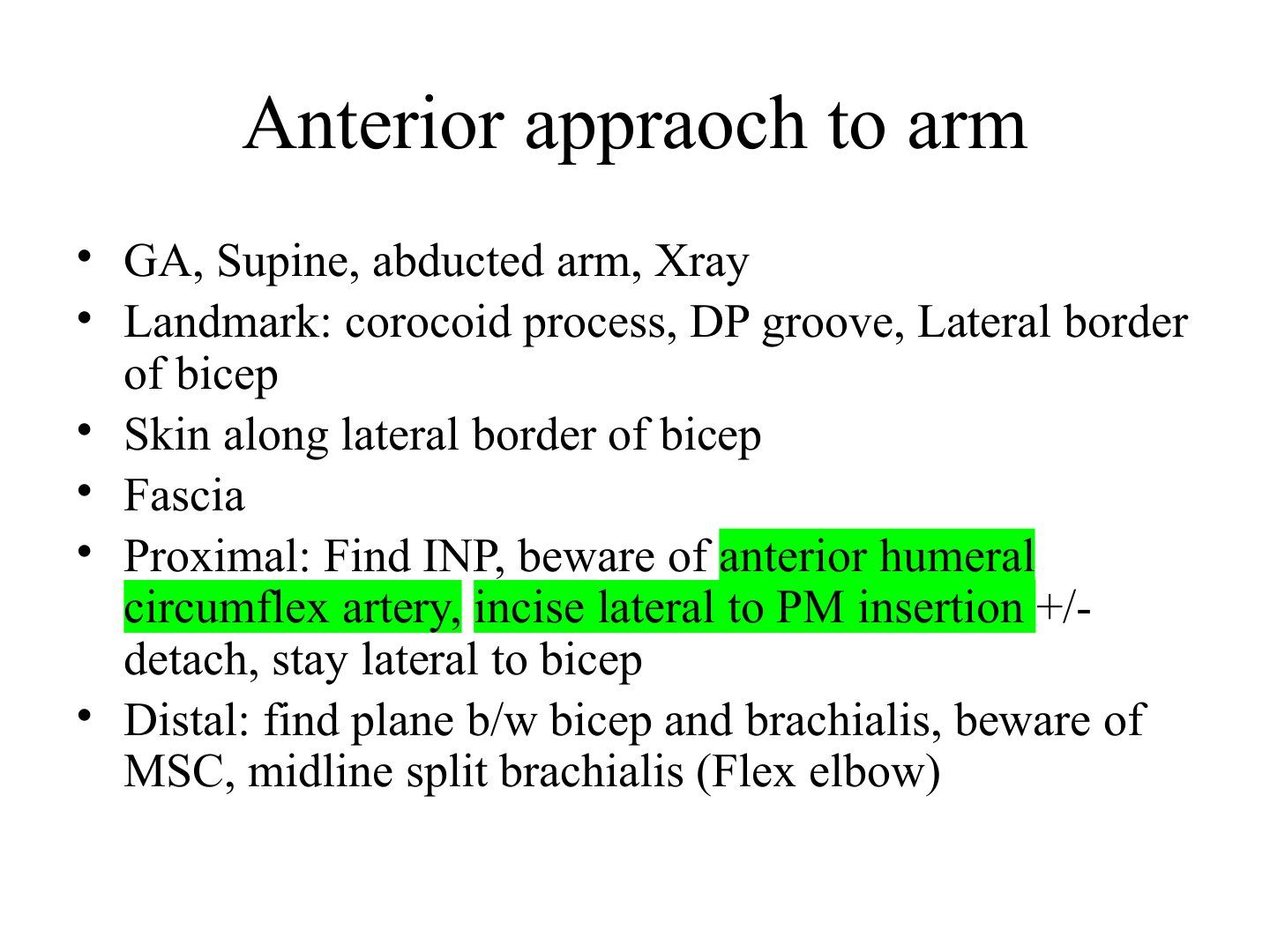
Q1-Q55 questions — tap to reveal all answerslist
- What is the anaesthesia, positioning and preparation for the anterior approach to the arm?
- What are the landmarks for the anterior approach to the arm?
- Where is the skin incision made in the anterior approach to the arm?
- What are the key steps and structures at risk in the proximal part of the anterior approach to the arm?
- What are the key steps and structures at risk in the distal part of the anterior approach to the arm?
Q1.What is the anaesthesia, positioning and preparation for the anterior approach to the arm?
- GA
- Supine with the arm abducted
- Xray available
Q2.What are the landmarks for the anterior approach to the arm?
- Coracoid process
- Deltopectoral (DP) groove
- Lateral border of biceps
Q3.Where is the skin incision made in the anterior approach to the arm?
- Skin incision along the lateral border of the biceps
- Then incise the fascia
Q4.What are the key steps and structures at risk in the proximal part of the anterior approach to the arm?
- Find the internervous plane (INP)
- Beware of the anterior humeral circumflex artery
- Incise lateral to the PM (pectoralis major) insertion +/- detach it
- Stay lateral to the biceps
Q5.What are the key steps and structures at risk in the distal part of the anterior approach to the arm?
- Find the plane between biceps and brachialis
- Beware of the musculocutaneous nerve (MSC)
- Midline split of brachialis with the elbow flexed
▸ Slide 375 · *Humeral shaft fractureTrauma · 13 questions expand

Q1-Q1313 questions — tap to reveal all answerslist
- Describe the X-ray on the left.
- What is acceptable alignment and conservative management for a humeral shaft fracture?
- Describe the X-ray on the right and why you would operate.
- What are the advantages and disadvantages of nailing a humeral shaft fracture?
- What are the indications for surgery in a humeral shaft fracture?
- How do you manage a radial nerve palsy with a humeral shaft fracture?
- Describe the technique of antegrade humeral nailing.
- What is a Sarmiento brace and how does it work?
- What are the contraindications to a Sarmiento brace?
- How is a hanging cast used for humeral shaft fractures?
- What are the deforming forces in a proximal humeral shaft fracture?
- How is radial nerve recovery monitored?
- How is secondary/iatrogenic radial nerve palsy after surgery managed?
Q1.Describe the X-ray on the left.
- Spiral fracture of the left midshaft humerus
- Mild angulation on attempted AP view
Q2.What is acceptable alignment and conservative management for a humeral shaft fracture?
- Acceptable: 30 degree angulation on AP, 20 degree on lateral, 3cm shortening
- Coaptation splint first for 2 weeks until swelling improves, then Sarmiento brace
- Tighten brace twice weekly; do not lean on the elbow
- Low threshold to ORIF as distal third fractures are prone to slip into varus
Q3.Describe the X-ray on the right and why you would operate.
- Short oblique midshaft fracture with minimal comminution, no shortening
- Less contact area - higher strain per unit length, high chance of nonunion if treated conservatively
- Hanging cast is contraindicated due to over-distraction
- ORIF via anterior approach; lag screw + neutralisation plate/DCP (broad 4.5mm, staggered holes, 7 cortices)
- Anterior approach: lateral to bicep, bicep to medial, split brachialis at lateral 1/3 b/w plane of radial nerve and MCN, risk if lateral cutaneous nerve of forearm, radial nerve at lateral border of brachialis
Q4.What are the advantages and disadvantages of nailing a humeral shaft fracture?
- Advantages: biologically friendly (does not disrupt fracture haematoma); mechanically more rigid, smaller bending strains, less fatigue failure
- Disadvantages: Limited by canal diameter and preexisting shoulder stiffness (7mm); iatrogenic comminution during reaming; cuff damage; radial nerve entrapment; axillary/radial nerve injury with locking screws
- Violates Perren's strain theory if no interfragmentary compression
- Heineman 2012: similar union/infection/reoperation but higher total complications with nail; Ouyang 2013 J SES 2013 metaanalysis: similar outcomes, plating less occurrence of shoulder problem
Q5.What are the indications for surgery in a humeral shaft fracture?
- Open fracture, compartment syndrome
- Floating elbow, vascular injury requiring repair
- Relative: 2nd radial nerve palsy after CR, Holstein-Lewis fracture, >20 degree angulation in 2 planes or >3cm shortening, unable to maintain acceptable closed reduction
Q6.How do you manage a radial nerve palsy with a humeral shaft fracture?
- Overall prevalence 12%; observe if alignment good, no open fracture, no vascular/soft tissue compromise, not iatrogenic
- Shao JBJS (Br) 2005 systemic review: spontaneous recovery 70%; no difference early exploration vs expectant management
- Ilyas JAAOS 2020 favours early exploration: expectant 77.2%, late >8 weeks 68.1%, surgical mx within 3 weeks 89.8%, 10.5% incarceration rate, 26.8% laceration rate
- Consider exploration + fracture fixation if no improvement after 3 weeks; monitor clinically (BR, ECRL/B, EDC/EPL, last EI) and with NCV
Q7.Describe the technique of antegrade humeral nailing.
- Anterolateral approach to the deltoid, enter through the rotator interval
- Split the rotator cuff and insert the nail at the greater tuberosity
- Proximal screws inserted laterally to avoid the axillary nerve
- Distal screws inserted anteroposteriorly to avoid the musculocutaneous nerve
Q8.What is a Sarmiento brace and how does it work?
- Functional orthosis giving stability while allowing movement of the joint above and below
- Uses the non-compressible nature of fluid and soft tissue
- Circumferential pressure generates hydraulic pressure that stabilises the bone
- Active muscle pull helps realign the fracture, promote secondary healing and provide blood supply
Q9.What are the contraindications to a Sarmiento brace?
- Shortening, length-unstable fracture, bone loss
- Open fracture/poor tissue envelope, fracture of other bones in the same limb
- Uncooperative patient, fracture too proximal or distal
- JBJS 2000: >80% union with good alignment; radial nerve palsy is not a contraindication
Q10.How is a hanging cast used for humeral shaft fractures?
- Realigns the fracture using the weight of the arm
- Long arm cast with holes at the wrist (dorsal to volar, radial side), hung on the neck by C&C
- Longer C&C extends, shorter C&C flexes
- More dorsal holes = apex medial; more volar holes = apex lateral
Q11.What are the deforming forces in a proximal humeral shaft fracture?
- Depends on the fracture position relative to the deltoid insertion
- Proximal to deltoid: proximal fragment adducted by pectoralis major
- Distal to deltoid: proximal fragment abducted by deltoid
Q12.How is radial nerve recovery monitored?
- Clinical: BR, ECRL/B, EDC/EPL, last EI
- NCV immediately after injury for documentation (absent signal may be pre-existing)
- Repeat at 2 weeks: conduction present = neuropraxia; absent = axonotmesis/neurotmesis
- Repeat at 2 months to differentiate axonotmesis from neurotmesis
Q13.How is secondary/iatrogenic radial nerve palsy after surgery managed?
- Entrapment 6-25%; laceration 20-42%
- Literature generally supports non-surgical management; no single algorithm
- No exploration: spontaneous recovery 70%, but 90% do not reach complete recovery
- Early exploration allows repair or early grafting; late exploration lets the neurilemmal sheath thicken
- Some studies show that function recovery is more complete and consistent with this approach - for me, I would choose early exploration, as the risk of bad result from postponement justifies early exploration in case of uncertain nerve damage
▸ Slide 376 · Describe Xray:Trauma · 7 questions expand

Q1-Q77 questions — tap to reveal all answerslist
- Describe the X-ray features of this distal third humeral shaft fracture.
- Why is the radial nerve prone to injury in distal third humeral fractures?
- Is radial nerve palsy an indication for surgery?
- Why operate on this fracture?
- What is a Holstein-Lewis fracture?
- What are the conservative options for a distal third humeral shaft fracture?
- What are the absolute indications for surgery in a humerus fracture?
Q1.Describe the X-ray features of this distal third humeral shaft fracture.
- Spiral fracture of the distal 1/3 humerus with butterfly fragment
- Fracture angulated into varus
Q2.Why is the radial nerve prone to injury in distal third humeral fractures?
- Tethered by the lateral intermuscular septum as it passes from posterior to anterior at the 1/3 of the humerus (15cm from lateral epicondyle)
- At risk of traction injury (22%)
Q3.Is radial nerve palsy an indication for surgery?
- Traditionally believed to be an absolute indication
- Found to have spontaneous recovery in 90% of cases (Elkholm JOT 2008), most due to neuropraxia
Q4.Why operate on this fracture?
- Alignment may put the nerve under more tension, affecting its capacity for good healing and recovery
- Difficult to immobilise the fracture without immobilising the elbow - causes elbow stiffness
- Posterior approach - split triceps or paratricipital
- 3.5mm extraarticular plate if very distal, or 4.5 narrow LCP
Q5.What is a Holstein-Lewis fracture?
- Distal third humeral shaft fracture associated with radial nerve palsy in 20%
- CR is contraindicated as it may tear the radial nerve
- If alignment unsatisfactory - plating via anterolateral approach, go through brachioradialis and brachalis, the plane in which the radial nerve lies when it goes anterior - can explore radial nerve
- Posterior approach: radial nerve lies between medial and lateral head of triceps then to the lateral intermuscular septum
Q6.What are the conservative options for a distal third humeral shaft fracture?
- Brace or cast
- Must immobilise the elbow as well
Q7.What are the absolute indications for surgery in a humerus fracture?
- Open fracture
- Vascular compromise
- Compartment syndrome
- Unacceptable alignment
▸ Slide 377 · Describe Xray:Trauma · 13 questions expand

Q1-Q1313 questions — tap to reveal all answerslist
- Describe the X-ray features of this intercondylar distal humeral fracture.
- What is the assessment and pre-op workup?
- What are the principles of surgery?
- What is the Jupiter classification?
- Describe the olecranon osteotomy.
- What are O'Driscoll's principles of distal humeral fixation?
- Describe the paratricipital approach to the distal humerus.
- Which posterior approaches are available for the distal humerus?
- What are the advantages of parallel plating?
- Should the ulnar nerve be transposed during distal humerus ORIF?
- What is the rate of heterotopic ossification and is prophylaxis indicated?
- When is total elbow arthroplasty considered for distal humerus fracture and what are its problems?
- Describe the ORIF sequence for a distal humerus fracture.
Q1.Describe the X-ray features of this intercondylar distal humeral fracture.
- Comminuted intercondylar fracture of the right humerus with varus angulation and metaphyseal comminution
- AO type C fracture
Q2.What is the assessment and pre-op workup?
- Age, function, hand dominance, mechanism; high energy - ATLS and rule out injury in the same limb
- Locally: skin wound/impingement, soft tissue condition, compartment, nerve palsy (radial nerve most at risk)
- Rest in a long arm slab and get CT before OT to assess the comminution
Q3.What are the principles of surgery?
- Anatomical reduction of the articular surface
- Restore the tiearch of the distal humerus
- Stable fixation with double plating to allow early mobilisation
Q4.What is the Jupiter classification?
- Low T, High T
- H, Y, medial/lateral lambda
Q5.Describe the olecranon osteotomy.
- Lateral decubitus, GA, Xray guidance; longitudinal incision at mid posterior curve to radial at olecranon process
- Elevate full thickness fasciocutanoeous flaps; identify and protect the ulnar nerve, release through cubital tunnel to first motor branch to FCU
- Proximally based V-shaped chevron osteotomy ~2cm distal to tip of olecranon, aiming at the bare area of the sigmoid notch
- Predrill holes; pass gauze inside the joint to protect cartilage; drill hole at the apex to prevent propagation; saw with irrigation and finish with osteotome
- JSES 2017: extraarticular step cut may be more stable with higher bone contact surface area
Q6.What are O'Driscoll's principles of distal humeral fixation?
- Respect O'Driscoll's principles: insert screws in an interdigitated manner, purchasing as many articular fragments as possible
- As many screws distally as possible; each screw should engage the other side; screw as long as possible
- Every distal screw should pass through the plate; plates apply compression at the supracondylar level
- Do not end plates at the same level, to avoid stress rise
- 90/90 plating: similar biomechanics to parallel plating but better resistance to torsional loading
Q7.Describe the paratricipital approach to the distal humerus.
- Midline posterior incision
- Identify the ulnar nerve and dissect 15cm proximal to the elbow and distally to the first branch to FCU
- Medially elevate triceps by freeing the medial intermuscular septum; distally elevate the posterior band of MCL
- Laterally identify the radial nerve if a long plate is planned; divide the anconeus distally for exposure
Q8.Which posterior approaches are available for the distal humerus?
- Olecranon osteotomy
- Paratricipital approach
- Triceps reflecting (TRAP) approach
Q9.What are the advantages of parallel plating?
- Can insert longer screws from the lateral side
- Orthogonal plating gives better torsional rigidity
- Better for coronal shear fragments
- Requires less stripping, preserving blood supply of the lateral condyle (SK Lee Eur J O&T 2013)
Q10.Should the ulnar nerve be transposed during distal humerus ORIF?
- Not routinely - transposition does not decrease ulnar nerve palsy rate
- Some studies record 4 times higher ulnar nerve dysfunction after transposition
- Decide based on impingement/subluxation when screening the full range after fixation
- Ilyas Hand Clinic 2018: overall incidence 19.3%; transposition 23.5%
Q11.What is the rate of heterotopic ossification and is prophylaxis indicated?
- HO reported rate 8%
- Routine prophylaxis is not warranted
- Indomethacin increased the rate of nonunion
Q12.When is total elbow arthroplasty considered for distal humerus fracture and what are its problems?
- For elderly patients with osteoporotic bone and comminution not amenable to stable fixation
- Dehghan 2019 JSES: Type C2/C3, female >65 years - TEA is an option
- Bryan Morrey approach: release triceps tendon and periosteum medial to lateral and elevate as a single unit off the olecranon, reflecting laterally
- Problems: columns and collaterals are not intact - need linked implants with bearing wear/loosening; periprosthetic fracture
Q13.Describe the ORIF sequence for a distal humerus fracture.
- Reduce articular fragments and fix large pieces with lag screws, or positional screws if comminuted
- Fix the articular fragment to the metaphyseal region
- Choose 90/90 plating (posterolateral and medial)
- Do not end the plates at the same level to avoid stress rise
▸ Slide 378 · Xray showing simple elbow dislocationTrauma · 8 questions expand
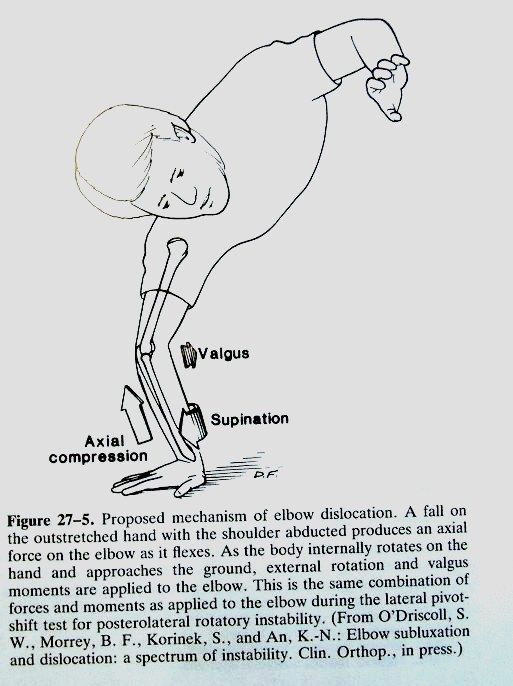
Q1-Q88 questions — tap to reveal all answerslist
- What is the acute management of a simple elbow dislocation?
- Describe the reduction technique for elbow dislocation.
- What do you check on post-reduction fluoroscopy?
- What is the management if the joint is congruent and stable?
- Describe PLRI and PMRI.
- When do you proceed to open reduction and what are the complications?
- How is the lateral pivot shift test performed?
- What physical examination tests suggest PMRI?
Q1.What is the acute management of a simple elbow dislocation?
- Stabilise patient and rule out other injuries
- Rule out local complications: open wound, compartment syndrome, NV injury
- Analgesics, immobilise
- Reduction + immobilise +/- operation
Q2.Describe the reduction technique for elbow dislocation.
- Under conscious sedation: traction, supination, direct manipulation
- Align forearm and humerus; traction with elbow flexed 90 degrees; direct pressure over olecranon; +/- supination
- Parvin's method: prone, traction over wrist for a few minutes, then lift arm
Q3.What do you check on post-reduction fluoroscopy?
- Joint congruity - drop sign = pseudosubluxation >=4mm ulnohumeral displacement (interposed soft tissue/instability)
- Range of stable reduction
- Collaterals - varus/valgus stress; pMCL only stable in pronation, aMCL+pMCL unstable in pronation; repeat in full extension and 30 degrees flexion
- Rotatory instability - lateral pivot shift, PL drawer, chair sign, table to relocation test
Q4.What is the management if the joint is congruent and stable?
- Immobilise for 2 weeks + early mobilisation
- Recurrence <2%, loss of terminal extension 5-15 degrees
- Stability is better in flexion and pronation
- Rehab: early ROM within 10 days, avoid immobilisation >3 weeks, aim full extension by 8 weeks
Q5.Describe PLRI and PMRI.
- Both involve axial load + forearm supination, with valgus stress in PLRI and varus stress in PMRI
- PLRI sequence: LUCL then pMCL then aMCL (Hori circle) +/- bone injury (coronoid tip, radial head)
- PMRI sequence: LUCL, aMCL +/- coronoid base/AM facet; radial head usually intact
- CT/MRI is the gold standard for diagnosis
Q6.When do you proceed to open reduction and what are the complications?
- Open reduction +/- repair if failed congruent reduction or unstable (dislocates in splint, dislocates with <30 degree flexion + pronation)
- Complications: HO, ulnar nerve irritation, OA, instability
Q7.How is the lateral pivot shift test performed?
- Forearm supinated, valgus and axial load
- In extension the radial head is dislocated
- Toward 40 degrees flexion the triceps reduces the radial head with a clunk
Q8.What physical examination tests suggest PMRI?
- Varus/valgus stress with the shoulder in max IR/ER
- Gravity assisted varus stress: shoulder 90 degrees abduction, elbow flexed and extended - pain on flexion due to lack of medial buttress by LUCL and coronoid
- Chair test
- Table top relocation test: radial head subluxes as the forearm presses the table in supination - apprehension relieved by posterior radial head pressure
▸ Slide 379 · C=terrible traidTrauma · 2 questions expand
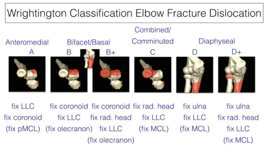
Q1-Q22 questions — tap to reveal all answerslist
- What does 'C' represent on this slide?
- What does 'B+' represent on this slide?
Q1.What does 'C' represent on this slide?
- C = terrible triad
- The slide gives no further detail
Q2.What does 'B+' represent on this slide?
- B+ = all column failure
- The slide gives no further detail
▸ Slide 380 · ORIGINTrauma · 2 questions expand
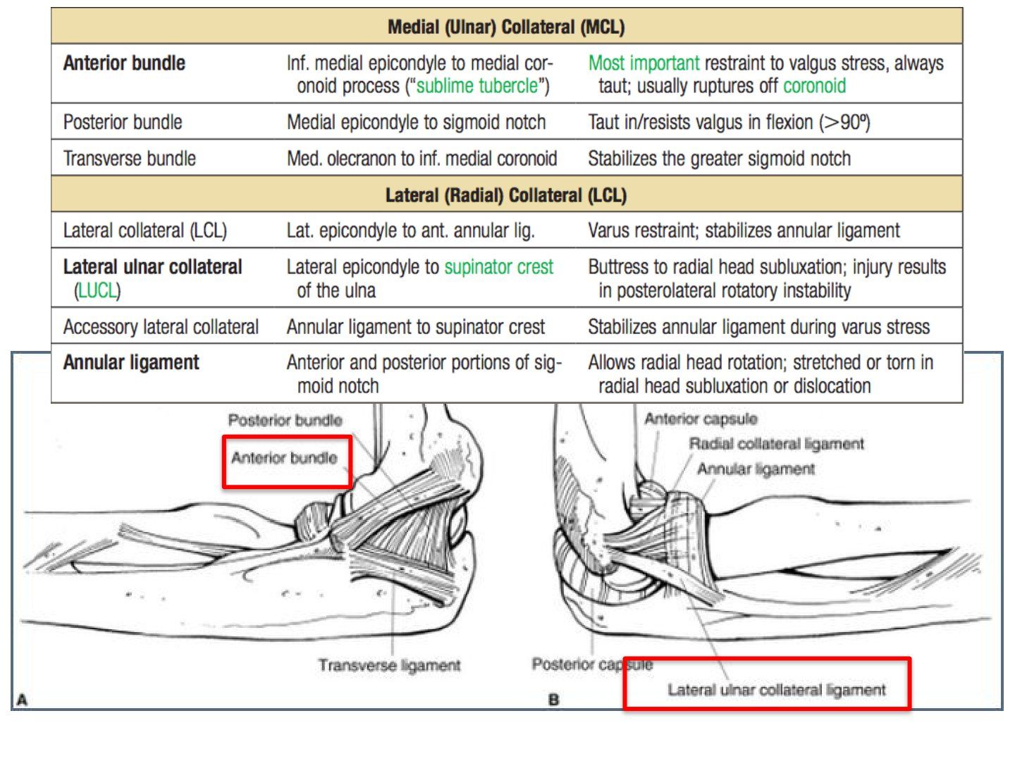
Q1-Q22 questions — tap to reveal all answerslist
- Where does the MCL originate?
- Where does the LCL originate?
Q1.Where does the MCL originate?
- Anteroinferior portion of the medial epicondyle
- MCL = medial collateral ligament
Q2.Where does the LCL originate?
- Inferior portion of a small tubercle on the lateral epicondyle
- LCL = lateral collateral ligament
▸ Slide 381 · Morrey stage of instability used in chronic conditionTrauma · 2 questions expand
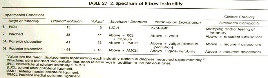
Q1-Q22 questions — tap to reveal all answerslist
- What is the Morrey staging of instability used for and how does it progress?
- List the structures involved in the Morrey stages of elbow instability, in order.
Q1.What is the Morrey staging of instability used for and how does it progress?
- Used in the chronic condition
- Progresses from lateral to medial
- LCUL --> RCL + capsule --> PMCL --> AMCL
Q2.List the structures involved in the Morrey stages of elbow instability, in order.
- 1. LCUL
- 2. RCL + capsule
- 3. PMCL
- 4. AMCL
▸ Slide 382 · XR showing left posterolateral elbow fracture dislocationTrauma · 14 questions expand
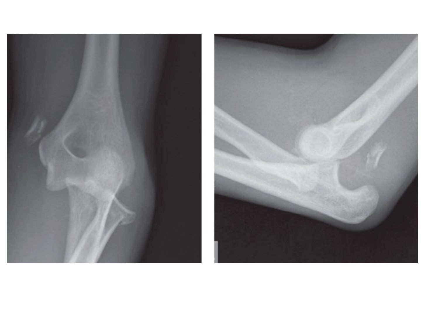
Q1-Q1414 questions — tap to reveal all answerslist
- What are the components of a terrible triad elbow injury?
- What initial assessment and management is described for a fracture dislocation of the elbow?
- What does Morrey's model describe as the primary static, secondary static and dynamic stabilizers of the elbow?
- What definitive planning is required after initial reduction of a terrible triad?
- What is the sequence of fixation in operative management of a terrible triad?
- Which approach is chosen in a terrible triad and what are the options?
- How can exposure be improved during the posterior utility approach?
- What medial approaches are available in a terrible triad and what are their pros and cons?
- How can PIN injury be avoided during the Kocher approach in terrible triad surgery?
- When is radial head ORIF preferred over replacement, and when is replacement indicated?
- What did the Sun 2016 meta-analysis find comparing arthroplasty with ORIF for radial head fractures?
- Describe the safe zones for applying a dynamic joint distractor (DJD).
- What rehabilitation and prophylaxis follow fixation of a terrible triad?
- For a transolecranon fracture dislocation, which radiographic parameters must be restored?
Q1.What are the components of a terrible triad elbow injury?
- Elbow dislocation
- Radial head fracture
- Coronoid fracture
- XR: fragments over the medial epicondyle and sigmoid notch (likely radial head), no soft tissue gas
Q2.What initial assessment and management is described for a fracture dislocation of the elbow?
- ATLS protocol, primary and secondary survey, AMPLE history + premorbid status
- Check NV status and compartments; examine one joint above and below (Essex lopresti)
- Analgesia with adequate sedation using midazolam and pethidine
- CR with elbow at 90 degrees flexion + gentle traction and direct olecranon manipulation, then splint
Q3.What does Morrey's model describe as the primary static, secondary static and dynamic stabilizers of the elbow?
- Primary static = UHJ, LUCL, MCL
- Secondary static = RCJ, CEO, CFO
- Dynamic = anconeus, triceps and biceps
- Indication for surgery = loss of dynamic and static stabilisers
- Aim = concentric stable reduction to allow early mobilisation
Q4.What definitive planning is required after initial reduction of a terrible triad?
- CT for planning: classify the coronoid fracture (O'Driscoll/Morrey) and radial head fracture (Mason)
- Indicated for surgical repair and ORIF due to loss of dynamic and static stabilisers
- Aim: concentric stable reduction to allow early mobilisation
- Position supine; approach = utility posterior approach
Q5.What is the sequence of fixation in operative management of a terrible triad?
- Fix the coronoid first through the radial head fracture window - small, use lasso transosseous repair
- Fixation or replacement of the radial head or neck
- Repair the LUCL with suture anchor or fibre tape
- Test stability; repair MCL via a medial approach if still unstable
- If still unstable, prepare radial head replacement or apply a dynamic joint distractor (DJD)
Q6.Which approach is chosen in a terrible triad and what are the options?
- Posterior utility approach, raising thick flaps on the medial and lateral sides to address the respective pathologies
- Laterally: Kocher interval (ECU-anconeus), Kaplan interval (ECRB-EDC) or Boyd interval (anconeus and FCU)
- Kaplan may preserve the remaining lateral ligamentous complex but has more PIN palsy
- Boyd if there is also an olecranon fracture
- Key point: watch out for the PIN and radial nerve
Q7.How can exposure be improved during the posterior utility approach?
- Release the ECRB along the supracondylar ridge
- Release some of the supinator distally
Q8.What medial approaches are available in a terrible triad and what are their pros and cons?
- Hotchkiss (flexor pronator mass, PL-FCU), split FCU, or Taylor Scham (elevate the entire flexor pronator mass from posterior to anterior)
- Watch the medial antebrachial cutaneous nerve on skin incision; deep, watch the ulnar nerve
- Hotchkiss: good access to the anterior elbow capsule, BUT limited access to the medial facet of the coronoid and poor access to its base
- Split FCU probably most versatile in terrible triad (addresses base and anteromedial facet)
- Taylor Sham best for accessing the base
Q9.How can PIN injury be avoided during the Kocher approach in terrible triad surgery?
- Fully pronate the forearm
- Avoid incising the capsule too anteriorly (PIN lies over it)
- Avoid dissection distal to the annular ligament (PIN lies within supinator)
- Do not place retractors around the radial neck
Q10.When is radial head ORIF preferred over replacement, and when is replacement indicated?
- ORIF best reserved for non-comminuted fractures with 3 or fewer fragments
- Attempted fixation of more comminuted fractures is prone to fixation failure and nonunion (Ring and Jupiter JBJS 2002)
- Replacement indications: Mason III + >3 articular fragments
- Replacement also for an unstable elbow (Mason IV / Essex-Lopresti injury)
- Prepare radial head replacement if still unstable after MCL + LUCL repair and ORIF
Q11.What did the Sun 2016 meta-analysis find comparing arthroplasty with ORIF for radial head fractures?
- Metaanalysis by Sun 2016 Eur J ortho trauma: arthroplasty yielded higher satisfaction, better elbow scores, shorter surgical time and lower incidence of recurrent instability
- Replacement has better ROM and is a faster operation
- ORIF has a higher reoperation rate and higher complication rate
- BUT functional score is similar
Q12.Describe the safe zones for applying a dynamic joint distractor (DJD).
- Distal 1/3 humerus: anterolateral or posterolateral
- Middle 1.3 humerus: anterolateral (middle third of the humerus)
- Proximal forearm: lateral/posterolateral proximal and middle 1/3 ulna, neutral rotation
- Axis guidewire: lateral just distal to lateral epicondyle, medial slightly anterior and distal to medial epicondyle; place hinge over the guidewire
Q13.What rehabilitation and prophylaxis follow fixation of a terrible triad?
- Long arm slab in pronation for 2 weeks
- Then progressive ROM exercises, starting with active assisted FE and SP
- HO prophylaxis
Q14.For a transolecranon fracture dislocation, which radiographic parameters must be restored?
- PUDA (proximal ulnar diaphyseal angle): 8-10 degrees
- OW (olecranon width)
- ODA (olecranon diaphyseal angle): 23 degrees between the olecranon tip and the diaphysis
▸ Slide 383Trauma · 3 questions expand
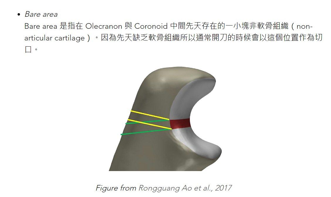
Q1-Q33 questions — tap to reveal all answerslist
- What is the topic of this slide?
- What are the key learning points on this slide?
- What source material is available for this slide?
Q1.What is the topic of this slide?
- Trauma
- Not covered in the speaker notes
Q2.What are the key learning points on this slide?
- Not covered in the speaker notes
Q3.What source material is available for this slide?
- Slide image only - no speaker notes
- Not in the speaker notes
▸ Slide 384 · Radial head replacementTrauma · 4 questions expand

Q1-Q44 questions — tap to reveal all answerslist
- Why is metal preferred over silicone for radial head replacement?
- What implant options are available for radial head replacement?
- How is the correct length of a radial head prosthesis determined?
- How is the correct size checked and what is assessed intra-operatively after radial head replacement?
Q1.Why is metal preferred over silicone for radial head replacement?
- Better biomechanics
- Decrease wear debris
- Decrease synovitis
Q2.What implant options are available for radial head replacement?
- Stems: loose, press fit or cemented
- Head: monopolar, bipolar or anatomic
- Modularity or monoblock
Q3.How is the correct length of a radial head prosthesis determined?
- Articular surface of the prosthesis at or slightly proximal to the lateral edge of the coronoid articular surface
- Check the DRUJ
Q4.How is the correct size checked and what is assessed intra-operatively after radial head replacement?
- Size should resemble the radial head fragment
- Check ROM in flexion/extension/supination/pronation
▸ Slide 385 · Radial head fractureTrauma · 9 questions 1 check expand
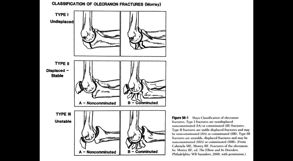
Q1-Q99 questions — tap to reveal all answerslist
- Describe the Mason classification of radial head fractures.
- When is operative treatment indicated for a radial head fracture?
- Describe the safe zone for fixation of a radial head fracture.
- What replacement options exist for a radial head fracture?
- What is the outcome of radial head excision?
- What are the functions of the radial head?
- What are the key anatomical relations of the coronoid process?
- Describe the Regan and Morrey classification of coronoid fractures.
- Describe the Mayo classification of olecranon fractures and the indications for surgery.
Q1.Describe the Mason classification of radial head fractures.
- Type 1: undisplaced (<2mm)
- Type 2: >2mm displacement
- Type 3: comminution
- Type 4: with dislocation (modified by Hotchkiss)
Q2.When is operative treatment indicated for a radial head fracture?
- Mechanical block to movement
- >30% articular surface involved
- Unstable elbow
Q3.Describe the safe zone for fixation of a radial head fracture.
- Keep hardware within a 100 degree arc centred on the dorsal aspect of the neutrally rotated forearm
- Safe zone lies between the radial styloid and Lister tubercle
- Hotchkiss: the posterior limit of the safe zone lies halfway between the reference marks made with the forearm in neutral rotation and full pronation; the anterior limit lies nearly two thirds of the distance between the neutral mark and the mark made in full supination
- Confirm by direct visualisation (thicker cartilage) or direct lateral XR until the radial tuberosity is tangential
Q4.What replacement options exist for a radial head fracture?
- Metallic vs silicone
- Bipolar vs monopolar
- Modular vs one piece
- Cemented vs non-cemented
Q5.What is the outcome of radial head excision?
- Good pain relief in Mason II and III (Antuna JBJS 2010)
- In patients <40 years old, >90% have good results
- OA but not functional deficits
- Can cause instability in varus, valgus and longitudinal directions
Q6.What are the functions of the radial head?
- Blocks proximal migration of the radius
- Stabilizes the elbow
- Reduces valgus stress
- Blocks PLR movement
Q7.What are the key anatomical relations of the coronoid process?
- Pathognomonic for elbow fracture (no attachment to the tip - shearing)
- AMCL attaches at the sublime tubercle, 18mm distal to the tip
- Anterior capsule attaches 6mm distal to the tip
- Midpoint = line from olecranon tip parallel to the ulnar longitudinal axis
Q8.Describe the Regan and Morrey classification of coronoid fractures.
- Type 1: tip
- Type 2: <1/2
- Type 3: >1/2
- O'Driscoll: address the anteromedial facet fracture - varus posteromedial rotatory instability
Q9.Describe the Mayo classification of olecranon fractures and the indications for surgery.
- Type 1 undisplaced, type 2 displaced stable, type 3 unstable
- Subtype A simple, B comminuted
- Operate for a disrupted joint or disrupted extensor mechanism
- Options: fix, or excise and advance triceps
Mason type 4 radial head fracture (with dislocation) is attributed to modification by Hotchkiss — misattribution — The dislocation type 4 was added by Johnston in 1962 (Mason-Johnston classification); the Hotchkiss modification redefines types 1-3 by operative indication — source
▸ Slide 386 · Kocher ApproachTrauma · 6 questions expand

Q1-Q66 questions — tap to reveal all answerslist
- What is the internervous plane of the Kocher approach and which nerves are at risk?
- What procedures is the Kocher (posterolateral) approach used for at the elbow?
- What are the landmarks, patient position and incision for the Kocher approach?
- Describe the superficial dissection of the Kocher approach.
- How is the PIN protected during the Kocher approach?
- What are the capsular cautions and limitation of the Kocher approach?
Q1.What is the internervous plane of the Kocher approach and which nerves are at risk?
- Between ECU (PIN) and anconeus (radial nerve)
- Danger: PIN and radial nerve
Q2.What procedures is the Kocher (posterolateral) approach used for at the elbow?
- Radial head - excision/ORIF/replacement
- LCL surgery
- Coronoid surgery
Q3.What are the landmarks, patient position and incision for the Kocher approach?
- Landmarks: lateral epicondyle, radial head, olecranon
- 5cm incision from lateral epicondyle to radial head (curved or straight)
- GA, supine with arm board, tourniquet
Q4.Describe the superficial dissection of the Kocher approach.
- Incise the fascia in line
- Distally find the plane between ECU and anconeus
Q5.How is the PIN protected during the Kocher approach?
- Pronate the arm to turn the PIN away from the field
- Do not place retractors around the radial neck
- Do not dissect anteriorly (radial nerve over the anterolateral capsule)
- Do not dissect distal to the annular ligament; split proximal fibers of supinator, staying on the posterior cortex of the radius, away from the PIN
Q6.What are the capsular cautions and limitation of the Kocher approach?
- If LCL intact: keep 1cm anterior to the crista supinatoris for the capsular incision
- If LCL torn: longitudinal capsular incision
- Stay at the midline of the radiocapitellar joint
- Cons: cannot extend distally
▸ Slide 387 · Xray lateral elbow showing fracture capitellumTrauma · 4 questions expand
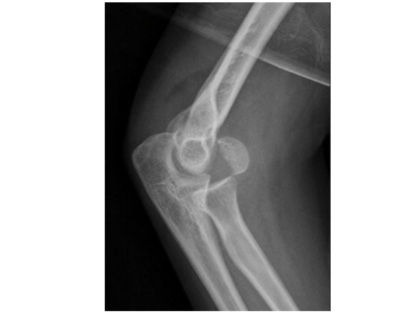
Q1-Q44 questions — tap to reveal all answerslist
- How do capitellum fractures present and why do they displace?
- Describe the Bryan and Morrey classification of capitellum fractures with eponyms.
- Describe the Dubberley classification of capitellum fractures.
- How are capitellum fractures managed?
Q1.How do capitellum fractures present and why do they displace?
- Coronal plane fracture after a fall on outstretched hand
- Little/no soft tissue attachment - often displaced
- Lateral X-ray shows the fragment displaced anteriorly; obtain AP view and CT
Q2.Describe the Bryan and Morrey classification of capitellum fractures with eponyms.
- Type 1: large osseous fracture (Hahn-Steinthal)
- Type 2: one cartilaginous piece with little osseous component (Kocher-Lorenz)
- Type 3: comminuted (Broberg-Morrey)
- Type 4: extension to the trochlea (McKee)
- Laugier - isolated trochlea fracture
Q3.Describe the Dubberley classification of capitellum fractures.
- Type 1: capitellum +/- lateral trochlear ridge
- Type 2: capitellum and trochlea as one single piece
- Type 3: capitellum and trochlea as separate pieces
Q4.How are capitellum fractures managed?
- Undisplaced: splint x 3 weeks
- Displaced: fix - Kaplan approach for type 4, posterior approach for type 1
- May consider excision (type 2) or arthroplasty
▸ Slide 388 · Olecranon fractureTrauma · 7 questions expand

Q1-Q77 questions — tap to reveal all answerslist
- What determines the olecranon fracture pattern and what injuries are associated?
- Describe the Mayo classification of olecranon fractures.
- What are the aims of olecranon fracture fixation?
- What is a tension band and what are the prerequisites for its use?
- Describe the tension band construct shown on these radiographs.
- Give other fractures where tension band fixation is used.
- Why is the lateral side of the femur the tension side for a tension band plate?
Q1.What determines the olecranon fracture pattern and what injuries are associated?
- Pattern: displacement, articular comminution, proximal fragment size
- Associations: UHJ dislocation, radial head dislocation, coronoid fracture
Q2.Describe the Mayo classification of olecranon fractures.
- Type 1 undisplaced, type 2 displaced and stable, type 3 displaced and unstable
- Subtype A = simple, B = comminuted
Q3.What are the aims of olecranon fracture fixation?
- Anatomical reduction, rigid fixation
- Restore the width of the trochlear notch
- Restore the proximal ulnar diaphyseal angle and olecranon diaphyseal angle
Q4.What is a tension band and what are the prerequisites for its use?
- A device that converts a tensile force into a compression force
- Bone must be eccentrically loaded; opposite cortex intact to withstand compression
- Fracture must be transverse and simple; construct applied on the tension side
- Implant must be able to withstand tensile force
Q5.Describe the tension band construct shown on these radiographs.
- AP and lateral radiographs of the right elbow
- Olecranon fracture fixed with two parallel pins and transosseous metal wire in a figure-of-eight configuration
- Restoration of the articular surface
Q6.Give other fractures where tension band fixation is used.
- Patella, calcaneum, medial malleolus
- Greater tuberosity, femur shaft
Q7.Why is the lateral side of the femur the tension side for a tension band plate?
- There is no lateral bowing of the femur; the tension side is determined by the anatomical axis
- Shifts the neutral axis from mid axis to the plate-bone junction
- Axial loading produces pure tension in the plate and pure compression across the fracture line
- Creates a bending close situation to reduce the working length of the plate
▸ Slide 389 · RTrauma · 6 questions expand
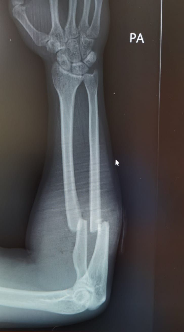
Q1-Q66 questions — tap to reveal all answerslist
- Describe the X-ray findings in this forearm injury.
- What history and examination are important in a both-bone forearm fracture?
- What is the acute management and aim of treatment for this forearm fracture?
- What is the patient setup and skin incision for the approach to the proximal radius?
- Describe the superficial dissection of the approach to the proximal radius.
- Describe the deep dissection of the approach to the proximal radius.
Q1.Describe the X-ray findings in this forearm injury.
- PA radiograph of the right forearm: fractures of the proximal 1/3 shaft of radius and ulna with translation
- DRUJ and elbow joint congruent
- Gas pocket in the subcutaneous layer - suspicious of open fracture
Q2.What history and examination are important in a both-bone forearm fracture?
- History: mechanism, associated injury, age, hand dominance, functional demand
- Examination: soft tissue, acute compartment, open wound, neurovascular status
- Examine the joint above and below
- 1mm inside out puncture wound
Q3.What is the acute management and aim of treatment for this forearm fracture?
- Analgesia, IV antibiotics, temporary stabilization with a long arm slab
- Prepare the patient for ORIF
- Aim: adequate debridement, compression plate fixation, early mobilization
Q4.What is the patient setup and skin incision for the approach to the proximal radius?
- Supine under GA
- Proximal landmark: biceps tendon
- Distal landmark: radial styloid process
Q5.Describe the superficial dissection of the approach to the proximal radius.
- Between BR and FCR
- Identify radial artery and SRN beneath BR
- Retract the radial artery medially, ligate recurrent branches to the mobile wad
- Retract the SRN laterally
Q6.Describe the deep dissection of the approach to the proximal radius.
- Supinate the forearm to displace the PIN away from the field
- Follow the lateral edge of the bicipital tuberosity, incise bursa
- Elevate the medial border of supinator from the radius
- Pronate the forearm (mid part) and partially detach the lateral border of pronator teres
▸ Slide 390 · Describe XRTrauma · 7 questions expand
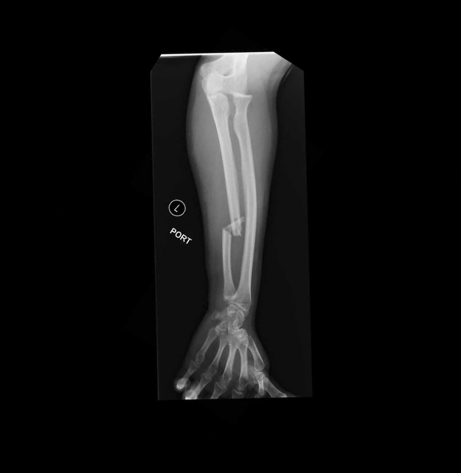
Q1-Q77 questions — tap to reveal all answerslist
- Describe the X-ray findings and diagnosis in this injury.
- What history and examination findings are important in a Monteggia fracture?
- How is a Monteggia fracture temporarily managed before operation?
- What is the significance of the Bado classification of Monteggia fractures?
- Describe the Jupiter classification subdivisions of Bado type II fractures.
- What are the aims and technique of ulnar fixation in this Monteggia fracture?
- How is the radiocapitellar joint assessed and managed after ulnar fixation?
Q1.Describe the X-ray findings and diagnosis in this injury.
- Fracture of the distal 1/3 ulnar shaft with comminution and displacement
- Anterior dislocation of the radial head
- Diagnosis: Monteggia fracture, Bado 1
Q2.What history and examination findings are important in a Monteggia fracture?
- History: time and mechanism of injury, associated injury
- P/E: soft tissue condition, open wound, acute compartment
- Palpate along the interosseous membrane and DRUJ for tenderness (Essex-Lopresti injury)
- Neurovascular injury especially PIN; associated injury in the ipsilateral limb
Q3.How is a Monteggia fracture temporarily managed before operation?
- Fracture dislocation - try CR, but unlikely to maintain reduction with a slab alone
- Temporary stabilisation with a long arm slab
- Prepare for operation under GA
Q4.What is the significance of the Bado classification of Monteggia fractures?
- Type I anterior: annular ligament
- Type II posterior: LUCL
- Type III lateral: PIN
Q5.Describe the Jupiter classification subdivisions of Bado type II fractures.
- A: ulnar fracture involves the distal olecranon and coronoid process
- B: fracture at the metadiaphyseal junction distal to the coronoid
- C: diaphyseal ulnar fracture
- D: fracture extends along the proximal third to half of the ulna
Q6.What are the aims and technique of ulnar fixation in this Monteggia fracture?
- Aim: anatomical reduction of the ulna (length, angulation, rotation), reduction of the RCJ, stable fixation for early mobilization
- Subcutaneous approach to the ulna
- Fix with a 3.5mm LCP in compression mode, aiming for primary bone healing
Q7.How is the radiocapitellar joint assessed and managed after ulnar fixation?
- True lateral view of the elbow with the forearm in full supination to assess congruency
- If suspicious: recheck the ulnar reduction and prepare open reduction of the RCJ via Kocher
- Look for interposition of soft tissue, e.g. annular ligament
- Persistent radial head subluxation - check for malreduction, capsuloligamentous causes, coronoid or radial head deficiency
- Postop: hinged elbow brace with the forearm in supination
▸ Slide 391 · Fracture proximal ulnar + posterior radial head dislocationTrauma · 5 questions expand
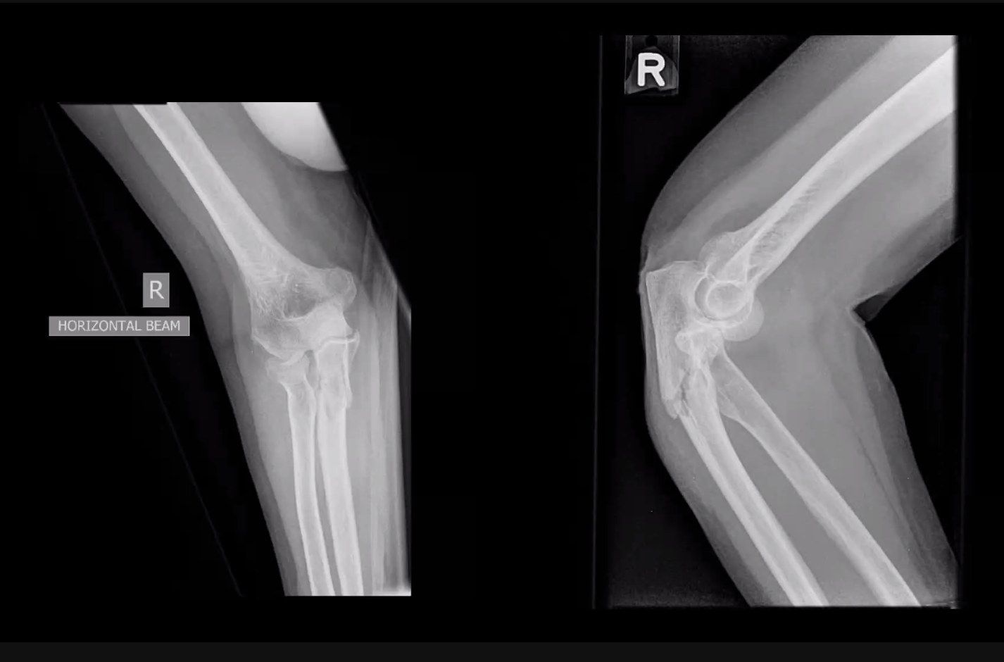
Q1-Q55 questions — tap to reveal all answerslist
- What is the diagnosis and classification of this injury?
- What history and examination are required in this Monteggia Bado II injury?
- What are the aims of surgery for this Monteggia fracture?
- Describe the steps of surgical management.
- How are Monteggia variants with radial head fracture addressed?
Q1.What is the diagnosis and classification of this injury?
- Fracture of the proximal ulna with posterior radial head dislocation
- Monteggia fracture
- Bado II
- Jupiter IIB
Q2.What history and examination are required in this Monteggia Bado II injury?
- History: premorbid status, level of activity
- Examination: soft tissue, wound, compartment
- Check PIN neurovascular status
Q3.What are the aims of surgery for this Monteggia fracture?
- Concentric reduction of the radiocapitellar joint
- Anatomical restoration of ulnar alignment - length, rotation and angulation
- Stable fixation for early mobilisation
Q4.Describe the steps of surgical management.
- Lateral decubitus position, arm board
- Posterior utilitarian approach, raise thick skin flaps both medially and laterally
- Radial head realigns with the capitellum after anatomical reduction of the ulna
- Temporary K-wire fixation; check PUDA, ODA, OW; then locking plate
Q5.How are Monteggia variants with radial head fracture addressed?
- Radial head fracture can be addressed through the ulnar fracture or via a Boyd approach
- Address the coronoid fracture to restore the bony buttress of the UHJ
- Then perform ligament repair/reconstruction