▸ Slide 261 · F&AFoot and Ankle · 2 questions expand

Q1-Q22 questions — tap to reveal all answerslist
- What does this F&A slide cover?
- What foot and ankle conditions or anatomy should be revised from this slide?
Q1.What does this F&A slide cover?
- Topic: Foot and Ankle
- No speaker notes provided - the slide image is the only source
- Not covered in the speaker notes
Q2.What foot and ankle conditions or anatomy should be revised from this slide?
- Not covered in the speaker notes
- Topic: Foot and Ankle
▸ Slide 262 · Describe photo:Foot and Ankle · 15 questions expand
Q1-Q1515 questions — tap to reveal all answerslist
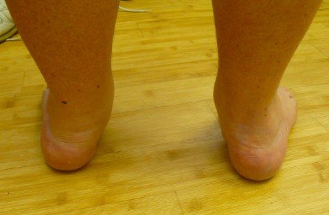
- Describe the clinical photo of an adult acquired flatfoot.
- What is the differential diagnosis of acquired flatfoot (bilateral vs unilateral)?
- What are the possible sources of pain in acquired flatfoot?
- How do you clinically assess a flatfoot?
- How is the foot arch maintained and what is the pathophysiology of PTTD?
- What is the aim of management in adult acquired flatfoot?
- What is the anatomy and action of the posterior tibialis tendon (PTT)?
- What is the Johnson classification of PTTD?
- How is stage 1 PTTD managed?
- How is stage 2 PTTD managed?
- Which tendon transfer is preferred for PTTD - FHL or FDL?
- How are stage 3 and stage 4 PTTD managed?
- What is the FAI 2020 consensus statement on adult acquired flatfoot?
- What is the role of arthroereisis in flatfoot?
- What are the positions of fusion in a triple arthrodesis?
Q1.Describe the clinical photo of an adult acquired flatfoot.
- Adult with forefoot abduction
- Decreased medial foot arch (midfoot supinated in relation to hindfoot)
- Hindfoot valgus
- Medial prominence
Q2.What is the differential diagnosis of acquired flatfoot (bilateral vs unilateral)?
- Bilateral/any: PTTD, tarsal coalition, congenital vertical talus
- Unilateral: post-traumatic (Lisfranc, spring ligament injury), degenerative, Charcot
- Bilateral systemic: rheumatoid arthritis, neuromuscular (spina bifida), cerebral palsy
- Generalised ligamentous laxity (Marfan, Ehlers-Danlos)
Q3.What are the possible sources of pain in acquired flatfoot?
- Medial: PTT, deltoid ligament, tarsal tunnel
- Lateral: subfibular impingement
- Heel: plantar aponeurosis, retrocalcaneal bursitis
- Diffuse: ankle, subtalar joint
- Proximal: knee, back; distal: bunion, metatarsal heads
Q4.How do you clinically assess a flatfoot?
- Determine whether the flatfoot and hindfoot valgus are flexible or rigid, and identify cause and complications (arthritis)
- Inspection: double heel raise and single leg heel raise (PT power, flexibility)
- Too many toes sign, navicular bulging signifying accessory navicular; look for scars (post trauma)
- Palpate sinus tarsi, deltoid ligament, PTT; test PT, FDL/ FHL power (transfer)
- Subtalar ROM and residual forefoot supination with correction; Silverskiold test
- Beighton score, check hands for RA, check spine if bilateral; assess gait
- X-ray: weight-bearing DP (Simmons angle, talocalcaneal angle, talus uncoverage) and lateral (Meary angle, calcaneal pitch, foot collapse at TN/NC/TTJ), No feature of previous fracture/ fragmentation, Saltzman view for hindfoot alignment, XR ankle mortise, Scannogram for LL alignment, MRI for PTT
- Bloods to rule out RA
Q5.How is the foot arch maintained and what is the pathophysiology of PTTD?
- Medial longitudinal arch static support: bony (calcaneum, talus, navicular, medial 3 rays) and soft tissue (spring ligament, plantar aponeurosis, deltoid ligament)
- Dynamic support: PTT, peroneus longus and intrinsics
- Lateral arch: calcaneum, cuboid, lateral 2 rays with long and short plantar ligaments, bifurcate ligament
- Transverse arch: keystone is 2nd MT and middle cuneiform, supported by the Lisfranc ligament
- Pathophysiology: PTT tenosynovitis causes loss of dynamic support, failure to lock the transverse tarsal joint --> further cause static stabilizer to fail
- Result: loss of one foot of the tripod, hindfoot valgus, MF/FF compensatory supination to maintain the tripod; PTT has a watershed blood supply 2-6cm proximal to insertion
Q6.What is the aim of management in adult acquired flatfoot?
- Stable, plantigrade, painless foot
- Rebalance the foot through bone and/or soft tissue to restore a balanced tripod
- Treat according to the patient's symptoms
Q7.What is the anatomy and action of the posterior tibialis tendon (PTT)?
- Origin: posterior fibula, tibia and interosseous membrane
- Inserts: navicular tuberosity, 1st – 3rd cuneiform, cuboid, 2-4 metatarsals, sustentaculum tali
- Invertor of the subtalar joint
- Plantarflexion of the ankle joint
- Adductor of the forefoot
- Watershed blood supply 2-6cm proximal to insertion
Q8.What is the Johnson classification of PTTD?
- 1: PTT power intact, no flatfoot
- 2: A flexible flatfoot, B abducted forefoot, C forefoot varus
- 3: Subtalar joint arthritis
- 4: Deltoid ligament insufficiency
Q9.How is stage 1 PTTD managed?
- Physio for TA stretching
- Orthosis: UCBL, moulded AFO, medial heel wedge, longitudinal arch support
- Tenosynovectomy
Q10.How is stage 2 PTTD managed?
- Combination of bony + soft tissue procedure, tailored to the individual’s deformities and muscle imbalance
- Soft tissue: FHL or FDL tendon transfer; TA lengthening also protects the transfer; +/- spring ligament reconstruction
- Coronal plane bone: calcaneal wedge osteotomy (85% pain relief/improved function at mean 15y, Chadwick BJJ 2015)
- Sagittal plane bone: 1st TMTJ fusion or Cotton medial cuneiform opening wedge osteotomy for forefoot varus - restores tripod
- Transverse plane bone: lateral column lengthening for forefoot abduction - protect FDL transfer
Q11.Which tendon transfer is preferred for PTTD - FHL or FDL?
- FHL is stronger
- FDL gives better functional outcomes because FHL is harder to mobilise
Q12.How are stage 3 and stage 4 PTTD managed?
- Stage 3: subtalar fusion +/- triple or double (TN + subtalar) fusion
- Double fusion (TN + subtalar) has similar functional scores and shorter OT time (Int Orthop 2021 Fadle)
- Stage 4: deltoid ligament reconstruction +/- ankle replacement or fusion
Q13.What is the FAI 2020 consensus statement on adult acquired flatfoot?
- Rename the condition Progressive Collapsing Foot Deformity (PCFD)
- New classification to stage the deformity
- A: hindfoot valgus deformity
- B: midfoot/forefoot abduction deformity
- C: forefoot varus/medial column instability
- D: peritalar subluxation/dislocation
- E: ankle instability
Q14.What is the role of arthroereisis in flatfoot?
- Subtalar arthroereisis induces a triplanar modification of the foot limiting pronation
- EFFORT review 2017 and AAOS 2021: no long-term studies, only poor quality evidence supporting its use
Q15.What are the positions of fusion in a triple arthrodesis?
- Subtalar joint 5 degrees valgus (TA is not a deforming force)
- TN joint: adequate coverage of the talar head; restore hindfoot-forefoot relationship so the 1st and 5th MT heads both touch the ground
- Restore the height of the medial arch
- CC joint: neutral (in situ)
▸ Slide 263 · High medial arch, hindfoot varus, midfoot cavus, forefoot adducted and pronated Foot and Ankle · 18 questions expand
Q1-Q1818 questions — tap to reveal all answerslist
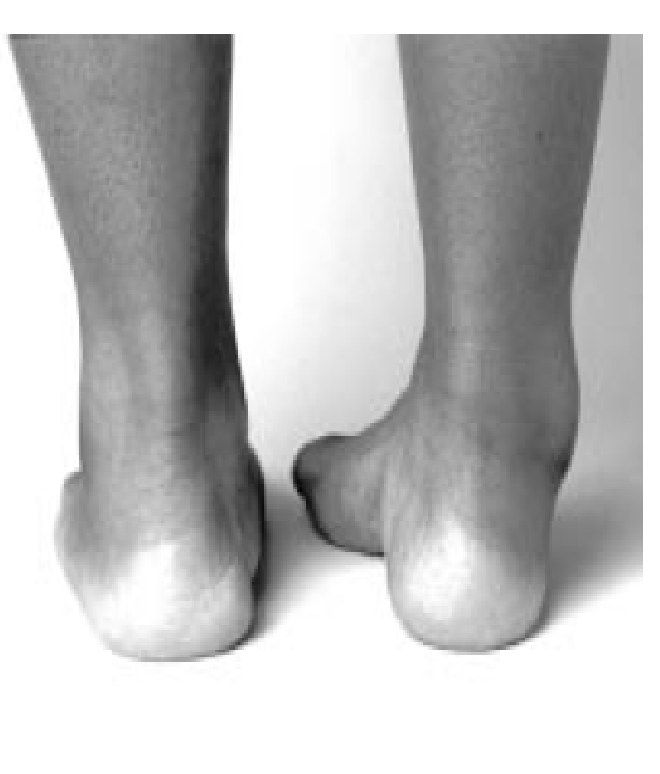
- Describe the clinical features of a cavovarus foot.
- What do you ask about in the history of a cavovarus foot?
- What are the examination findings in cavovarus foot?
- Explain the Coleman block test.
- How is cavovarus foot managed surgically?
- What additional examination findings are checked in cavovarus foot?
- What is a cavus foot?
- What are the causes of cavus foot?
- What are the X-ray findings in cavovarus foot?
- When is a neurologist referral made in cavovarus foot and what tests are requested?
- What is the aim and conservative management of cavovarus foot?
- How is surgical management of cavovarus foot tailored to flexibility?
- What soft tissue procedures are used in cavovarus foot?
- What bony procedures are used in cavovarus foot?
- How are claw toes in cavovarus foot managed?
- What is the Jones procedure for the big toe in cavovarus foot?
- How is equinus managed in cavovarus foot surgery?
- Explain forefoot-driven versus hindfoot-driven cavovarus.
Q1.Describe the clinical features of a cavovarus foot.
- High medial arch with midfoot cavus
- Hindfoot varus, forefoot adducted and pronated in relation to hindfoot
- Claw toes, peek-a-boo heel sign
Q2.What do you ask about in the history of a cavovarus foot?
- History of neurological disease, family history (CMT)
- Instability, metatarsalgia, fatigue, shoewear problems
- Callus, ulcers, pain (OA), stress fractures, Muller Weiss
- Functional limitation and expectations
Q3.What are the examination findings in cavovarus foot?
- LL muscle wasting - inverted champagne bottle appearance
- Forefoot clawing, adduction, pronation; midfoot cavus; hindfoot varus equinus
- Coleman block test for flexibility and forefoot-driven deformity
- Gait: broad based ataxic gait; check lateral ankle instability (ATFL, CFL)
- Full neuro exam (cerebellar), sensation, spine and hands
Q4.Explain the Coleman block test.
- Decides whether cavovarus is hindfoot or forefoot driven and whether flexible
- Block supports the 5th MT and heel, freeing the 1st MT
- Hindfoot corrects if the deformity is forefoot driven and flexible
- Rationale: hindfoot varus is compensatory to keep the lateral rays on the ground
Q5.How is cavovarus foot managed surgically?
- Combination of bony procedure with soft tissue procedure, tailored to the individual’s deformities and muscle imbalance
- Flexible: bony + soft tissue procedures for forefoot and midfoot +/- TA lengthening
- Rigid: add midtarsal and hindfoot bony procedures
- Arthritis/progressive disease: fusion
- Soft tissue: PL to PB transfer, Steindler plantar aponeurosis release
- Bone: Dwyer lateral closing wedge calcaneal osteotomy, 1st ray dorsiflexion osteotomy, triple arthrodesis
Q6.What additional examination findings are checked in cavovarus foot?
- Sole: callosities and ulcers
- Scars and correctability
- Muscle power: PT, PB, tib ant, PL
- Lateral ankle instability (ATFL and CFL)
- Silverskiold test
Q7.What is a cavus foot?
- Fixed plantar flexion deformity of the forefoot in relation to the hindfoot
- Results in an abnormally high arch
- 2/3 are neurological
Q8.What are the causes of cavus foot?
- Neurological: CP/stroke/Friedreich's ataxia
- Neurological: syringomyelia/diastematomyelia/spina bifida
- Neurological: SMA, polio, CMT, DMD
- Non-neurological: post-traumatic (Volkmann contracture, talar neck malunion)
- Non-neurological: residual club foot
Q9.What are the X-ray findings in cavovarus foot?
- DP: talocalcaneal angle and talo-1st MT angle
- Lateral: see-through sign, calcaneal pitch, Meary angle, calcaneal-1st MT angle (Hibb's angle N>150)
- Lateral: lateral kite angle <25 (parallelism), double talar head sign, bell-shaped cuboid
- Look for degeneration; obtain ankle mortise and Saltzman view
- MRI spine if neurological disease is suspected
Q10.When is a neurologist referral made in cavovarus foot and what tests are requested?
- Refer if neurological disease is suspected
- ECG NCS
- Sural nerve biopsy, muscle biopsy, CK
Q11.What is the aim and conservative management of cavovarus foot?
- Systemically delineate and treat the underlying cause
- Locally provide a plantigrade, stable, pain-free, shoeable, preferably mobile foot
- Conservative: physio for stretching and accommodative shoewear to relieve pressure
Q12.How is surgical management of cavovarus foot tailored to flexibility?
- Flexible: bony + soft tissue procedure for forefoot and midfoot; hindfoot should correct with these +/- TA lengthening
- Rigid: add midtarsal + hindfoot bony procedure
- Arthritis/progressive disease: fusion
- +/- lateral ankle ligamentous reconstruction if ankle instability
Q13.What soft tissue procedures are used in cavovarus foot?
- Hindfoot varus: PL to PB transfer
- Hindfoot varus: PT to cuneiform transfer
- Hindfoot varus: tib ant to cuneiform transfer
- Midfoot cavus: Steindler plantar aponeurosis release
Q14.What bony procedures are used in cavovarus foot?
- Hindfoot: Dwyer lateral closing wedge calcaneal osteotomy
- Hindfoot: lateral calcaneal slide osteotomy
- Midfoot cavus: 1st ray dorsiflexion osteotomy
- Midfoot cavus: TMTJ (Jahss) or tarsal (Japas) osteotomy
Q15.How are claw toes in cavovarus foot managed?
- PIPJ flexible: Girdlestone-Taylor FDL to EDL transfer
- PIPJ rigid, MTPJ flexible: DuVries (P1 head + P2 base resection/fusion) + EDL tenotomy
- PIPJ rigid, MTPJ rigid not subluxed: DuVries + EDB tenotomy + MTPJ dorsal capsulotomy + EDL lengthening
- PIPJ rigid, MTPJ rigid and subluxed: Weil osteotomy
Q16.What is the Jones procedure for the big toe in cavovarus foot?
- EHL to MT neck transfer
- Fuse the IPJ
Q17.How is equinus managed in cavovarus foot surgery?
- Usually corrects after the other deformities are corrected
- TA lengthening is left until other corrections are done
Q18.Explain forefoot-driven versus hindfoot-driven cavovarus.
- FF driven: PL >> ATT causes 1st MT plantarflexion; PT >> PB causes hindfoot varus
- 1st MT plantarflexion + forefoot pronation -> medial forefoot strikes ground first
- Subtalar joint inverts to bring the lateral forefoot down, so hindfoot varus is compensatory
- Recruited EHL causes claw toe; MTPJ extension tightens the plantar aponeurosis via the windlass, worsening cavus and varus
- Hindfoot cavus (calcaneocavus): weak triceps surae with relatively strong PTT/FHL plantarflexing the entire forefoot
▸ Slide 264 · Charcot Marie Tooth DiseaseFoot and Ankle · 7 questions expand
Q1-Q77 questions — tap to reveal all answerslist
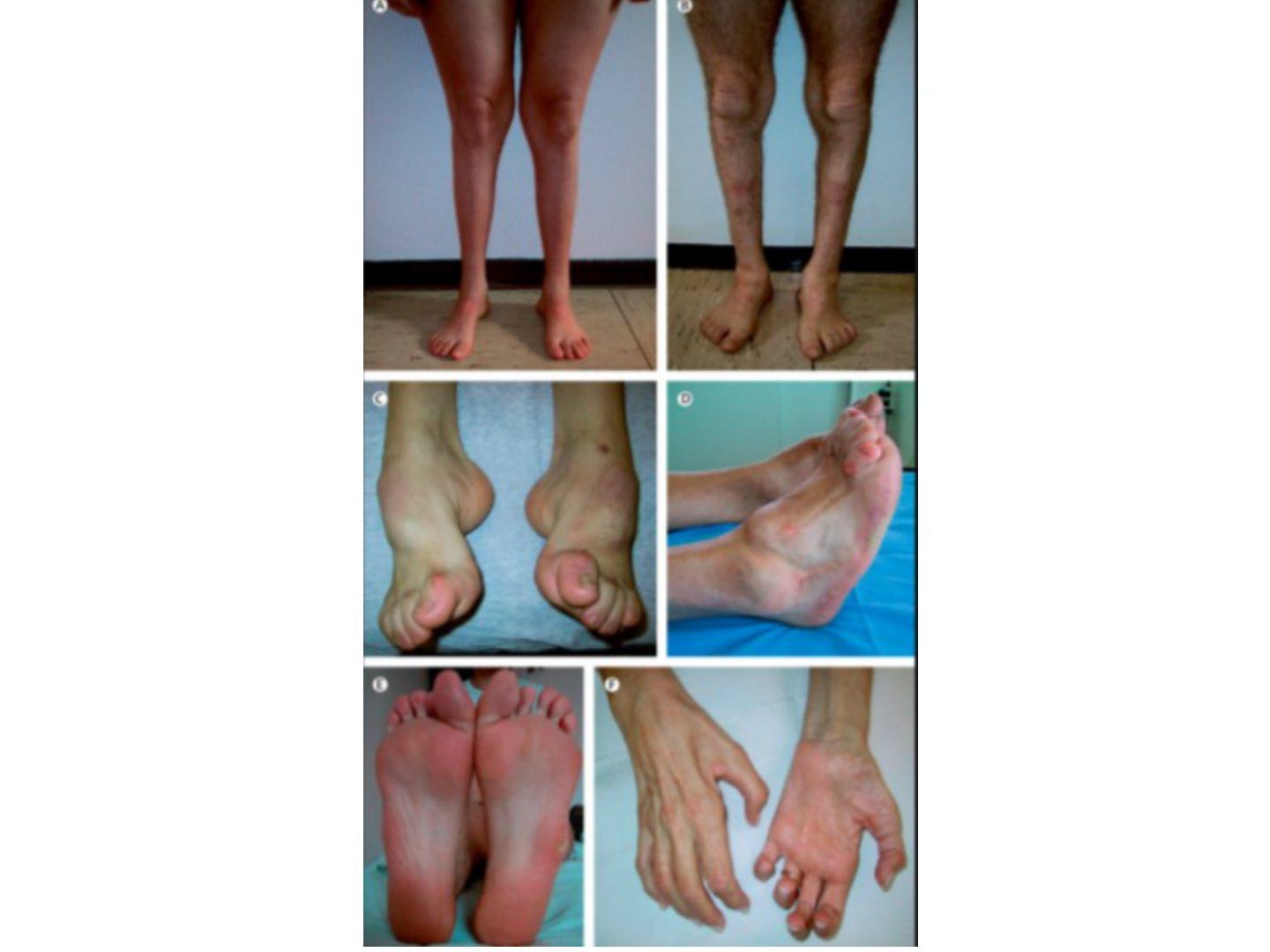
- What is Charcot-Marie-Tooth disease?
- What is the genetic basis of CMT?
- What are the three main types of CMT?
- What are the clinical features and diagnosis of CMT?
- How is CMT managed non-operatively?
- How is CMT managed surgically?
- What are the differential diagnoses of CMT?
Q1.What is Charcot-Marie-Tooth disease?
- Hereditary motor sensory neuropathy
- Peroneal muscle atrophy
- Prevalence 1:2500 - most common inherited neuropathy
- Usually autosomal dominant (can be AR or X-linked)
- Affects motor more than sensory
Q2.What is the genetic basis of CMT?
- Peripheral myelin protein 22 - defective Schwann cell membrane protein
- Causes defective myelin sheath formation
- Duplication of chromosome 17
Q3.What are the three main types of CMT?
- Type 1: demyelinating, presents 10-20s, AD, cavus foot
- Type 2: axonopathy, 20s+, flaccid foot, less disabling
- Type 3: X-linked recessive, demyelinating + axonal degeneration
Q4.What are the clinical features and diagnosis of CMT?
- Equinocavovarus +/- rigid hindfoot
- Weakened TA and peroneal muscles - foot drop during swing phase
- Check the hands
- Diagnosis: NCV/EMG, DNA/chromosome analysis
- No role for nerve biopsy
Q5.How is CMT managed non-operatively?
- Always non op first
- Stretching
- Orthosis: need a solid AFO
- No posterior leaf AFO due to inversion and eversion weakness
Q6.How is CMT managed surgically?
- Follow cavovarus principles
- Traditionally fusion as the disease is progressive
- Reconstruction has lower rates of degeneration and reoperation compared with triple arthrodesis (Ward JBJS 2008)
Q7.What are the differential diagnoses of CMT?
- Spina bifida
- SMA
- Polio
- Friedreich ataxia
▸ Slide 265 · Friedreich's AtaxiaFoot and Ankle · 5 questions expand

Q1-Q55 questions — tap to reveal all answerslist
- What is the genetic basis of Friedreich's ataxia?
- How is Friedreich's ataxia diagnosed?
- How does Friedreich's ataxia present clinically?
- What are the orthopaedic and systemic associations of Friedreich's ataxia?
- What is the prognosis of Friedreich's ataxia?
Q1.What is the genetic basis of Friedreich's ataxia?
- Autosomal recessive
- Frataxin mutation affecting mitochondria production
Q2.How is Friedreich's ataxia diagnosed?
- Clinical + family history + genetic testing
- It is the commonest spinocerebellar degenerative disease
Q3.How does Friedreich's ataxia present clinically?
- Staggering, wide-based gait
- Triad: ataxia, areflexia, upgoing plantar
Q4.What are the orthopaedic and systemic associations of Friedreich's ataxia?
- Cavovarus foot
- Scoliosis
- Cardiomyopathy - assess before planning surgery
Q5.What is the prognosis of Friedreich's ataxia?
- Wheelchair-bound around 30 years
- Death around 50 years
▸ Slide 266 · HF calcaneus (not varus)Foot and Ankle · 12 questions expand

Q1-Q1212 questions — tap to reveal all answerslist
- Describe the deformity in this spina bifida patient.
- How does a calcaneus foot present and how is it managed?
- What is spina bifida?
- What are the risk factors for spina bifida?
- What prenatal screening is done for spina bifida?
- What is the postnatal treatment of spina bifida and what must you beware of?
- Classify spina bifida.
- What are the features of a pre-L3 spina bifida lesion?
- What are the features of an L4 (L5 defect) spina bifida lesion?
- What are the features of an L5 (S1 defect) spina bifida lesion?
- What are the features of S1 or below spina bifida lesions?
- Why must VP shunt function be checked before spine surgery in spina bifida?
Q1.Describe the deformity in this spina bifida patient.
- Hindfoot calcaneus (not varus)
- Midfoot cavus with 1st ray plantarflexion
- Claw toes
- Scar at heel from previous TA lengthening
- Calf muscle wasting
Q2.How does a calcaneus foot present and how is it managed?
- History of instability and pain, previous TA lengthening
- Neuromuscular causes: spina bifida and polio
- Gait: slapping (lack of gastrocnemius eccentric contraction); heel ulcer
- XR: pistol grip deformity
- Mx: non-operative; tibialis anterior to TA transfer; dome calcaneal osteotomy with posterior tuberosity proximalisation
Q3.What is spina bifida?
- Neural tube defect
- Failure of neural tube closure at 3 weeks
Q4.What are the risk factors for spina bifida?
- Folate deficiency
- Maternal diabetes
- Valproic acid
Q5.What prenatal screening is done for spina bifida?
- Maternal AFP level (high = bad)
- Amniotic fluid AFP
- Structural scan
Q6.What is the postnatal treatment of spina bifida and what must you beware of?
- Neurosurgery: untethering of cord, drainage of syrinx, screen for Arnold-Chiari syndrome
- Beware malignant hyperthermia
- Beware latex allergy
- Beware neurogenic bladder
Q7.Classify spina bifida.
- Occulta: vertebral arch failure
- Meningocele: meninges protrusion, no neural element
- Myelomeningocele: meninges and neural elements protrusion
- Rachischisis: neural elements protrusion with no coverings
Q8.What are the features of a pre-L3 spina bifida lesion?
- Hip FABER contracture
- Knee flexion contracture (FFC)
- Feet equinovarus + claw toe
- Non-ambulatory
Q9.What are the features of an L4 (L5 defect) spina bifida lesion?
- Hip: full quadriceps, Trendelenburg gait
- Knee: recurvatum (unopposed knee extension)
- Feet: calcaneus
- Limited community ambulator
Q10.What are the features of an L5 (S1 defect) spina bifida lesion?
- High normal
- Knee normal
- Feet: calcaneus
- Community ambulator
Q11.What are the features of S1 or below spina bifida lesions?
- Only claw toe
- Near normal
Q12.Why must VP shunt function be checked before spine surgery in spina bifida?
- Check VP shunt function before spine surgery
- Otherwise it may result in death
▸ Slide 267 · Unilateral knee recurvatum + genu valgumFoot and Ankle · 17 questions expand

Q1-Q1717 questions — tap to reveal all answerslist
- Describe the limb deformity in this polio patient.
- What do you assess in a polio patient with deformity?
- What is the current disability history in this polio patient?
- How does quadriceps weakness cause knee flexion contracture and how is it managed?
- How is knee recurvatum managed in polio?
- How does exaggeration of the passive stance phase cause knee recurvatum in polio?
- What are the aims of management in polio deformity?
- How does gluteal weakness present in polio and how is it managed?
- What is the FABER contracture in polio and what problems does it cause?
- How is FABER contracture managed in polio?
- What is poliomyelitis?
- What is the pattern of poliomyelitis infection?
- What are the phases of poliomyelitis?
- What is post-polio syndrome?
- What are the diagnostic criteria for post-polio syndrome?
- What is the pathophysiology of post-polio syndrome?
- What are the 5 potential problems after the residual paralysis stage?
Q1.Describe the limb deformity in this polio patient.
- Right lower limb: hip extension, knee extension +/- recurvatum
- Ankle plantarflexed, tibial external rotation, genu valgum
- Contralateral hip and knee flexion compensating for LLD
- Pelvic obliquity; no claw toes
Q2.What do you assess in a polio patient with deformity?
- Check shoewear
- Gait: Trendelenburg, short limb, recurvatum, hand-knee gait
- Sitting: inspect for neuropathic ulcers and callosity
- Contractures: Silverskiold (Achilles), knee FFC (recurvatum), hip FABER (Ober)
- Power of antigravity muscles: ankle plantarflexion, knee extension, hip extension
Q3.What is the current disability history in this polio patient?
- Pain from ulcers, callosity, stress fracture
- Instability
Q4.How does quadriceps weakness cause knee flexion contracture and how is it managed?
- Knee collapse and unopposed hamstrings perpetuate flexed knee posture
- Ankle PF + hip flexion bring CG forward to lock knee; FFC becomes contracture over time
- Hand-knee gait; first line hinged KAFO
- Flexible: biceps femoris to quadriceps transfer (needs normal hip extension/ankle PF, no recurvatum)
- Rigid: <15 degrees serial casting, 15-30 degrees hamstring/ITB/posterior capsule release, 30-70 degrees distal femoral extension osteotomy, >70 degrees staged release then osteotomy
Q5.How is knee recurvatum managed in polio?
- <15 degrees: observe
- Solid AFO
- Triple tenodesis (gracilis, hamstrings, ITB)
- Posterior capsular advancement; gracilis/semitendinosus or biceps femoris/tibialis tenodesis
Q6.How does exaggeration of the passive stance phase cause knee recurvatum in polio?
- By exaggeration of passive stance phase: knee recurvatum assists in locking of the knee (ITB becomes extensor, CG falls in front of knee)
- Posterior knee capsules and hamstrings act as restraints, counterbalanced by hip extension and ankle PF
- Over time the knee loses its posterior constraint causing instability; pain occurs over the anterior knee from excessive hyperextension (perpetuated by fixed equinus)
Q7.What are the aims of management in polio deformity?
- Maximise gait efficiency
- Prevent deformity with growth
- Prevent upper limb contractures and scoliosis
Q8.How does gluteal weakness present in polio and how is it managed?
- Gluteus medius/minimus weakness -> Trendelenburg gait
- Gluteus maximus weakness -> back lurch
- Abductors: external oblique transfer
- Glut max (iliopsoas to GT transfer)
Q9.What is the FABER contracture in polio and what problems does it cause?
- Frog posture: hamstring, hip flexors, TFL spasm -> ITB contracture
- Secondary pelvic obliquity
- Leads to scoliosis and increase contralateral hip dislocation
Q10.How is FABER contracture managed in polio?
- Mild: fasciotomy
- Moderate: Ober release
- Ober release: ASIS - sartorius + TFL; AIIS - rectus femoris; GT - anterior fibres of glut med/min; LT - iliopsoas
- Severe: Campbell procedure
- Campbell: outer table glut med/min + TFL; inner table iliacus; AIIS rectus femoris; ASIS distalisation of sartorius
Q11.What is poliomyelitis?
- Non progressive motor paralysis secondary to polio virus infection at anterior horn cells
- Enterovirus, faecal-oral route
Q12.What is the pattern of poliomyelitis infection?
- 80% asymptomatic
- 10% flu-like
- 10% neurological symptoms, of which 2% paralysis
Q13.What are the phases of poliomyelitis?
- Acute: flu-like symptoms, flaccid paralysis
- Recovery: no more muscle ache, some recovery of power
- Residual: >2 years, deformity
Q14.What is post-polio syndrome?
- New onset weakness after a prolonged period of recovery
- Occurs in 50% of patients
Q15.What are the diagnostic criteria for post-polio syndrome?
- History of paralytic poliomyelitis by clinical/NCT
- Complete recovery >15 years with neurological and functional stability
- New onset weakness
- New onset muscle/joint pain, deformity, cold intolerance
- Exclude other causes
Q16.What is the pathophysiology of post-polio syndrome?
- Immunological/viral reactivation
- Aging/degeneration of neurons
Q17.What are the 5 potential problems after the residual paralysis stage?
- Isolated muscle weakness - splintage, passive mobilisation
- Deformity
- Shortening (LLD)
- Flail joint - may need arthrodesis or splintage
- Vascular dysfunction
▸ Slide 268 · Describe Xray:Foot and Ankle · 12 questions expand
Q1-Q1212 questions — tap to reveal all answerslist
- Describe the X-ray findings in a Charcot foot.
- What is a Charcot joint and what is its pathophysiology?
- What examinations differentiate infection from Charcot in the foot?
- What are the Eichenholtz and Brodsky classifications of Charcot?
- What are the principles, applications and contraindications of total contact casting?
- What are the surgical options and long-term aims in Charcot foot?
- What is the history in a suspected Charcot foot?
- What investigations are performed to differentiate Charcot from infection?
- What is the mid-term management of Charcot foot?
- When is surgery indicated acutely in Charcot foot and what are the difficulties?
- What is a superconstruct in Charcot foot surgery?
- What is the long-term management of Charcot foot?
Q1.Describe the X-ray findings in a Charcot foot.
- Destruction of the Chopart joint with subluxation and deformity
- Increased density with bone debris
- +/- dislocation, +/- soft tissue swelling
- Ddx: infection, post-trauma, Charcot (DM, syringomyelia, syphilis, leprosy)
- The 5 Ds: destruction, debris, dislocation, deformity, density
Q2.What is a Charcot joint and what is its pathophysiology?
- Neuropathic arthropathy: progressive, non-infectious destructive bone and joint disorder with sensory neuropathy
- Neurotraumatic: loss of protective sensation and repeated microtrauma
- Neurovascular: abnormal autonomic vessel tone, increased blood flow causing bone resorption
- Causes: DM, Hansen’s disease (Leprosy), syphilis, syringomyelia, spina bifida
Q3.What examinations differentiate infection from Charcot in the foot?
- Look for sinus, ulcer, deformities
- Heel raise test differentiates infection from Charcot (OM has less deformity)
- Neuro exam: 128Hz tuning fork, 10g monofilament, proprioception
- Palpate pulses; toe pressures, ABI, transcutaneous O2 for healing potential
Q4.What are the Eichenholtz and Brodsky classifications of Charcot?
- Eichenholtz: stage 0 (joint oedema), fragmentation, coalescence, consolidation
- Brodsky I: 60% TMTJ
- Brodsky II: Chopart/subtalar
- Brodsky III: 20% ankle/calcaneal tuberosity, IV mixed, V forefoot
Q5.What are the principles, applications and contraindications of total contact casting?
- Immobilise immediately; admit for infection, dislocation or debridement
- Principles: increase contact surface area, evenly distribute forces, control oedema
- Well padded, limit toe movement, change every 2-4 weeks, allow WBAT
- Duration: forefoot 3-6 months, midfoot 6-12 months, hindfoot 12-24 months; 70-80% wound healing
- Contraindications: passed coalescence, severe deformity, uncontrolled oedema, active infection, severe obesity
Q6.What are the surgical options and long-term aims in Charcot foot?
- Aim: take patient through Eichenholtz stages while minimising the deformity and subsequent skin breakdown and infection; stable plantigrade shoeable foot
- Patient education!; multidisciplinary input (endocrinologist, vascular surgeons, P&O, occu physio); Systemic: control DM (better outcomes in surgical and non-surgical treatment), optimise nutrition
- Some centres may give bisphosphonates, but evidence is not strong; ongoing RCTs investigating denosumab in treatment of Charcot
- Acute surgery indications: fracture, dislocation or impending skin breakdown - exostectomy + TAL
- Superconstruct: fusion beyond zone of injury, shorten to reduce, strongest tolerated device
- Long term: prevention, identify foot at risk, early podiatry referral
Q7.What is the history in a suspected Charcot foot?
- Ask about DM, trauma, other peripheral neuropathies
- 50% of patients have pain - dorsal columns are affected but the spinothalamic tract remains intact
- Assess functional limitations
Q8.What investigations are performed to differentiate Charcot from infection?
- Bloods
- WBC scan
- Biopsy
- Infection: C/ST +ve
- Charcot: bone and cartilage debris within synovium
Q9.What is the mid-term management of Charcot foot?
- Midterm management: CROW boot after contact casting: bivalved AFO with full foot enclosure, hard outer shelf to prevent deformity, rigid rocker bottom base, custom foam padding
- Double rocker shoe modifications reduce ulceration risk at the plantar apex of the deformity
Q10.When is surgery indicated acutely in Charcot foot and what are the difficulties?
- Fracture, dislocation or impending skin breakdown
- Usually exostectomy + TAL; deformity correction with rigid fixation and immobilisation is risky; final option is amputation
- Difficulties: bleeding, wound infection, failed fixation
Q11.What is a superconstruct in Charcot foot surgery?
- Fusion extends beyond the zone of injury
- Bone resection shortens the extremity to allow adequate reduction
- Use the strongest device tolerated by the soft tissue envelope
- Applied in a position that maximises mechanical function
Q12.What is the long-term management of Charcot foot?
- Aim at prevention
- Identify the foot at risk
- Early referral and frequent podiatry assessment
▸ Slide 269 · *Tendon achilles ruptureFoot and Ankle · 11 questions 1 check expand
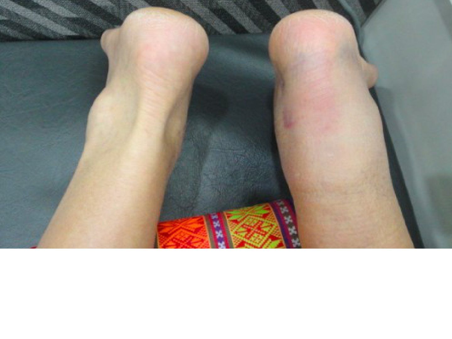
Q1-Q1111 questions — tap to reveal all answerslist
- Describe the clinical findings of an Achilles tendon rupture.
- What risk factors and mechanisms do you ask about in Achilles rupture?
- What investigations are performed for Achilles rupture?
- How is an acute Achilles rupture treated and what is the evidence?
- How is a delayed presentation with a gap managed?
- What are the complications and rehabilitation after Achilles repair?
- What is the non-operative treatment for an old, low-demand patient with Achilles rupture?
- What is the essence of accelerated rehabilitation after Achilles repair?
- What anatomy must be known for an FHL transfer?
- Describe the anatomy of the Achilles tendon.
- Describe the Cook and Purdam model of Achilles degeneration.
Q1.Describe the clinical findings of an Achilles tendon rupture.
- Ankle swelling with absence of the normal Achilles tendon contour
- Palpate the gap (size of gap, distance between insertion and gap)
- Thompson's test positive
Q2.What risk factors and mechanisms do you ask about in Achilles rupture?
- Mechanism: eccentric loading of gastrosoleus in middle-aged patients
- Hx: AS, steroid, fluroquinolones, flat foot, gout, hyperlipidemia, chronic renal disease
- Chronicity of injury and pre-existing pain
- Poor wound healing risks: PVD, smoking, diabetes
Q3.What investigations are performed for Achilles rupture?
- USG to delineate site and size, complete versus partial rupture
- X-ray: calcinosis suggests pre-existing tendinosis - may need intraop debridement and preparation for tendon gap
- Kager's triangle is distorted when the tendon is ruptured
Q4.How is an acute Achilles rupture treated and what is the evidence?
- Treatment is individualised: age, functional demand, tendon retraction, timing
- Repair aims for intrinsic healing + aggressive rehab
- JBJS 2001 meta-analysis: less rerupture with surgery (2% vs 20%)
- Accelerated functional rehab concept: Willits RCT (JBJS 2010) - no significant difference in rerupture rate or power
- Later confirmed with (Soroceanu JBJS 2012 metanalysis)
- NEJM 2022 Myhrvold Norway multicentre RCT MIS vs open vs nonop: Rupture rate: conservative 6.2% vs 0.6%; Infection MIS 5.2% vs open 2.8% vs 0.6% nonop; Overall surgery was not associated with better outcomes
- Should be shared decision making, weighing avoiding surgery against higher rerupture risk
Q5.How is a delayed presentation with a gap managed?
- Aim to reconstruct tension and length - shorten first, then retension
- Gap <3cm: direct repair
- Gap 3-5cm: turn-down flap, VY plasty +/- augmentation (Lindholm, plantaris)
- Gap >5cm: FHL transfer (dispensable, good vascularity, in phase with gastroc)
- FHL released at the Knot of Henry, retrieved to repair site +/- bone tunnel at calcaneal tuberosity
Q6.What are the complications and rehabilitation after Achilles repair?
- Complications: skin necrosis 2%, rerupture 2%, deep infection 1%, sural nerve injury (especially percutaneous)
- Accelerated rehab: 0-2wk CAM NWB 2cm raise; 2-6wk protected weight bear, 1cm raise, active PF/DF to neutral; 6-8wk WBAT, no heel raise, gradual DF ex, OKC/CKC ex; 8-12wk strengthening + proprioception; >12wk plyometrics
- Return to athletic activity at 4-6 months
Q7.What is the non-operative treatment for an old, low-demand patient with Achilles rupture?
- Conservative with an accelerated rehab program
- UKSTAR trial Lancet 2020: cast vs functional bracing - no difference in functional outcomes
Q8.What is the essence of accelerated rehabilitation after Achilles repair?
- Early weight bearing and early mobilisation
- allow collagen fibril crosslinking, also healing tendons undergo intrinsic healing, increasing load to failure
Q9.What anatomy must be known for an FHL transfer?
- FHL crosses superficial to FDL at the Knot of Henry
- The Knot of Henry lies below the bony prominence of the navicular
Q10.Describe the anatomy of the Achilles tendon.
- Collagen accounts for 70% of dry weight, of which 95% is type I, with a very small amount of elastin
- Fibres rotate 90 degrees toward insertion into the calcaneal tuberosity (gastrocnemius laterally, soleus medially)
- Watershed blood supply at 5-6cm from insertion
Q11.Describe the Cook and Purdam model of Achilles degeneration.
- Reactive: myofibroblast proliferation and collagen synthesis in response to repeated mechanical stress
- Reparative: inflammation of the paratenon as an attempt at soft tissue repair
- Degenerative: vascular constriction leading to ischaemia
NEJM 2022 trial infection rates: minimally invasive surgery 5.2%, open 2.8%, nonoperative 0.6% — misattributed — Those percentages are sensory nerve injury rates; deep infection was 1.1% nonoperative, 1.1% open and 1.7% minimally invasive surgery — source
▸ Slide 270 · Xr showing bony spur over the posterosuperior aspect of calcaneal tuberosity, alFoot and Ankle · 2 questions expand
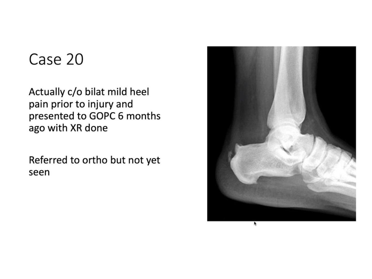
Q1-Q22 questions — tap to reveal all answerslist
- What is the diagnosis from this X-ray?
- What is Haglund syndrome?
Q1.What is the diagnosis from this X-ray?
- Bony spur over the posterosuperior aspect of the calcaneal tuberosity
- Calcification over the insertion of the Achilles tendon
- Diagnosis: Haglund deformity with insertional Achilles tendinopathy
Q2.What is Haglund syndrome?
- Triad of retrocalcaneal pain
- Insertional tendinopathy
- Haglund deformity
▸ Slide 271 · Clinical photo showing plantar aspect of foot with 2 ulcers over medial MT headFoot and Ankle · 14 questions 1 check expand
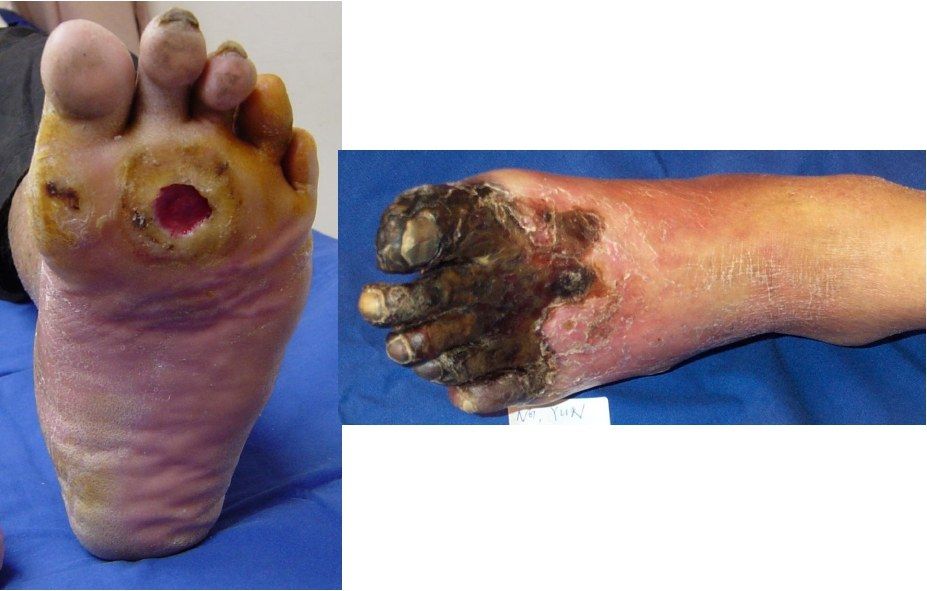
Q1-Q1414 questions — tap to reveal all answerslist
- Describe the ulcer shown on the plantar aspect of the foot.
- What is the Brodsky depth-ischemia classification of diabetic foot ulcers?
- What neurological and vascular examination would you perform?
- How do you differentiate a neuropathic from an arterial ulcer?
- How would you manage this patient with a neuropathic ulcer and absent pulses?
- When and at what level would you amputate?
- What blood tests and X-ray would you request for this foot ulcer?
- Which laboratory tests assess healing potential?
- What further vascular investigations would you perform?
- What is the pathophysiology of the diabetic foot ulcer?
- How would you risk-stratify a diabetic foot?
- What is the principle of total contact casting?
- What local wound care is used?
- What long-term surveillance does an at-risk foot need?
Q1.Describe the ulcer shown on the plantar aspect of the foot.
- Erythematous base with punched out edges
- No exposed tendon or bone, no pus
- Surrounded by callosities
- Associated hallux deformity with the 2nd and 3rd toes in clawed position
- Site and deformities suggest a DM neuropathic ulcer
Q2.What is the Brodsky depth-ischemia classification of diabetic foot ulcers?
- Depth: 0 at risk, 1 superficial ulcer, 2 deep ulcer (tendon or joint exposed), 3 extensive ulcer or abscess
- Ischemia: A not ischemic, B ischemia without gangrene, C partial forefoot gangrene, D complete gangrene
- 4+4 system combining depth and ischemia
Q3.What neurological and vascular examination would you perform?
- Proprioception
- Vibration with 128 Hz tuning fork
- Semmes Weinstein 5.07 for protective sensation
- Palpate pulses, ABI
- Check muscle power PL, PB, TA, PT; Silverskiold test; shoewear
Q4.How do you differentiate a neuropathic from an arterial ulcer?
- Neuropathic: plantar, not painful, foot deformity, pulse likely present, punched out lesion
- Arterial: tip of foot or edges, painful, atrophic changes, absent pulse, fibrotic base, no surrounding callosity
Q5.How would you manage this patient with a neuropathic ulcer and absent pulses?
- Multidisciplinary: medical, vascular, P&O, ortho; patient education
- Goal: ulcer free, functional, plantigrade foot fitting a brace or shoe, with minimal complications and hospital stay
- Systemically control/correct risk factors eg DM, PVD; improve nutrition
- 1. unload prominent area: total contact casting (increases contact area, transmits load proximally, typically 6 months)
- Prevent infection: daily dressing (Medihoney), debridement or amputation
Q6.When and at what level would you amputate?
- Indications: dangerous (infected), dead (gangrene), damn nuisance
- Level balances healing potential against energy expenditure
- Longer residual limb is more energy conserving; the exception is Symes
- Order: Transmetatarsal > Symes > Lisfranc > Chopart > transtibial
- Lisfranc and Chopart need TA lengthening
Q7.What blood tests and X-ray would you request for this foot ulcer?
- WCC (>1.5), CRP, ESR, albumin (lecturer quotes >30 g/dL; see factcheck for unit correction)
- XR to look for fragmentation signifying Charcot arthropathy or osteomyelitis
Q8.Which laboratory tests assess healing potential?
- Serum albumin >3.0 g/dL
- Total lymphocyte count >1,500/mm3
Q9.What further vascular investigations would you perform?
- ABI >0.45
- Ischemic index (Doppler pressure at intended level / brachial sBP) >0.5
- Absolute toe pressure: minimum for healing 40 mmHg, normal 100 mmHg
- Transcutaneous O2 pressure of toe >40 mmHg
- In this patient the pulse is not palpable; DM patients can have mixed cause of ulcer
Q10.What is the pathophysiology of the diabetic foot ulcer?
- Neurology (most important), vasculopathy, or mixed
- Sensory: loss of protective sensation; 90% who fail to sense 10 g with the 5.07 Semmes-Weinstein monofilament for 1 s; 128 Hz vibration at the base of the big toe nail
- Motor: muscle imbalance causing deformity (most common common peroneal nerve)
- Autonomic: drying of skin from glandular dysfunction
- Angiopathy present in 60%
- Net effect: mechanical stress on skin that is more prone to injury due to drying, and lacks protective sensation
Q11.How would you risk-stratify a diabetic foot?
- By sensation, pulse, deformity and ulcer
- Low if all normal; medium if one impaired; high if >1 impaired or previous ulcer; active ulcer
- High risk: screening every 3 months; immediate attention if Charcot
Q12.What is the principle of total contact casting?
- Increase contact surface area and transmit vertical load proximally
- Cast in plantigrade to prevent weight bearing and limit toe movement
- Typically cast for 6 months
- Also corrects deformity
Q13.What local wound care is used?
- Daily dressing; wound nurse uses Medihoney - hygroscopic to dehydrate the wound
- Contains glucose oxidase producing small amounts of H2O2 and lowers pH to speed healing
- Debridement or amputation
Q14.What long-term surveillance does an at-risk foot need?
- Screen the at-risk foot: NV status, deformity, footwear, ulcers
- Refer medical for home glucose monitoring control
- Refer P&O for unloading orthosis over bony prominences; refer podiatry
Serum albumin threshold for healing potential is Alb >30g/dL — wrong units; 30 g/dL is physiologically impossible — Threshold is serum albumin >3.0 g/dL (= 30 g/L); total lymphocyte count >1,500/mm3 (>1.5 x10^9/L)
▸ Slide 272 · Symes amputationFoot and Ankle · 3 questions expand
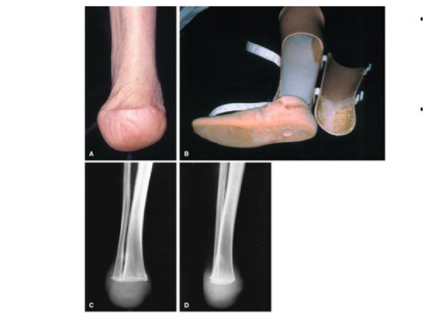
Q1-Q33 questions — tap to reveal all answerslist
- What is a Symes amputation?
- Describe the technique of a Symes amputation.
- What are the common complications of a Symes amputation?
Q1.What is a Symes amputation?
- Amputation at the level of the talotibial joint
- End-bearing amputation
- More energy saving than midfoot amputation
- Requires a patent posterior tibial artery to keep the heel pad viable
Q2.Describe the technique of a Symes amputation.
- Fish mouth incision starting 1 cm distal and anterior to both malleoli, one along anterior ankle joint and one at 90 deg along the CCJ
- Ligate posterior tibial artery as distal as possible
- Expose the ankle joint
- Resect bone 0.5 cm above the joint line
- Drill holes over anterior tibia and fibula and attach the heel pad
Q3.What are the common complications of a Symes amputation?
- Posterior migration of the heel pad
- Skin slough from overly vigorous trimming or circulation compromise over the heel pad
▸ Slide 273 · Stress view Xray showing significant talar tilt suggesting lateral ankle ligamenFoot and Ankle · 14 questions 1 check expand

Q1-Q1414 questions — tap to reveal all answerslist
- What types of ankle sprain do you know of?
- Which lateral ankle ligaments are injured and by what mechanism?
- How do you distinguish low from high ankle sprain on examination?
- When would you X-ray a patient with an ankle sprain?
- What is the Chorley classification of low ankle sprain?
- What is the treatment for a low ankle sprain?
- What does the stress view X-ray show?
- What history and examination features determine functional vs mechanical instability?
- What concomitant pathologies must be ruled out in an ankle sprain?
- What X-ray views and MRI are used in ankle sprain?
- How is an acute high ankle sprain (syndesmosis injury) treated?
- How is chronic syndesmosis instability treated?
- What are the surgical options for recurrent ankle sprain with mechanical instability?
- What is the evidence for lateral ligament augmentation?
Q1.What types of ankle sprain do you know of?
- Low (90%) or high
- Eversion or inversion
Q2.Which lateral ankle ligaments are injured and by what mechanism?
- ATFL is commonest (plantarflexion + inversion)
- CFL is 2nd most common (dorsiflexion + inversion)
- PTFL is the strongest
Q3.How do you distinguish low from high ankle sprain on examination?
- Low: anterior drawer (dorsiflexion tests CFL, plantarflexion ATFL), talar tilt indicates CFL torn
- High: syndesmosis tenderness, squeeze test, cotton test, Frick test (external rotation), cross leg test
Q4.When would you X-ray a patient with an ankle sprain?
- Follow the Ottawa rules
- Pain within 6 cm of the malleolar tip or unable to bear weight: X-ray ankle
- Pain over the 5th MT base or navicular or unable to bear weight: X-ray foot
Q5.What is the Chorley classification of low ankle sprain?
- I: partial ATFL
- II: complete ATFL +/- partial CFL (anterior drawer I)
- III: complete ATFL + CFL +/- capsule +/- PTFL (anterior drawer II, talar tilt)
Q6.What is the treatment for a low ankle sprain?
- Conservative: RICE, physio, functional training, peroneal and proprioception training
- 20% develop symptoms of chronic ankle instability
- Surgery if bony avulsion, both medial and lateral ligament rupture, or talar tilt in athlete > 15deg
- Recurrent instability: anatomic repair (Brostrom +/- Gould modification), anatomic reconstruction, or non-anatomic (Chrisman Snook, Watson Jones, Evans)
Q7.What does the stress view X-ray show?
- Significant talar tilt suggesting lateral ankle ligamentous disruption
Q8.What history and examination features determine functional vs mechanical instability?
- History: age, body build, sport, occupation
- History of hypermobility, mechanism of injury, recurrence
- Examination aims to determine functional vs mechanical instability and high vs low sprain
Q9.What concomitant pathologies must be ruled out in an ankle sprain?
- Tendon: peroneal split tear or subluxing tendons
- Bone: fractures of the anterior calcaneal process, lateral or posterior talar process, 5th MT base
- Cartilage/joint: OCD, loose bodies
- Predisposing factor: cavovarus
Q10.What X-ray views and MRI are used in ankle sprain?
- Ankle AP, lateral, mortise weight bearing, Saltzman view (syndesmosis and OCD)
- Foot DP, oblique, weight-bearing lateral
- Stress view with ER/ varus: talar tilt 5 deg+ (or absolute 10) abnormal; anterior drawer same
- Scannogram; MRI for OCD, cartilage status, degree of tear and tissue quality
Q11.How is an acute high ankle sprain (syndesmosis injury) treated?
- No diastasis: immobilize 1-3 weeks, then functional rehab + strengthening
- Diastasis: obtain MRI
- Isolated AITFL: immobilization + NWB 4-6 weeks
- AITFL + others: 2 syndesmosis screws, then NWB 6-12 weeks
Q12.How is chronic syndesmosis instability treated?
- Debridement + 2 syndesmosis screws or suture button
- Reconstruction by peroneus longus: half PL harvested from the LM tip
- Free end secured by a 3.5 mm screw + washer, protected with a syndesmosis screw
Q13.What are the surgical options for recurrent ankle sprain with mechanical instability?
- Anatomic repair with endogenous tissue when adequate (Brostrom +/- Gould modification: anatomical shortening and reinsertion of ATFL + CFL with inferior extensor retinaculum and fibular periosteum); fewer complications, quicker rehab
- Anatomic reconstruction: semiT or plantaris autograft or synthetic graft replicating ATFL/CFL anatomy
- Non-anatomic checkrein: Chrisman Snook, Watson Jones, Evans
- Consider diagnostic arthroscopy: intraarticular pathology amenable to arthroscopy was found in 83% of Brostrom repairs
Q14.What is the evidence for lateral ligament augmentation?
- AJSM 2019 RCT: primary repair + LARS gave better FAOS at 5-year follow-up and high Tegner scores vs MBG
- Foot and Ankle Surg 2021 meta-analysis: no difference
Ankle X-ray is indicated if pain is within 6 cm to the tip of the malleolus — imprecise phrasing of the Ottawa ankle rules — Rule = pain in the malleolar zone AND bone tenderness along the distal 6 cm of the posterior edge/tip of either malleolus, or inability to bear weight 4 steps — source
▸ Slide 274 · Shepred fracture: posterolateral talar tubercle #Foot and Ankle · 2 questions expand
Q1-Q22 questions — tap to reveal all answerslist
- What is a Shepherd fracture?
- What is a Cedell fracture and what structure lies between the two tubercles?
Q1.What is a Shepherd fracture?
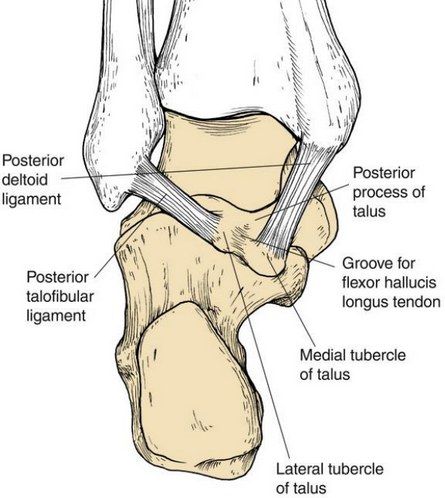
- Fracture of the posterolateral talar tubercle
- Contrast with a Cedell fracture = posteromedial talar tubercle
Q2.What is a Cedell fracture and what structure lies between the two tubercles?
- Posteromedial talar tubercle fracture
- The FHL tendon runs between them
▸ Slide 275 · Describe Xray:Foot and Ankle · 19 questions expand

Q1-Q1919 questions — tap to reveal all answerslist
- What are the causes of ankle arthritis?
- What is the Takakura classification of ankle osteoarthritis?
- What are the prerequisites for a joint-preserving supramalleolar osteotomy?
- What are the advantages and disadvantages of ankle fusion?
- What is the optimal position for ankle fusion?
- What are the contraindications to total ankle replacement?
- Describe the X-ray findings on this slide.
- What history, examination and imaging workup is needed?
- What patient and disease factors guide management?
- What is the overall management principle and what conservative options exist?
- What joint-preserving options exist for ankle arthritis?
- What are the contraindications to ankle fusion?
- What techniques and fixation are used for ankle fusion?
- Which features do you comment on in a total ankle replacement X-ray?
- What are the specific complications and commonly used implants in TAR?
- What is the survival of the STAR prosthesis and what trial is ongoing?
- What are the properties of ankle cartilage and why is ankle arthritis uncommon?
- Describe the three generations of total ankle replacement.
- What is used if both the ankle and subtalar joints have OA?
Q1.What are the causes of ankle arthritis?
- Post-traumatic (70%): altered load following injury; 2/3 fracture, 1/3 sports-related ligamentous injury (Iowa study)
- Primary OA
- Inflammatory arthritis (RA, gout)
- Neuropathic
- Post-infection
Q2.What is the Takakura classification of ankle osteoarthritis?
- I: early sclerosis
- II: medial joint space narrowing, no subchondral bone contact
- III: subchondral bone contact
- IIIA: medial malleolus joint space obliteration; IIIB: talar dome roof obliteration with bone contact
- IV: all joint space with complete bone contact
Q3.What are the prerequisites for a joint-preserving supramalleolar osteotomy?
- Medially focused ankle arthritis (stage 2/3a)
- Nearly normal ROM
- Minimal talar tilting
- Realignment targets: TAS(tibial anterior surface angle)=LDTA (lateral distal tibial angle)=89, talar tilt <10, TLS(tibial lateral surface angle)=ADTA(anterior distal tibial angle)=80
Q4.What are the advantages and disadvantages of ankle fusion?
- Advantages: more stable and durable than replacement, corrects deformity, less infection
- Disadvantages: longer rehab (3-6 months), adjacent joint degeneration, motion loss, stress fracture, require shoe modification if active
- Complications: adjacent joint arthritis 50% in 10 year, non union 20%
Q5.What is the optimal position for ankle fusion?
- Neutral dorsiflexion
- 5 deg external rotation (similar to the other side)
- 5 deg hindfoot valgus
- Talus translated 5mm posteriorly (anterior edge of talus same level as anterior edge of tibia - normalize gait and decrease stress at knee)
Q6.What are the contraindications to total ankle replacement?
- Young and active patients
- Poor bone stock or talar AVN
- Infection
- Poor ligamentous stability or absent malleoli
- Significant malalignment
Q7.Describe the X-ray findings on this slide.
- Osteoarthritic change at the tibiotalar joint: decreased joint space, subchondral sclerosis
- Evidence of healed tibia and fibula fracture; syndesmosis appears intact
- Diagnosis: post-traumatic ankle arthritis
Q8.What history, examination and imaging workup is needed?
- Hx: risk factors, pain, functional limitations
- PE: skin status, tenderness, ROM, deformity, adjacent joints, overall alignment
- XR: LL scannogram, foot and ankle for subtalar/other joint degeneration, Saltzman view
- MRI if early stage
Q9.What patient and disease factors guide management?
- Patient: age, functional status, rehab potential, expectations
- Disease: cause (post-traumatic, inflammatory - may be bilateral/osteopenic)
- Adjacent joint OA/bilateral disease, extra-articular malalignment, bone stock
- Joint congruency and correctability, OA stage
Q10.What is the overall management principle and what conservative options exist?
- Principle: resume stability, correct alignment and preserve cartilage if possible
- Conservative: physio, NSAID, rocker bottom sole, AFO to simulate fusion, off-loading brace
Q11.What joint-preserving options exist for ankle arthritis?
- Realignment: supramalleolar osteotomy +/- soft tissue
- Joint resurfacing: MACI, stem cells
- Ankle scope: synovectomy, cheilectomy
- Distraction diastasis: Exfix for 3/12 (3 months), stimulates PG production
Q12.What are the contraindications to ankle fusion?
- Bilateral disease
- Adjacent joint degeneration
Q13.What techniques and fixation are used for ankle fusion?
- Arthroscopic arthrodesis in minimal deformity
- Anterior +/- transfibular approach (fibula can be used as onlay graft)
- Fixation: plate/screw, Blair's fusion, or retrograde hindfoot nail if subtalar degeneration
Q14.Which features do you comment on in a total ankle replacement X-ray?
- Cement, bearing, syndesmosis fixation, alignment, sizing
- Bone cut (just below the syndesmosis)
Q15.What are the specific complications and commonly used implants in TAR?
- Specific complications: malleolar impingement, medial malleolus fracture
- Common implants: STAR (no tibiofibular fusion) and Agility
Q16.What is the survival of the STAR prosthesis and what trial is ongoing?
- STAR 70.7% survival 10 yrs; 80% at 5 years; 50% at 15 years
- TARVA trial ongoing
Q17.What are the properties of ankle cartilage and why is ankle arthritis uncommon?
- Thinnest cartilage, smaller contact area, stiffer
- 1 mm shift of a fracture decreases contact surface area by 42%; medial instability by 15%
- Uncommon because the joint is highly congruent/stable with rolling (not rolling + shear)
- Cartilage is well nourished, with resistance to inflammatory mediators like MMP, IL1 --> stiffness does not decrease with age
Q18.Describe the three generations of total ankle replacement.
- 1st: two components, highly constrained, cemented tibia and talus, high incidence of loosening
- 2nd: two components, polyethylene bearing, more conservative bone cuts, cementless
- 3rd: three components, independent PE mobile bearing, cementless, More rely on ligamentous stability
Q19.What is used if both the ankle and subtalar joints have OA?
- Hindfoot arthrodesis nail
▸ Slide 276 · Clinical photo showing severe bilateral hallux valgus deformity with big toe devFoot and Ankle · 12 questions expand
Q1-Q1212 questions — tap to reveal all answerslist
- Describe the deformity shown in the clinical photo.
- What would you examine in this patient?
- What X-ray parameters do you assess in hallux valgus?
- What determines the choice of surgery in hallux valgus?
- Compare chevron and scarf osteotomies for hallux valgus.
- Explain the pathophysiology of hallux valgus.
- What history would you take in hallux valgus?
- What conservative management is offered for hallux valgus?
- Describe the modified McBride procedure.
- Describe the scarf osteotomy technique and aftercare.
- What are the risk factors for hallux valgus?
- What are the deforming forces in hallux valgus?
Q1.Describe the deformity shown in the clinical photo.
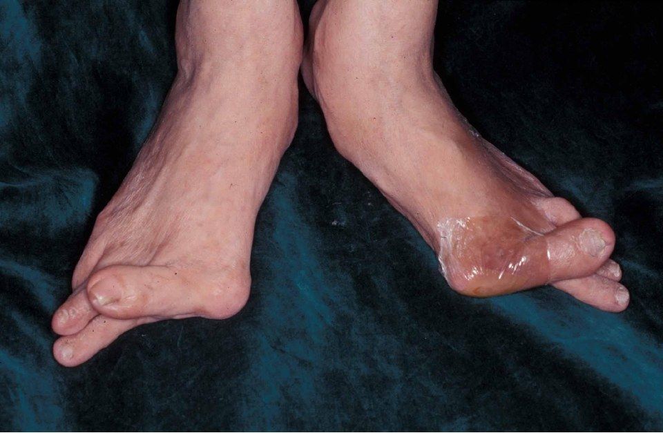
- Severe bilateral hallux valgus: big toe deviated laterally, 1st MT medially
- Hallux also in a pronated position
- Lesser toe deformity is present
- Bunion with no callosity, no scars
Q2.What would you examine in this patient?
- Standing foot arch and hindfoot alignment; sole for callosity
- Single and double heel raise
- Palpate bunion, 2nd MT plantar and 5th MT head; check correctability; Check TMTJ hypermobility
- NV exam, Silverskiold test, gait (loss of third ankle rocker)
- Look for RA features and generalized ligamentous laxity
Q3.What X-ray parameters do you assess in hallux valgus?
- DP foot: HVA, IMA, HVI, DMAA, 1st MTPJ congruence, sesamoid station, MTPJ/TMTJ arthritis, Maestro's cascade
- Lateral: calcaneal pitch and Meary angle
Q4.What determines the choice of surgery in hallux valgus?
- 1. severity, 2. congruence, 3. TMTJ laxity, 4. 1st MTPJ arthritis
- This case needs a combined soft tissue and bony procedure: modified McBride (DSTP) + 1st MT realignment osteotomy (scarf osteotomy)
Q5.Compare chevron and scarf osteotomies for hallux valgus.
- Chevron: no more than 1/3 shift, 1 mm ~ 1 degree of IMA correction, not for severe deformity
- Scarf: versatile, corrects deformity in all three planes, stable allowing early weight bearing
- Scarf complications: troughing, nonunion, hallux varus
Q6.Explain the pathophysiology of hallux valgus.
- Defined as lateral deviation of the great toe with medial deviation of the 1st MT
- Overall: destabilization of 1st MTPJ
- Medial capsular attenuation; the 1st PP deviates laterally while the 1st MT drifts medially off the sesamoids
- Plantar-lateral migration of abductor hallucis plantarflexes and pronates the phalanx
- EHL and FHL subluxate laterally and become deforming forces; secondary contracture of the lateral capsule, adductor hallucis and intermetatarsal ligament
- The 1st ray defunctions, the 2nd MT head takes body weight and the plantar plate eventually become attenuated
- Sagittal imbalance causes claw toe from EDL overpull
Q7.What history would you take in hallux valgus?
- RA, family history
- Pain, walking tolerance
- Expectations
Q8.What conservative management is offered for hallux valgus?
- Wide toe box, advise against high heels
- NSAIDs
- Toe spreader or spacer
Q9.Describe the modified McBride procedure.
- Lateral: release adductor hallucis from the sesamoid
- Lateral: release the intermetatarsal ligament (watch the common digital nerve), capsulotomy, release the suspensory sesamoid-metatarsal ligament
- Medial: capsulorrhaphy
- Medial: bunionectomy (cut 2 mm medial to the sulcus)
Q10.Describe the scarf osteotomy technique and aftercare.
- Medial incision; protect the dorsal medial cutaneous branch of the superficial peroneal nerve and the medial cutaneous branch of the hallux (medial plantar nerve)
- Z-shaped osteotomy: longitudinal cut parallel to the sole, transverse cut perpendicular to the 2nd MT; distal limb points dorsal, proximal limb plantar
- Translate the distal fragment laterally, fix with 2 x 2 mm headless compression screws
- Rehab: heel walking for 6 weeks
- Complications: troughing, nonunion, hallux varus
Q11.What are the risk factors for hallux valgus?
- Multifactorial: female, genetic predisposition
- Extrinsic: shoewear
- Intrinsic systemic: RA, ligamentous laxity
- Intrinsic local: flat foot, amputated 2nd toe
Q12.What are the deforming forces in hallux valgus?
- Dynamic: adductor hallucis - pronation and adduction of the proximal phalanx
- Dynamic: abductor hallucis - plantarflexion and pronation of the proximal phalanx
- Dynamic: EHL and FHL subluxed laterally in relation to the MTPJ
- Static restraint loss: plantar plate (sesamoid pronation), lateral capsule contracture, intermetatarsal ligament contracture, crist erosion
▸ Slide 277 · Xray bilateral foot showing severe hallux valgus deformityFoot and Ankle · 2 questions expand

Q1-Q22 questions — tap to reveal all answerslist
- Describe the X-ray findings and their significance.
- What are the indications for 1st MTPJ fusion?
Q1.Describe the X-ray findings and their significance.
- Severe bilateral hallux valgus deformity
- OA changes at the 1st MTPJ
- Rule out an underlying systemic cause
- Operative management if symptomatic
Q2.What are the indications for 1st MTPJ fusion?
- OA
- RA
- Spasticity
- Fusion corrects the IMA with realignment of the hallux
▸ Slide 278 · Xray showing patient with bilateral hallux valgusFoot and Ankle · 5 questions expand
Q1-Q55 questions — tap to reveal all answerslist
- What are the radiographic findings?
- What is the operative plan?
- What are the complications of hallux valgus surgery?
- How do MTPJ, TMTJ, severity, DMAA and HVI guide the operation?
- What conservative management would you try first?
Q1.What are the radiographic findings?
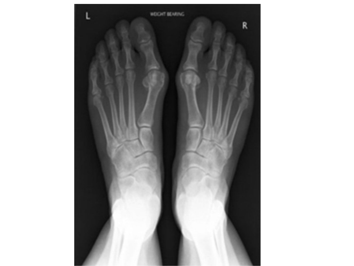
- Bilateral hallux valgus
- Eyeballing HVA >40 deg, IMA >13 deg
- DMAA <10 deg
- No 1st MTPJ arthritic changes
- Clinically need to check 1st TMTJ laxity
Q2.What is the operative plan?
- Proximal metatarsal osteotomy
- Distal soft tissue release
- Medial eminence resection and capsular repair
Q3.What are the complications of hallux valgus surgery?
- Overcorrection causing hallux varus
- Undercorrection (OT factor, patient factor)
- Malunion or nonunion
- AVN (medial artery to metatarsal neck), recurrence
- Troughing leading to elevation or rotation of the 1st MT
Q4.How do MTPJ, TMTJ, severity, DMAA and HVI guide the operation?
- 1st MTPJ pathological: fuse
- 1st TMTJ lax: fuse
- Severe HVA/IMA: proximal osteotomy; mild: distal
- Abnormal DMAA: proximal + distal medial closing wedge; both severe: biplanar closing wedge
- HVI >10: Akin
Q5.What conservative management would you try first?
- Shoewear advice
- Orthosis
- Operate if symptomatic despite conservative measures
▸ Slide 279 · Physeal bracketFoot and Ankle · 5 questions expand
Q1-Q55 questions — tap to reveal all answerslist
- What is the pathophysiology of a physeal bracket?
- What conditions are associated with a physeal bracket?
- What is the radiographic staging?
- How is a physeal bracket managed?
- What are the complications of nonoperative and operative treatment?
Q1.What is the pathophysiology of a physeal bracket?
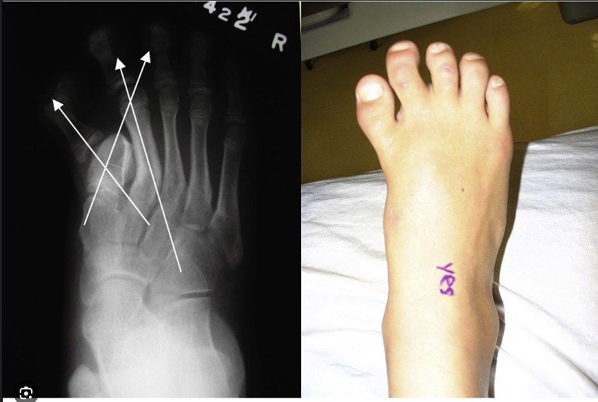
- Incomplete development of primary ossification centres
- Aberrant physeal cartilage extends along the diaphysis and interferes with normal growth
- Leads to a short, wide, triangular or trapezoidal phalanx
- Longitudinally oriented cartilage bracket follows a C-shaped curve
- Autosomal dominant when present in the hand, otherwise sporadic
Q2.What conditions are associated with a physeal bracket?
- Rubinstein-Taybi syndrome
- Fibrodysplasia ossificans progressiva
- Hallux varus in the feet
- Clinodactyly and polydactyly in the hands
- Apert's syndrome
Q3.What is the radiographic staging?
- Stage 1: delta-shaped diaphysis with radiolucent cartilaginous epiphysis
- Stage 2: secondary ossification centres at proximal and distal ends
- Stage 3: complete ossification of the bracket epiphysis along one side of the diaphysis
- Stage 4: closure of the physis
- MRI may be needed
Q4.How is a physeal bracket managed?
- Splinting to loosen soft tissues prior to surgery
- Osteotomies (opening or closing wedge) in older children after closure of the bracket epiphysis
- Physiolysis + interposition (PMMA or fat): earlier intervention allows more correction of angulation and more longitudinal growth
Q5.What are the complications of nonoperative and operative treatment?
- Nonoperative: structural deformation of the forefoot
- Nonoperative: incomplete longitudinal growth of the affected bone
- Nonoperative: clinodactyly at the interphalangeal joint
- Operative: recurrence of deformity
- Operative: incomplete correction of deformity
▸ Slide 280 · Juvenile HVFoot and Ankle · 2 questions expand

Q1-Q22 questions — tap to reveal all answerslist
- How does juvenile hallux valgus differ from adult hallux valgus?
- What is the management of juvenile hallux valgus?
Q1.How does juvenile hallux valgus differ from adult hallux valgus?
- Usually large IMA, large DMAA +/- HVI, congruent MCPJ
- Metatarsus primus varus
- Ligamentous laxity with hypermobile 1st ray
- Physis not yet closed
- High recurrence
Q2.What is the management of juvenile hallux valgus?
- Address ligamentous laxity: TMT arthrodesis after physis closure for a hypermobile 1st ray
- If physis open: osteotomy at medial cuneiform or wait until closure
- Double osteotomy: medial cuneiform opening wedge + 1st MT osteotomy +/- Akin
- Associated with flexible flatfoot and tight TA
▸ Slide 281 · Same as HV as aboveFoot and Ankle · 6 questions expand

Q1-Q66 questions — tap to reveal all answerslist
- What is the management principle for RA foot deformity?
- Why does RA foot surgery favour fusion over reconstruction?
- What are the fusion positions and procedures used in the RA foot?
- What is the pathophysiology of RA foot deformities?
- If the IMA is not severe but the HVA is severe, what does that suggest?
- How does the X-ray localize a flatfoot deformity?
Q1.What is the management principle for RA foot deformity?
- Relieve symptoms, preserve function, minimize complications
- Goal: stable, pain-free plantigrade foot
- Multidisciplinary: rheumatology, medical, anaesthetics, physio, OT
- Conservative: physio, shoe wear (large toe box, rockerbottom), metatarsal pads or bars, NSAIDs
Q2.Why does RA foot surgery favour fusion over reconstruction?
- Primary disease is progressive, so reconstruction has a high chance of recurrence
- need to bear in mind the risk of complications as a result of osteopenia, reduced vascularity, and immunosuppression
- Look for other RA complications e.g. C1/2 subluxation, other lower limb arthritis
Q3.What are the fusion positions and procedures used in the RA foot?
- 1st MTPJ arthrodesis: 20 deg dorsiflexion, 10 deg valgus, neutral rotation
- Lesser toes: principle is to decompress the joint to allow relocation, while maintaining the MT length cascade (Maestro's cascade)
- MTPJ: Fowler (proximal P1 excision) or Stainsby (+ flexor-extensor interposition); IPJ: DuVries
- Midfoot: TN +/- CC fusion; hindfoot: triple fusion
- Ankle: synovectomy; fusion vs arthroplasty (JBJS 2007 systematic review, 70% satisfaction rate in both groups)
Q4.What is the pathophysiology of RA foot deformities?
- Stage I: synovitis
- Stage II: joint erosion and tendon dysfunction
- Stage III: progressive deformity
- Forefoot: hallux valgus, claw toe, splay foot
- Midfoot: rockerbottom + forefoot abduction; hindfoot: PTT insufficiency and disruption of the talocalcaneal interosseous ligament causing hindfoot valgus
Q5.If the IMA is not severe but the HVA is severe, what does that suggest?
- MCL rupture (usually acute, sesamoid not subluxed)
- Metatarsus adductus
Q6.How does the X-ray localize a flatfoot deformity?
- X-ray localization - if at TMTJ, then it is a midfoot driven flatfoot. If at TNJ, is it s hindfoot driven flatfoot
▸ Slide 282 · Hammer toesFoot and Ankle · 5 questions expand
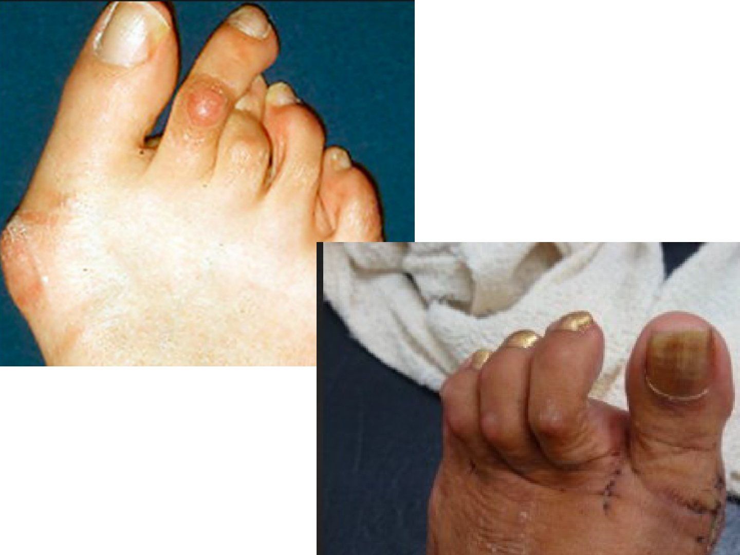
Q1-Q55 questions — tap to reveal all answerslist
- What is the deformity in hammer toes and what causes it?
- How are hammer toes treated?
- What is the deformity in claw toes and what causes it?
- How do you treat claw toes?
- What is the function of the lesser toes?
Q1.What is the deformity in hammer toes and what causes it?
- PIPJ flexed, DIP extended, MTP neutral or extended
- Due to simultaneous contraction of extrinsic flexors and extensors without the modifying action of intrinsics
- Weak intrinsics or plantar plate rupture causes MTPJ extension and PIPJ flexion
Q2.How are hammer toes treated?
- Flexible: tendon transfer (FDL to EDL, EDL lengthening)
- Fixed: tendon + bone procedure (PIPJ excisional arthroplasty, PIPJ arthrodesis)
Q3.What is the deformity in claw toes and what causes it?
- Intrinsic minus deformity: MTPJ hyperextended, PIPJ and DIPJ flexed
- Causes: neurological, synovitis, trauma, compartment syndrome
Q4.How do you treat claw toes?
- Conservative: plantar padding at MTPJ, orthosis to offload MT heads
- PIPJ flexible: Girdlestone-Taylor flexor to extensor hood transfer +/- EBD tenotomy
- PIPJ fixed, MTPJ flexible: distal PP resection + PIPJ fusion (DuVries) + EDL tenotomy
- PIPJ fixed, MTPJ fixed (not subluxed): DuVries + EDL lengthening, EBD tenotomy, dorsal capsulotomy + collateral release
- PIPJ fixed, MTPJ fixed and subluxed: Weil osteotomy
Q5.What is the function of the lesser toes?
- Dissipate forefoot pressure
- Maintain the arch through the windlass mechanism
▸ Slide 283 · Describe Xray:Foot and Ankle · 8 questions expand

Q1-Q88 questions — tap to reveal all answerslist
- Describe the X-ray finding in this patient.
- What is a Haglund deformity and how is it treated?
- How is insertional Achilles tendinitis treated?
- What is the pathophysiology of paratenonitis and Achilles tendinosis?
- How is Achilles tendinosis treated?
- How is the parallel pitch line constructed?
- What conditions make up the Achilles tendinopathy group?
- What is the etiology and presentation of insertional Achilles tendinitis?
Q1.Describe the X-ray finding in this patient.
- Spur at the calcaneal tuberosity protruding from the parallel pitch line
- Consistent with a Haglund deformity
Q2.What is a Haglund deformity and how is it treated?
- Bony spur at the posterosuperior tubercle of the calcaneum, measured by the parallel pitch line
- Tenderness 2-3 cm proximal to the Achilles insertion
- Compresses the bursa on ankle dorsiflexion causing retrocalcaneal bursitis
- Conservative: NSAID, padding; NOT steroid injection (may cause rupture)
- Operative: excision of bursa and Haglund lesion
Q3.How is insertional Achilles tendinitis treated?
- Treat the underlying cause, e.g. removal of a Haglund lesion
- Conservative: physiotherapy with TA stretching, small heel raise
- Operative depends on the size of the diseased tendon
- <50%: debridement +/- reattach tendon to bone
- >50%: reattach tendon to bone +/- FDL transfer if gap >5 cm
Q4.What is the pathophysiology of paratenonitis and Achilles tendinosis?
- Overuse with repetitive stress on the Achilles
- Cook and Purdam continuum model (reactive, reparative, degenerative)
- Myofibroblasts proliferate and synthesize more collagen III at the paratenon causing scarring and shrinkage
- Causes ischemic damage to the tendon at the watershed region
Q5.How is Achilles tendinosis treated?
- Physiotherapy: eccentric training + stretching (Alfredson protocol)
- Padding + small heel raise
- Operative: 50% debride; >50% FHL transfer
Q6.How is the parallel pitch line constructed?
- 1st line: on the calcaneal tuberosity to the anterior calcaneal tubercle
- 2nd line: parallel to this and touching the posterior lip of the posterior facet
- A spur protruding from the line indicates a Haglund deformity
Q7.What conditions make up the Achilles tendinopathy group?
- Haglund deformity + retrocalcaneal bursitis = Haglund syndrome
- Insertional Achilles tendinitis
- Achilles tendinosis
Q8.What is the etiology and presentation of insertional Achilles tendinitis?
- Etiology: retrocalcaneal bursitis, RA/AS with enthesopathy
- Repeated trauma leads to inflammation then metaplasia of tendon to cartilage/ bone
- Tenderness at the insertion, may have bony swelling
▸ Slide 284 · Mortons neuroma = interdigital neuromaFoot and Ankle · 6 questions expand

Q1-Q66 questions — tap to reveal all answerslist
- What is a Morton's neuroma?
- How does Morton's neuroma present and how is it examined?
- What is the differential diagnosis and how do you rule it out?
- What are the treatment options for Morton's neuroma?
- Describe the dorsal neurectomy and its commonest complication.
- What causes Morton's neuroma?
Q1.What is a Morton's neuroma?
- A neuroma caused by compressive neuropathy
- Most common over the 2nd inter-digital nerve between the 3rd and 4th metatarsal heads
- Confluence of branches of the lateral and medial plantar nerves
- Compression by the intermetatarsal ligament
- More common in women
Q2.How does Morton's neuroma present and how is it examined?
- 60% pain, 40% numbness; worse with tight shoes and high heels
- Palpation: plantar tenderness
- Squeeze test: compressing the forefoot while palpating the web space reproduces symptoms
- Mulder click: squeeze elicits an occasional bursal click
- MTPJ drawer test for instability; nerve block test is diagnostic
Q3.What is the differential diagnosis and how do you rule it out?
- XR and MRI to rule out stress fracture, MTPJ arthritis, metatarsalgia, MTP synovitis and Freiberg disease
- Ddx for a mass in the sole: bursitis, fibroma over tendon sheath, vascular lesion
Q4.What are the treatment options for Morton's neuroma?
- Conservative: shoewear, MT pads proximal to the focus of pain (decompress), short-term steroid
- Surgical: neurectomy + bury end
- Dorsal approach: less plantar scar
- Plantar approach: less recurrent neuroma, no need to incise the ligament
Q5.Describe the dorsal neurectomy and its commonest complication.
- Incise the transverse intermetatarsal ligament
- Identify the common digital nerve and resect it 2-3 cm proximal to the intermetatarsal ligament, proximal to the small plantar branches
- Commonest complication: stump neuroma due to inadequate retraction or inadequate resection
Q6.What causes Morton's neuroma?
- Compressive neuropathy; compression by the intermetatarsal ligament
- Physiology poorly understood: tension/compression/microtrauma
- More common in women; associated with tight shoe wear and high heels
▸ Slide 285 · Describe Xray (left): bony spur at anterior distal tibia and dorsal aspect of taFoot and Ankle · 6 questions expand

Q1-Q66 questions — tap to reveal all answerslist
- Describe the X-ray and the diagnosis.
- What causes anterior and posterior ankle impingement?
- What is an os trigonum and how is it managed?
- What are the indications for ankle arthroscopy?
- What are the ankle arthroscopy portals and the structures at risk?
- How is ankle arthroscopy set up?
Q1.Describe the X-ray and the diagnosis.
- Bony spur at the anterior distal tibia and dorsal aspect of the talar neck
- Anterior ankle pain on dorsiflexion with limited dorsiflexion: anterior ankle impingement
Q2.What causes anterior and posterior ankle impingement?
- Anterior: overuse and repeated trauma with osteophyte formation at the anterior ankle
- Common in athletes playing on grass or turf, footballers and dancers
- Posterior: overuse and repeated plantarflexion, common in ballet dancers
- Posterior impingement may cause FHL tendinopathy; X-ray may show an os trigonum
Q3.What is an os trigonum and how is it managed?
- Accessory ossicle of the separated posterolateral talar tubercle
- Usually asymptomatic; symptomatic in ballet dancers from repeated extreme plantarflexion
- May associate with FHL tendinitis
- Ddx: Shepherd's fracture (fracture of the posterior process of the talus)
- Conservative treatment; excision if it fails
Q4.What are the indications for ankle arthroscopy?
- OCD
- Impingement
- Removal of loose body
- Debridement of Haglund lesion
Q5.What are the ankle arthroscopy portals and the structures at risk?
- Anteromedial: primary portal, between tibialis anterior and saphenous vein
- Anterolateral: primary portal, lateral to peroneus tertius (superficial peroneal nerve at risk)
- Posterolateral: 2 cm proximal to lateral malleolus tip, medial to peroneal tendons, lateral to Achilles (sural nerve)
- Posteromedial: just medial to Achilles (posterior tibial artery and tibial nerve)
- Anterocentral: between EDC and EHL (DP artery and deep peroneal nerve)
Q6.How is ankle arthroscopy set up?
- GA, supine, tourniquet
- Traction strap of the foot and heel
▸ Slide 286 · Tarsal tunnel syndromeFoot and Ankle · 4 questions expand
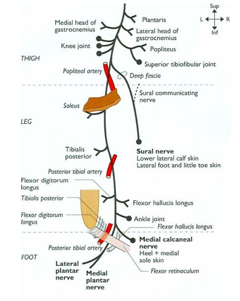
Q1-Q44 questions — tap to reveal all answerslist
- What is tarsal tunnel syndrome and what are the tunnel boundaries?
- What is the usual presentation and what clinical signs do you look for?
- What is the heel pain triad?
- How is tarsal tunnel syndrome investigated and treated?
Q1.What is tarsal tunnel syndrome and what are the tunnel boundaries?
- Compressive neuropathy of the tibial nerve posterior and inferior to the medial malleolus
- Superficial: flexor retinaculum
- Deep: talus, calcaneus, sustentaculum tali
- Inferior: abductor hallucis
Q2.What is the usual presentation and what clinical signs do you look for?
- Vague medial foot pain exacerbated by prolonged walking or standing, burning sensation
- Tinel sign
- Diminished two-point discrimination
- Wasting of abductor hallucis (medial plantar nerve) or abductor digiti quinti (lateral plantar nerve)
- Look for pes planus which further stretches the nerve
Q3.What is the heel pain triad?
- PTTD
- Tarsal tunnel syndrome
- Plantar fasciitis
Q4.How is tarsal tunnel syndrome investigated and treated?
- Dx: NCV better than EMG; MRI for space-occupying lesion
- Conservative: NSAID, vitamin B6, tricyclic antidepressant, physio, massage, desensitization for 3-6m
- Surgical: nerve release +/- excision of SOL
- Release investing fascia, flexor retinaculum, and superficial and deep fascia of abductor hallucis
▸ Slide 287 · Anterior tarsal tunnel syndromeFoot and Ankle · 4 questions expand

Q1-Q44 questions — tap to reveal all answerslist
- What is anterior tarsal tunnel syndrome?
- What are the symptoms of anterior tarsal tunnel syndrome?
- Describe the course and branches of the deep peroneal nerve at the ankle.
- How is anterior tarsal tunnel syndrome treated?
Q1.What is anterior tarsal tunnel syndrome?
- Compressive neuropathy of the deep peroneal nerve
- Tunnel formed by the Y-shaped inferior extensor retinaculum
- Superficial boundary: inferior extensor retinaculum
- Deep boundary: fascia overlying the talus and navicular
- Contents: deep peroneal nerve and branches, EHL, EDL, dorsalis pedis artery
Q2.What are the symptoms of anterior tarsal tunnel syndrome?
- Burning pain and paresthesias or vague dorsal foot pain
- Worse at night as the ankle assumes a plantarflexed posture
Q3.Describe the course and branches of the deep peroneal nerve at the ankle.
- Passes medial to EHL proximal to the ankle, then laterally between EHL and EDC
- Divides into lateral motor and medial sensory branches within the tunnel
- Accompanied by the dorsalis pedis artery
Q4.How is anterior tarsal tunnel syndrome treated?
- Conservative: NSAID, night splints, footwear
- Surgical release: incise the inferior extensor retinaculum
- Protect the DP artery and release both branches of the nerve
- Remove SOL and repair the capsule to protect the nerve from bleeding bone
▸ Slide 288 · Hallux rigidusFoot and Ankle · 7 questions expand
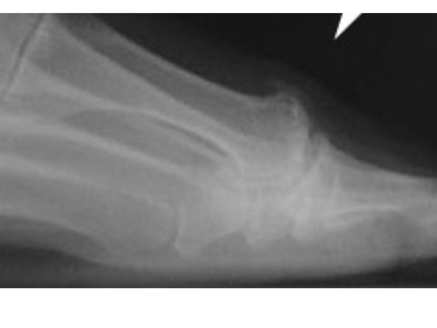
Q1-Q77 questions — tap to reveal all answerslist
- Describe the X-ray and clinical findings.
- What is the Coughlin staging of hallux rigidus?
- What are the conservative and joint-preserving options?
- What are the joint-sacrificing options and their problems?
- Why is some valgus needed in fusion, and what is the evidence for fusion vs replacement?
- What is the pathophysiology of hallux rigidus?
- What are the history and examination findings in hallux rigidus?
Q1.Describe the X-ray and clinical findings.
- Osteophytes at the dorsal side of the 1st MTPJ
- Clinically limited dorsiflexion: hallux rigidus
Q2.What is the Coughlin staging of hallux rigidus?
- 1: mild pain with ROM
- 2: moderate pain with ROM
- 3: stiffness + pain with end ROM
- 4: stiffness + pain through ROM
- Grades 1-2: osteophyte; grades 3-4: joint space narrowing
Q3.What are the conservative and joint-preserving options?
- Conservative: NSAID, Morton's extension stiff foot plate limiting toe dorsiflexion, high box/rocker bottom shoes
- Arthroscopic debridement/synovectomy for grades I-II
- Cheilectomy (also remove 25% of dorsal MT head) for grades I-II
- Dorsal closing wedge osteotomy for grades II-III
Q4.What are the joint-sacrificing options and their problems?
- Fusion
- Interpositional arthroplasty
- Keller excision arthroplasty - problem is cock up deformity
- Arthroplasty
Q5.Why is some valgus needed in fusion, and what is the evidence for fusion vs replacement?
- Valgus lessens the chance of IPJ arthritis
- JBJS Rev 2017 Stevens systematic review: arthrodesis is superior for clinical outcome and pain with fewer complications and revisions
- 20.9% loosening rate in the replacement group
Q6.What is the pathophysiology of hallux rigidus?
- MT primus elevatus
- Degenerative change at the 1st MTPJ resulting in limited dorsiflexion
Q7.What are the history and examination findings in hallux rigidus?
- Hx: pain on push off or forced dorsiflexion of the great toe
- Shoe irritation due to dorsal osteophytes compressing the medial dorsal cutaneous nerve (MDCN); metatarsalgia; bunion
- PE: swelling, dorsal bunion, antalgic supinated gait, grind test
- Grading depends on pain upon ROM, osteophyte size and joint space
▸ Slide 289 · Freiberg's diseaseFoot and Ankle · 4 questions expand
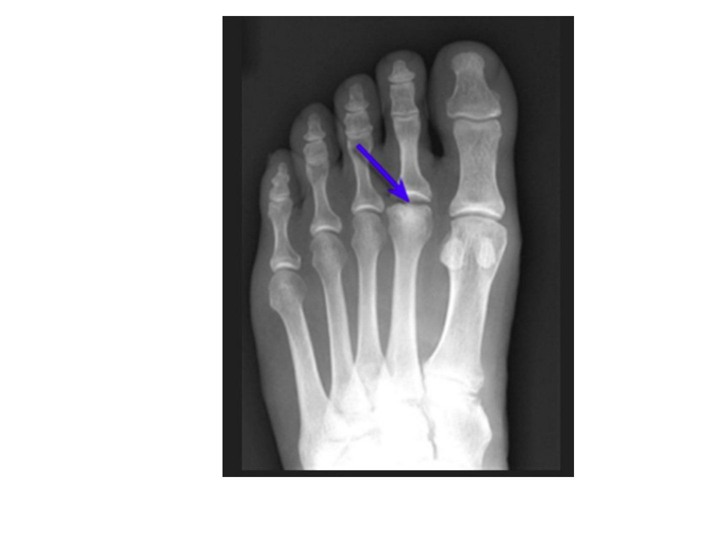
Q1-Q44 questions — tap to reveal all answerslist
- What is Freiberg's disease?
- How does Freiberg's disease present?
- What is Smillie's classification?
- What are the operative options for Freiberg's disease?
Q1.What is Freiberg's disease?
- Infarction and fracture of a metatarsal head
- Common in female adolescents and athletes
- Most commonly the second MT
- Thought due to microtrauma, stress overload, osteonecrosis
Q2.How does Freiberg's disease present?
- Foot dorsum pain and swelling over the 2nd MTPJ on weight bearing
- Diagnosis is by X-ray and graded with Smillie's classification
Q3.What is Smillie's classification?
- 1: normal X-ray, abnormal MRI (subchondral fracture)
- 2: collapse on the dorsal side of the articular surface
- 3: further collapse with bony projections on the medial and lateral head
- 4: collapse involving the whole head distorting anatomy
- 5: arthritic; grades 4-5 impossible to reconstruct
Q4.What are the operative options for Freiberg's disease?
- Goal: pain relief + pressure relief
- Non-operative: activities modification, rest, NSAID, short leg cast, MT pad
- Early/salvageable: arthrotomy for loose body, dorsal closing wedge osteotomy, Shortening osteotomy to unload joint, Bone graft/ drilling
- Late/non-salvageable: DuVries arthroplasty (partial MT head resection + capsular interposition), excisional arthroplasty
▸ Slide 290 · Iselin disease is a benign and self-limiting condition, defined as apophysitis oFoot and Ankle · 2 questions expand
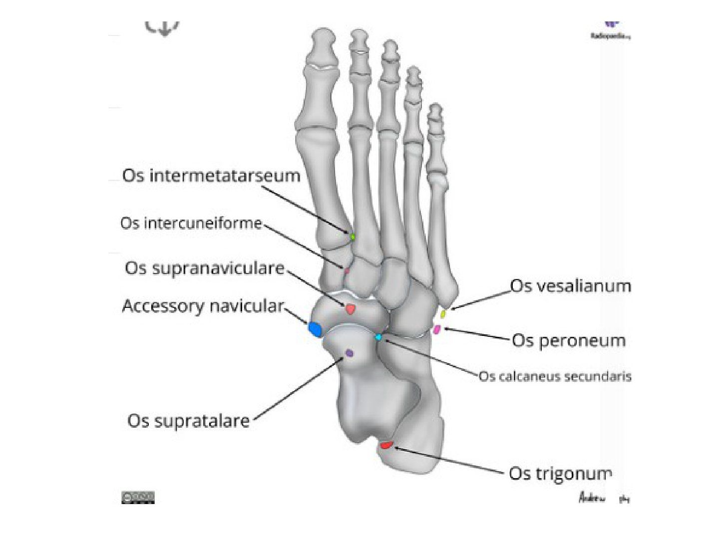
Q1-Q22 questions — tap to reveal all answerslist
- What is Iselin disease?
- How would you counsel a patient with Iselin disease?
Q1.What is Iselin disease?
- Apophysitis of the base of the 5th metatarsal
- A benign and self-limiting condition
Q2.How would you counsel a patient with Iselin disease?
- Reassure the patient: the condition is benign
- The condition is self-limiting
▸ Slide 291 · Bunionette deformityFoot and Ankle · 3 questions 1 check expand

Q1-Q33 questions — tap to reveal all answerslist
- What is the Coughlin classification of bunionette deformity?
- How does a bunionette present?
- How is a bunionette treated?
Q1.What is the Coughlin classification of bunionette deformity?
- Type I: large MT head or lateral exostosis
- Type II: congenital bowing with a normal 4-5 IMA
- Type III: 4-5 IMA >12 deg
Q2.How does a bunionette present?
- Cosmesis
- Pain from prominence, lateral bunion, plantar callosity
- Check shoewear
Q3.How is a bunionette treated?
- Nonoperative first - 70-90% success (shoewear modification, analgesics, callosity shaving)
- Surgery if nonoperative treatment fails
- Type I: lateral condylectomy
- IMA <12: distal MT osteotomy
- IMA >12: proximal MT osteotomy
Type III bunionette is defined as a 4-5 intermetatarsal angle >12 deg — conflates the classification with a surgical threshold — Coughlin type III is simply an increased 4-5 IMA; abnormal is commonly cited as >8-10 deg, while >12 deg is used to select a proximal osteotomy — medium confidence — source
▸ Slide 292 · Harris axial viewFoot and Ankle · 3 questions expand
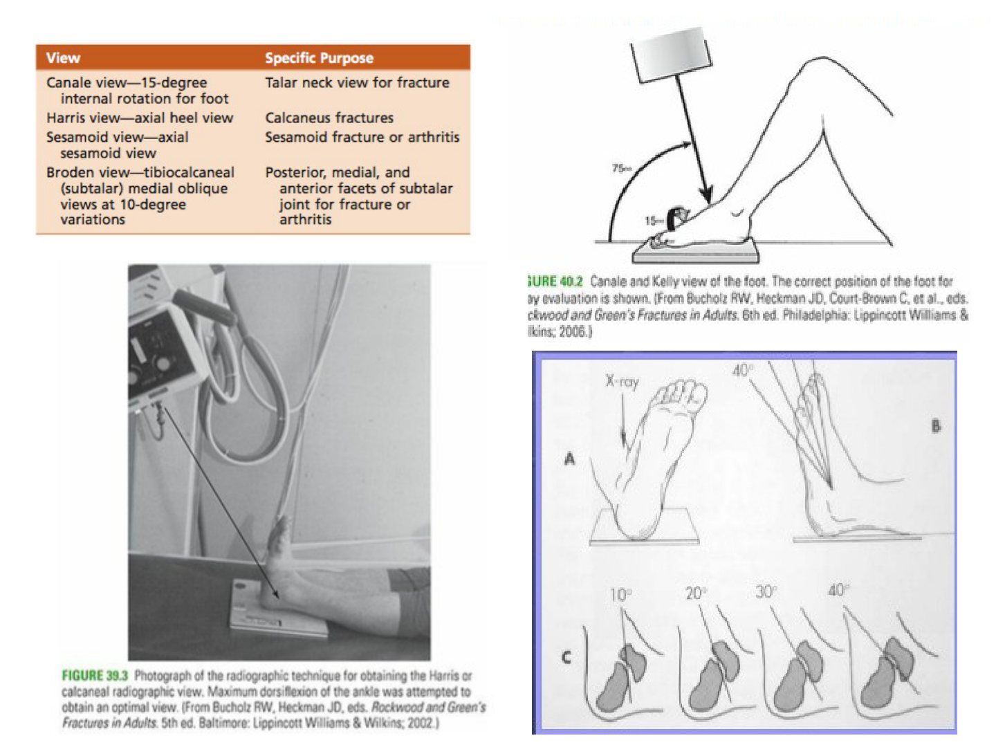
Q1-Q33 questions — tap to reveal all answerslist
- How is the Harris axial view taken and what does it show?
- How is the Canale view taken?
- How is the Broden view taken?
Q1.How is the Harris axial view taken and what does it show?
- Foot in dorsiflexion with the beam angled 45 deg cephalad
- Visualizes the joint surface, loss of height, increasing width and angulation of the tuberosity fragment (usually varus)
Q2.How is the Canale view taken?
- Ankle in maximal equinus
- Foot on the cassette pronated 15 deg
- X-ray beam 15 deg cephalad
Q3.How is the Broden view taken?
- Patient supine, cassette under the leg and ankle
- Foot in neutral flexion with the leg internally rotated 15 deg (mortise)
- X-ray centred over the lateral malleolus
- Beam directed toward the head at 40, 30, 20 and 10 deg
▸ Slide 293 · Proper shoe wearFoot and Ankle · 4 questions expand
Q1-Q44 questions — tap to reveal all answerslist
- What are the three key features of proper shoe wear?
- What does rotational rigidity in a shoe protect?
- What does heel counter stability provide?
- What does flexion stability in a shoe protect?
Q1.What are the three key features of proper shoe wear?
- Rotational rigidity (protects the flatfoot)
- Heel counter stability
- Flexion stability (protects the forefoot)
Q2.What does rotational rigidity in a shoe protect?
- It protects the flatfoot
Q3.What does heel counter stability provide?
- Heel counter stability is one of the three key features of proper shoe wear
- The notes do not specify the structure it protects
Q4.What does flexion stability in a shoe protect?
- It protects the forefoot
▸ Slide 294 · Subluxation of peroneal tendonFoot and Ankle · 13 questions expand
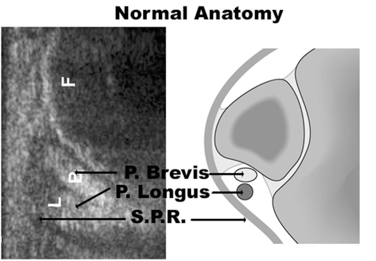
Q1-Q1313 questions — tap to reveal all answerslist
- Describe the Oden classification of superior peroneal retinaculum (SPR) injury.
- Which Oden grade is most common and what is its pathoanatomy?
- What is the pathoanatomy of an Oden grade 2 lesion?
- What is the pathoanatomy of an Oden grade 3 lesion?
- What is the pathoanatomy of an Oden grade 4 lesion?
- What other classifications are used for peroneal tendon pathology?
- Describe the anatomy of the peroneal tendon groove.
- How do you assess suspected peroneal tendon subluxation?
- How is the apprehension test for peroneal tendon subluxation performed?
- What imaging is used for peroneal tendon subluxation?
- How is acute peroneal tendon subluxation managed nonoperatively?
- What is the operative management of acute peroneal tendon subluxation?
- How is chronic or recurrent peroneal tendon dislocation managed?
Q1.Describe the Oden classification of superior peroneal retinaculum (SPR) injury.
- Grade 1: SPR elevated off the fibula, tendons lie between bone and periosteum (most common, >50%)
- Grade 2: fibrocartilaginous ridge avulses with the retinaculum; tendons displaced beneath the ridge
- Grade 3: avulsion of a small cortical osseous fragment from the fibular insertion; tendons dislocate beneath the fragment
- Grade 4 (rarest): complete avulsion/rupture of the SPR; tendons lie external and superficial to the retinaculum
Q2.Which Oden grade is most common and what is its pathoanatomy?
- Grade 1, >50% of cases
- SPR elevated off the fibula
- Tendons come to lie between the bone and the periosteum
Q3.What is the pathoanatomy of an Oden grade 2 lesion?
- The fibrocartilaginous ridge behind the lateral insertion of the SPR avulses together with the retinaculum
- The peroneal tendons are displaced beneath the ridge
Q4.What is the pathoanatomy of an Oden grade 3 lesion?
- Avulsion of a small cortical osseous fragment from the fibular insertion
- The tendons dislocate beneath the fibular fragment
Q5.What is the pathoanatomy of an Oden grade 4 lesion?
- The rarest type of lesion
- Complete avulsion or rupture of the superior peroneal retinaculum
- Tendons lie external and superficial to the retinaculum
Q6.What other classifications are used for peroneal tendon pathology?
- Raikin for intrasubstance subluxation
- Redfern for peroneal tendon tear (both okay/partial tear, one torn, both torn; A not retracted, B retracted)
Q7.Describe the anatomy of the peroneal tendon groove.
- Peroneal tendon sheath splits at the level of the peroneal tubercle
- Groove over the posterolateral aspect of the fibula
- Bounded by the superior peroneal retinaculum, fibrocartilaginous ridge and Talus/ calcaneum
Q8.How do you assess suspected peroneal tendon subluxation?
- History: timing, recurrence
- Apprehension test (resisted dorsiflexion and eversion, or plantarflexion + inversion to dorsiflexion + eversion)
- Check for voluntary subluxation and any hindfoot varus
Q9.How is the apprehension test for peroneal tendon subluxation performed?
- Resisted dorsiflexion and eversion, or
- Move the ankle from plantarflexion + inversion to dorsiflexion + eversion
Q10.What imaging is used for peroneal tendon subluxation?
- XR: look for the fleck sign
- Dynamic USG
- CT
- MRI
Q11.How is acute peroneal tendon subluxation managed nonoperatively?
- Short leg cast and protected weight bearing for 6 weeks
Q12.What is the operative management of acute peroneal tendon subluxation?
- Acute repair of the superior peroneal retinaculum
- Deepening of the fibular groove
Q13.How is chronic or recurrent peroneal tendon dislocation managed?
- Groove deepening with soft tissue transfer and/or osteotomy
▸ Slide 295 · Tarsal tunnel syndromeFoot and Ankle · 11 questions expand

Q1-Q1111 questions — tap to reveal all answerslist
- What is tarsal tunnel syndrome?
- Describe the anatomy and contents of the tarsal tunnel.
- Where does the tarsal tunnel begin and end?
- What are the causes of tarsal tunnel syndrome?
- What is the differential diagnosis of tarsal tunnel syndrome?
- How does tarsal tunnel syndrome present?
- What are the signs of tarsal tunnel syndrome?
- What is the workup of tarsal tunnel syndrome?
- What is the conservative treatment of tarsal tunnel syndrome?
- What are the indications and options for surgery in tarsal tunnel syndrome?
- What is Baxter's nerve?
Q1.What is tarsal tunnel syndrome?
- Compressive neuropathy of the tibial nerve or its terminal branches (medial and lateral plantar nerves) in the tarsal canal
- Impingement within the fibro-osseous tunnel or as it passes into the abductor hallucis muscle
Q2.Describe the anatomy and contents of the tarsal tunnel.
- Roof: flexor retinaculum (laciniate ligament)
- Floor (medial): superior calcaneus, medial wall of talus, distal-medial tibia
- Inferior: abductor hallucis
- Contents: TibP, FDL, FHL, tibial nerve, posterior tibial A&V
Q3.Where does the tarsal tunnel begin and end?
- Begins a few cm proximal to the tip of the medial malleolus, where the crural fascia condenses to form the unyielding roof - the flexor retinaculum (laciniate ligament)
- Ends where the medial and lateral plantar nerves enter or pass deep to the abductor hallucis
Q4.What are the causes of tarsal tunnel syndrome?
- Space-occupying lesion: lipoma, nerve sheath tumour, ganglion, varicose vein, bony prominence, exostosis
- Valgus hindfoot
Q5.What is the differential diagnosis of tarsal tunnel syndrome?
- Peripheral neuropathy
- HMSN
- S1 radiculopathy
- Plantar fasciitis
- Lateral plantar nerve compression neuropathy
- Heel pad atrophy/hypermobility
Q6.How does tarsal tunnel syndrome present?
- Vague foot pain
- Commonly confused with plantar fasciitis
Q7.What are the signs of tarsal tunnel syndrome?
- Atrophy of intrinsics
- Numbness in the sole
- Tinel sign posterior to the medial malleolus
- Aggregated numbness upon eversion and dorsiflexion
Q8.What is the workup of tarsal tunnel syndrome?
- XR and MRI
- NCT/EMG: latency to abdH and abdDM, slow velocity, decreased amplitude
Q9.What is the conservative treatment of tarsal tunnel syndrome?
- Steroid injection
- PT for stretching
- Orthosis in plantarflexion + varus
Q10.What are the indications and options for surgery in tarsal tunnel syndrome?
- Indications: space-occupying lesion or failed conservative treatment
- Decompression - open or arthroscopic
Q11.What is Baxter's nerve?
- First branch of the lateral plantar nerve
- Implicated as one of the cause of heel pain, which can be similar to plantar fasciitis
- Provides motor innervation for the abductor digiti minimi