Hip
Early post-op delirium and nerve palsy
Post-operative confusion and management of foot drop after hip replacement
11 questions
2 source pages
1 images
Images appear with the first question taken from each source page — tap a question to open it.
Q1What are the causes of confusion after TKR?▸
- Drug causes
- Neurological: stroke, pre-existing dementia
- Metabolic: hyponatraemia, hypoglycaemia
- Hypoperfusion: shock (cardiogenic/hypovolaemic), anaemia, PE
- Infection: UTI, pneumonia
Q2Which metabolic and infective causes must be excluded?▸
- Hyponatraemia and hypoglycaemia
- UTI and pneumonia
Q3How would you structure assessment of post-TKR confusion?▸
- Review drugs, then screen neurological causes including stroke and pre-existing dementia
- Correct metabolic causes - hyponatraemia and hypoglycaemia
- Assess perfusion: shock (cardiogenic/hypovolaemic), anaemia, PE
- Screen for infection: UTI and pneumonia
Q4What are the goals in managing foot drop after THR?📷▸
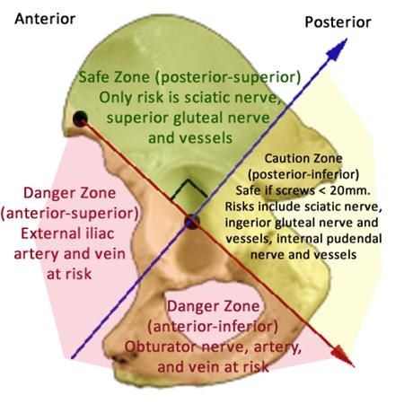
- 1. Establish the diagnosis
- 2. Delineate the level of the nerve lesion
Q5What history is relevant in foot drop after THR?▸
- Primary pathology requiring THR
- Review the operation record: approach, any limb lengthening, intra-operative complications
- Risk factors: female, DDH, limb lengthening, revision surgery, spinal stenosis, post-traumatic
Q6What examination is performed for foot drop after THR?▸
- Check drains/output and any wound complication
- Remove tight dressing, splint and abduction pillow over the knee and allow knee flexion in case of common peroneal nerve injury
- Examine ankle dorsiflexion (common peroneal component) and ankle plantarflexion (posterior tibial component)
- Examine hamstrings to look for above or below knee palsy
Q7What investigations are used and what is the timing of nerve assessment?▸
- XR/CT to confirm screw placement and acetabular component position
- USG for haematoma; NCT for extent, severity and monitoring of recovery
- D2: assess distal segment acute or chronic palsy
- 2 weeks: assess axonotmesis, neurotmesis, neuropraxia
- 3 months: assess recovery of nerve
Q8What non-operative measures are given for foot drop?▸
- AFO to prevent equinus deformity
- Physiotherapy to prevent joint contracture
Q9What are the causes of sciatic nerve palsy after THR?▸
- Traction injury - retractor placement, acute limb lengthening >4cm (Kavanagh 1991: <4cm no palsy, >4cm 28% sciatic nerve palsy)
- Pressure injury - post-operative haematoma, hip dislocation
- Direct injury - acetabular screw placement, diathermy, cement or prosthesis impingement, thermal injury from cement
Q10Will you explore the sciatic nerve, and what is the incidence of nerve palsy?▸
- If no structural lesion (screw malposition, haematoma compression), observe recovery; ~40% recovery rate at 21 months (Farrell JBJS 2005)
- Incidence 3.5% in primary, 7% in revision cases; 90% sciatic nerve, 50% common peroneal nerve
- Risk factors: acute leg lengthening >4cm, revision surgery, DDH, female, pre-existing spinal stenosis
- Avoidance: extend hip, flex knee, subtrochanteric osteotomy; post-op flex hip to 45 deg and bend knee to 90 deg
Q11How do you differentiate a sciatic nerve palsy from a common peroneal nerve palsy?▸
- Sciatic palsy involving both tibial and peroneal components affects biceps femoris, semi-T, semi-M and the posterior compartment of the calf
- Peroneal-only sciatic palsy is difficult to differentiate clinically from common peroneal palsy below the knee - EMG tests the short head of biceps femoris
- Common peroneal component is densely packed, more lateral and fixed at the fibular neck