▸ Slide 186 · HipHip · 2 questions expand
Q1-Q22 questions — tap to reveal all answerslist
- What are the key topics covered in this hip slide?
- What learning objectives relate to the hip in this section?
Q1.What are the key topics covered in this hip slide?
- Not covered in the speaker notes
Q2.What learning objectives relate to the hip in this section?
- Not covered in the speaker notes
▸ Slide 187 · Goal and principle - THRHip · 3 questions expand
Q1-Q33 questions — tap to reveal all answerslist
- What is the goal of total hip replacement (THR)?
- Which biomechanical parameters should be restored at THR?
- What are the surgical principles of THR relating to leg length and soft tissues?
Q1.What is the goal of total hip replacement (THR)?
- Painless, stable, mobile hip
Q2.Which biomechanical parameters should be restored at THR?
- Restore hip centre and offset
- Equalization of LLD
- Correct orientation of components
Q3.What are the surgical principles of THR relating to leg length and soft tissues?
- Equalization of LLD
- Good soft tissue balance
▸ Slide 188 · Total hip survivalHip · 3 questions expand

Q1-Q33 questions — tap to reveal all answerslist
- What registry data are referenced for total hip survival?
- What are the 15-year revision rates for THR by fixation type in the UK joint registry?
- What are the 15-year revision rates in the AUS joint registry?
Q1.What registry data are referenced for total hip survival?
- Refer to AONJRR
- UK joint reg – revision at 15 yrs
- AUS joint reg – revision at 15 yrs
Q2.What are the 15-year revision rates for THR by fixation type in the UK joint registry?
- Overall: cemented 4.85%, cementless 5%, hybrid 4%
- <55: hybrid 4%, cementless 6%, cemented 7%
- >75: cemented 2%, cementless 4%
Q3.What are the 15-year revision rates in the AUS joint registry?
- Overall: cemented 5.7%, cementless 6%, Hybrid 5.5 %
▸ Slide 189 · Consent of THRHip · 5 questions 1 check expand

Q1-Q55 questions — tap to reveal all answerslist
- What general and anaesthetic risks would you discuss when consenting for THR?
- What thromboembolic risks are quoted for THR?
- What are the intraoperative risks of THR?
- What are the postoperative risks of THR?
- What are the consent figures for revision THR?
Q1.What general and anaesthetic risks would you discuss when consenting for THR?
- Anaesthesia risk --> CVA, AMI, death
- Wound complications; bleeding requiring transfusion
- Clinical DVT ~2%
- UTI and chest infection ~10%
- Mortality ~0.5% in hospital
Q2.What thromboembolic risks are quoted for THR?
- Symptomatic PE ~2.5%
- Fatal PE <0.5%
Q3.What are the intraoperative risks of THR?
- Fracture: primary cement 1%, cementless 10%
- Fracture: revision cement 3%, cementless 30%
- Nerve palsy 1-3 %
- Vascular injury 0.2%
- LLD: 1.5 cm lengthening in 18%
Q4.What are the postoperative risks of THR?
- Infection ~1% (Lidwell BMJ 1982)
- Dislocation 3%
- HO 2-90%
- Periprosthetic fracture
- Loosening: 10-year survivorship ~90%
Q5.What are the consent figures for revision THR?
- Mortality 2.5% in 90 days
- 1% not satisfied
Consent for THR: clinical DVT ~2%, symptomatic PE ~2.5% and fatal PE <0.5% — outdated / higher than modern series — Contemporary THA data report symptomatic DVT ~0.3-0.6%, symptomatic PE ~0.2-0.5% and fatal PE ~0.03-0.1% with modern prophylaxis and early mobilisation — medium confidence — source
▸ Slide 190 · Low DDH for osteotomyHip · 6 questions expand

Q1-Q66 questions — tap to reveal all answerslist
- Describe the X-ray findings in a young adult with a dysplastic hip.
- What causes the pain in a dysplastic hip?
- What further investigations are needed in a dysplastic hip?
- What is the aim of management in an early dysplastic hip with no OA in a young patient?
- What are the treatment options for early dysplastic hip with no OA in a young patient?
- What are the prerequisites and prognostic factors for PAO?
Q1.Describe the X-ray findings in a young adult with a dysplastic hip.
- Undercoverage of the femoral head and underdeveloped acetabulum
- Acetabular angle ~60 (N 33-38), CEA ~0 (N 25-40), Tonnis angle ~20 (N <10)
- Acetabular index = Hilgenreiner line to acetabular edge
- Superior migration of femur, Shenton's line broken
- Tear drop
- No OA change; also comment on the femur
Q2.What causes the pain in a dysplastic hip?
- Cartilage wear due to small weight-bearing surface and abnormal loading
- Labral injury
Q3.What further investigations are needed in a dysplastic hip?
- False profile X-ray (true lateral of hip) for anterior coverage (anterior centre edge angle normal 25-50)
- X-ray with hip in abduction + IR to check congruency (can consider osteotomy)
- MRI for cartilage condition and labral tear, r/o AVN (rule out AVN)
Q4.What is the aim of management in an early dysplastic hip with no OA in a young patient?
- Relief of symptoms and prevent progression to early osteoarthritis
- Treatment options aim at pain relief +/- improving coverage to reduce contact stress
Q5.What are the treatment options for early dysplastic hip with no OA in a young patient?
- Conservative - disadvantage: later joint preservation surgery not possible
- Hip arthroscopy: indication labral/chondral pathology; controversial for persistent pain, ?instability after labral debridement
- Periacetabular osteotomy (Ganz): increased coverage and congruency, decreased pain, hyaline cartilage
- Salvage (Chiari, shelf): fibrocartilage, 84% at 17 years
- VDRO if PAO cannot provide sufficient coverage
Q6.What are the prerequisites and prognostic factors for PAO?
- Prerequisites: congruent joint, no OA, good range, young
- Ganz osteotomy: anterior rotation, lateral rotation, medialisation, intact posterior column
- Poor prognostic factor: >30 y.o
- Bone cyst is not a contraindication in PAO
- Outcomes 60% survivorship in 20yrs
▸ Slide 191 · Xray AP PelvisHip · 12 questions expand
Q1-Q1212 questions — tap to reveal all answerslist
- Describe the AP pelvis X-ray findings.
- What is the classification of this hip and why?
- What history and examination findings are relevant in a dysplastic hip?
- What investigations and templating technique are used?
- What are the aims and counselling points for THR in a dysplastic hip?
- What anatomical abnormalities and natural history are expected in a dysplastic hip?
- Why does a dysplastic hip develop early OA?
- What implant and bearing choices are suitable for THR in a dysplastic hip?
- Describe the exposure and soft tissue release in THR for a dysplastic hip.
- What are the key points of acetabular bone work in a dysplastic hip?
- What are the femoral considerations in THR for DDH?
- What is the rehabilitation after THR in a dysplastic hip?
Q1.Describe the AP pelvis X-ray findings.
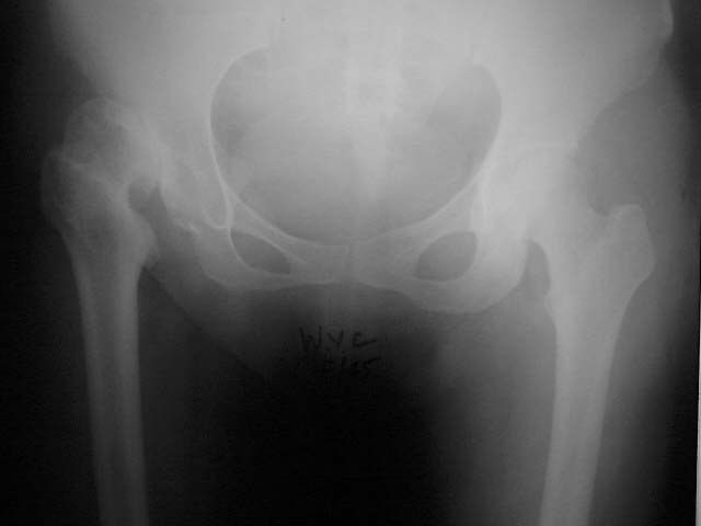
- Right dysplastic hip with superior and lateral dislocation (broken Shenton's line)
- Femoral head loss of sphericity with coxa plana and coxa breva, increased ER (prominent LT), suspected increased NSA, hypoplastic femoral shaft
- False acetabulum in contact with the native acetabulum; anterior superior rim bone loss; teardrop poorly formed
- Tonnis angle >10, acetabular angle of Sharp >45, CEA >45
- Contralateral hip normal; significant LLD; no evidence of previous PAO or VDRO
Q2.What is the classification of this hip and why?
- Hartofilakidis Type B (low dislocation)
- Lower lip of the false acetabulum is in contact with the upper lip of the true acetabulum
Q3.What history and examination findings are relevant in a dysplastic hip?
- Hx: age, occupation, premorbid walking, PMHx, risk factors including childhood DDH; pain and limitations
- Local PE: skin, scar, sinus, ROM, FFC, abductor power
- Systemic: gait, nerve, LLD --> scoliosis, pelvic obliquity, contralateral knee flexion contracture, ipsilateral foot equinus
Q4.What investigations and templating technique are used?
- Bloods to rule out infection +/- joint aspiration
- CT for bone stock, morphology, dimensions, acetabular orientation and femoral anteversion
- Scannogram; +/- MRI/EMG for abductors
- Template: use Ranawat triangle technique to template for anatomical hip centre, measure the superolateral bone defect
- Isocheles triangle, h=1/5 of pelvic height. Hip Center= midpoint of the diagonal line (isosceles triangle, h = 1/5 pelvic height, hip centre = midpoint of the diagonal line)
Q5.What are the aims and counselling points for THR in a dysplastic hip?
- Offer conservative management, if failed advise THR
- Counsel: overall survival worse (10 yr 87%, 20 yr 55%, Hip Int 2017)
- Aim: restore offset and anatomical hip centre, equalise LLD, correct alignment, good soft tissue tension
- Anatomical centre benefits: better biomechanics (less JRF, abductor function), more bone stock, restore LLD, less impingement
Q6.What anatomical abnormalities and natural history are expected in a dysplastic hip?
- Soft tissue: elongated redundant capsule, psoas hypertrophy, contracted transversely orientated abductors, shortened rectus/adductor/hamstring
- Nerve: femoral and sciatic nerve shortened
- Bone: shallow, excessively anteverted acetabulum with superanterior bone defect
- Femur: ER, excessive anteversion, increased neck shaft angle, small posteriorly displaced GT, canal stenosis
- Natural history: subluxation --> pain at 20-30; dislocation --> well till 50 (adjacent joint problems)
Q7.Why does a dysplastic hip develop early OA?
- Loss of coverage of the femoral head + lateralised head
- Decreased abductor lever arm and small contact area --> early OA
Q8.What implant and bearing choices are suitable for THR in a dysplastic hip?
- Cementless THR or hybrid: both have good longevity in joint registries
- Bearing: metal-on-PE or ceramic-on-PE (good wear profile without the risk of CoC)
- Dual mobility if stability is a concern
- Combined anteversion of 35 degrees
Q9.Describe the exposure and soft tissue release in THR for a dysplastic hip.
- Larger incision, beware the sciatic nerve
- Contracted soft tissue, pulvinar in the acetabulum, thickened ligamentum teres
- Release sequence: short rotators, capsule, reflected head of rectus, gluteus maximus, iliopsoas (beware descending branch of MFC), adductors
- Finding the acetabulum: use TAL and pulvinar, trace the ligamentum teres; standby X-ray
- Consider transtrochanteric/transfemoral approach (not commonly done)
Q10.What are the key points of acetabular bone work in a dysplastic hip?
- Usually soft bone - be careful when reaming or broaching
- Acetabulum hypoplastic, deficient anterior/superolateral bone stock
- Aim for cementless fixation with 66% rim fit or 80% host bone contact, standby cemented cup
- Address bone defect: biological or metallic options (trabecular metal, bulk autogenous bone graft, reinforcement rings, cup cage)
- Cotyloplasty: deliberate medial wall fracture to increase coverage, then graft + cementless - difficult revision as bone stock not restored
Q11.What are the femoral considerations in THR for DDH?
- Small head, short anteverted valgus neck, posterior GT attachment, narrow straight tapered canal
- Subtrochanteric osteotomy if LLD >4 cm - step cut for rotational stability; derotation osteotomy/modular implant if anteversion >40 degrees
- Attempting an uncemented stem in a deformed femur risks fracture
- Cementless modular DDH stem if subtroch osteotomy (press-fit distal fixation, cable/wires + graft); cemented if no osteotomy
- GT osteotomy if abductors need to be advanced and retensioned; proximal femoral resection sacrifices bone stock and makes rotation hard to determine
Q12.What is the rehabilitation after THR in a dysplastic hip?
- Keep hip in extension, abduction and knee in flexion
- Full weight bearing, hip precautions
- Watch for HO
▸ Slide 192 · Crowe classificationHip · 3 questions expand

Q1-Q33 questions — tap to reveal all answerslist
- What is the Crowe classification based on?
- What are the Crowe grades by head/neck junction to femoral head diameter?
- What are the alternative Crowe grades using vertical height?
Q1.What is the Crowe classification based on?
- Radiological classification based on proximal migration of the femoral head compared with pelvic height
- Pelvic vertical height = distance from inferior ischial tuberosity to superior iliac crest
- Measure distance between head-neck junction and inter-teardrop line
Q2.What are the Crowe grades by head/neck junction to femoral head diameter?
- 1 <50%
- 2 50-75%
- 3 75-100%
- 4 >100%
- 4 = Hartofilakidis B/C
Q3.What are the alternative Crowe grades using vertical height?
- 1 <10%
- 2 10-15%
- 3 15-20%
- 4 >20%
▸ Slide 193 · DDX:Hip · 6 questions 1 check expand

Q1-Q66 questions — tap to reveal all answerslist
- What are the differential diagnoses for a previously dysplastic or deformed hip?
- How do you look for evidence of a septic or TB hip?
- What preoperative planning is needed for THR in DDH?
- What bone and soft tissue considerations exist in high or dislocated hips?
- What is the quoted risk of sciatic nerve injury with limb lengthening?
- What does the teardrop indicate?
Q1.What are the differential diagnoses for a previously dysplastic or deformed hip?
- Infection - septic hip / TB
- Dysplastic hip (treated DDH with childhood surgery)
- Post-traumatic
Q2.How do you look for evidence of a septic or TB hip?
- History: fever, antibiotics, surgical drainage
- PE: look for sinus
- Ix: inflammatory markers, +/- bone scan, +/- USG for effusion
- If TB hip: 3 months preop + 9 months postop antibiotics
Q3.What preoperative planning is needed for THR in DDH?
- Consult senior joint surgeon
- PE for LLD, ROM, abductor
- Templating
- CT for bone defect and femur morphology
- MRI / EMG for abductor; prepare implant; ?intra-op culture
Q4.What bone and soft tissue considerations exist in high or dislocated hips?
- Acetabulum: decide high vs low hip centre
- High hip centre = reconstruction 35 mm higher than inter-teardrop line or 15 mm higher than femoral head centre
- Femur: hypoplastic, narrow and straight, excessive anteversion --> small straight stem / subtrochanteric osteotomy
- Soft tissue: contracture of surrounding soft tissue, sciatic nerve
Q5.What is the quoted risk of sciatic nerve injury with limb lengthening?
- Kavanagh 1991: <4 cm --> 0% palsy
- >4 cm --> 28% palsy
Q6.What does the teardrop indicate?
- Tear drop normal if onset of pathology is after 8 years
Sciatic nerve injury after THA: 0% palsy if limb lengthening <4 cm and 28% if >4 cm (Kavanagh 1991) — source attribution unverified; length threshold contested — Modern evidence is mixed: a 508-hip DDH series (Eggli 1999) found no correlation between amount of lengthening and nerve palsy, while other series report risk mainly with lengthening >5 cm; a 2017 systematic review notes the 3-4 cm threshold is suggested but debated — medium confidence — source
▸ Slide 194 · Whats this?Hip · 10 questions expand

Q1-Q1010 questions — tap to reveal all answerslist
- What is a Brittain ischiofemoral arthrodesis?
- What other hip fusion techniques exist?
- What are the indications and contraindications for hip fusion?
- What is the ideal position for hip fusion and its biomechanics?
- What history and examination findings are relevant before taking down a fused hip?
- What are the options and outcomes for taking down a fused hip?
- What are the difficulties in exposure and identifying the hip centre when taking down a fused hip?
- What soft tissue difficulties and overall results are expected when taking down a fused hip?
- Is abductor weakness an absolute contraindication to take-down, and how is it tackled?
- What do you need to know about TB hip when considering take-down?
Q1.What is a Brittain ischiofemoral arthrodesis?
- Extra-articular arthrodesis of the hip
- Subtrochanteric osteotomy and medial displacement of the femoral shaft with fibular graft to the ischium just below the acetabulum
- Principle: compression force by adductor
- Extracapsular (away from TB infection and progressive femoral head destruction)
- Disadvantages: needs postop immobilisation and difficult revision
Q2.What other hip fusion techniques exist?
- Extraarticular: iliofemoral fusion
- Intraarticular (Cabenela): dislocate hip, ream away articular cartilage, DHS across joint; preserve abductor, minimal change in anatomy
- Combined: medializing acetabular osteotomy, intra/extraarticular arthrodesis, Davis muscle pedicle graft, cobra plate - stability but stress riser at plate end
Q3.What are the indications and contraindications for hip fusion?
- Indication: young + recurrent infection
- CI: ipsilateral LL fusion
- CI: contralateral hip fusion
- CI: persistent infection
- CI: poor cardiorespiratory tolerance; poor bone stock
Q4.What is the ideal position for hip fusion and its biomechanics?
- 25 degrees flexion (balance of sitting/standing)
- 5 degrees adduction/neutral (restore apparent LLD; abduction causes pelvic obliquity and back pain)
- 5 degrees external rotation
- 50% reduction in gait efficiency (increased contralateral pelvic rotation, increased adjacent knee JRF)
- Increased O2 demand and 30% increase in energy consumption
Q5.What history and examination findings are relevant before taking down a fused hip?
- Hx: age + function, initial indication for fusion, current disability, neurological deficits
- Walking aids + shoe raise
- Posture --> position of fusion; short limb gait + ipsilateral vaulting
- LLD + compensatory deformities; ROM; Thomas test
- Abductor power and adductor jerk test; sinus/scar; gluteal and thigh wasting; Trendelenburg test
Q6.What are the options and outcomes for taking down a fused hip?
- Options: take down/ corrective osteotomy --> fused in better position; take down + arthroplasty
- Indications: adjacent joint pain (ipsilateral knee, contralateral hip), increasing back/radicular pain
- Also suboptimal fusion position, painful pseudarthrosis
- CI to take down: poor function, residual infection, abductor function (clinical/EMG/MRI)
- Preop: sitting-standing pelvic XR, CT bone stock, MRI/ EMG for abductor, Biopsies or aspiration for potential for reactivation of dormant infection
- Outcome worse than routine THR (74% at 15 yr, Allan Gross JBJS 2021); up to 1/3 revision in 10 yr
Q7.What are the difficulties in exposure and identifying the hip centre when taking down a fused hip?
- Cannot mobilise the hip - need in-situ dissection, high risk of neurovascular injury
- In-situ double cut osteotomy / trochanteric slide osteotomy (cut neck closer to trochanter to save bone stock; landmarks GT, pubofemoral arch)
- Tilt table for visualisation; identify femoral neck orientation and protect with 2 Hoffman
- Hip centre: pulvinar, transverse acetabular ligament, AIIS, greater sciatic notch
- Cup malposition common due to spine/pelvic deformity; XR or robotic guidance; Trimming of residual bone after cup placement; +/- screw augmentation
Q8.What soft tissue difficulties and overall results are expected when taking down a fused hip?
- Soft tissue: capsule, adductor tenotomy, reflected head of rectus femoris, psoas, HO
- Aim: pain relief, mobile joint, restore hip centre and offset, equalise LLD, balance soft tissue tension, accurate component placement
- High risk surgery (up to 1/3 revision in 10 years)
- Gait will worsen due to inadequate abductor function
- Will improve gradually over months/ years
- Implant choice: hybrid THR
Q9.Is abductor weakness an absolute contraindication to take-down, and how is it tackled?
- No - but it causes abnormal gait and increases dislocation rate
- Tenodesis of ITB at the greater trochanter (similar to transfer)
- Constrained liner to decrease dislocation
Q10.What do you need to know about TB hip when considering take-down?
- Traditionally Hardinge and Charnley: THR when no sinus discharge >20 yr, fusion >10 yr, previous medically treated TB
- Kim SJ BJJ 2013 meta-analysis: THR in active TB may be offered before this with extensive debridement + perioperative TB meds
- Pre op 1-3 months Anti TB med
- Post op 9 months anti TB med (in accordance with Infectious Disease Society of America guidelines)
- Sepsis rate post TB ~ 12 %
- Shanmugasundaram classification: stable (normal/atrophic/Perthes) and unstable forms (subluxed/dislocated, protrusio, pestle and mortar)
- Phemister triad: peripheral osseous erosion, joint space narrowing, juxta-articular osteopenia; Tuli stages 1-4
▸ Slide 195 · Describe Xray:Hip · 16 questions expand
Q1-Q1616 questions — tap to reveal all answerslist
- Describe the X-ray findings and the likely stage of AVN.
- What is the Ficat classification of AVN?
- What are the causes and risk factors for AVN of the hip?
- What is the role of MRI and classification in pre-collapse AVN?
- What history and examination findings are needed in AVN of the hip?
- What is the aim of AVN management and which parameters guide it (CARE)?
- When do you observe, decompress or graft an AVN hip?
- What is the pathophysiology of idiopathic AVN of the hip?
- What is the pathophysiology of steroid-induced AVN?
- What is the crescent sign and why does it occur?
- What is the conservative and joint-preserving management of AVN?
- Describe the technique of core decompression for AVN.
- Describe the principles, technique and complications of vascularised bone grafting for AVN.
- What newer stem cell techniques exist for AVN?
- What are the joint-sacrificing options for AVN?
- How common is bilateral AVN and which part of the head is usually affected?
Q1.Describe the X-ray findings and the likely stage of AVN.
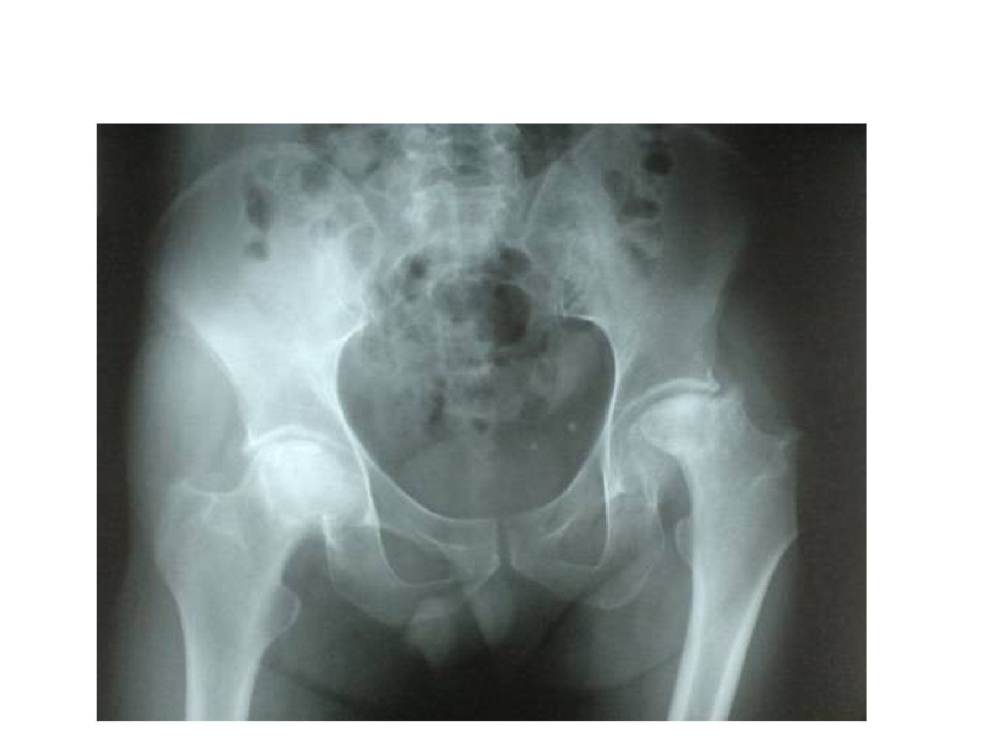
- Collapse of femoral head bilaterally R>L; left Shenton's line broken
- Right hip positive crescent sign
- Positive OA change at the left hip; acetabulum well formed; SIJ normal
- Suggests Ficat IV left hip and Ficat 2B right hip; ask for frog leg lateral if collapse not seen
- Bilateral in 40-80% of cases; Ddx tumour, septic arthritis, BMES (self limiting)
Q2.What is the Ficat classification of AVN?
- I normal
- II sclerosis or cyst (A focal changes, B crescent sign without flattening)
- III flattening of femoral head without joint space narrowing
- IV flattening of femoral head + OA
Q3.What are the causes and risk factors for AVN of the hip?
- Traumatic: # NOF or hip dislocation; risk: femoral head fracture 75-100%, NOF I/II <10%, III/IV >80%, basicervical 50%, trochanteric 25%, hip dislocation 5-40% (reduction within 6 hr 2-10%)
- Non-traumatic: idiopathic; intra/extra-vascular and mixed (commonest is mixed)
- Alcohol: 400 ml/week --> RR 9.8-fold (1 unit = 10 ml/8 g alcohol, i.e. 40 units/week, 20 beers/week)
- Steroid: >20 mg/day prednisolone for >30 days or >2 g over 3 months
- Intravascular: protein S/C, SLE, fat embolism, caisson disease, sickle cell anaemia
- Extravascular (marrow adipocyte hypertrophy): Gaucher, radiation
Q4.What is the role of MRI and classification in pre-collapse AVN?
- Double line sign on T2: hypodense line = edge of necrosis, hyperdense line = hypervascular granulation tissue
- Steinberg (1-6, A <15% / B 15-30% / C >30%) predicts need for head-preserving procedure; <15% observe, >15% offer core decompression
- Kerboul angle: <190 no collapse, 190-240 50% collapse, >240 100% collapse; >200 predicts poor result with preserving procedures
- Shimizu: >1/4 head diameter and >2/3 weight-bearing area --> 74% collapse in <3 years
- Risk of collapse: position, size, presence of bone edema; overall 50-80% progress (symptomatic 70-80%)
- +/- bone scan: increased uptake at the femoral head in AVN, at femoral head and acetabulum in OA
Q5.What history and examination findings are needed in AVN of the hip?
- Hx: risk factors/causes - drinker? steroid use? SLE? diver? trauma?; current symptoms; functional limitations (HHS)
- PE local: sinus, ROM, FFC, abductor
- Systemic: gait, LLD, document NV status
- Bloods to rule out infection; +/- CT; +/- bone scan
- MRI in pre-collapse stage: size, site, prognostication (Kerboul angle, Shimizu)
Q6.What is the aim of AVN management and which parameters guide it (CARE)?
- Aim: pain relief, improve function via joint salvage or joint replacement depending on stage
- Care parameters: Collapse of head (Ficat), Age, Reversible cause, Extent of disease (Steinberg ABC, Kerboul, Shimizu)
Q7.When do you observe, decompress or graft an AVN hip?
- Pre-collapse + small necrotic area + not in weight-bearing zone --> observe
- Stage 1 or 2 + predictor of collapse (large necrotic area in weight-bearing zone by Kerboul, Shimizu, Steinberg) --> core decompression (5-7 yr survival 70-90%)
- Stage 3 + young --> vascularised bone graft (5-7 yr survival 70-80%)
Q8.What is the pathophysiology of idiopathic AVN of the hip?
- Biological and mechanical
- Intraosseous microcirculation coagulation --> venous thrombosis --> retrograde arterial occlusion --> increased intraosseous pressure --> decreased blood flow to femoral head --> AVN
- Mechanical load on necrotic bone incapable of repair --> mechanical failure and collapse
- Modes of failure: fine cracking of subchondral bone, linear tangential fracture, shearing fracture
Q9.What is the pathophysiology of steroid-induced AVN?
- Fat cell hypertrophy --> femoral head compartment syndrome
- Hyperlipidaemic state - fat emboli causing endothelial damage
- Direct toxicity - damage to endothelial and smooth muscle cells in the vasculature
- Direct osteocyte death
Q10.What is the crescent sign and why does it occur?
- Overlying cartilage remains viable from synovial fluid, while subchondral bone is necrotic and collapses
- Ficat IIB = crescent sign without flattening of the femoral head
Q11.What is the conservative and joint-preserving management of AVN?
- Counsel on natural history: Rate of preservation of the femoral head by stage: stage 1 35%, stage 2 31%, stage 3 13% (Hungerford JBJS 1995)
- Protective weight bearing has no evidence (Fairbank CORR 1996)
- Alendronate may prevent collapse in early disease; LMWH for coagulopathy; statin for steroid-induced (decrease adipogenesis)
- Core decompression (stage I, IIA; preferably reversible cause): 5-7 yr survival 70-90%, # risk 1%; poor results in steroid induced
- Hungerford 1995 JBJS 10-year survival: 90% stage 1, 70% stage 2, 30% stage 3
- Vascularized bone graft for stage 3 young: survival 70-80%; trapdoor for Ficat III/early IV (73% good-excellent at 5 yr)
- Rotational intertrochanteric osteotomy for small lesions, Kerboul <200; fails if >30-50% head involved
Q12.Describe the technique of core decompression for AVN.
- Indication: pre-collapse lesion, preferably reversible cause (Ficat I, IIA)
- Mechanism: relief of intraosseous hypertension, remove necrotic material, stimulate angiogenesis (mainly pain relieving; effect on natural history unknown)
- Method 1: 8 mm trephine after guide pin, within 5 mm of the articular surface, additional channel by 5 mm trephine
- Method 2: 3.2 mm pin 2-3 times for decompression
- +/- structural support (tantalum rod/strut graft) +/- marrow/BMP to stimulate bone growth
Q13.Describe the principles, technique and complications of vascularised bone grafting for AVN.
- Indication: pre-collapse or collapsed AVN in young patient (<45 y), preferably reversible cause (Ficat II B/C)
- Principles: decompression, removal of necrotic bone, replacement with corticocancellous graft for structural support, Revascularization
- Graft: fibular (peroneal --> ascending branch of lateral circumflex) or iliac crest (deep circumflex iliac artery); AL approach
- Postop: LMWH then aspirin + NWB walking 6/52
- Complications: subtrochanteric fracture, donor site morbidity (FHL/big toe contracture), harvest fibular graft >6 cm from knee and ankle, superficial peroneal nerve injury
- VBG vs core decompression (CORR 2017 Cao RCT): VBG better functional score (not reaching MCID) and vascularity on SPECT; no difference in progression to THR (10%)
Q14.What newer stem cell techniques exist for AVN?
- Three-stage procedure: stem cell culture, then cultured stem cells reinjected into a previous core decompression site
- Bone marrow from the pelvis centrifuged in the OR to yield bone marrow concentrate rich in stem cells, transplanted into the necrotic area under X-ray control after core decompression
Q15.What are the joint-sacrificing options for AVN?
- THR is the mainstay: previous incisions, steroid osteoporosis, acetabular defect, Altered anatomy and bone stock, higher wear/osteolysis, infection, Post op dislocation (alcoholic)
- Aus registry 20y revision rate 12.3%; THR > bipolar (Lee CORR 2004)
- Hip resurfacing: indication AVN of femoral head with no acetabular damage
- Resurfacing CI: poor proximal femoral bone stock (AVN >25% of head), coxa vara/breva, small cup + large neck, osteoporosis, childbearing
- Resurfacing pros: preserve bone stock, less osteolysis, less dislocation; risks: femoral neck fracture 1-2%, metallosis, impingement
- Hip arthrodesis for very young patient
Q16.How common is bilateral AVN and which part of the head is usually affected?
- Bilateral in 40-80% of cases overall
- 50% of idiopathic cases and 80% of steroid-induced cases
- Usually affects the anterolateral femoral head
▸ Slide 196 · Classification and treatmentHip · 5 questions expand
Q1-Q55 questions — tap to reveal all answerslist
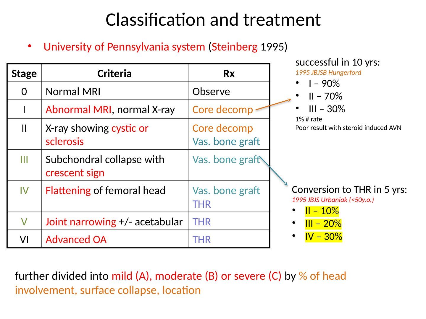
- Which classification system is used for AVN of the hip, and who described it?
- What are the criteria and treatment for each stage of the University of Pennsylvania (Steinberg) system?
- How are the stages further subdivided in this system?
- What is the conversion rate to THR in 5 years (1995 JBJS Urbaniak, <50 years old)?
- Which stages are successful in 10 yrs and at what rates (1995 JBJS Hungerford)?
Q1.Which classification system is used for AVN of the hip, and who described it?
- University of Pennsylvania system (Steinberg 1995)
Q2.What are the criteria and treatment for each stage of the University of Pennsylvania (Steinberg) system?
- 0: Normal MRI → observe
- I: Abnormal MRI, normal X-ray → core decompression
- II: X-ray showing cystic or sclerosis → core decompression, vascularized bone graft
- III: subchondral collapse with crescent sign → vascularized bone graft
- IV: flattening of femoral head → vascularized bone graft, THR
- V: joint narrowing +/- acetabular → THR
- VI: advanced OA → THR
Q3.How are the stages further subdivided in this system?
- Mild (A), moderate (B) or severe (C) by % of head involvement, surface collapse and location
Q4.What is the conversion rate to THR in 5 years (1995 JBJS Urbaniak, <50 years old)?
- Stage II – 10%
- Stage III – 20%
- Stage IV – 30%
Q5.Which stages are successful in 10 yrs and at what rates (1995 JBJS Hungerford)?
- Stage I – 90%
- Stage II – 70%
- Stage III – 30%
- 1% fracture rate
- Poor result with steroid-induced AVN
▸ Slide 197 · G g spondylitisHip · 20 questions expand

Q1-Q2020 questions — tap to reveal all answerslist
- Describe the X-ray findings in ankylosing spondylitis with hip arthropathy.
- How do you diagnose ankylosing spondylitis?
- How common is hip involvement in AS and why is the pelvis tilted?
- Describe the distraction provocation test for the SIJ.
- Describe the compression provocation test for the SIJ.
- Describe the thigh thrust test for the SIJ.
- Describe the sacral thrust test for the SIJ.
- Describe Gaenslen's test.
- What are the surgical difficulties in THR for AS and how can you tackle them?
- What is the sequence and rationale for surgery in an RA patient?
- What do you assess in a patient with AS?
- What are the ASAS classification criteria for spondyloarthritis?
- What are the medical and anaesthetic pre-operative considerations in THR for AS?
- What positioning and draping issues arise in THR for AS?
- How do you manage exposure and soft tissues in THR for AS?
- How do you identify the true acetabulum in a fused AS hip?
- What femoral and post-operative problems occur in THR for AS?
- Hip or spine first in AS? What is the rationale?
- How do spinal flexibility and balance guide hip vs spine surgery and cup position (Phan, JBJS 2015)?
- What are the risk factors for heterotopic ossification?
Q1.Describe the X-ray findings in ankylosing spondylitis with hip arthropathy.
- Decreased joint space over bilateral hip joints, R > L, with OA changes
- Pelvis extended, like an outlet view
- SIJ fused
- Diagnosis: AS with hip arthropathy
Q2.How do you diagnose ankylosing spondylitis?
- Modified New York criteria 1984: 1 clinical + 1 radiological criterion
- Back pain > 3 months improving with exercise, not relieved by rest
- Limited chest expansion; limited lumbar spine ROM in coronal and sagittal planes
- SIJ involvement: bilateral grade 2-4 or unilateral grade 3-4
- SIJ XR grades: 0 normal, 1 suspicious, 2 erosion/sclerosis, 3 narrowed joint space, 4 fused
Q3.How common is hip involvement in AS and why is the pelvis tilted?
- 30% of AS have ankylosis of both hips; 90% have bilateral involvement
- Loss of lumbar lordosis and fixed thoracic kyphosis -> pelvis hyperextended (outlet view)
- Fixed hip contracture -> pelvis flexed (inlet view)
Q4.Describe the distraction provocation test for the SIJ.
- Patient supine
- Examiner applies posterolateral directed pressure to bilateral ASIS
- Positive = reproduction of pain
Q5.Describe the compression provocation test for the SIJ.
- Patient side-lying
- Examiner compresses the pelvis, pressure over the iliac crest directed at the opposite iliac crest
- Positive = reproduction of symptoms
Q6.Describe the thigh thrust test for the SIJ.
- Patient supine, hip placed in 90 deg flexion and adduction
- Examiner applies posteriorly directed force through the femur at varying abduction/adduction angles
- Positive = reproduction of buttock pain
Q7.Describe the sacral thrust test for the SIJ.
- Patient prone
- Examiner delivers an anteriorly directed thrust over the sacrum
- Positive = reproduction of pain
Q8.Describe Gaenslen's test.
- Patient supine with the symptom leg hyperextended below the bed, asymptomatic leg flexed
- Press on the hyperextended leg
- Positive = reproduction of pain
Q9.What are the surgical difficulties in THR for AS and how can you tackle them?
- Pre-op: stop biological agent, assess lung and heart function, ulcer prophylaxis; C-spine XR r/o C1/2 instability, may need fibre-optic intubation
- Ortho: decide hip or spine first - usually hip first; expect more bleeding, inform anaesthetist +/- cell saver
- Intra-op: careful positioning (trunk vertical axis parallel to table), posterior approach, sliding trochanteric osteotomy, protect sciatic nerve
- Acetabulum: identify true floor using TAL (Beverland); graft protrusio; set version for tilted pelvis - if PT<20, decrease cup 5 deg for every 10 degrees beyond 20 deg PT; aim combined anteversion 35 deg
- Post-op: HO risk 10%, anterior dislocation, pneumonia, DVT prophylaxis
Q10.What is the sequence and rationale for surgery in an RA patient?
- 1. Life saving (C1/2 instability)
- 2. Function (ruptured tendon, acute carpal tunnel, most painful joints)
- 3. Sure win procedure
- 4. Lower limb: foot > hip > knee - Foot – eliminate ulcers as source of infection; THR improves flexion range for TKR (typically delay TKR for 6 month due to risk of dislocation)
- 5. Upper limb: proximal to distal
Q11.What do you assess in a patient with AS?
- Treatment and control of AS
- Associated conditions - Ortho: spine; Others: chest expansion, eye, heart
- Function: walking, sitting, gaze
Q12.What are the ASAS classification criteria for spondyloarthritis?
- > 3 months back pain + age < 45
- SI joint on imaging (MRI+ or definite XR by modified NY criteria) + >= 1 SpA feature, OR HLA-B27 + >= 2 SpA features
- Sensitivity 82.9%, specificity 84.4%
- SpA features: inflammatory back pain, arthritis, enthesitis, uveitis, psoriasis, dactylitis, IBD, good NSAID response, family history, HLA-B27, elevated CRP
Q13.What are the medical and anaesthetic pre-operative considerations in THR for AS?
- Medical: stop biological agent; assess chest (lung function test) and heart (echocardiogram); ulcer prophylaxis
- Anaesthetist: C-spine X-ray to rule out C1/2 instability
- Limited C-spine range may need fibre-optic assisted intubation; assess TMJ condition
- Expect more bleeding - inform anaesthetist +/- cell saver
Q14.What positioning and draping issues arise in THR for AS?
- Cautious during positioning, especially over the neck
- Trunk vertical axis (ear to greater trochanter) parallel to the OT table with eye level perpendicular -> functional axis used as reference for anteversion
- Good padding of all pressure points
- Draping difficult in an adducted position with immobility of the other side (may need to break table)
Q15.How do you manage exposure and soft tissues in THR for AS?
- Consider posterior approach (better exposure, lower risk of anterior dislocation)
- Difficult exposure: sliding trochanteric osteotomy to increase exposure (needs extensive soft tissue release; hip dislocation + soft bone = fracture risk; consider double cut osteotomy)
- Sequential soft tissue release for scarring; protect the sciatic nerve
Q16.How do you identify the true acetabulum in a fused AS hip?
- Use TAL (Beverland) as landmark
- Drill through the acetabulum and measure
- Intra-op image intensifier
- Protrusio may need bone graft/cage
Q17.What femoral and post-operative problems occur in THR for AS?
- Femoral preparation: risk of iatrogenic fracture; osteopenic bone may need a cemented implant
- Post-op: high risk of heterotrophic ossification (heterotopic ossification) - previous hip surgery, complete ankylosis - 10%
- Risk of anterior dislocation - hip precautions; contralateral fused hip causes compensatory flexed position -> stiffness and dislocation
- Pneumonia with limited chest expansion; DVT prophylaxis
Q18.Hip or spine first in AS? What is the rationale?
- Controversial; lecturer usually does hip first - regains hip mobility to compensate for a stiff spine, may regain horizontal gaze, avoids high-risk spine surgery
- Downsides of hip first: THR dislocation; kyphotic spine + fall -> spine fracture; catastrophic neurological complications during THR rehab
- If spinal imbalance is severe such that THR will not restore horizontal gaze, restore spinopelvic harmony first
- Individualized decision involving multidisciplinary input
Q19.How do spinal flexibility and balance guide hip vs spine surgery and cup position (Phan, JBJS 2015)?
- Depends on spinal flexibility and overall spinal balance
- Stiff spine, normal balance: increase acetabular anteversion (avoid anterior impingement/posterior dislocation when sitting - the pelvis will not retrovert)
- Stiff spine, unbalanced (retroverted pelvis): decrease anteversion (avoid posterior impingement/anterior dislocation when standing) OR do spine osteotomy first
- Flexible, unbalanced spine: same as stiff unbalanced, but lean more towards hip first (more likely to regain horizontal gaze after THR)
- Flexible, normal balance: unlikely in AS
Q20.What are the risk factors for heterotopic ossification?
- Patient: age > 60, male, previous HO, diseases (AS, DISH, hypertrophic OA, Paget's disease)
- Injury: head injury, burns
- Surgery: SP > Hardinge, extensive dissection, revision; posterior approach = lowest risk
- Post-op: prolonged immobilisation
▸ Slide 198 · Xray showing bilateral protrusio acetabuli (femoral head medial to ilioischial lHip · 9 questions 1 check expand
Q1-Q99 questions — tap to reveal all answerslist
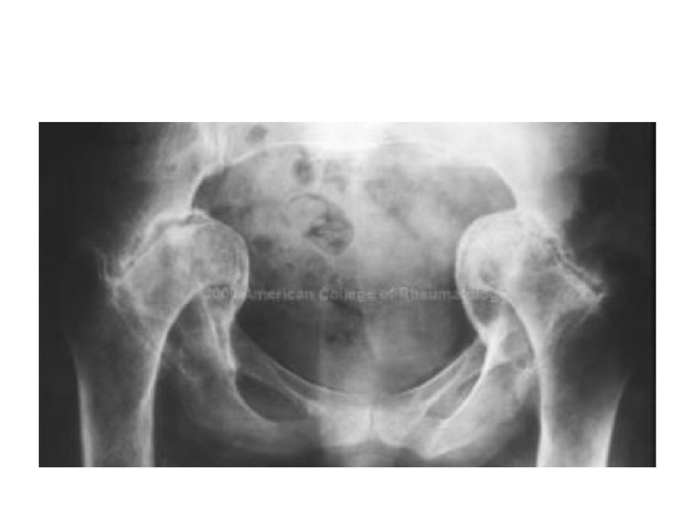
- What are the radiographic features of protrusio acetabuli?
- How is protrusio acetabuli classified and how does it differ from coxa profunda?
- What are the causes of protrusio acetabuli?
- What are the aims and operative plan for THR in protrusio acetabuli?
- How do you manage the acetabular bone defect in protrusio acetabuli?
- What history and examination findings are relevant in protrusio acetabuli?
- What investigations are needed in protrusio acetabuli?
- What general technical principles apply to THR in protrusio acetabuli?
- How is reduction and rehabilitation managed after THR for protrusio acetabuli?
Q1.What are the radiographic features of protrusio acetabuli?
- Femoral head medial to the ilioischial line
- Violation of the ilioischial line (> 3mm in men, > 6mm in women)
- Decreased CEA (centre-edge angle)
- LLD (leg length discrepancy)
Q2.How is protrusio acetabuli classified and how does it differ from coxa profunda?
- Hirst classification by degree of medial migration (distance between acetabulum and ilioischial line)
- Lecture ranges: 5-10, 10-15, > 15mm
- Not equal to coxa profunda: lateral border of the acetabular teardrop in line with or medial to the ilioischial line
Q3.What are the causes of protrusio acetabuli?
- Uncommon; rule out other pathologies
- Primary: Otto's disease (arthrokatadysis)
- Congenital: Marfan, OI
- Metabolic: osteomalacia, rickets, osteoporosis
- Inflammatory: RA, psoriasis, AS; Infection: TB
- Others: migration of hemiarthroplasty, Paget's disease
Q4.What are the aims and operative plan for THR in protrusio acetabuli?
- Aims: stable, painless, mobile joint; restore vertical and horizontal offset and restore LLD
- Choose hybrid THR with ceramic-on-PE articulation
- Posterior approach, protect sciatic nerve; difficult dislocation - extensive capsular incision, may need in-situ osteotomy
- Acetabulum: bone graft the floor and lateralise the hip centre; use TAL as reference; cementless cup with line-to-line fit; cemented standby
- Femur: low level neck cut with enhanced offset to restore abductor tension; Dorr cc ratio > 75% -> cemented
Q5.How do you manage the acetabular bone defect in protrusio acetabuli?
- Essentially a IIc defect
- Contained: morselized autograft from the femoral head
- Uncontained: mesh + impaction bone graft technique, aim for peripheral fit
- Large reamer first (hourglass deformity), do not deepen the acetabulum further
Q6.What history and examination findings are relevant in protrusio acetabuli?
- Hx: age, occupation, premorbid walking, PMHx
- Hx: pain and functional limitations
- Local PE: soft tissue, scar, sinus, ROM, FFC
- Systemic PE: LLD, gait, nerve exam, scoliosis, pelvic obliquity, knee FFC, ankle equinus
Q7.What investigations are needed in protrusio acetabuli?
- Bloods
- CT
- Scannogram
- Mx: conservative or operative
Q8.What general technical principles apply to THR in protrusio acetabuli?
- Soft bone - careful reaming and broaching
- Bone graft the floor and lateralise the hip centre; use TAL as reference
- Cementless cup line-to-line; cemented standby
- Restore abductor tension: low level neck cut with enhanced offset femoral component
Q9.How is reduction and rehabilitation managed after THR for protrusio acetabuli?
- Release soft tissue (gluteus maximus, anterior capsule, iliopsoas) before reduction
- Rehab: hip precautions
- Rehab: FWB (full weight bearing)
HIRST classification grades protrusio acetabuli by medial migration of 5-10, 10-15 and >15mm — incorrect ranges — Hirst grading is gender-specific by acetabulum-ilioischial line distance: men 3-8/8-13/>13mm (grade III with fragmentation); women 6-11/12-17/>17mm. The 5-10/10-15/>15 grouping resembles other classifications — source
▸ Slide 199 · Hip resurfacingHip · 7 questions expand
Q1-Q77 questions — tap to reveal all answerslist
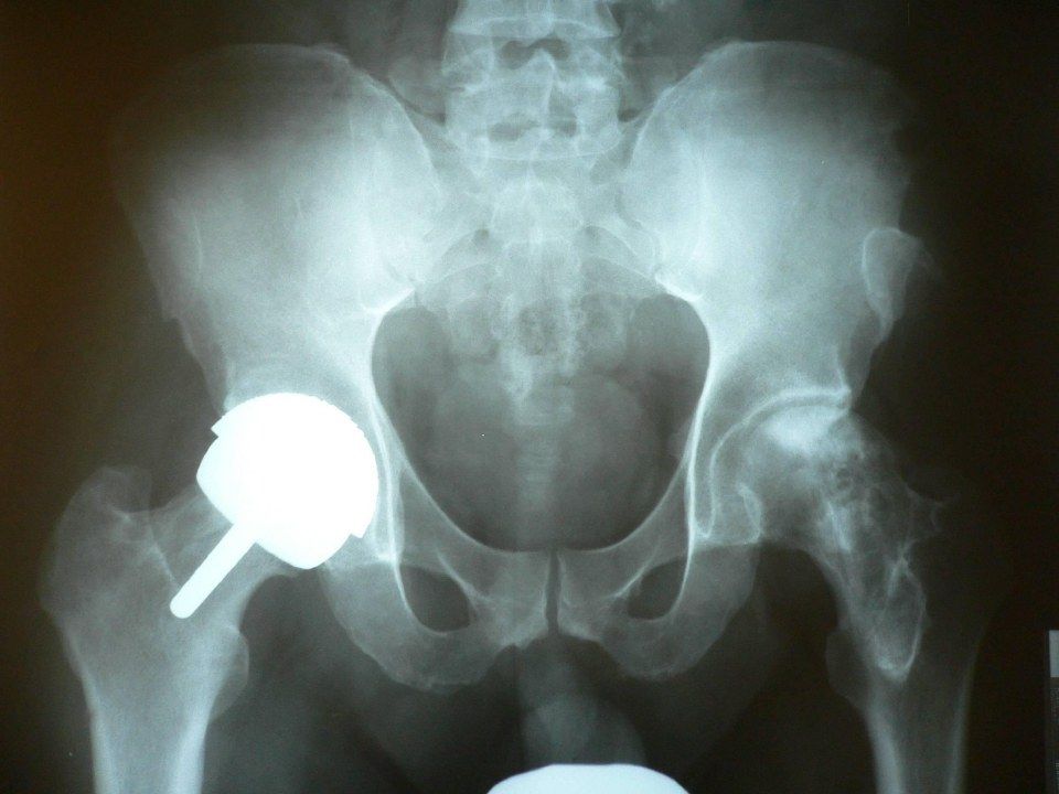
- What are the indications for hip resurfacing?
- What are the benefits and risks of hip resurfacing?
- What head size is required for hip resurfacing and why does a small head matter?
- What do you look for on follow-up MARS MRI after hip resurfacing?
- Describe the Anderson classification of ARMD.
- What are the contraindications to hip resurfacing?
- What proportion of hip resurfacings have a favourable outcome?
Q1.What are the indications for hip resurfacing?
- Large diameter metal-on-metal bearing
- Best in young, high-demand individuals with end-stage arthritis and good bone stock
Q2.What are the benefits and risks of hip resurfacing?
- Benefits: less particulate-induced osteolysis, low dislocation rate, normal hip kinematics
- Benefits: preserve proximal femoral bone stock, self-healing
- Risks: femoral neck fracture
- Risks: metal ion toxicity
Q3.What head size is required for hip resurfacing and why does a small head matter?
- Small acetabulum or abnormal anatomy with head size < 48mm is a contraindication
- Small heads are associated with more impingement and edge loading
Q4.What do you look for on follow-up MARS MRI after hip resurfacing?
- Pseudotumour
- Abductor detachment
- Presence and extent of osteolysis
- Periprosthetic fluid collections
Q5.Describe the Anderson classification of ARMD.
- A = normal appearances including seromas
- B = infection
- C1 = mild MOM disease, soft tissue mass < 5cm
- C2 = moderate MOM disease, mass > 5cm or muscle atrophy / marrow oedema
- C3 = severe MOM disease: fracture, tendon avulsion, bone marrow signal change
Q6.What are the contraindications to hip resurfacing?
- Inadequate neck bone stock
- Small acetabulum/abnormal anatomy with head size < 48mm
- Small heads -> more impingement and edge loading
- Relative: coxa vara, LLD
Q7.What proportion of hip resurfacings have a favourable outcome?
- 80-90% favourable
▸ Slide 200 · XR pelvisHip · 9 questions expand
Q1-Q99 questions — tap to reveal all answerslist
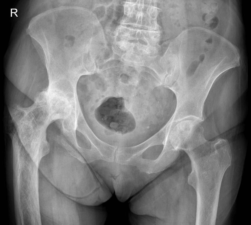
- Describe the X-ray findings in this patient.
- What is the differential diagnosis of these X-ray changes?
- What investigations are helpful in Paget's disease?
- What is Paget's disease?
- What causes the increased number and activity of Paget's osteoclasts?
- What are the perioperative considerations for THR in Paget's disease?
- What history and examination findings should be sought in Paget's disease of the hip?
- What is the pathophysiology of Paget's disease?
- What are the issues with cemented stems in Paget's disease and what is the current trend?
Q1.Describe the X-ray findings in this patient.
- Coarse trabeculation over the right femur, enlargement of bone and varus deformity
- Thickened iliopectineal and ilioischial lines (Brim sign)
- OA change; subluxation with disruption of Shenton's line
- Diagnosis: Paget's disease
Q2.What is the differential diagnosis of these X-ray changes?
- Sclerotic bony metastases
- Renal osteodystrophy
- Fibrous dysplasia, multiple myeloma, lymphoma
- Myelofibrosis
Q3.What investigations are helpful in Paget's disease?
- Raised ALP with normal calcium
- Urine hydroxyproline also raised
- XR of whole lower limb: look for stress fracture
- CT: morphology
Q4.What is Paget's disease?
- Disorder of high bone turnover
- Abnormal osteoclastic bone resorption with uncoupled osteoblastic bone formation
- Unknown cause: ?paramyxovirus infection in genetically susceptible individuals
- New bone is irregular and woven in nature, less resistant and prone to deformity and fracture
Q5.What causes the increased number and activity of Paget's osteoclasts?
- Osteoclasts hypersensitive to RANKL and calcitriol
- Marrow stromal cells have increase RANKL expression
- Increased osteoclast precursor recruitment by IL6
- Antiapoptotic oncogene Bcl 2 overexpressed
- Increased proto-oncogene cfos
Q6.What are the perioperative considerations for THR in Paget's disease?
- Pre-op: ensure the pain generator is the hip (not spinal stenosis, sarcoma or fracture); optimise high output cardiac failure; bisphosphonate (pamidronate) and calcitonin reduce disease activity and bleeding; templating: morphology, ? Need osteotomy
- Intra-op: cell saver, permissive hypotension; bone brittle - risk of iatrogenic fracture
- Acetabulum: protrusio - bone graft, anti-protrusion cage
- Femur: prepare burr for sclerotic and dense bone; Bowing & wide canal: osteotomy/ modular stem; cemented stem has poor interdigitation - trend towards cementless (Parvizi CORR 2002)
- Post-op: HO prophylaxis; Increase risk of osteolysis owing to the increase metabolic turnover of the pathological bone
Q7.What history and examination findings should be sought in Paget's disease of the hip?
- Clarify symptom: generalised pain / referred pain / Paget's sarcoma / fracture
- Medical treatment; age and functional demand
- PE: ROM, LLD, abduction power
Q8.What is the pathophysiology of Paget's disease?
- Unknown cause: ?paramyxovirus infection in genetically susceptible individuals; also high arsenic exposure
- Primary abnormality: intense focal resorption by abnormal osteoclasts (abnormal size, activity and quantity) -> large resorption cavities
- Osteoblasts recruited in response but activity is rapid -> disorganised, irregular woven bone
- Woven bone is less resistant and more elastic than lamellar bone -> prone to deformity and fracture
Q9.What are the issues with cemented stems in Paget's disease and what is the current trend?
- Pathological bone -> poor ossointegration for cementless; bleeding not good for cement interdigitation
- Risk of cement extrusion through an osteotomy site
- Trend towards use of cementless in recent years - previous concerns for the problem with osseous integration are mostly unfounded
- Bone ingrowth biology is similar to the early phases of fracture healing; In Pagets, fracture healing is normal (Parvizi CORR 2002)
▸ Slide 201 · Mode of failure of bipolar constrain linerHip · 2 questions expand
Q1-Q22 questions — tap to reveal all answerslist
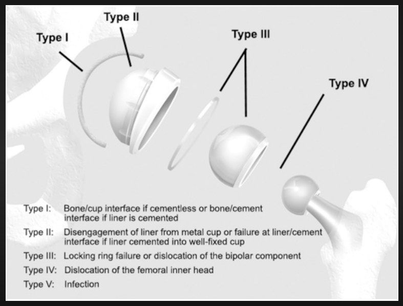
- Describe the modes of failure of a bipolar constrained liner.
- If the liner is cemented, at which interfaces can failure occur?
Q1.Describe the modes of failure of a bipolar constrained liner.
- Type I: bone/cup interface if cementless, or bone/cement interface if the liner is cemented
- Type II: disengagement of the liner from the metal cup, or failure at the liner/cement interface if cemented into a well-fixed cup
- Type III: locking ring failure or dislocation of the bipolar component
- Type IV: dislocation of the femoral inner head
- Type V: infection
Q2.If the liner is cemented, at which interfaces can failure occur?
- Type I: bone/cement interface (compared with bone/cup interface when cementless)
- Type II: liner/cement interface if cemented into a well-fixed cup
- Type II also includes disengagement of the liner from the metal cup
▸ Slide 202 · AJR basic scienceHip · 4 questions expand

Q1-Q44 questions — tap to reveal all answerslist
- What materials are listed under AJR basic science for hip arthroplasty?
- What are the key THR implant design features listed under AJR basic science?
- What modes of fixation are listed for THR implants?
- What complications of THR are listed under AJR basic science?
Q1.What materials are listed under AJR basic science for hip arthroplasty?
- PE (polyethylene)
- Cement
- Metal
- Ceramic
Q2.What are the key THR implant design features listed under AJR basic science?
- Stem
- Cup design
- Morse taper
Q3.What modes of fixation are listed for THR implants?
- Cementless
- Cemented
Q4.What complications of THR are listed under AJR basic science?
- Infection
- Wear
- Osteolysis
- Dislocation
- Protrusio
- DVT
▸ Slide 203 · BiomaterialHip · 4 questions expand
Q1-Q44 questions — tap to reveal all answerslist
- Define biomaterial.
- What are the basic requirements of a biomaterial?
- Define biocompatibility.
- What is forging?
Q1.Define biomaterial.
- Non-viable material used in a medical device
- Intended to interact with biological systems
Q2.What are the basic requirements of a biomaterial?
- Inert, non-allergic
- Mechanically sound
- Special properties (e.g. bioabsorbable)
- Cheap to manufacture
Q3.Define biocompatibility.
- Ability of a material to perform with an appropriate host response
- In a specific application
Q4.What is forging?
- Original heated material with coarse grain structure + pressure impaction
- Causes plastic deformation and recrystallization
- Forms new fine grain (reduce flaws in material)
▸ Slide 204 · Polyethylene is a thermoplastic polymer made of long hydrocarbon chain [(C2H4)n]Hip · 10 questions expand

Q1-Q1010 questions — tap to reveal all answerslist
- What is polyethylene and how is it structured?
- What are the advantages and disadvantages of polyethylene?
- What happens to polyethylene on irradiation?
- What is UHMWPE and how does it wear in TKR?
- What are the effects of highly cross-linked polyethylene?
- What is the evidence for highly cross-linked PE?
- How is polyethylene manufactured?
- What is the difference between direct compression moulding and ram bar extrusion?
- What is the effect of thermal treatment on polyethylene?
- What are the intrinsic factors affecting PE wear?
Q1.What is polyethylene and how is it structured?
- Thermoplastic polymer of long hydrocarbon chains (C2H4)n of ethylene monomers held by covalent bonds
- Molecular weight 3-5 million Daltons
- Two phases: disorganised amorphous and organised crystalline
- Formed by addition polymerization and sintering (addition = breaking covalent bond; condensation = byproduct)
Q2.What are the advantages and disadvantages of polyethylene?
- Advantages: tough, ductile, resistant to wear (low coefficient of friction)
- Disadvantage: susceptible to abrasion (not hard)
- Thermoplastic - cannot autoclave
- Anisotropic, weak in tension
Q3.What happens to polyethylene on irradiation?
- Irradiation generates free radicals
- In the presence of O2: oxidative degradation -> chain scission -> reduced fatigue strength (brittle) -> delamination
- In the absence of O2: cross-link
- Other fates: recombination and unsaturation
Q4.What is UHMWPE and how does it wear in TKR?
- Each molecule contains > 200,000 units of ethylene; molecular weight 3-5 million
- Better static mechanical properties due to crystalline phase: high ultimate stress, ductile, reduces fatigue crack propagation
- In TKR (less conforming, point loading) it wears by fatigue - functions above the endurance limit on the SN curve
Q5.What are the effects of highly cross-linked polyethylene?
- High dose irradiation (5-15 Mrad) in an oxygen-free environment
- Cross-linking limits mobility of chains in amorphous regions -> less creep
- Improves wear, surface hardness and stiffness; smaller, fewer PE particles
- But worse static properties: tensile strength, yield stress, ductility, fatigue strength, fracture toughness
Q6.What is the evidence for highly cross-linked PE?
- THR: SICOT 2020 Langlois - reduction in revision rate; Australian registry 6 vs 12%, better < 55yo; Swedish and NZ agree
- TKR controversial: JBJS 2020 Partridge - no difference at 12 years
- Australian registry: HXLPE lower revision than CPE at 10 years (5.8 vs 3.6%)
Q7.How is polyethylene manufactured?
- Condensation polymerization and sintering (Ziegler process): ethylene gas polymerised into PE resin powder at low temperature and low pressure using titanium chloride catalyst
- Sintering: heating and pressurising powder causes atomic diffusion to form a homogenous solid
- Fabrication +/- machining: ram bar extrusion (calcium stearate) / direct compression mould / sheet compression mould / isostatic moulding
- High dose irradiation (5-10 Mrad) in inert gas for cross-linking
- Thermal treatment: annealing < 137 or remelting at 137 (fewer free radicals but mechanical property affected; optimal crystallinity 45-60%)
- Additive vitamin E; mechanical compression (anisotropic); sequential irradiation/annealing; 2-MPC photoinduced graft polymerisation for hydrophilicity
- Sterilisation (2.5 MRad irradiation, gas plasma or ethylene oxide); storage under vacuum/argon/nitrogen
Q8.What is the difference between direct compression moulding and ram bar extrusion?
- Direct compression moulding is better than ram bar extrusion - less linear and volumetric wear (Bankston CORR 2005)
- Ram bar extrusion uses calcium stearate -> risk of non-consolidation of the centre of the bar
- Cutting a ram bar stretches the amorphous phase -> more susceptible to radiation -> more free radicals
Q9.What is the effect of thermal treatment on polyethylene?
- Promotes recombination of free radicals caused by radiation
- Annealing (< 137): potential for oxidation and osteolysis
- Remelting (137): eliminates free radicals in both crystalline and amorphous phases but affects mechanical properties more
- Optimal crystallinity 45-60%
Q10.What are the intrinsic factors affecting PE wear?
- (1) PE manufacturing process: direct compression moulding reduces volumetric/linear wear (no calcium stearate); high dose irradiation in inert gas; thermal treatment; additives; packaging/storage
- (2) Conformity: more conforming reduces fatigue wear (increases adhesive and abrasive wear but less fatigue wear)
- (3) PE thickness > 8mm (including metal back; Bartel et al: 4-6mm without metal back) reduces fatigue
▸ Slide 205 · This is cementHip · 10 questions expand
Q1-Q1010 questions — tap to reveal all answerslist
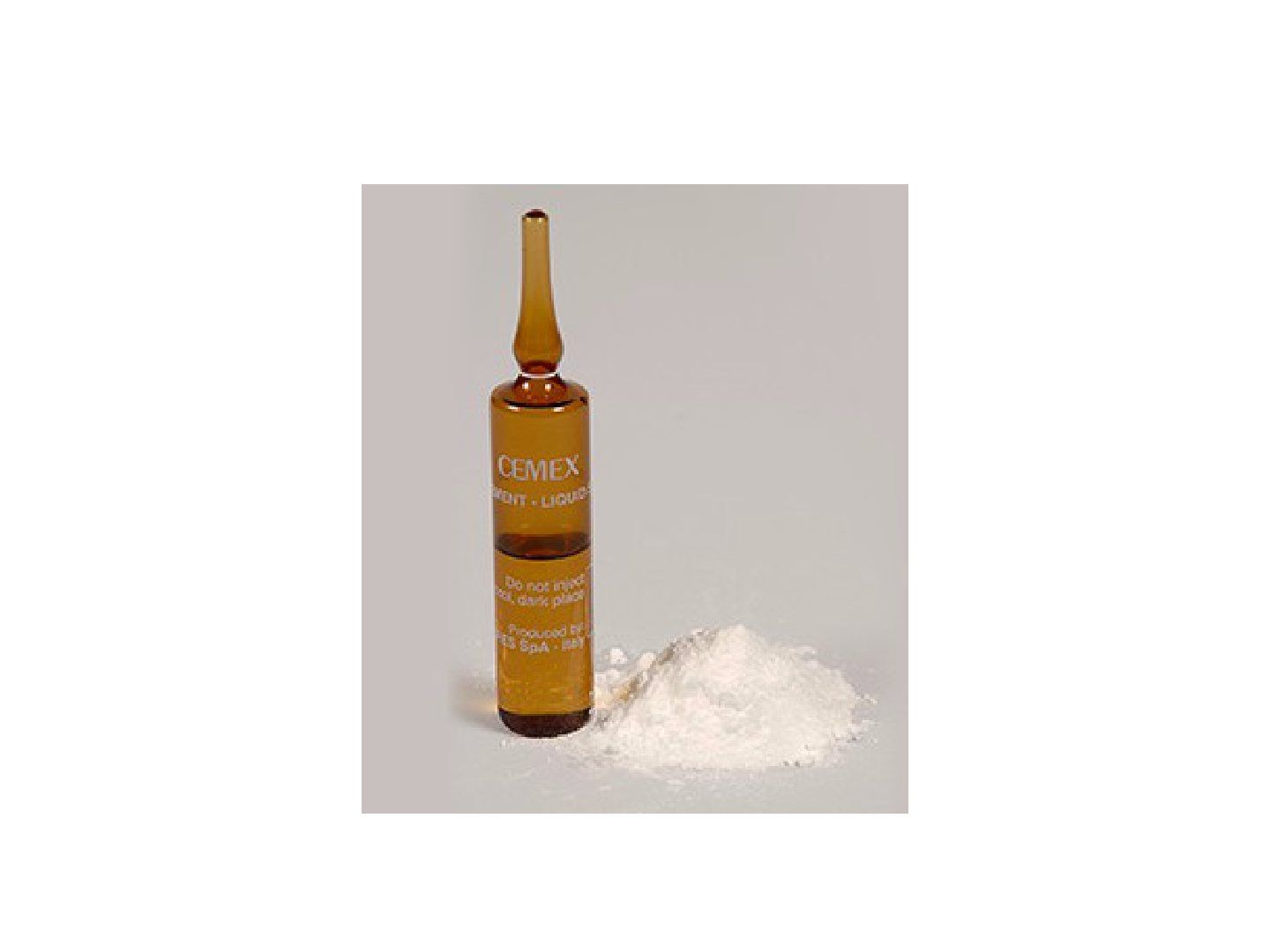
- What is PMMA and how does it work?
- What are the uses of PMMA cement?
- What are the components of bone cement powder and liquid?
- Define dough time, working time and setting time.
- How do you improve the cement mantle?
- What is bone cement implantation syndrome?
- How does cement viscosity affect its use?
- What are the local and systemic complications of bone cement?
- What is the pathophysiology of bone cement implantation syndrome?
- What are the risk factors and prevention of bone cement implantation syndrome?
Q1.What is PMMA and how does it work?
- Grout, not glue - no adhesive property; fills voids for load transfer or local antibiotic delivery
- Forms a stable static mechanical bond by interdigitation into cancellous bone
- Preferred in osteoporotic bone (deep penetration) and irradiated bone
- Brittle, strong in compression, weak in tension, notch sensitive and viscoelastic
Q2.What are the uses of PMMA cement?
- Fixation of arthroplasties
- Kyphoplasty to stabilise a vertebral compression fracture
- Cement spacer in total joint infections
Q3.What are the components of bone cement powder and liquid?
- Powder: PMMA polymer; initiator benzoyl peroxide
- Powder: radio-opacifier 10% barium sulphate or zirconium dioxide
- Powder: antibiotic tobramycin or gentamycin (10% maximal -> vancomycin 4g in 40g cement)
- Liquid: MMA monomer; catalyst N,N dimethyl-P-toluidine
- Liquid: stabiliser hydroquinone to avoid premature polymerisation; colourant
Q4.Define dough time, working time and setting time.
- Dough time: start mixing until not sticky
- Working time: not sticky until setting (50% max temperature)
- Setting time = dough + working time
- Phases: mixing -> stringy -> working -> hardening
- Rapid/vacuum mixing, increased temp/humidity, less monomer -> decreased setting time
Q5.How do you improve the cement mantle?
- Fully controllable: vacuum mixing, mixing speed, antibiotic inclusion, insertion pressurisation (cement gun, cement restrictor), centraliser, stiff stem without stress riser
- Partially controllable: canal prep to minimise blood and fat inclusion; broach to leave 2mm cancellous bone proximally; pulsatile lavage; brush and dry; adrenaline gauze/H2O2; suction
- Aim: interdigitating, homogenous, uniform cement mantle
Q6.What is bone cement implantation syndrome?
- Monomer leakage on pressurisation -> vasodilatation and cardiosuppression
- Triad: desaturation, hypotension, arrythmia
- Donaldson classification: Grade 1: SpO2 <94%/fall in SBP 20%; Grade 2: SpO2 <88%/fall in SBP 40%/unconsciousness; Grade 3: requiring CPR
- Prevention: increase O2 at cementation, medullary lavage, meticulous haemostasis, do not inject before dough time
Q7.How does cement viscosity affect its use?
- Viscosity = internal friction of the fluid; mainly a concern in THR (TKR is pressurised easily)
- Low viscosity: better interdigitation into cancellous bone but cannot stop bleeding; poorer clinical result in THR (less effective marrow displacement, haemodynamic backflow)
- High viscosity: may not be usable in a cement gun for medullary insertion
- Medium viscosity: usable with cement gun pressurisation and can stop bleeding; modified by temperature, humidity, mixing conditions; Simplex-P is medium viscosity
Q8.What are the local and systemic complications of bone cement?
- Systemic: cardiopulmonary suppression, emboli
- Local: bone necrosis
- Systemic syndrome: bone cement implantation syndrome
Q9.What is the pathophysiology of bone cement implantation syndrome?
- Monomer leakage into the bloodstream upon pressurisation -> vasodilatation and cardiosuppression
- Release of endothelial mediators, histamine and complement activation
- Mechanical blockage / multiple embolisation theory: high pressure during insertion + exothermic cement reaction -> cement expands; air and marrow contents embolise
Q10.What are the risk factors and prevention of bone cement implantation syndrome?
- Patient: ASA 3-4, known pHT/IHD, osteoporosis; pathological fracture/fracture TOF, long stem
- Anaesthetic prevention: close monitoring and increase O2 at cementation; avoid volume depletion
- Surgical prevention: medullary lavage, vent tube, meticulous haemostasis
- Do not inject before dough time
▸ Slide 206 · Factors affecting cement strengthHip · 5 questions expand
Q1-Q55 questions — tap to reveal all answerslist
- How do you classify the factors affecting cement strength?
- What is good cementation?
- How do you classify the cementing technique?
- Describe the Barrack classification of the cement mantle.
- Which factors increase or decrease cement strength?
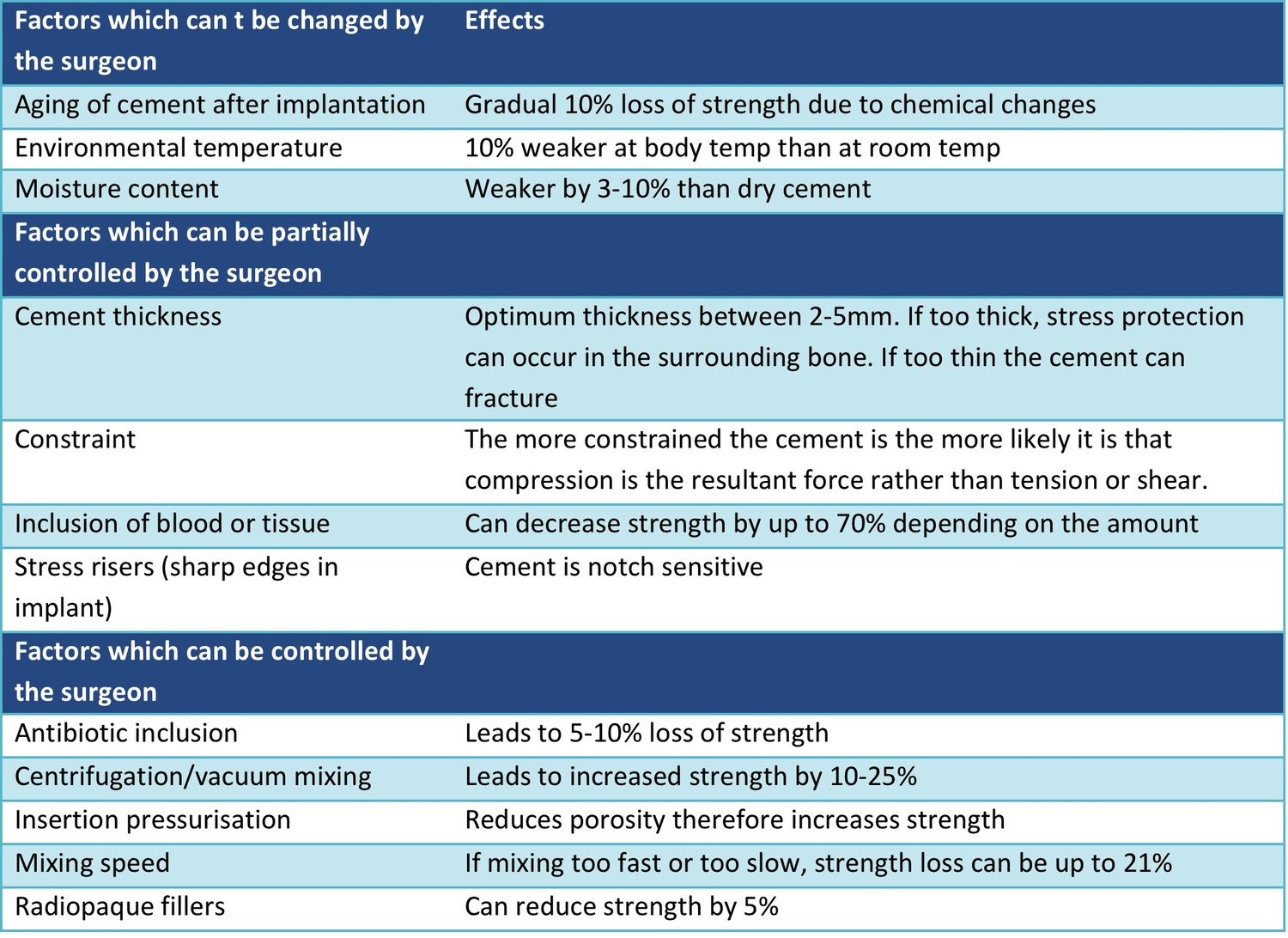
Q1.How do you classify the factors affecting cement strength?
- Uncontrollable: cement change after implantation - aging, higher body temperature, moisture
- Partially controllable: canal preparation and achieving a well-shaped cement mantle
- Controllable: ingredients and how we prepare the cement
Q2.What is good cementation?
- Uniform, homogenous and interdigitating cement mantle
- Plus a stiff and smooth stem
Q3.How do you classify the cementing technique?
- By generation of cementation technique
- By radiological appearance of the cement mantle (Barrack classification)
- Generations: 1 thumbing; 2 canal preparation (rasping, brush, cement gun, cement plug); 3 porosity reduction (CPPV: vacuum mixing, pulsatile lavage, pressurisation, centraliser)
- 4th generation: proximal and distal centralisation, proximal pressurisation (Exeter polished surface - Taper lock effect by controlled subsidence)
Q4.Describe the Barrack classification of the cement mantle.
- Grade A = medullary canal completely filled with cement (white out)
- Grade B = 0-50% radiolucency
- Grade C = > 50% radiolucency (C1 incomplete proximal cement; C2 mantle < 1mm)
- Grade D = 100% radiolucency / absence of cement distal to stem
Q5.Which factors increase or decrease cement strength?
- Increase: optimal mixing speed, vacuum mixing (reduces porosity), pressurised insertion
- Increase: good canal preparation without blood, mantle at least 2mm thick without stress riser
- Decrease: additives - radio-opaque and antibiotic
- Decrease: cement aging, higher body temperature and moisture after implantation
▸ Slide 207 · CeramicHip · 10 questions expand
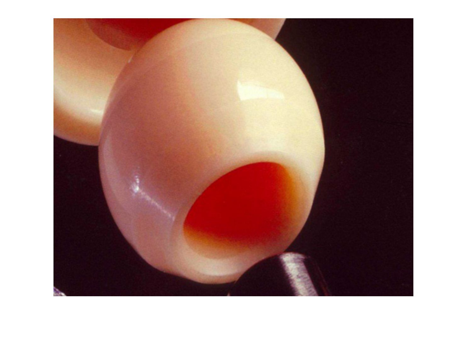
Q1-Q1010 questions — tap to reveal all answerslist
- What are the pros and cons of ceramic bearings?
- What is the fracture rate of ceramic bearings?
- How is ceramic manufactured and what affects its strength?
- Describe the generations of ceramic.
- What are the causes of ceramic liner fracture and how do you avoid it?
- How do you revise a ceramic fracture?
- What are the limitations of ceramic head and neck options?
- What is oxidized zirconium?
- What are the special wear patterns of a ceramic head?
- What is HIP and what is sintering?
Q1.What are the pros and cons of ceramic bearings?
- Pros: low coefficient of friction (0.11-0.12), hard, wettable, inert, corrosion resistant
- Cons: brittle, notch sensitive, anisotropic thus position sensitive -> edge loading and fracture with catastrophic failure; difficult revision
- Cons: squeaking (painless); limited head size and neck length options; expensive
Q2.What is the fracture rate of ceramic bearings?
- 0.013 to 1.1%
- Biolox delta 0.2%
Q3.How is ceramic manufactured and what affects its strength?
- Mix powder and water -> pressed in prefabricated cast -> sintering -> hot isostatic pressing (HIP) -> finishing
- Porosity affected by sintering time; grain size affected by particle size
- Strength inversely proportional to porosity and grain size
Q4.Describe the generations of ceramic.
- 1st: alumina oxide sintered in air; long sintering -> large grain size and impurity -> reduced strength
- 2nd: Yttria stabilized tetragonal zirconium for toughening, phase change absorbs fissure energy; Ca + Mg decrease grain size
- 3rd: HIP after sintering to reduce grain size, limit grain boundaries, increase purity
- 4th: alumina matrix composite - 82% alumina, 17% zirconia, 0.3% chromium, 0.6% strontium; chromium improves hardness, strontium platelets deflect cracks
Q5.What are the causes of ceramic liner fracture and how do you avoid it?
- 4 Ms: malposition, mishandling, instability (microseparation), manufacturing
- Avoid: 4th generation ceramic, correct bore-trunnion match, improved surface finishing
- Surgically: no debris, good fit (liner and trunnion), no malposition, no hammering or trial with ceramic liner, good soft tissue balance
Q6.How do you revise a ceramic fracture?
- Pre-op: revise asap, avoid weight bearing, confirm proper implant position
- Intra-op: thorough debridement and extensive synovectomy
- Avoid further damage of metal component during removal of ceramic component and protect it after removal; prepare to revise metal component if grossly damaged
- Clear all debris from taper and shell before implanting new ceramic
Q7.What are the limitations of ceramic head and neck options?
- Head size limited - ceramic-on-ceramic must be placed within a metal shell
- Small head: less stable, less fluid film
- Limited neck length options -> limits hip offset and increases impingement
Q8.What is oxidized zirconium?
- Metallic alloy with a ceramic surface
- Resistant to abrasion and brittle fracture
- Undetectable nickel ions
Q9.What are the special wear patterns of a ceramic head?
- Stripe wear
- Polar wear with impingement (levering effect)
- Peripheral wear without impingement - microseparation during swing phase and relocation during stance phase
Q10.What is HIP and what is sintering?
- HIP: gas pressure applied isostatically at high temperature to enhance sintering and produce dense bodies
- Sintering: powder product becomes a strong dense ceramic body with heating and pressure, removing voids between particles
- Heating is below melting point
▸ Slide 208 · CoCr: Cobalt 61%, Chromium 20-30%, Molybdenum 6-10%, Nickel, Carbon, TungstenHip · 3 questions expand
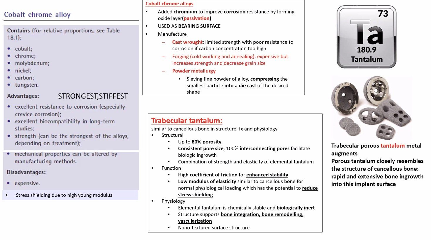
Q1-Q33 questions — tap to reveal all answerslist
- What is the composition of cobalt chrome?
- What is the composition of 316L stainless steel?
- What is the composition of titanium alloy 6Al4V?
Q1.What is the composition of cobalt chrome?
- Cobalt 61%, chromium 20-30%, molybdenum 6-10%, plus nickel, carbon and tungsten
- Young's modulus 210
- Better corrosion profile than stainless steel but susceptible to galvanic corrosion compared with titanium
Q2.What is the composition of 316L stainless steel?
- Iron 62%, chromium 18%, molybdenum 3%, nickel 16%, carbon 0.03%
- L = low carbon
- Young's modulus 190
Q3.What is the composition of titanium alloy 6Al4V?
- Titanium 89%, aluminium 6%, vanadium 4%, others 1%
- Young's modulus 100
- Vanadium toxicity -> newer reiterations contain niobium and zirconium
▸ Slide 209 · MetalHip · 5 questions expand

Q1-Q55 questions — tap to reveal all answerslist
- How are the properties of metals classified?
- What are the features of 316L stainless steel?
- What are the features of cobalt chrome?
- What are the features of titanium?
- What are the features and uses of tantalum?
Q1.How are the properties of metals classified?
- Surface: hardness, wettability, finishing
- Mechanical: stress-strain curve, endurance limit, viscoelasticity
- Biological property
- Manufacturing property
Q2.What are the features of 316L stainless steel?
- 3% molybdenum reduces pitting and crevice corrosion; 16% nickel stabilises FCC lattice -> ductility, fatigue strength, toughness
- Low carbon < 0.03%: carbon forms compound with chromium improving corrosion resistance but decreasing stiffness
- Less hard than chromium, smooth, low wettability; stiff, high tensile strength, tough, ductile
- Relatively biocompatible, readily available, cheap; stress and crevice corrosion -> stress corrosion cracking
- Used in metal plating and screws
Q3.What are the features of cobalt chrome?
- Smooth finishing, hard; stiff, strong, tough; biocompatible
- Resistant to wear and corrosion
- Used for joint replacement articulating surfaces
- Problem of stress shielding
Q4.What are the features of titanium?
- More elastic (lower Young's modulus, similar to bone) -> less stress shielding as a femoral stem
- Ductile; low wear resistance (poor hardness) and notch sensitive
- Biphasic precipitation -> more fatigue resistance
- Biocompatible; excellent corrosion resistance due to self-passivating oxide layer
Q5.What are the features and uses of tantalum?
- Osteoconductive property, biocompatible
- Stiffness similar to bone
- Used as a coating surface
- Rod most used in core decompression
▸ Slide 210 · Biological FixationHip · 7 questions expand

Q1-Q77 questions — tap to reveal all answerslist
- What are the aims and prerequisites of cementless biological fixation?
- How is initial rigid fixation achieved?
- What are the requirements for bone ingrowth?
- How does ongrowth differ from ingrowth?
- What is the role of hydroxyapatite (HA)?
- Describe the Khanuja classification of cementless stems.
- How are plasma spray and grit blasting performed?
Q1.What are the aims and prerequisites of cementless biological fixation?
- Aim: rigid internal fixation + cortical bone contact
- Prerequisite: viable bone
- Macroloc = initial fixation
- Microloc = long term fixation
Q2.How is initial rigid fixation achieved?
- Press fit: reaming 1-2mm less than implant (hoop stress)
- Line-to-line fit: supplemented with screw (cup) or porous coating (stem)
- Cup: 70% cortical contact, 2/3 rim fit, dome intact
- Stem: at least 4cm scratch fit
Q3.What are the requirements for bone ingrowth?
- Porous proximally or extensively coated
- Pore size 50-150um; volume 50% (too much may shear)
- Micromotion < 50-150um (too much = fibrous); gap < 50um
- May be sintered beads, fiber mesh or tantalum
Q4.How does ongrowth differ from ingrowth?
- Ongrowth: grit blasted / plasma spray / HA; always titanium; always extensive (weaker method of fixation)
- Roughness = average distance from peak to valley, proportional to interface shear strength
- Theoretically ingrowth is better, but no clinical difference
- Manufacturing process for porous ingrowth stems can result in diminished fatigue strength properties
Q5.What is the role of hydroxyapatite (HA)?
- Ca10(PO4)6(OH)2, an osteoconductive adjunct
- Benefits shown only in animal models, not in humans
- Deposited by plasma spray (better than electrochemical); optimal 50um
- May cause 3rd body wear
Q6.Describe the Khanuja classification of cementless stems.
- I single wedge (Taperloc); II double wedge (Summit)
- IIIA tapered round (Mallory); IIIB tapered spline/conical (Wagner); IIIC tapered rectangular (Zweymuller)
- IV cylindrical fully coated (Restoration); V modular; VI anatomical
- Proximally coated rely on metaphyseal press fit; extensively coated on diaphyseal scratch fit; type III at metadiaphyseal junction
Q7.How are plasma spray and grit blasting performed?
- Plasma spray: hot air + titanium alloy projected onto the surface
- Grit blasted: stream of abrasive material -> abrasive wear
- Ongrowth implants are always titanium and always extensive (weaker fixation method)
▸ Slide 211 · Cemented fixationHip · 4 questions expand
Q1-Q44 questions — tap to reveal all answerslist
- How does cement achieve fixation and when is it preferred?
- What are the disadvantages of cemented fixation and where is it used?
- What are the fully and partially controllable factors for optimal cement fixation?
- What are the uncontrollable factors affecting cement fixation?
Q1.How does cement achieve fixation and when is it preferred?
- PMMA acts as a grout, transmitting load from implant to bone via a static stable interface with mechanical interlock into cancellous bone
- Good in osteoporotic bone (better penetration) and irradiated bone (not reliant on bone ingrowth)
- e.g. Dorr C, poor bone quality
Q2.What are the disadvantages of cemented fixation and where is it used?
- Bone heat necrosis
- Cardiopulmonary suppression
- PMMA is strong in compression but weak in shear and tension - not preferred in the cup
- Used in the stem
Q3.What are the fully and partially controllable factors for optimal cement fixation?
- Fully controllable: antibiotic and barium inclusion, vacuum mixing to reduce porosity
- Fully controllable: insertion pressurisation, avoid vigorous mixing, type of stem used
- Partially controllable: 2-5mm thick cement mantle, blood and fat inclusion
- Partially controllable: canal preparation - pulsatile lavage, stress riser
Q4.What are the uncontrollable factors affecting cement fixation?
- Cement aging
- Strain rate
- Temperature and humidity in the body
▸ Slide 212 · Ideal THR implantHip · 6 questions expand
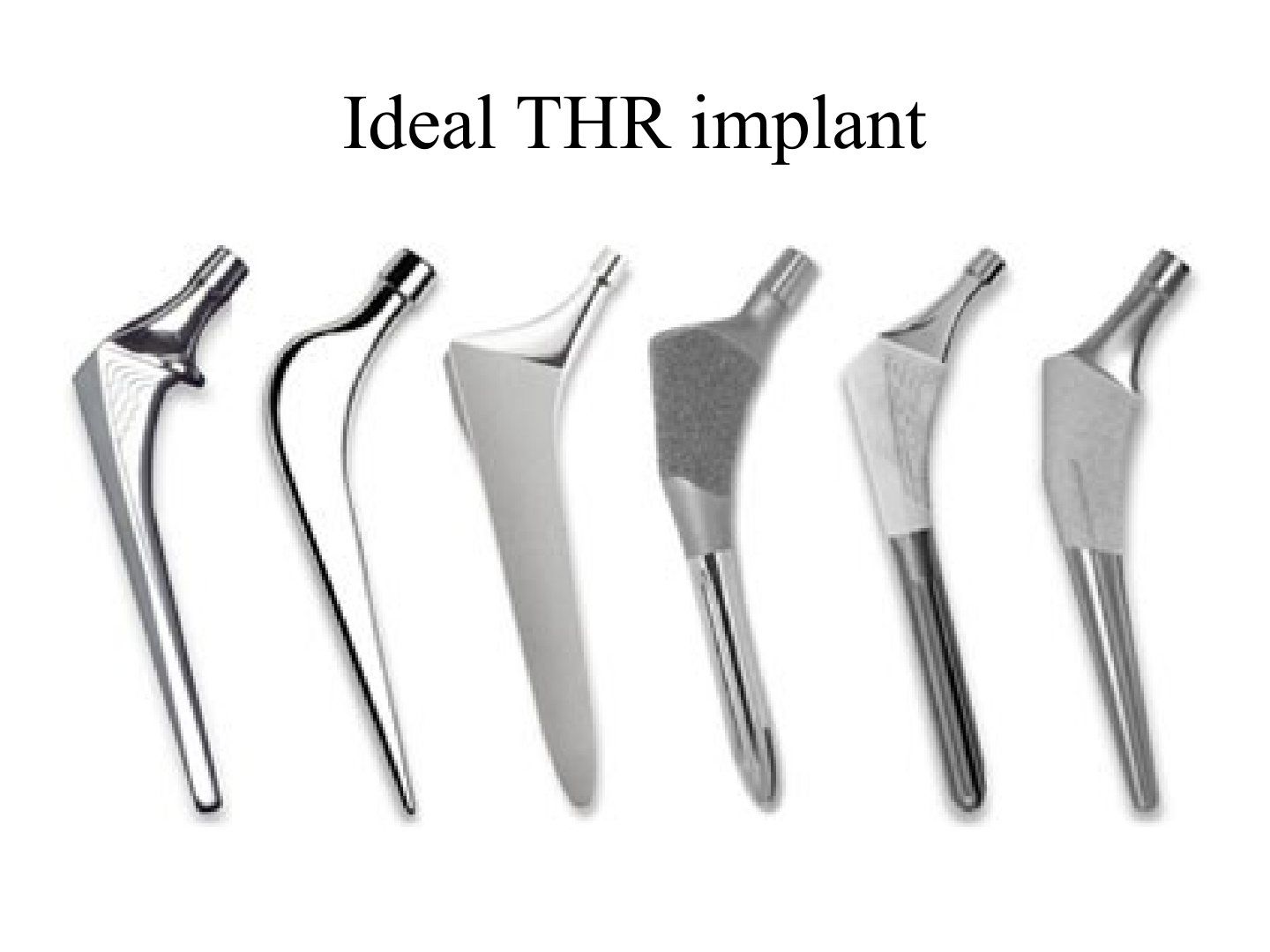
Q1-Q66 questions — tap to reveal all answerslist
- Is there an ideal THR implant?
- What are the 6 headings for choosing a THR implant?
- How does material and fixation differ in young versus old patients?
- What head size is preferred and why?
- Which bearing surface would you choose and why?
- How is stability achieved with cementless fixation?
Q1.Is there an ideal THR implant?
- No ideal implant suits every patient population
- Largely depends on age and quality of bone - should be individualised
- Use registry data to help make decisions
- Lecturer's choice: hybrid, ceramic-on-PE THR
Q2.What are the 6 headings for choosing a THR implant?
- Material (stiff material, high fatigue strength, corrosion resistant)
- Modularity (pros and cons)
- Femoral stem geometry (cementless or cemented)
- Fixation to bone
- Head size
- Bearing surface
Q3.How does material and fixation differ in young versus old patients?
- Young: cementless titanium biological fixation; avoid cement (cyclic loading fails in shear); titanium reduces stress shielding; rule of 50; Swedish registry better 50-70M
- Old: cemented stainless steel stiff stem to avoid bending in the cement mantle, otherwise will fail like the 3M capital THR
- Old: immediate stability with proven longevity, 3rd generation cementation, even 2mm mantle (Barrack A); registry better > 70
Q4.What head size is preferred and why?
- 32mm
- Large head increases head-neck ratio -> decreased impingement, better range, decreased dislocation rate
- Too large -> thin PE thickness (traditionally > 8mm), increased volumetric wear
Q5.Which bearing surface would you choose and why?
- Metal on PE has proven longevity
- Avoids ceramic complications (fracture with malposition, squeaking) and MoM complications (metallosis, ALVAL, pseudotumour)
- Use highly cross-linked PE of at least 5mm thickness
Q6.How is stability achieved with cementless fixation?
- Initial stability: macroloc; press fit vs line to line (extensive coated stem for better frictional fit)
- Long term: microloc - porous coated (ingrowth) vs grit blasted (ongrowth) +/- HA coating
- Consider pore size, porosity, gap, micromotion
- Obtain cortical contact for biological interdigitation and long term fixation
▸ Slide 213 · Implant (hip)Hip · 7 questions 1 check expand

Q1-Q77 questions — tap to reveal all answerslist
- When given a stem, what features do you assess?
- Who was Charnley and what is he famous for?
- What is Charnley's low friction hip arthroplasty design?
- Describe the classical Charnley stem and its generations.
- Compare the Muller and Exeter stems.
- What are the uses of a collar and what was the McKee Farrar prosthesis?
- Describe a cementless stem as presented in the lecture.
Q1.When given a stem, what features do you assess?
- Modularity; material
- Geometry: taper, stem shape (anatomical/straight/curved)
- Surface: polish, ingrowth or ongrowth mechanism, HA coated
- Collar; and what/when it is used for
Q2.Who was Charnley and what is he famous for?
- Father of modern hip replacement
- Charnley low friction arthroplasty (CLFA)
- Introduction of antibiotics into cement, clean air enclosure, total body exhaust suits, instrument trays
- Importance of bony compression in arthrodesis; closed treatment of common fractures
Q3.What is Charnley's low friction hip arthroplasty design?
- 1962
- ultra high molecular weight polyethylene cup (UHMWPE; pioneer to use); small femoral head 7/8 inch = 22.225mm based on the low frictional torque principle
- Trochanteric osteotomy with lateral and distal repositioning
- Medialisation of the cup; use of acrylic cement for load distribution
Q4.Describe the classical Charnley stem and its generations.
- Monobloc, 22.225mm head, collar, single taper with sharp corner, polished
- Flat back -> round back (more cross-sectional area, matted) -> cobra flange -> C stem/triple taper
- Materials: EN58J stainless steel -> 316 low carbon stainless steel -> Orton
- Gen 1 polished flat back; gen 2 round back matt; gen 3 matt with cobra flange; gen 4 polished triple taper C stem
Q5.Compare the Muller and Exeter stems.
- Muller: monobloc, collared, curved, single taper, not polished, sharp anterior edge (stress riser)
- Muller: bigger head and larger head-neck ratio (more volumetric wear, less impingement)
- Exeter: modular, collarless, highly polished, double tapered, round edge
- Exeter: head cobalt chrome, stem orthinox (low carbon SS, nitrogen replaces nickel); designed for controlled subsidence; PMMA centraliser
Q6.What are the uses of a collar and what was the McKee Farrar prosthesis?
- Collar: reference of implant position
- Collar: load medial side of femur to prevent stress shielding (contact may not be enough)
- Collar: initial stability, prevent subsidence
- McKee Farrar: metal-on-metal, 10-year survival 27%
Q7.Describe a cementless stem as presented in the lecture.
- Modular stem
- Collared
- Proximal rough surface with porous coating
- Not tapered; designed to encourage proximal ingrowth
McKee Farrar metal-on-metal prosthesis has a 10-year survival of 27% — likely timeframe error — The 27.5% survivorship figure is from the 808-hip Norwich series at 20 years (August et al. 1986); other series report 76% at 10 years and 74-84% at 20-28 years — medium confidence — source
▸ Slide 214 · This is a charnley stemHip · 6 questions expand
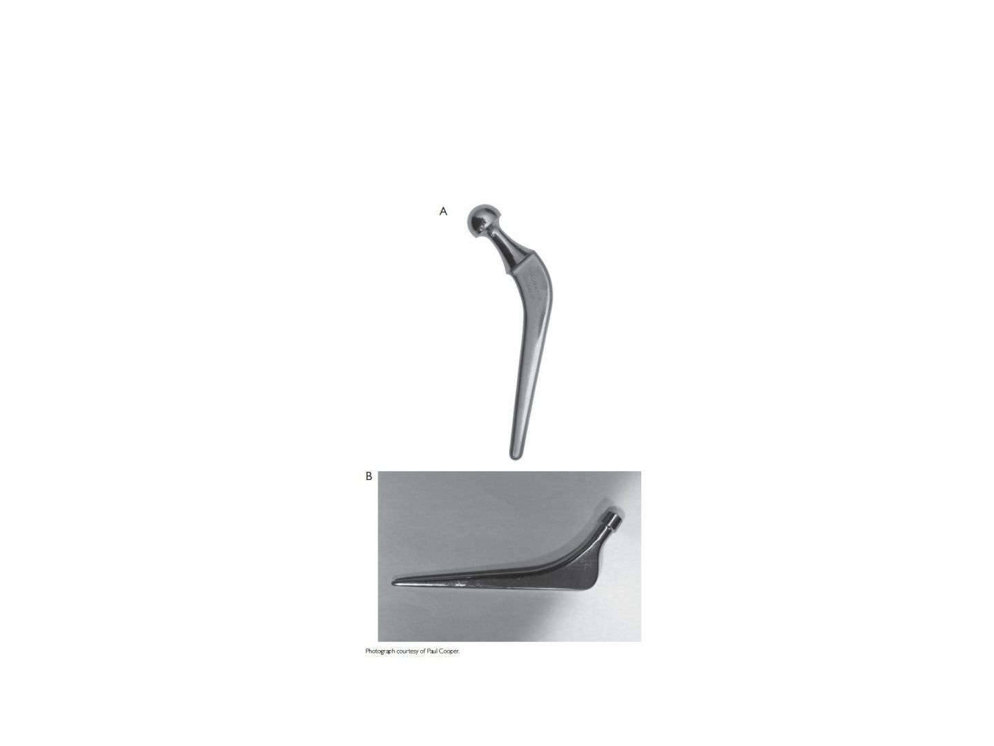
Q1-Q66 questions — tap to reveal all answerslist
- How would you describe this stem?
- Why does the Charnley stem fail?
- Describe the evolution of the Charnley stem.
- How would you describe the Exeter stem?
- Compare the composite beam and taper slip design philosophies.
- What is the evidence comparing Exeter and Charnley survival?
Q1.How would you describe this stem?
- Monobloc stem used in cemented total hip arthroplasty
- Made of Orton, a stainless steel alloy
- Head 22.225mm with a collar to prevent subsidence
- Looks like a round back generation with a matted surface
- Charnley principle: low frictional torque arthroplasty, medialisation of hip centre, lateralisation of GT
Q2.Why does the Charnley stem fail?
- Small head neck ratio
- Sharp edges
- Fatigue failure at the anterolateral tension site in earlier generations
- Loosening from micromotion between implant and cement (Gruen 1a failure)
Q3.Describe the evolution of the Charnley stem.
- Flat back (sharp corner, single tapered) -> round back (increased cross-sectional area to resist fatigue, matted surface) -> cobra flange -> C stem/triple taper
- EN58J stainless steel -> 316 low carbon stainless steel -> Orton
- Four generations: polished - matt - matt - polished
Q4.How would you describe the Exeter stem?
- Modular, collarless stem used in THR or hemiarthroplasty
- Made of orthinox
- Double taper with a polished surface and round edges
- Allows for controlled subsidence
Q5.Compare the composite beam and taper slip design philosophies.
- Charnley (composite beam): a rod in two tubes, relying on mechanical interlock at cement-bone and implant-cement interfaces
- Load travels from the femoral head, bypasses the stem to the tip and to the bone; rough surface and collar minimise micromotion
- The mechanism of failure is different: composite beam fails when micromotions occur at the prosthesis-cement interface; the rough stem will piston within the cement mantle, leading to wear debris circulation around the effective joint space and osteolysis
- Exeter (taper slip): viscoelastic cement dissipates vertical force into hoop stress as the stem subsides, transferred along the whole stem; polished taper stems are inherently stable and fail when they rotate in the axial plane (RSA and retrieval studies)
Q6.What is the evidence comparing Exeter and Charnley survival?
- Kiran Acta Orthop Belg 2019
- Minimum 15-year follow-up, 876 THR
- Exeter 98%, Charnley 97%
▸ Slide 215 · Centraliser made of PMMA with air filled distal voidHip · 6 questions expand
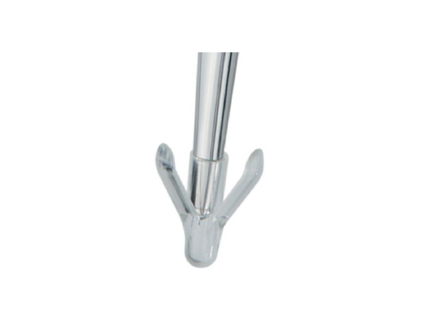
Q1-Q66 questions — tap to reveal all answerslist
- What is the centraliser made of and what is its key design feature?
- What are the functions of a PMMA centraliser with an air-filled distal void?
- How does the air-filled distal void affect stresses at the stem-cement interface?
- What is the consequence of end bearing of the stem tip on the cement mantle?
- How does the centraliser improve the distal cement mantle?
- What does the centraliser seal off and why does this matter?
Q1.What is the centraliser made of and what is its key design feature?
- Made of PMMA
- Contains an air-filled distal void
Q2.What are the functions of a PMMA centraliser with an air-filled distal void?
- Allows the stem to subside into the void in a controlled fashion
- Converts shear stresses into compressive forces with almost no tensile stress
- Prevents end bearing of the stem tip on the cement mantle and cement fracture
- Improves stem position and centralises the tip for an even cement mantle distally
- Seals off the effective joint space and stem-cement interface
Q3.How does the air-filled distal void affect stresses at the stem-cement interface?
- Stem can subside into the void in a controlled fashion
- Converts shear stresses into compressive forces
- Almost no tensile stress on the mantle
Q4.What is the consequence of end bearing of the stem tip on the cement mantle?
- Direct end bearing causes cement fracture
- Prevented by the air-filled distal void allowing controlled subsidence
Q5.How does the centraliser improve the distal cement mantle?
- Improves stem position
- Centralises the tip of the stem
- Creates an even cement mantle distally
Q6.What does the centraliser seal off and why does this matter?
- Seals off the effective joint space
- Seals off the stem-cement interface
- Prevents fluid flow that may lead to loosening
▸ Slide 216 · Muller stemHip · 5 questions expand

Q1-Q55 questions — tap to reveal all answerslist
- What are the design features of the Muller stem?
- What material is the Muller stem made of and is it modular?
- Why does the Muller stem use a 32mm head?
- What is the purpose of the collar on the Muller stem?
- Why is the Muller stem curved and how is it inserted?
Q1.What are the design features of the Muller stem?
- Modular
- Stainless steel
- 32mm head - relatively large head/neck ratio to decrease impingement; alumina ceramic head
- Curved stem - easier insertion in anterior approach
- Sharp edges with diamond-shaped cross section - 1st generation cementation technique
- Collar increases stress transfer to proximal medial femur
Q2.What material is the Muller stem made of and is it modular?
- Stainless steel
- Modular design
Q3.Why does the Muller stem use a 32mm head?
- Relatively large head/neck ratio
- To decrease impingement
Q4.What is the purpose of the collar on the Muller stem?
- Increases stress transfer to proximal medial femur
- Decreases stress shielding
Q5.Why is the Muller stem curved and how is it inserted?
- Curved - easier insertion in the anterior approach
- Sharp edges with diamond-shaped cross section
- Inserted using the 1st generation cementation technique
▸ Slide 217 · Acetabular implants:Hip · 4 questions expand
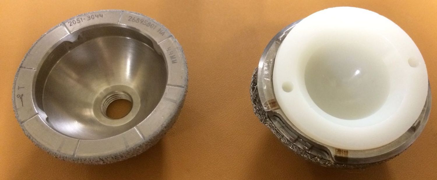
Q1-Q44 questions — tap to reveal all answerslist
- What are the features of cementless acetabular implants?
- What are the disadvantages of cemented acetabular implants?
- Which DeLee and Charnley zone is the most common area for osteolysis?
- What are the features and proposed advantages of a bipolar acetabular implant?
Q1.What are the features of cementless acetabular implants?
- Allow addition of screw fixation
- Screw holes increase the effective joint space
- Potential for backside wear
- Thinner liner
Q2.What are the disadvantages of cemented acetabular implants?
- Decrease cancellous bone stress
- Difficult zone 1 pressurization - increases loosening
Q3.Which DeLee and Charnley zone is the most common area for osteolysis?
- Zone 3 is the most common area for osteolysis
- DeLee and Charnley divided the acetabulum into 3 zones
Q4.What are the features and proposed advantages of a bipolar acetabular implant?
- 2 interfaces
- Advantage is theoretical - possibly less dislocation and less acetabular erosion
- Immediate stability from screw fixation
- Quadrant
▸ Slide 218 · PCA cup (porous coated anatomical cup):Hip · 4 questions expand

Q1-Q44 questions — tap to reveal all answerslist
- What fixation does the PCA cup use initially and long term?
- What are the problems with the PCA polyethylene liner?
- What problems arise from the other design features of the PCA cup?
- What is the 12-year survivorship of the PCA stem and cup?
Q1.What fixation does the PCA cup use initially and long term?
- Initial fixation: 2 pegs
- Long term: CoCr sintered beads
- Porosity is not great for bone ingrowth
Q2.What are the problems with the PCA polyethylene liner?
- Irradiated in air - prone to generating PE particles
- Uneven thickness - thinnest at the locking mechanism with sharp edges, causing stress concentration
Q3.What problems arise from the other design features of the PCA cup?
- Central peg and antirotation notch at the rim increase the effective joint space
- Protrusion beyond the cup leads to impingement
- Poor locking mechanism causes backside wear
Q4.What is the 12-year survivorship of the PCA stem and cup?
- Stem 97%
- Cup 87.7%
▸ Slide 219 · Ganz reinforcement ringHip · 3 questions expand
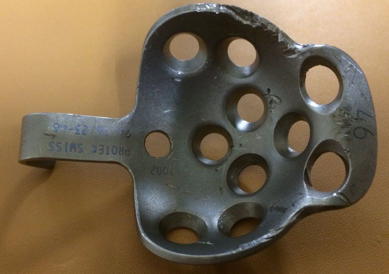
Q1-Q33 questions — tap to reveal all answerslist
- What is a Ganz reinforcement ring?
- How is a Ganz reinforcement ring fixed?
- Why does fixation of the Ganz ring depend only on screws?
Q1.What is a Ganz reinforcement ring?
- A reinforcement ring with a hook
- Has screw holes for fixation
Q2.How is a Ganz reinforcement ring fixed?
- Bone graft on the defect
- Fix the ring with screws
- Then cement PE onto the ring
Q3.Why does fixation of the Ganz ring depend only on screws?
- There is no ingrowth or ongrowth onto the ring
- Ring fixation depends only on the screws
▸ Slide 220 · Isoelastic stem is a type of cementless monobloc stem made of polyacetal resin wHip · 6 questions expand

Q1-Q66 questions — tap to reveal all answerslist
- What type of stem is the isoelastic stem and what is it made of?
- What is the geometry of the isoelastic stem?
- What is the surface of the isoelastic stem and how does it achieve fixation?
- Why is the isoelastic stem designed to have a Young's modulus similar to bone?
- How does the isoelastic stem transmit force?
- What are the disadvantages of the isoelastic stem?
Q1.What type of stem is the isoelastic stem and what is it made of?
- Cementless monobloc stem
- Polyacetal resin with a stainless steel core
Q2.What is the geometry of the isoelastic stem?
- Quadrilateral cross section + fluted for rotational stability
- Straight conical stem
- Collared
Q3.What is the surface of the isoelastic stem and how does it achieve fixation?
- Crosshatched surface and conical recess for bone ingrowth
- Skirted head, usually used with a cemented PE cup
Q4.Why is the isoelastic stem designed to have a Young's modulus similar to bone?
- Young's modulus similar to bone
- Reduces stress shielding
Q5.How does the isoelastic stem transmit force?
- Medially through the collar to the medial calcar
- Laterally through 2 screws at the greater trochanter
Q6.What are the disadvantages of the isoelastic stem?
- Less strong material - porosity cannot be small - poor bone ingrowth and micromotion
- Generates polyacetal molecules which increase wear
- Small head-neck ratio (thick neck to prevent fracture) - impingement
▸ Slide 221 · MonoblockHip · 6 questions expand

Q1-Q66 questions — tap to reveal all answerslist
- What material is the monoblock stem made of?
- What is the geometry of the monoblock stem?
- What surface features does the monoblock stem have?
- How is the monoblock stem fixed?
- What are the modes of failure of the monoblock stem?
- How can failure of the monoblock stem be prevented? (Yau, Injury 2003)
Q1.What material is the monoblock stem made of?
- Cobalt chrome – Molybdenum alloy
Q2.What is the geometry of the monoblock stem?
- Curved stem tapering in coronal and sagittal profile
- Transverse dimension rectangular
Q3.What surface features does the monoblock stem have?
- Collared with fenestrations
- Matted surface
Q4.How is the monoblock stem fixed?
- Cementless fixation with the 3 point fixation principle
- No bone ingrowth unless bone graft is inserted into the fenestrations
Q5.What are the modes of failure of the monoblock stem?
- Loosening
- Subsidence
Q6.How can failure of the monoblock stem be prevented? (Yau, Injury 2003)
- Only use in patients >75 years
- Metaphyseal filling >75%
▸ Slide 222 · LooseningHip · 8 questions expand
Q1-Q88 questions — tap to reveal all answerslist
- What are the Harris criteria for definite femoral loosening?
- What are the Harris criteria for probable femoral loosening?
- What are the Harris criteria for possible femoral loosening?
- What are the reported percentages for the DeLee and Charnley zones?
- What are the radiological signs of cementless acetabular loosening?
- What are the features of bony ingrowth in the Engh classification?
- What is the radiological feature of stable fibrous ingrowth (Engh)?
- What are the radiological features of an unstable cementless femoral stem (Engh)?
Q1.What are the Harris criteria for definite femoral loosening?
- Stem fracture, subsidence, cement fracture
- Loosening at the stem-cement interface
Q2.What are the Harris criteria for probable femoral loosening?
- 100% cement-bone interface radiolucency
Q3.What are the Harris criteria for possible femoral loosening?
- 50-100% cement-bone interface radiolucency
- Not present immediately post-op
Q4.What are the reported percentages for the DeLee and Charnley zones?
- 3 zone: 94%
- 2 zone: 71%
- 1 zone: 7%
Q5.What are the radiological signs of cementless acetabular loosening?
- >8 degrees change in opening angle
- 3mm translation
- Shedding of porous coating
- Halo around screw
Q6.What are the features of bony ingrowth in the Engh classification?
- Cortical hypertrophy at stem tip
- Spot welding
- Proximal stress shielding
Q7.What is the radiological feature of stable fibrous ingrowth (Engh)?
- Parallel line along the stem at the bone-implant junction
- Indicates stable fibrous ingrowth in the Engh classification
Q8.What are the radiological features of an unstable cementless femoral stem (Engh)?
- Distal pedestal
- Calcar hypertrophy
- Divergent line
- Migration/subsidence
- Porous coating shedding
▸ Slide 223 · Patient walking with trendelenberg gait after THRHip · 6 questions expand

Q1-Q66 questions — tap to reveal all answerslist
- What is the common cause of Trendelenburg gait after THR?
- How can a defective abductor mechanism cause Trendelenburg gait after THR?
- How is head offset measured?
- How do you restore abductor function in THR?
- What are the problems of increased offset after THR?
- What are the problems of decreased offset after THR?
Q1.What is the common cause of Trendelenburg gait after THR?
- Defective abductor mechanism - think about the lever system
Q2.How can a defective abductor mechanism cause Trendelenburg gait after THR?
- Instability to generate force: nerve/muscle weakness or inhibition from pain
- Shortened lever arm from altered vertical/horizontal offset - head-neck shortening, LLD with poor length-tension
- Unstable fulcrum (dislocated hip)
Q3.How is head offset measured?
- From the hip centre to the anatomical axis of the femur
- Relevant when restoring offset in THR
Q4.How do you restore abductor function in THR?
- Restore the hip centre
- Restore offset
- Restore neck length
Q5.What are the problems of increased offset after THR?
- Increased lever arm of the neck - increased bending moment at the stem - risk of neck fracture
- If increase with long neck may need larger neck which decreased head neck ratio
- Trochanteric bursitis
Q6.What are the problems of decreased offset after THR?
- Abductor tension decreased - power decreased (length-tension relationship)
- Decreased abductor lever arm
- Increased joint reaction force
▸ Slide 224 · Morse taperHip · 9 questions 1 check expand
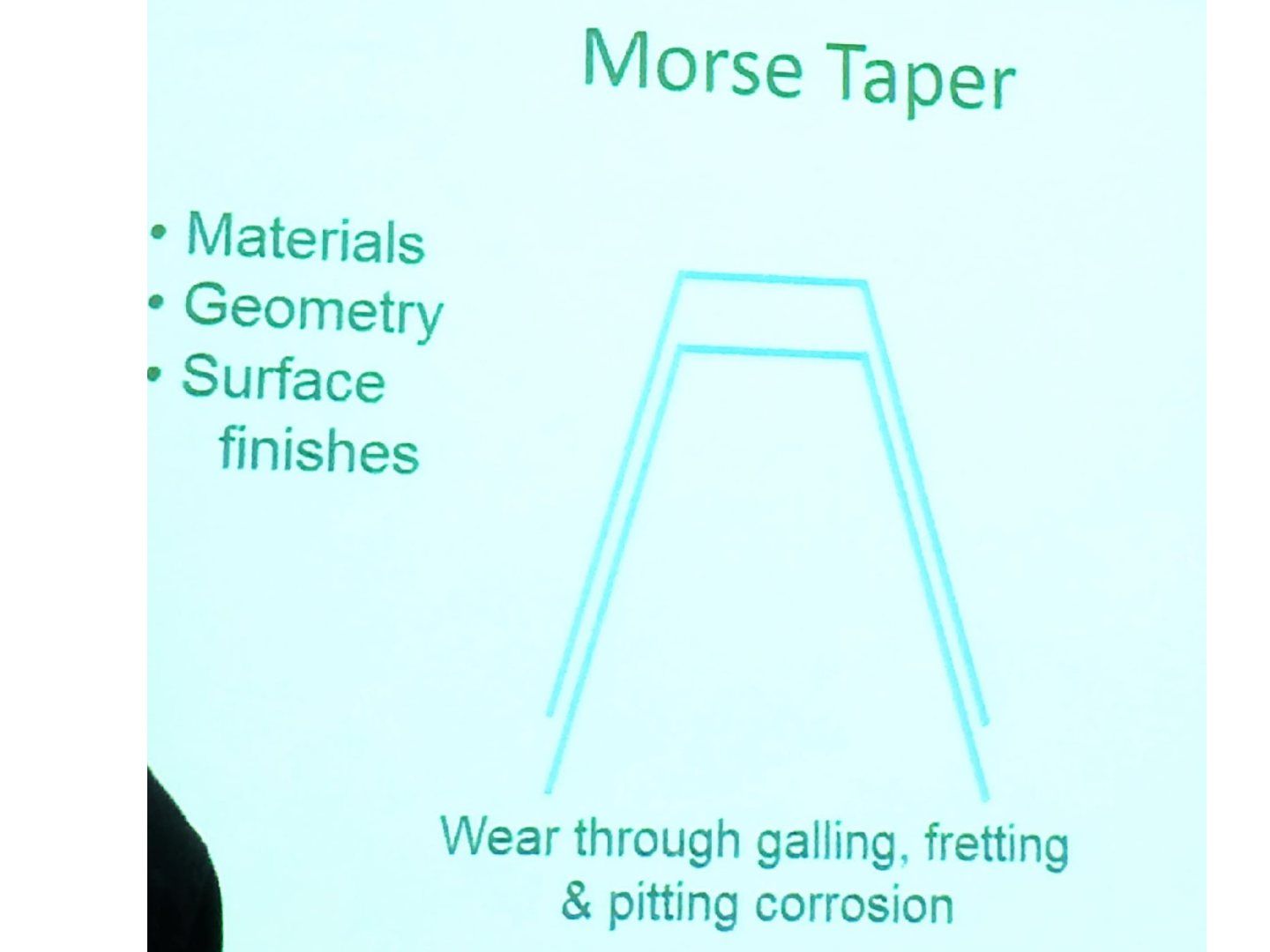
Q1-Q99 questions — tap to reveal all answerslist
- What is a Morse taper?
- What material factors affect Morse taper performance?
- Describe the taper angles of the C taper and the V40 taper.
- What is the critical locking angle of a Morse taper?
- What taper geometry gives more ROM, and what is the downside of a short taper?
- What surgeon factors affect Morse taper fixation?
- What is the difference between positive and negative taper mismatch?
- What produces the side bearing stability at a Morse taper?
- Why can head and neck of different brands not be mixed?
Q1.What is a Morse taper?
- Trunnion (stem) and bore (head) - a cone in a cone
- Some mismatch between the cones allows an interference fit
- Impaction expands the bore walls and causes cold welding (van der Waals force)
Q2.What material factors affect Morse taper performance?
- Titanium allows higher pull-off and torsional strengths than cobalt chrome
- Mixed alloy tapers corrode more and have lower flexural rigidity
- Surface roughness also affects performance
Q3.Describe the taper angles of the C taper and the V40 taper.
- C taper 12/14; V40 has 8% less taper length and 20% lower surface area
- Both have a 5 degrees 40' (5.67 degree) taper angle
Q4.What is the critical locking angle of a Morse taper?
- <7 degrees half taper angle mismatch = self-locking
Q5.What taper geometry gives more ROM, and what is the downside of a short taper?
- Shorter taper + small diameter with trapezoidal neck gives more ROM than a large diameter long taper with circular neck
- A short taper sits entirely within the bore, leading to edge loading
Q6.What surgeon factors affect Morse taper fixation?
- Angle and force of impaction
- Repeated impactions - single strike of >6000N (20cm drop with 1.4kg hammer, Pennock 2006)
- Fluid contamination - dry environment preferred
- Femoral head size
Q7.What is the difference between positive and negative taper mismatch?
- Female head angle > trunnion angle = proximal contact (positive mismatch)
- Female head angle < trunnion angle = distal contact (negative mismatch)
- Cannot mix head and neck of different brands
Q8.What produces the side bearing stability at a Morse taper?
- Taper angles differ between the spigot and the bore, creating an angle mismatch
- This causes integration between the two tapers
- A smaller angle difference between male and female components gives a high level of stability
Q9.Why can head and neck of different brands not be mixed?
- Taper angles and geometry differ between manufacturers
- Cannot mix head and neck of different brands
Morse taper: taper angle 2-12' — garbled/incorrect angle — The original Morse taper is 2 degrees 50 minutes; orthopaedic tapers generally range 5-18 degrees (e.g. 12/14 taper 5 degrees 40 minutes) — medium confidence — source
▸ Slide 225 · PCL Retained implantHip · 8 questions expand
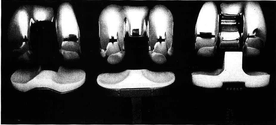
Q1-Q88 questions — tap to reveal all answerslist
- What are the features of a PCL-retaining knee design?
- What are the contraindications to a PCL-retaining knee?
- What are the advantages of a PCL-retaining knee?
- What are the disadvantages of a PCL-retaining knee?
- What is the sacrificing (total condylar) design - features, advantages and disadvantages?
- What is the stabilizing (posterior-stabilized) design - mechanism and advantages?
- What complications are associated with the stabilizing design?
- What is the survivorship difference between PCL-retaining and stabilized designs (Australian joint registry, 15-year revision)?
Q1.What are the features of a PCL-retaining knee design?
- Tibia box leaves space for the PCL (notch)
- Tibia insert has space for the PCL
- Relatively flat and not conforming to the femoral component
- Asymmetrical on the tibia insert - must differentiate the side
Q2.What are the contraindications to a PCL-retaining knee?
- Inflammatory arthritis
- PCL injury
- Patellectomy
Q3.What are the advantages of a PCL-retaining knee?
- Better flexion
- Better proprioception
- Preserves bone stock
- Preserves joint line level
- PCL takes some varus/valgus stress otherwise transferred to the bone-implant junction
Q4.What are the disadvantages of a PCL-retaining knee?
- Difficult to balance
- PCL may not function afterwards - failure gives 40% decrease in rollback (paradoxical roll forward)
- Flat PE - low contact area, high contact stress, point contact, seesaw effect, wear
- Mayo 3907 TKRs, 10 year survivorship 91%
Q5.What is the sacrificing (total condylar) design - features, advantages and disadvantages?
- Symmetrical femoral component; insert no cam and post, conformed
- Advantages: preserves bone; technically easier than CR knee
- No rollback, poor range
- High conformity - large contact area, low contact stress but abrasive/adhesive wear - PE particles
- Dislocation/subluxation if gaps not balanced
- Insall 215 TKRs, 21 year 91 % success
Q6.What is the stabilizing (posterior-stabilized) design - mechanism and advantages?
- Controlled rollback by cam-post mechanism; better conformity than PCL-retained
- Advantages: easier to balance, less point loading/fatigue wear, no reliance on PCL, better exposure in revision/deformity
- Disadvantages: more constraint - loosening at bone-implant interface, reduced ROM, more bone loss, post wear, raised joint line
- Insall 2036 metal-backed, 14 year 98 % success
Q7.What complications are associated with the stabilizing design?
- Patellar impingement from thicker PE and joint elevation
- Patella clunk, jump cam, posterior dislocation
- Peg wear and fracture
- More bone cut in the femur; more constraint increases stress at the prosthesis-bone interface
Q8.What is the survivorship difference between PCL-retaining and stabilized designs (Australian joint registry, 15-year revision)?
- Minimally stabilized 6.0
- PS 7.2
- Medial pivot 6.2
- CR > medial pivot > PS
▸ Slide 226 · Principle: uncouple flexion/extension from axial rotation --> less stress on fixHip · 4 questions expand
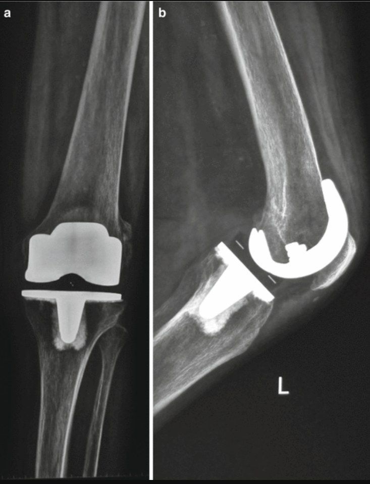
Q1-Q44 questions — tap to reveal all answerslist
- What is the principle of a rotating platform knee design?
- What are the pros of a rotating platform?
- What are the cons of a rotating platform?
- What other rationale supports the rotating platform and what is the evidence?
Q1.What is the principle of a rotating platform knee design?
- Uncouples flexion/extension from axial rotation - less stress on the fixation interface, longer survival
- Solves the kinematic conflict: more conformity means less wear but less ROM and more implant-bone stress
Q2.What are the pros of a rotating platform?
- Decreased stress to bone-cement/cement-implant interface
- PE and femoral component can be made more conformed
- Rotation occurs through the PE and tibia tray
Q3.What are the cons of a rotating platform?
- If the gap is not well balanced - PE spin out
- Backside wear
Q4.What other rationale supports the rotating platform and what is the evidence?
- Restore normal knee kinematics of posterior rollback + medial pivot
- KSSTA 2019 Park: no difference in midterm clinical or radiological results
▸ Slide 227 · How do you reduce total joint replacement infection rate?Hip · 6 questions 1 check expand
Q1-Q66 questions — tap to reveal all answerslist
- How do you reduce total joint replacement infection rate pre-operatively?
- What intra-operative measures reduce infection risk?
- What intra-operative soft tissue and planning measures reduce infection risk?
- What post-operative measures reduce infection?
- What prophylactic antibiotic is used and what is the dose?
- What were the infection rates in the Lidwell MRC trial?
Q1.How do you reduce total joint replacement infection rate pre-operatively?
- Patient selection; medical optimization (HbA1c <7)
- Screen for active infection (UTI, tinea pedis, LL ulcer, MRSA, dental caries)
- MRSA carrier: nasal decolonization with mupirocin 2%
- Preop skin cleansing with chlorhexidine (WHO, ICM, CDC)
- Same day admission to a clean ward
Q2.What intra-operative measures reduce infection risk?
- Theatre design: HEPA filter, exponential airflow (at least 20x air exchange per hour), room-in-room
- Limit traffic and personnel
- Systemic prophylaxis within 1hr of skin incision
- Impermeable drape and gown, body exhaust system
- Iodine impregnated drapes (NICE)
- Antibiotic-loaded cement, haemostasis, watertight closure
Q3.What intra-operative soft tissue and planning measures reduce infection risk?
- Decrease soft tissue trauma
- Avoid prolonged procedure with meticulous preop planning
Q4.What post-operative measures reduce infection?
- Antibiotic coverage to 24 hours (Wilson and Savati: infection reduced from 11% to 1%)
- No drain
- Optimize preop Hb to minimize the need for postop transfusion
- Minimise dressing changes
Q5.What prophylactic antibiotic is used and what is the dose?
- Cefazolin 2g 2 hours before skin incision, extra 1g if operation >4 hours
- Bactericidal against Streptococcus and S. aureus, good tissue penetration
Q6.What were the infection rates in the Lidwell MRC trial?
- Clean air 1.7%
- Antibiotics 0.85%
- Antibiotics + clean air 0.4%
- Antibiotics + clean air + body exhaust suit 0.2%
Cefazolin 2g 2 hours before skin incision — timing incorrect and contradicts the same slide's 'within 1hr' rule — Cefazolin 2g (3g if >120kg) is given within 60 minutes before skin incision; a 2-hour window applies to vancomycin/fluoroquinolone infusions — source
▸ Slide 228 · 1. What does this photo demonstrate? 2. How to design a theatre?Hip · 11 questions 1 check expand

Q1-Q1111 questions — tap to reveal all answerslist
- How is a theatre divided into zones?
- What are the environmental requirements of a theatre?
- How does theatre ventilation decrease infection?
- What is plenum (positive pressure) ventilation?
- What is laminar flow ventilation?
- What is exponential airflow in theatre ventilation?
- What does HEPA filtration remove?
- How is theatre air monitored?
- What is the definition of ultraclean air?
- What is the literature evidence for laminar flow?
- What is the aim of theatre design?
Q1.How is a theatre divided into zones?
- 4 zones: outer, clean, aseptic, disposal
- Outer = rest of hospital
- Clean = reception to theatre door
- Aseptic ends at entry of the anaesthetic room
- Disposal zone
Q2.What are the environmental requirements of a theatre?
- Temperature 22 degrees; radiant warming blankets (bearhugger shown to increase infection rate)
- Humidity 40-60%
- Light without shadow - minimum 40,000 lux at incision site
Q3.How does theatre ventilation decrease infection?
- 95% of SSI are from airborne infection
- Two components: pressured air and clean air
Q4.What is plenum (positive pressure) ventilation?
- 15-20x air changes/hour
- Effective at <35 CFU/m3
Q5.What is laminar flow ventilation?
- Parallel flow lines, uniform velocity, single direction (horizontal or vertical)
- 200-400 air change/hr
Q6.What is exponential airflow in theatre ventilation?
- Inverted trumpet flow
- Vertical flow then radially directly outwards
- Prevents entrainment
Q7.What does HEPA filtration remove?
- Filters particles >0.5 microns with 99.997% efficiency (newer HEPA filters >0.1 micron)
- Smallest bacteria 40 microns; coronavirus 0.125 microns (the droplet, not the virus itself)
Q8.How is theatre air monitored?
- Measured by colony forming units (CFU)/m3, sampled every 3 months
- Passive monitoring: settle plate
- Active monitoring: volumetric Casella slit sampler, 30L/min air through sampler, grow in 37C for 2 days
Q9.What is the definition of ultraclean air?
- <20 CFU/m3 in the periphery of the theatre
- <10 CFU/m3 in the centre of the theatre
Q10.What is the literature evidence for laminar flow?
- Charnley 1972: deep infection reduced from 7% to 0.5%
- Lidwell 1982: non-clean air 3.4%, clean air 1.7%, antibiotics 0.85%, clean air + antibiotics 0.4%, + exhaust suit 0.2%
- Hooper NZ registry 2011 JBJS: laminar flow/space suits did not reduce revision for early deep infection
- BUT BJJ 2018 Thomas: ultraclean air still valid
- Other cited work: Bischoff 2017 Lancet and the Rillio trial
Q11.What is the aim of theatre design?
- Decrease infection rate
- Provide optimal conditions for patient and staff
HEPA filters particles >0.5 microns with 99.997% efficiency — wrong particle size/efficiency benchmark — HEPA is rated at >=99.97% removal of particles at the most penetrating size of about 0.3 microns; smaller and larger particles are removed more efficiently — source
▸ Slide 229 · How to reduce wear in THRHip · 8 questions expand
Q1-Q88 questions — tap to reveal all answerslist
- What is wear and what is it proportional to?
- What is McKellop's classification of wear?
- How can first mode wear be reduced? (volumetric wear = sliding distance x load / hardness)
- What are the patient, implant and surgeon factors in wear?
- How can friction be reduced and lubrication improved (lambda ratio)?
- What is the Stribeck curve used for?
- What is tribology and what is the friction equation?
- What coefficient of friction values are quoted?
Q1.What is wear and what is it proportional to?
- Progressive loss of a bearing substance caused by mechanical or chemical action
- Proportional to friction and inversely proportional to lubrication
Q2.What is McKellop's classification of wear?
- First mode: between the two bearing surfaces intended by the designer
- Second mode: wear through
- Third body: cement debris and loose bone
- Modularity: at the trunnion interface
Q3.How can first mode wear be reduced? (volumetric wear = sliding distance x load / hardness)
- Reduce load (weight, activity; increase offset and medialize cup to decrease JRF; correct cup orientation to avoid edge loading)
- Reduce sliding distance (head size)
- Increase hardness, reduce roughness, improve PE intrinsic properties (manufacturing, conformity, thickness)
Q4.What are the patient, implant and surgeon factors in wear?
- Patient: BMI, activity level
- Implant: head size, articulation material, PE thickness, PE manufacturing
- Surgeon: implant position and alignment, soft tissue tension, cup and trunnion locking, cement debris
Q5.How can friction be reduced and lubrication improved (lambda ratio)?
- Aim for fluid film lubrication - a thin layer of lubricant separating the bearing surfaces
- Lambda ratio = fluid film thickness / asperities
- Increase film thickness: bearing wettability, large head for entrainment velocity (but higher frictional torque = friction x radius), radial clearance 90-200um
- Decrease asperities: hard material, low coefficient of friction
Q6.What is the Stribeck curve used for?
- Sommerfeld parameters: x axis = viscosity x speed / load, y axis = coefficient of friction
- Shows transition from boundary to mixed to fluid film lubrication
- Load distribution over the bearing surface also affects film thickness
Q7.What is tribology and what is the friction equation?
- Tribology = science dealing with the interaction between surfaces in contact and the consequences of that interaction
- Friction = load between surfaces x coefficient of friction (F = uL)
- Friction is independent of surface area and speed
- Depends on roughness of the two surfaces and the presence of lubrication
Q8.What coefficient of friction values are quoted?
- Knee 0.005-0.02
- Hip 0.01-0.04
- Metal on PE 0.02
- Metal on metal 0.8
▸ Slide 230 · This is an explanted PE cup with evidence of eccentric wear. It may have been reHip · 6 questions expand

Q1-Q66 questions — tap to reveal all answerslist
- What is aseptic loosening?
- What is osteolysis?
- What is wear?
- What are the mechanisms of wear?
- Why might an explanted PE cup show eccentric wear?
- How do stress shielding, osteolysis and infection differ radiologically?
Q1.What is aseptic loosening?
- Failure of fixation leading to macromotion and micromotion at the implant-bone interface
- Due to inadequate initial mechanical fixation or biological loss of fixation secondary to particle-induced osteolysis
Q2.What is osteolysis?
- A histiocytic response to wear debris
- Causes bone resorption and resultant implant loosening
Q3.What is wear?
- Progressive loss of a bearing substance caused by mechanical or chemical action
Q4.What are the mechanisms of wear?
- Abrasive: asperites on the hard bearing carve ridges into the soft bearing (cheese grater effect)
- Adhesive: opposing asperites of two surfaces bond with each other to form a junction; the junction is held by intermolecular bonds and generates friction. If the bonds are stronger than the cohesive strength of the weaker material, the weaker material is sheared off
- Fatigue: cyclic loading below the ultimate tensile load but above the endurance limit leads to small cracks in subsurface; propagation leads to delamination of surface
- Erosive (third body): extraneous material enters the interfacial region
Q5.Why might an explanted PE cup show eccentric wear?
- Aseptic loosening
- Infection
- Recurrent dislocation
Q6.How do stress shielding, osteolysis and infection differ radiologically?
- Stress shielding - round off
- Osteolysis - punched out
- Infection - endosteal scalloping + periosteal reaction
▸ Slide 231 · Photo showing removed total knee polyethylene liner (hood et al)Hip · 8 questions expand
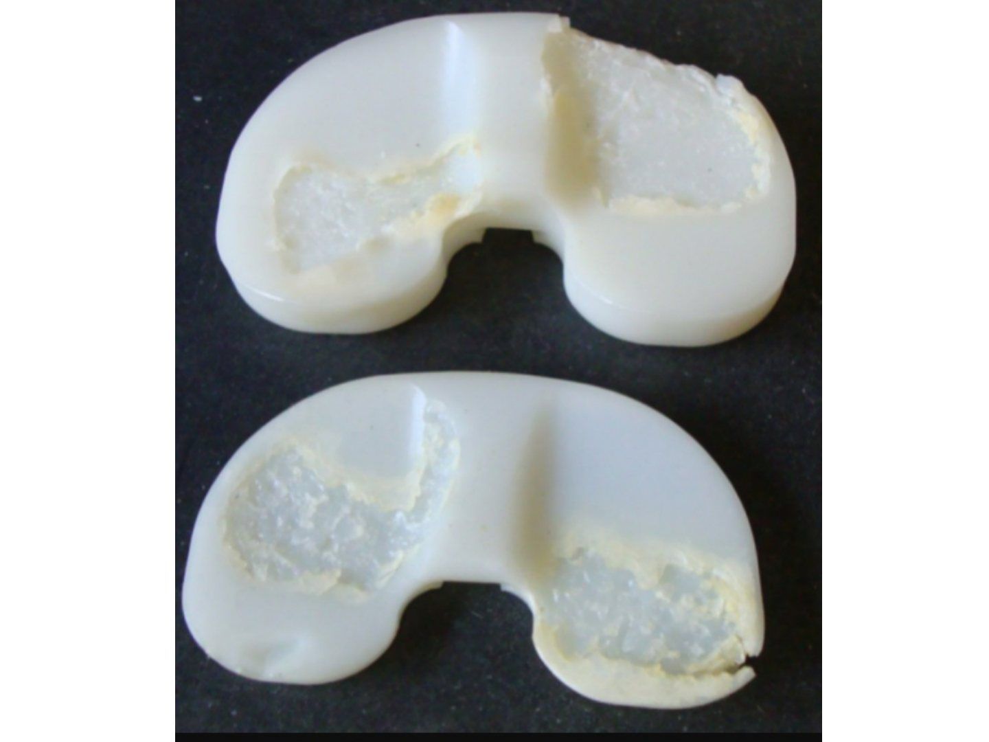
Q1-Q88 questions — tap to reveal all answerslist
- What features are seen on an explanted total knee polyethylene liner?
- What are the mechanisms of wear seen in the removed TKR polyethylene liner?
- What is wear in the context of TKR?
- Why is fatigue wear important in TKR?
- How can wear in TKR be reduced?
- What patient, implant and surgeon factors affect wear in TKR?
- What is the Lambda ratio and what determines the quality of lubrication in TKR?
- What is the evidence for computer navigation and robotic-assisted TKR?
Q1.What features are seen on an explanted total knee polyethylene liner?
- White bands of subsurface delamination and fatigue cracking
- Yellowing as a result of oxidation
Q2.What are the mechanisms of wear seen in the removed TKR polyethylene liner?
- Burnishing (polishing) - combination of adhesive and abrasive wear, generates submicron particles
- Scratching - abrasive wear
- Pitting - fatigue wear from repetitive tensile and compressive surface stresses
- Third body wear (embedded debris)
- Creep (surface deformation) - if severe, may indicate severe malalignment
Q3.What is wear in the context of TKR?
- Removal of material from two surfaces under load due to the sliding motion between them
- Proportional to friction and inversely proportional to lubrication
Q4.Why is fatigue wear important in TKR?
- TKR is not conforming - point loading and shearing stress in PE
- Repetitive cycling causes fatigue failure below UTS
- TKR functions above the endurance limit and below UTS
- PE mechanical properties/manufacturing matter most (strength, fatigue resistance, vita E, thickness)
Q5.How can wear in TKR be reduced?
- First mode: reduce load, restore mechanical alignment to avoid edge loading, soft tissue balancing (coronal and F/E gaps)
- Implant design: bearing conformity (PS or mobile bearing)
- Second mode: proper locking mechanism or monobloc tibial component
- Third body: remove cement debris and loose bone
- Modularity: tight trunnion fitting, minimal fluid at interfaces
Q6.What patient, implant and surgeon factors affect wear in TKR?
- Patient: weight, activity level
- Implant: conformity, material, PE thickness and manufacturing
- Surgeon: soft tissue balance, mechanical axis, coupling of PE and modular components, implant position, third bodies
Q7.What is the Lambda ratio and what determines the quality of lubrication in TKR?
- Aim for fluid film lubrication - a thin layer of lubricant separating the bearing surfaces
- Lambda ratio = fluid film thickness / asperities
- Increase film thickness: bearing wettability, lubricant viscosity, radial clearance (Sommerfeld parameters)
- Decrease asperities: hard material, low coefficient of friction
Q8.What is the evidence for computer navigation and robotic-assisted TKR?
- Navigation: most mid/long-term studies show no substantial functional benefit
- Recent Aus JR: small advantage, especially in younger patients - small reduction in revision for loosening
- Robotics (BJJ 2018 Kayani): less pain, improved early functional recovery, reduced time to discharge
▸ Slide 232 · SN curveHip · 5 questions expand
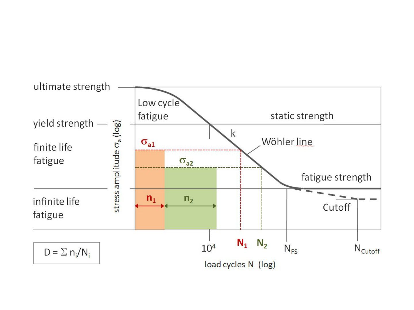
Q1-Q55 questions — tap to reveal all answerslist
- What does an SN curve show?
- What is fatigue failure?
- What is the endurance limit?
- What is fatigue strength/limit?
- What fracture and fatigue types are described on the SN curve?
Q1.What does an SN curve show?
- Number of cycles leading to fracture at a specific stress
- Describes fatigue failure from finite-life to infinite-life fatigue
Q2.What is fatigue failure?
- Failure due to repetitive loading at a stress less than UTS
- Plotted on the SN curve
Q3.What is the endurance limit?
- Stress at which a material can withstand 10 million cycles without fatigue failure
- Divides finite-life from infinite-life fatigue
Q4.What is fatigue strength/limit?
- Stress at which fracture occurs after a specified number of loading cycles
- Also called the fatigue limit
Q5.What fracture and fatigue types are described on the SN curve?
- Brittle fracture
- Fatigue fracture
- Creep fracture
- Low cycle fatigue and high cycle fatigue
▸ Slide 233 · Theoretical benefits:Hip · 2 questions expand

Q1-Q22 questions — tap to reveal all answerslist
- What are the theoretical benefits of an all-poly tibial component?
- What is the evidence for all-poly versus metal-backed tibial components?
Q1.What are the theoretical benefits of an all-poly tibial component?
- More even distribution of weight bearing stresses
- Same amount of tibial resection allows a thicker PE
- Reduction in potential polyethylene deformity caused by creep
- Less tibial bone resection - large metaphyseal area to share stresses
Q2.What is the evidence for all-poly versus metal-backed tibial components?
- KSSTA 2017 Longo systematic review
- No significant difference in outcome scores
- High revision and complication rates in all poly
▸ Slide 234 · Zirconium oxide ceramic coating on a zirconium metal alloy femoral componentHip · 2 questions expand
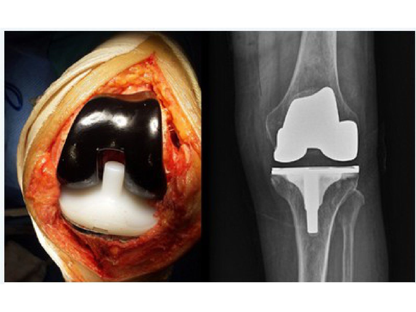
Q1-Q22 questions — tap to reveal all answerslist
- What are the benefits of a zirconium oxide ceramic-coated femoral component?
- How is the zirconium oxide ceramic coating manufactured?
Q1.What are the benefits of a zirconium oxide ceramic-coated femoral component?
- Zirconium oxide ceramic coating on a zirconium metal alloy component
- More scratch resistant
- Less wear debris production
- Safe in nickel sensitivity
Q2.How is the zirconium oxide ceramic coating manufactured?
- Zirconium alloy heated in air
- Oxygen diffuses into the alloy
- The surface transforms into ceramic
▸ Slide 235 · What is osteolysis?Hip · 9 questions expand
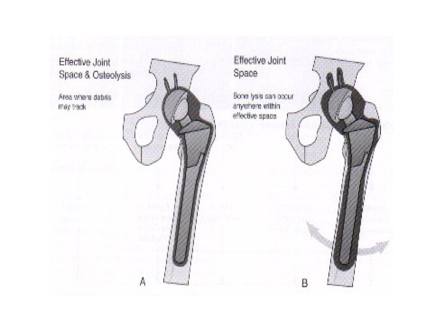
Q1-Q99 questions — tap to reveal all answerslist
- What is osteolysis and what is its radiological definition?
- Describe the pathway of particle-induced osteolysis.
- What is submicron debris and why is it significant?
- What particle characteristics influence osteolysis?
- What are the quoted wear rates for different bearings?
- What is aseptic loosening and where do the particles come from?
- What is the effective joint space and how does osteolysis differ in cemented versus cementless fixation?
- What are the causes/factors of osteolysis (implant, patient, surgical)?
- How do macrophages respond to phagocytosed wear debris?
Q1.What is osteolysis and what is its radiological definition?
- Histiocytic response to submicron wear debris - vicious cycle of bone resorption, debris generation and loosening
- Radiological definition: irregular radiolucent zone in the periprosthetic region >5mm
- Groin pain suggests cup loosening; thigh pain suggests femoral loosening
Q2.Describe the pathway of particle-induced osteolysis.
- Particulate debris formation - submicron sized particles >10billion particles/gram
- Debris phagocytosed by macrophages
- Release of IL-1B, IL-6, TNFa, RANKL - upregulate RANKL on osteoblasts
- RANKL binds RANK on macrophages - differentiate into osteoclasts - bone resorption
- Prosthesis micromotion
- Particulate debris dissemination - build up hydrostatic pressure, spread through the effective joint space
Q3.What is submicron debris and why is it significant?
- Particles <1 micrometer
- Travel into the effective joint space with joint fluid and trigger a biological reaction
- Phagocytosed by macrophages - cytokine release activates osteoclasts - osteolysis and loosening
- Determinants: volume, number, size and immune response of the particles
Q4.What particle characteristics influence osteolysis?
- Size, morphology (elongated more active than round) and volume (critical 140mm3/yr)
- Particles 0.2-0.7 micron induce osteolysis
- Ceramic 0.7 micron, PE 0.5 micron, metal <0.1 micron
Q5.What are the quoted wear rates for different bearings?
- MoPE 0.1mm/yr (100um)
- CoPE 0.05mm/yr (50um)
- MoM 0.005mm/yr (5um)
- CoC 0.0005-0.001mm/yr (0.5um)
Q6.What is aseptic loosening and where do the particles come from?
- Failure of fixation - macromotion and micromotion at the implant-bone interface
- Inadequate initial fixation or particle-induced osteolysis; activated macrophages/osteoclasts are key
- Mode 1: femoral head and PE articulating surfaces
- Mode 3: third body wear, cement fragments
- Mode 4: cement-bone, cement-implant, bone-implant interfaces
Q7.What is the effective joint space and how does osteolysis differ in cemented versus cementless fixation?
- All periprosthetic regions accessible to joint fluid and particulate debris by the pumping action of the joint
- Factors affecting the effective joint space: fixation, screw holes in shell
- Reduce by avoiding screw augmentation, cementing (Exeter centralizer), Circumferential proximal coated cementless stem, better liner locking
- Cemented: linear osteolysis over the cement-bone interface - subchondral plate formation around cement - early loosening as the whole rim is osteolyzed
- Cementless: focal expansile osteolysis - patchy bony ingrowth - osteolysis from peripheral gap extending into non-ingrown fibrous region - may fail catastrophically
Q8.What are the causes/factors of osteolysis (implant, patient, surgical)?
- Implant: head size, choice of articulation, PE thickness
- Patient: BMI, activity level
- Surgical: malalignment causing edge loading/impingement, offset and soft tissue tension affecting JRF, third body, locking of modular parts
- Plus cementing technique
Q9.How do macrophages respond to phagocytosed wear debris?
- 1. Secrete mediators (TNF alpha, IL-6, PGE2) that induce osteoblast proliferation/activation and RANKL induction
- 2. Differentiate directly into osteoclasts via the RANKL pathway
▸ Slide 236Hip · 2 questions expand

Q1-Q22 questions — tap to reveal all answerslist
- What subject does slide 236 cover?
- What is the key learning point of this slide?
Q1.What subject does slide 236 cover?
- Not covered in the speaker notes
- The slide has no accompanying speaker notes
Q2.What is the key learning point of this slide?
- Not covered in the speaker notes
- The slide image is the only source
▸ Slide 237 · Mechanism of taper corrosion?Hip · 6 questions expand
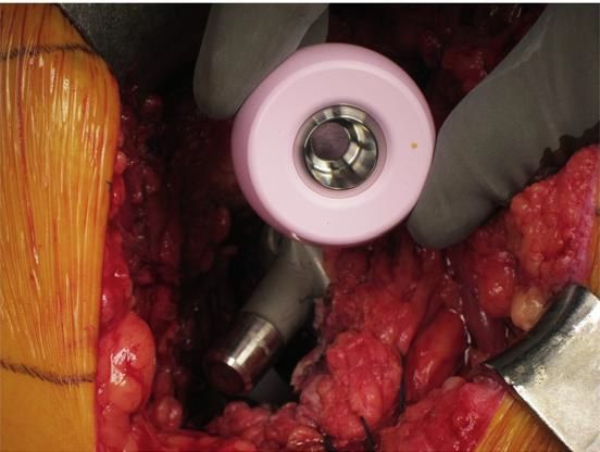
Q1-Q66 questions — tap to reveal all answerslist
- What is the mechanism of taper corrosion in modular hip implants?
- What factors are proposed to increase taper corrosion?
- How do design factors minimise the risk of taper corrosion?
- What is the trade-off between short and long taper/neck designs?
- How do surgeon factors minimise the risk of taper corrosion?
- What is done at revision surgery for taper corrosion?
Q1.What is the mechanism of taper corrosion in modular hip implants?
- Mechanically assisted crevice corrosion (MACC)
- Micromotion and shear stresses at the taper interface cause oxide film disruption, exposing fresh metal to oxygen-rich fluid
- Oxidation of underlying metal creates an acidic, low-oxygen crevice environment, destabilising the passive oxide film and increasing corrosion rate
Q2.What factors are proposed to increase taper corrosion?
- Larger head size
- High offset stem
- Smaller taper design
- Taper cleaning
- Assembly force
Q3.How do design factors minimise the risk of taper corrosion?
- Material: titanium allows higher pull-off and torsional strengths than cobalt chrome; mixed alloy tapers corrode more and have lower flexural rigidity
- Surface roughness
- Taper angle
- Taper mismatch: <7 deg half taper angle mismatch = self locking
- Short taper sits entirely within the bore, leading to edge loading
Q4.What is the trade-off between short and long taper/neck designs?
- Short taper + small diameter with a trapezoidal neck gives more ROM than a large diameter, long taper with circular neck
- A short taper sits entirely within the bore, leading to edge loading
Q5.How do surgeon factors minimise the risk of taper corrosion?
- Angle and force of impaction
- Avoid repeated impactions: single strike >6000N = 20cm drop with a 1.4kg hammer (Pennock J Arthroplasty 2006)
- Dry environment - avoid fluid contamination
- Femoral head size
- High offset stem causes more taper corrosion
Q6.What is done at revision surgery for taper corrosion?
- Complete synovectomy
- Head and taper cleaned to remove corrosion debris
- Ceramic head used whenever possible to change the taper junction interface and remove the potential cobalt source
- Titanium adapter sleeve to prevent head fracture when placed against a damaged taper
▸ Slide 238 · CoCHip · 6 questions expand

Q1-Q66 questions — tap to reveal all answerslist
- Describe the X-ray findings in this ceramic-on-ceramic THR.
- In an asymptomatic patient with an improperly seated ceramic liner, what do you offer and why?
- What are the advantages of a ceramic-on-ceramic bearing?
- What are the disadvantages of a ceramic-on-ceramic bearing?
- What ceramic material characteristics reduce the risk of fracture?
- What are the OT precautions in a CoC bearing to prevent fracture?
Q1.Describe the X-ray findings in this ceramic-on-ceramic THR.
- Cementless THR with ceramic head and ceramic liner
- Liner is improperly seated
- Edge loading between liner and metal back increases the chance of catastrophic failure
Q2.In an asymptomatic patient with an improperly seated ceramic liner, what do you offer and why?
- Offer exchange of the liner even with no symptoms
- Edge loading between liner and metal back increases the chance of catastrophic failure
Q3.What are the advantages of a ceramic-on-ceramic bearing?
- Decreased wear due to surface properties: low surface roughness, wettability (allowing fluid film formation) and hardness
- Biologically inert - no increase in serum metal ion or local reaction
Q4.What are the disadvantages of a ceramic-on-ceramic bearing?
- Brittle with no plastic deformity - edge loading from a malpositioned cup causes catastrophic failure
- Stiff - catastrophic failure debris causes significant third body wear after revision
- Squeaking
Q5.What ceramic material characteristics reduce the risk of fracture?
- Choose ceramic with smaller grain size and less porosity
- Choose a later generation of ceramic, e.g. Biolox delta with alumina, zirconia and yttria to prevent zirconia phase transformation
- Strontia increases toughness
Q6.What are the OT precautions in a CoC bearing to prevent fracture?
- Ensure no debris and no protruded screw head - ceramic is sensitive to abrasive wear
- Accurate positioning of the ceramic liner to prevent edge loading
- Minimal hammering - brittleness of ceramic can cause catastrophic wear
▸ Slide 239 · Hard on hard bearingHip · 8 questions expand
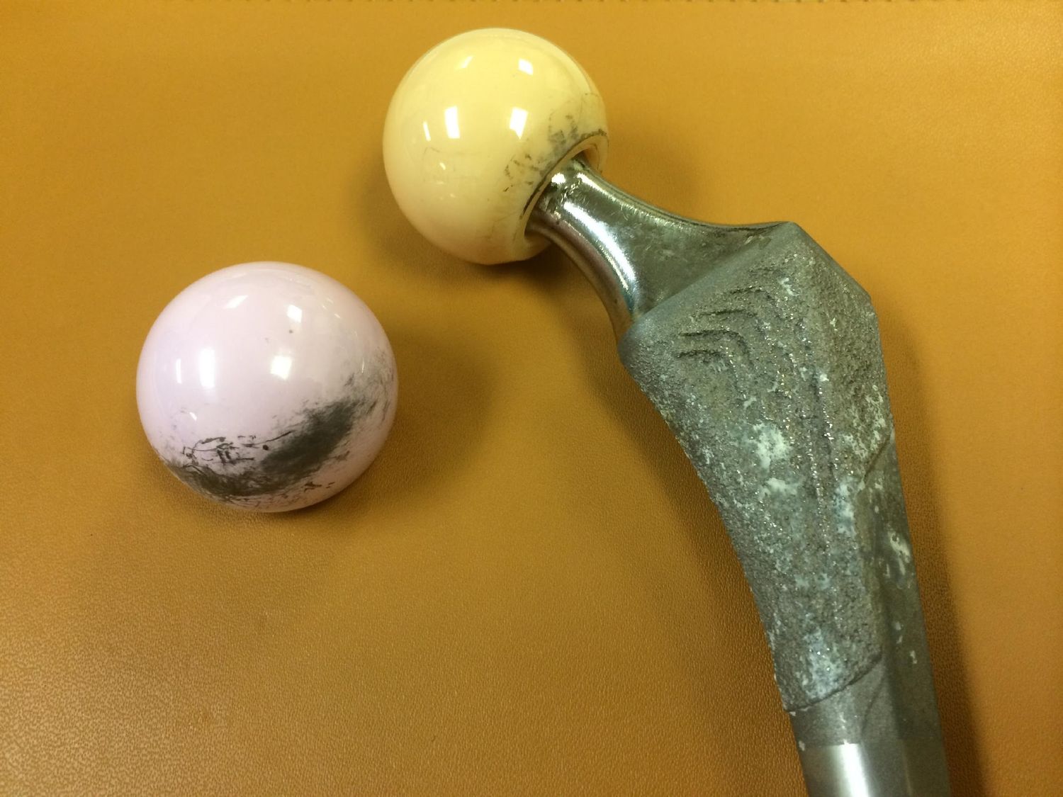
Q1-Q88 questions — tap to reveal all answerslist
- What are the pros of ceramic-on-ceramic bearings?
- What are the cons of ceramic-on-ceramic bearings?
- What causes squeaking in ceramic-on-ceramic bearings and who is affected?
- What are the two wear patterns in ceramic-on-ceramic bearings?
- What do the colours pink and orange indicate for ceramic bearings?
- What is the problem with zirconia and how is it controlled?
- What is phase transformation toughening?
- How can the fracture rate of ceramic bearings be decreased?
Q1.What are the pros of ceramic-on-ceramic bearings?
- Tribological properties - hard, wettable, low coefficient of friction
- Biocompatible and inert
- No corrosion
Q2.What are the cons of ceramic-on-ceramic bearings?
- Not ductile, notch sensitive - fracture leads to difficult revision
- Squeaking
- Need metal shell, decrease head size and stability
- Limited neck length options - limits hip offset and increases impingement
Q3.What causes squeaking in ceramic-on-ceramic bearings and who is affected?
- Usually in tall, heavy patients
- Impingement leading to lever range wear with stripe line formation
- Poor tension
- Failed locking mechanism
- Third body
Q4.What are the two wear patterns in ceramic-on-ceramic bearings?
- Polar wear - impingement at end range motion; femoral neck levers out, top of head against acetabular rim
- Peripheral wear (stripe wear) - microseparation during swing phase, inferior head impingement on rim, heel strike, reduction, superior head impinges
Q5.What do the colours pink and orange indicate for ceramic bearings?
- Pink = 4th generation
- Orange = 3rd generation
Q6.What is the problem with zirconia and how is it controlled?
- Zirconia is a metastable ceramic (monoclinic, tetragonal and cubic phases)
- Temperature, humidity and stress cause tetragonal to monoclinic phase transformation with a volume change
- Phase transformation toughening resists crack growth, but uncontrolled transformation is disastrous
- For this reason, yttrium is added to stabilize the tetragonal phase of zirconium
Q7.What is phase transformation toughening?
- Stress causes tetragonal to monoclinic transformation with a volume change
- Generates a compression stress field at the tip of a propagating crack, giving crack growth resistance
- If improperly controlled, the volumetric transformation has disastrous consequences
Q8.How can the fracture rate of ceramic bearings be decreased?
- Alumina with strontium increases toughness (elongated grains act as a barrier to deflect subcritical cracks)
- Yttrium added for stabilisation
- Smaller granules by hot isostatic pressing (HIPing)
- Higher purity and decreased porosity
▸ Slide 240 · Hip resurfacingHip · 8 questions 1 check expand
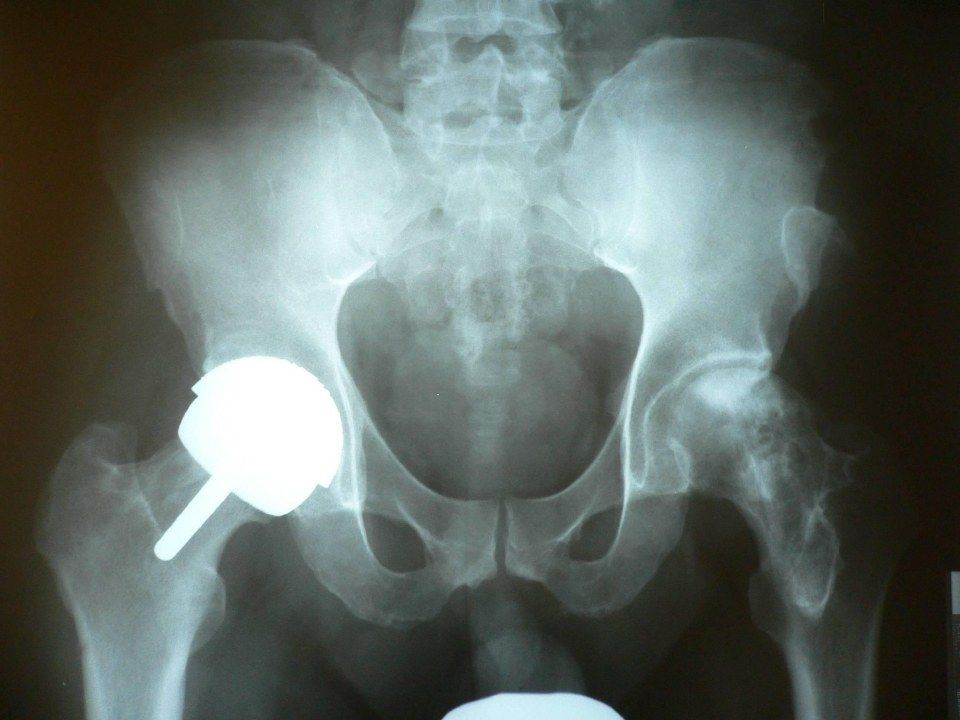
Q1-Q88 questions — tap to reveal all answerslist
- What is hip resurfacing and who is it best for?
- What are the functional benefits of hip resurfacing?
- What are the tribological and other benefits of hip resurfacing?
- What are the risks of hip resurfacing?
- What is ARMD in metal-on-metal hip resurfacing?
- What are the contraindications to hip resurfacing?
- What determines whether a fluid film is achieved in a metal-on-metal bearing?
- How do you work up a symptomatic patient with MoM THR?
Q1.What is hip resurfacing and who is it best for?
- Large diameter metal-on-metal bearing
- Best in young, high demand individuals with end stage arthritis and good bone stock
Q2.What are the functional benefits of hip resurfacing?
- Low dislocation rate (large head, bigger jump distance)
- Increased ROM before impingement
- Normal hip kinematics
Q3.What are the tribological and other benefits of hip resurfacing?
- Less wear (fluid film formation, low coefficient of friction per lecture notes)
- Less particulate-induced osteolysis
- Self healing
- Preserves proximal femoral bone stock
- Less risk of infection, DVT/PE
Q4.What are the risks of hip resurfacing?
- Femoral neck fracture
- Metal ion toxicity, highest at 1-2 years (ARMD)
- Local soft tissue reaction: pseudotumour (ALVAL - aseptic lymphocyte-dominant vasculitis-associated lesion)
- T-cell mediated hypersensitivity with macrophage activation causing a vicious cycle
Q5.What is ARMD in metal-on-metal hip resurfacing?
- Adverse reaction to metal debris, related to metal ion toxicity which is highest at 1-2 years
- Manifests as pseudotumour (ALVAL) and T-cell mediated hypersensitivity activating macrophages in a vicious cycle
Q6.What are the contraindications to hip resurfacing?
- Inadequate neck bone stock
- Small or abnormal acetabulum
- Relative: coxa vara, LLD
- Pregnant women
- Renal failure
Q7.What determines whether a fluid film is achieved in a metal-on-metal bearing?
- Optimal clearance 90-200um
- Conformity (high vs low)
- Contact (polar, midpolar, equatorial)
- Position sensitivity - high conformity with polar contact is good
Q8.How do you work up a symptomatic patient with MoM THR?
- History and PE: time and brand of THR, current ROM, any mass, rule out infection (WCC, CRP, ESR)
- MRI/USG for metallosis; blood Cr/Co ions at 0 and 3 months
- Consider revision
- Follow MHRA recommendations: 4 groups - MoM resurfacing, stemmed MoM <36mm head, stemmed MoM >36mm head, DePuy ASR
Metal-on-metal hip resurfacing has a low coefficient of friction of 0.8 — decimal error — In vitro studies report an average coefficient of friction of about 0.079-0.098 (~0.08) for MoM resurfacing bearings, not 0.8 — source
▸ Slide 241 · ComplicationHip · 4 questions expand
Q1-Q44 questions — tap to reveal all answerslist
- List the complications of total hip replacement shown on this slide.
- Which nerve palsy is listed on the slide as a complication of total hip replacement?
- Which complications listed on the slide are thrombotic and neuropathic respectively?
- What does HO stand for in this complication list?
Q1.List the complications of total hip replacement shown on this slide.
- Dislocation
- HO (heterotopic ossification)
- Infection
- DVT (deep vein thrombosis)
- Confusion
- Sciatic nerve palsy
Q2.Which nerve palsy is listed on the slide as a complication of total hip replacement?
- Sciatic nerve palsy - listed on the slide; no further detail in the speaker notes
Q3.Which complications listed on the slide are thrombotic and neuropathic respectively?
- Thrombotic: DVT
- Neuropathic: Sciatic nerve palsy
Q4.What does HO stand for in this complication list?
- Heterotopic ossification
- It is listed on the slide; no further detail is given in the speaker notes
▸ Slide 242 · XR pelvis showing a cement THR complicated with dislocationHip · 12 questions expand
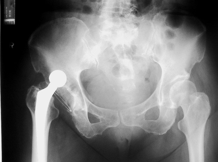
Q1-Q1212 questions — tap to reveal all answerslist
- Describe the X-ray findings in this cemented THR with dislocation.
- What is the initial management of a dislocated THR?
- What are the causes of dislocation after THR (Knutsor Lancet 2019)?
- What is the safe zone for acetabular component position?
- What is the natural history and incidence of dislocation after THR?
- What are the treatment options for recurrent dislocation after excluding infection, trauma and loosening?
- What does the stability of a hip replacement depend on?
- What are the resting positions for anterior and posterior dislocation?
- How are cup anteversion and stem anteversion defined?
- What is the likely direction of dislocation in this patient and how is it confirmed?
- When is a constrained liner indicated and what are its failure modes?
- What is the principle of a dual mobility (MDM) construct?
Q1.Describe the X-ray findings in this cemented THR with dislocation.
- Cemented THR with dislocation; hard-on-soft bearing
- Acetabular opening angle and anteversion appear within the safety range of Lewinneck, with no signs of loosening by DeLee criteria
- Cemented modular femoral component with small head-neck ratio, no loosening by Harris criteria
- Bony outgrowth at AIIS may cause impingement and dislocation; spine looks degenerated
Q2.What is the initial management of a dislocated THR?
- Rule out other injuries; AMPLE history; check NV status
- CR in OT under GA to allow muscle relaxation
- Use modified Allis manoeuvre
- Post-reduction check safety range, give abduction pillow, recheck NV status
- CT to check for congruency and any fracture, also implant loosening and alignment (femoral and acetabular version)
Q3.What are the causes of dislocation after THR (Knutsor Lancet 2019)?
- Preop patient: neuromuscular/neurodegenerative disease (dementia, PD), psychiatric disease (alcoholism, schizophrenia), history of spinal fusion
- Preop disease: revision, DDH, inflammatory arthropathy, fracture
- Intraop technique: approach, soft tissue tension (offsets, releases, capsule repair), impingement (osteophytes, cement, AIIS), component malposition
- Intraop implant: head size, head-neck ratio, neck geometry, neck skirts, constrained cups
- Postop: non-compliance, provocative activities (anterior shoelacing; posterior squat/low chair), infection, wear/loosening, GT nonunion
Q4.What is the safe zone for acetabular component position?
- Lewinnek 1978: 40 deg abduction + 15 deg anteversion - 6% dislocation inside the zone, 15% outside
- Barrack 2003: anteversion 10-20 deg of both stem and cup minimises impingement and dislocation
- Dorr combined anteversion 2009: 37.6
Q5.What is the natural history and incidence of dislocation after THR?
- 60% occur <6 weeks, usually due to technical error
- Late dislocation suggests impingement, wear or increased ROM
- Incidence 3% primary, 15% revision (Woo & Morrey JBJS Am 1982)
Q6.What are the treatment options for recurrent dislocation after excluding infection, trauma and loosening?
- Revision if the hip dislocates in the patient's normal functional range
- Indications: recurrent >=2, late (postop 1 year), malalignment, implant fracture/loosening, Impingement/ PE wear
- Correct the cause: malposition revision, impingement resection, GT distal advancement/increased horizontal offset
- Femoral side: larger head, longer neck, larger offset, GT advancement
- Acetabular side: check anteversion, lipped liner, constrained liner, MDM, resection arthroplasty
Q7.What does the stability of a hip replacement depend on?
- Component design - head size, neck morphology, head-neck ratio, constrained liner, neck skirt
- Alignment - anteversion 15-30, opening angle 35-45
- Soft tissue tensioning - offset and neck length
- Soft tissue function - abductor complex
Q8.What are the resting positions for anterior and posterior dislocation?
- Anterior: extension + external rotation
- Posterior: flexion + internal rotation + adduction
Q9.How are cup anteversion and stem anteversion defined?
- Cup anteversion: angle between the plane of the acetabular cup opening and the sagittal plane
- Stem anteversion: angle between the transcondylar plane including the femoral shaft and the axis of the femoral neck
Q10.What is the likely direction of dislocation in this patient and how is it confirmed?
- Suspect anterior dislocation by the size of the head, and the rotation and position of the femur
- Confirmed by lateral X-ray
Q11.When is a constrained liner indicated and what are its failure modes?
- Indication: bone stock okay but soft tissue dysfunction with a well fixed acetabulum
- Covers the femoral head beyond the equator, decreasing dislocation at the expense of decrease ROM
- 4 modes of failure (Dorr J Arthroplasty 2005): fixation failure, biomaterial failure, femoral head dislocation, liner dissociation
- Now superseded by MDM - increases survival and decreases redislocation; theoretical increase in wear due to more bearing surface
Q12.What is the principle of a dual mobility (MDM) construct?
- Increases the functional arc of motion
- Large head principle (McKee Farrar) vs small head (Charnley)
- 3 primary goals: increase stability, restore nearly physiological ROM, reduce wear
- Anatomical and modular dual mobility - modular risks mal-seating, but can add screws
▸ Slide 243 · Total knee periprosthetic fractureHip · 9 questions expand

Q1-Q99 questions — tap to reveal all answerslist
- How are periprosthetic knee fractures classified?
- What is the Lewis and Rorabeck classification for femoral periprosthetic fractures?
- What is the Su classification for femoral periprosthetic fractures?
- What is the Felix classification for tibial periprosthetic fractures?
- What is the Keating classification for patellar periprosthetic fractures?
- What is assessed in patient evaluation before treating a knee periprosthetic fracture?
- What pre-operative workup is required?
- What is the management principle for knee periprosthetic fractures?
- What are the general management options?
Q1.How are periprosthetic knee fractures classified?
- Site: patella > femoral > tibial
- Timing: intra-operative or post-operative
Q2.What is the Lewis and Rorabeck classification for femoral periprosthetic fractures?
- 1: non-displaced, component intact
- 2: displaced, component intact
- 3: displaced, component loose
- Anterior notching is a debatable risk factor
Q3.What is the Su classification for femoral periprosthetic fractures?
- I: proximal to the component
- II: at the level of the anterior flange
- III: distal to the anterior flange
Q4.What is the Felix classification for tibial periprosthetic fractures?
- 1: plateau
- 2: adjacent to tibial stem
- 3: tibial shaft distal to component
- 4: tibial tubercle
- A = implant well fixed; B = implant loose
Q5.What is the Keating classification for patellar periprosthetic fractures?
- 1: vertical fracture, stable implant, extensor mechanism intact
- 2: horizontal fracture, stable or unstable implant, extensor mechanism disrupted
- 3: intact extensor mechanism but loose implant
- Goldberg: 1 not involving implant, 2 involving implant/quadriceps, 3 inferior pole, 4 fracture dislocation
Q6.What is assessed in patient evaluation before treating a knee periprosthetic fracture?
- General condition of the patient plus comorbidities
- Function status before fracture
- Prosthesis history: previous OT record, model and size, postop complications, preceding pain before fracture
Q7.What pre-operative workup is required?
- Bloods
- Nutritional assessment
- +/- joint aspiration if infection is suspected
Q8.What is the management principle for knee periprosthetic fractures?
- Usually geriatric patients with osteopenic bone
- Stable fixation of fracture and implant in good alignment to allow early weight bearing
- Prevent malunion, which creates abnormal loading and early implant loosening
- Consider fracture site, implant stability and bone stock
Q9.What are the general management options?
- Conservative (all)
- Long stem fixation spanning the fracture by at least 2 cortical diameters (tibia/femur)
- Nail or plate (femur) - double plate preferred; nail needs at least 2 distal locking screws and may be blocked by notch or patella baja (quote Jones Injury 2016 paper, which describes commonly used knee implants that may or may not allow a nail to pass through)
- Distal femoral replacement (femur)
- ORIF/revision/patellectomy +/- repair (patella)
- Medial approach dissection in the plane of vastus medialis and sartorius
▸ Slide 244 · <Left>Hip · 10 questions expand
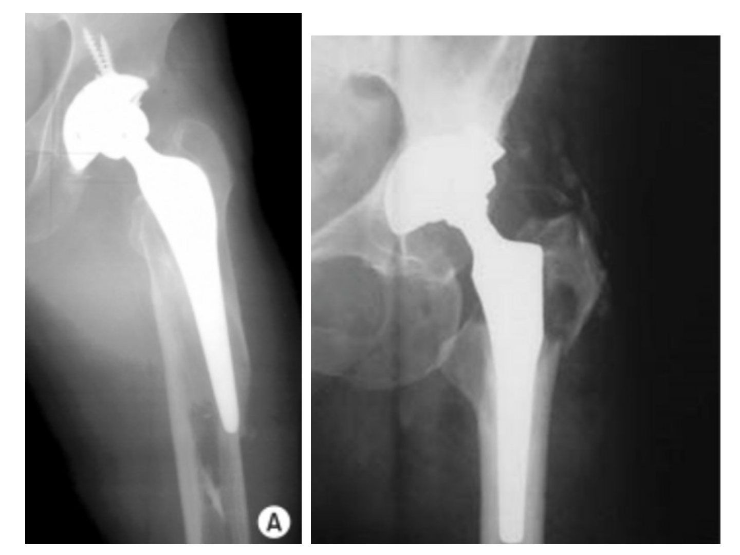
Q1-Q1010 questions — tap to reveal all answerslist
- Describe the fracture shown and its classification.
- What is the Vancouver classification of periprosthetic femoral fractures?
- What history, examination and investigations are needed for this fracture?
- What are the aims and surgical principles of definitive management?
- How are Vancouver type A fractures managed?
- How is a Vancouver B1 fracture managed?
- How are Vancouver B2 and B3 fractures managed?
- What are the key steps when revising the femoral stem for B2/B3?
- How is the acetabulum addressed and what bearing is chosen?
- What other techniques are described for these fractures?
Q1.Describe the fracture shown and its classification.
- X-ray left proximal femur showing periprosthetic femoral fracture in a previous uncemented THR
- Fracture extends from the middle of the stem to distal to the stem
- Suspected loosening of the stem; compare with previous X-ray
- Vancouver type B2 or B3
Q2.What is the Vancouver classification of periprosthetic femoral fractures?
- A: trochanteric region
- B: around the stem - 1 stable, 2 unstable, 3 unstable with bone loss
- C: away from the stem
Q3.What history, examination and investigations are needed for this fracture?
- History and PE: mainly rule out infection and other injuries
- Check previous OT record to know the implant brand and sizes
- Postoperatively also work up for osteoporosis
- Investigations: X-ray lateral, CT for bone stock
Q4.What are the aims and surgical principles of definitive management?
- Aim: stable, painless, mobile hip allowing early weight bearing
- Restore offset and hip centre; equalise LLD
- Correct positioning of components and soft tissue tension
- Acute setting: seek advice and support from other senior hip surgeons
Q5.How are Vancouver type A fractures managed?
- Often requires treatment that addresses the osteolysis
- AG fractures <2cm displacement: nonoperative, partial weight bearing, allow fibrous union
- AG fractures >2cm: ORIF with trochanteric claw/cables (loss of abductor function leads to instability)
Q6.How is a Vancouver B1 fracture managed?
- ORIF using cerclage cables and locking plates
- Subvastus approach; reduction with reduction forceps and colinear clamp; temporary cerclage wires
- LCP: unicortical screws proximally not to violate cement mantle + cerclage wires; distal bicortical screws
- Cortical defect or marked comminution: femoral cortical struct allograft on the anterior aspect of the femur to bypass the defect by at least 2 cortical widths
Q7.How are Vancouver B2 and B3 fractures managed?
- Revise the stem for B2 and above - ORIF alone in B3 has high reoperation rates: BJJ 2017 Khan showed almost 30% revision rate compared to 15% revision for revision + ORIF
- Femoral component revision with proximal femoral allograft/bone graft or proximal femoral replacement + ORIF
Q8.What are the key steps when revising the femoral stem for B2/B3?
- Posterior approach - extensile and familiar
- Bypass the fracture with a long, titanium, modular, fluted conical revision stem (e.g. modular restoration stem) - flexible control of leg length, offset and version
- ORIF of the fracture with or without an allograft to maintain the femoral tube
- Decide whether to also revise the acetabulum: compatibility with new stem, premorbid status, fixation and PE wear
Q9.How is the acetabulum addressed and what bearing is chosen?
- If revising: cementless socket with supplementary screws, with augments available for significant bone defects
- Bearing of choice: ceramic 36mm head on XLPE with a lipped PE liner to minimise posterior dislocation
Q10.What other techniques are described for these fractures?
- Distally locking, cementless HA-coated long femoral stem (Cannulok; El-Bakoury 2017 100% survivorship at 4 years)
- Exeter group: impaction bone grafting supported by a plate, with a long cemented femoral stem for B2 and B3
- Proximal femoral replacement (Parvizi) with constrained liner as abductors are usually poor; keep as much proximal femoral bone as possible and reapproximate it onto the PFR
▸ Slide 245 · New evidenceHip · 3 questions expand

Q1-Q33 questions — tap to reveal all answerslist
- What is the new evidence on B2 periprosthetic fractures in cemented stems?
- What is the rationale for ORIF without stem revision in B2 fractures?
- What are the criteria for ORIF without stem revision?
Q1.What is the new evidence on B2 periprosthetic fractures in cemented stems?
- Smitham 2019, Journal of Arthroplasty (Solomon's group, Adelaide)
- B2 fractures in cemented femoral implants can be treated with ORIF without revision of the stem
Q2.What is the rationale for ORIF without stem revision in B2 fractures?
- Patients too frail to undergo lengthy revision surgery
- CPDT stem can regain stability in the cement mantle after anatomical reduction, provided the cement-bone interface is maintained
- Stem revision can be performed later as a planned elective procedure
Q3.What are the criteria for ORIF without stem revision?
- Cemented polished double tapered stem
- Cement-bone interface maintained - fracture only disrupts the stem-cement interface
- Stable anatomical reduction achieved at surgery
- Bone stock is adequate
▸ Slide 246 · HOHip · 6 questions 1 check expand
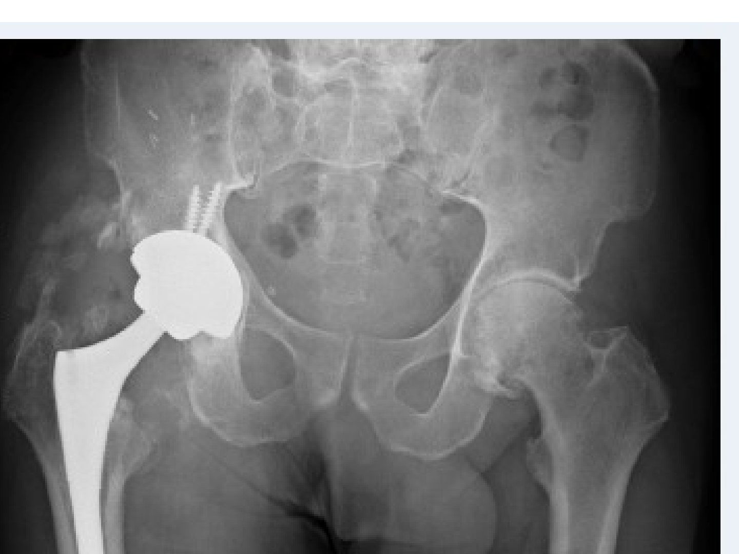
Q1-Q66 questions — tap to reveal all answerslist
- What is heterotopic ossification and what are its causes?
- What are the risk factors for HO?
- What is the pathophysiology of HO?
- What is the Brooker classification of HO, and what grade is shown on this X-ray?
- What laboratory findings are seen in HO and what is their limitation?
- How is HO prevented and when is it excised?
Q1.What is heterotopic ossification and what are its causes?
- Ectopic lamellar bone formation in soft tissues
- Traumatic
- Neurogenic - inflammatory neuropeptides including substance P and CGRP are highly upregulated
Q2.What are the risk factors for HO?
- Patient: hypertrophic OA, history of HO, AS, DISH, Paget's disease
- General: head trauma, spinal cord injury, burn, massive trauma
- Local: approach (Hardinge, anterior > posterior), haemostasis, marrow spilling, immobilisation
- Forceful passive manipulation after a period of immobilisation
Q3.What is the pathophysiology of HO?
- Unknown trigger with tissue expression of BMPs
- Primitive mesenchymal cells migrate to the injured area, become fibroblasts and secrete collagen/ECM (fibroblastic metaplasia)
- Primitive mesenchymal cells migrate to injured area and transform into fibroblast --> secrete collagen and ECM --> fibroblastic metaplasia --> fibroblast transform into chondrocytes (similar to enchondral ossification) --> some chondrocytes continue to deposit collagen into cartilage matrix while the remaining chondrocytes transform into osteoblasts --> osteoid deposition
- Fibroblasts become chondrocytes (like enchondral ossification); chondrocytes become osteoblasts with osteoid deposition
- Starts within 16 hours after the index operation
Q4.What is the Brooker classification of HO, and what grade is shown on this X-ray?
- Grade 1: island
- Grade 2: bone spur with >1cm gap
- Grade 3: <1cm gap
- Grade 4: complete osseous ankylosis
- Also mentioned: Alonzo classification
- This X-ray shows HO around a right THR - Brooker III
Q5.What laboratory findings are seen in HO and what is their limitation?
- ALP, CRP and ESR are elevated
- ALP cannot determine maturity of the heterotopic ossification
Q6.How is HO prevented and when is it excised?
- Preop: RT 800 Gy within 4-6 hours of surgery (per lecture notes)
- Intraop: minimal subperiosteal stripping, decrease pericapsular trauma
- Postop: RT single dose <72hr; indomethacin 25mg tds x 6 weeks decreases HO by ~10% and decreases severity
- Excise when mature: X-ray trabecular formation/mature rim, normal ALP and ESR, no increased uptake on bone scan
- Indocid MOA: decreases insulin-like growth factor
Preoperative radiotherapy 800 Gy within 4-6 hours of surgery prevents heterotopic ossification — unit error / unsafe dose — Single-fraction HO prophylaxis is 700-800 cGy (7-8 Gy), given within 24 hours preoperatively or within 72 hours postoperatively; 800 Gy would be a lethal dose — source
▸ Slide 247 · Post op 3y, hip pain focus of femoral sideHip · 12 questions expand

Q1-Q1212 questions — tap to reveal all answerslist
- Describe the X-ray findings and what you would compare with.
- What are the risk factors for infection after THR?
- What should you look for in history and examination?
- What is the Tsukayama classification of total joint infection?
- What blood tests are used to investigate THR infection?
- How useful is hip aspiration and how is it performed?
- What are the MSIS criteria and common pathogens for PJI?
- What are the principles of management of a chronic infected THR?
- How does the timing of infection determine management?
- How do the KLIC and CRIME 80 scores guide DAIR?
- What is the antibiotic-loaded cement recipe used for spacers?
- What are the reported success rates of one-stage and two-stage revision?
Q1.Describe the X-ray findings and what you would compare with.
- Previous cemented THR with femoral lucency over Gruen zones 2, 3, 5, 6, 7, 8, 9, 11, 14 (8-14 anterior to posterior)
- Multiple cortical lytic lesions with endosteal scalloping; no periosteal new bone; no soft tissue calcification
- Compare previous X-ray for stem loosening: subsidence, implant migration, cement breakage, radiolucent line at bone-cement interface
Q2.What are the risk factors for infection after THR?
- Patient: elderly, immunocompromised - DM, CRF, steroids, alcoholism, recent infection
- Disease: psoriasis, RA, prior hip operation, prior hip infection
- Surgical: prolonged operation; post-operative complications UTI, skin necrosis, haematoma
Q3.What should you look for in history and examination?
- Immunocompromised conditions, cause of THR, early wound infection, repeated antibiotics, pain-free interval, preceding bacteraemia
- Expect non-mechanical pain
- PE: erythema, sinus
- Classify using Tsukayama (positive intraop c/st, early postop, acute haematogenous, late chronic)
Q4.What is the Tsukayama classification of total joint infection?
- 1: positive intra-operative cultures
- 2: early acute, <1 month post-op
- 3: acute haematogenous (late acute)
- 4: chronic
Q5.What blood tests are used to investigate THR infection?
- Leukocytosis, CRP >10mg/L, ESR >30; CRP sensitivity and specificity ~90%
- IL-6 (precursor of CRP) is more accurate but may not be available
- Berbari JBJS Am 2010: accuracy IL-6 > CRP > ESR > WBC
- ESR 75%/70%, CRP 88%/74%, IL-6 97%/91% (sensitivity/specificity)
Q6.How useful is hip aspiration and how is it performed?
- Spangehl: sensitivity 0.86, specificity 0.94, PPV 0.67, NPV 0.98 (level 1); suggest in high clinical suspicion with abnormal ESR/CRP
- Anterior approach: 1 inch lateral to femoral pulse, 1 inch below inguinal ligament; lateral approach at GT under XR guidance; US or XR guided
- Off antibiotics for at least 1 week; no intra-articular LA (bacteriostatic)
- Send immediately for GS/C+ST, PCR and total cell count (>3000 cells/ml, >80% polymorph); different instruments from 3 different sites, 2/3 samples positive
Q7.What are the MSIS criteria and common pathogens for PJI?
- MSIS: any one major or 3/5 minor
- Acute <6 weeks: ESR not reliable, CRP >100, WBC >10000 cells/mL, neutrophil >90%
- Chronic: ESR >30, CRP >10, WBC >3000 cells/mL, neutrophil >80%
- Staph aureus and Staph epidermidis account for 2/3; also E. coli, enterococcus
Q8.What are the principles of management of a chronic infected THR?
- MDT approach with MSK radiologist, microbiologist, histopathologist, plastic surgeon and experienced revision arthroplasty surgeon
- 2-stage revision: removal of implant, debridement and antibiotic-loaded cement spacer
- Systemic antibiotics 6-12 weeks with monitoring; repeat hip aspiration at 6 weeks; CT to reassess bone stock
- Second stage if infection free and serology negative x 2 weeks; must use cemented components (no ingrowth); frozen section negative if <5 polymorphs per 400x HPF
- One stage only if host not immunocompromised, low-virulence organism, no sinus tract, no OM/loosening
- 6 vs 12 weeks antibiotics non-inferior (NEJM 2021 Bernard RCT)
Q9.How does the timing of infection determine management?
- <4 weeks: acute infection with less biofilm - radical debridement, exchange of modular bearing (thorough debridement), IV antibiotics
- >4 weeks: chronic infection, biofilm present - implant removal and antibiotic-loaded cement spacer
Q10.How do the KLIC and CRIME 80 scores guide DAIR?
- KLIC for Tsukayama type 2 early acute: kidney 2, liver 1.5, indication (fracture/revision) 1.5, cemented 2, CRP>115 2.5; score >4 = 55% failure rate
- CRIME 80 for type 3 acute haematogenous: COPD 2, CRP>150 1, RA 3, indication fracture 3, male 1, exchange mobile component -1, age >80 2; score >3 = 60% failure
Q11.What is the antibiotic-loaded cement recipe used for spacers?
- Options are gentamicin, vancomycin, tobramycin
- 1 pack cement (40g) with 1 bottle antibiotic (4g vancomycin); ~10% (~1.x g gentamicin)
- No vacuum, no drain; local concentration falls to 1/2 after 10 days
Q12.What are the reported success rates of one-stage and two-stage revision?
- UCLH experience: TKR 1 stage 100%, 2 stage 93.2%
- THR 1 stage 94.5%, 2 stage 91.8%
- Ongoing ccc
▸ Slide 248Hip · 2 questions expand

Q1-Q22 questions — tap to reveal all answerslist
- What hip condition is presented in this slide?
- What are the key learning points for this hip condition?
Q1.What hip condition is presented in this slide?
- Not covered in the speaker notes
- The slide image is the only source for this slide
Q2.What are the key learning points for this hip condition?
- Not covered in the speaker notes
- No management or investigation details are provided in the notes
▸ Slide 249 · Why is it difficult to eradicate in infected THR?Hip · 5 questions expand
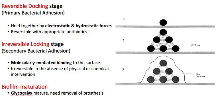
Q1-Q55 questions — tap to reveal all answerslist
- Why is infection difficult to eradicate in an infected THR?
- What are the steps of biofilm formation?
- How does bacterial resistance arise?
- What are the mechanisms of antibiotic resistance?
- What is the Coventry classification (1975)?
Q1.Why is infection difficult to eradicate in an infected THR?
- Biofilm formation over the implant
- Biofilm is a structural community of bacteria encased in glycocalyx adhering to inert or living surfaces
- 3 stages: reversible docking / irreversible locking / maturation
- Acts as a physical and chemical barrier against the host immune system and antibiotics (prevents penetration, inactivation (opsonization) of drugs, acidic and anaerobic conditions)
Q2.What are the steps of biofilm formation?
- Bacterial adherence
- Microcolony quorum formation
- Quorum sensing - bacterial load reaches a predetermined concentration
- Colony expresses biofilm; polysaccharide coating and proliferation
- Mature biofilm formation (4 weeks)
- Planktonic bacteria dissemination
Q3.How does bacterial resistance arise?
- Spontaneous/sporadic mutation
- Horizontal gene transfer via conjugation, transduction, transformation or transmissible plasmid
Q4.What are the mechanisms of antibiotic resistance?
- Drug inactivation or modification (e.g. beta-lactamase deactivation of penicillin G)
- Alteration of target site (penicillin binding protein in MRSA)
- Alternative metabolic pathway (sulfonamide)
- Active efflux of drugs
Q5.What is the Coventry classification (1975)?
- Stage I: acute post-operative infection (<6 weeks) - contamination during operation or wound complication
- Stage II: delayed deep infection (6 weeks to 2 years) - deep seated with healed wound
- Stage III: late haematogenous infection (>2 years)
▸ Slide 250 · Antibiotics properties:Hip · 3 questions expand
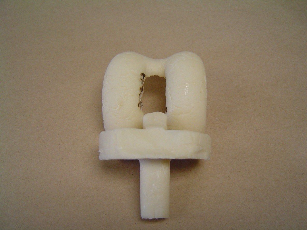
Q1-Q33 questions — tap to reveal all answerslist
- What are the ideal properties of antibiotic-loaded cement?
- What is the technique for mixing antibiotic-loaded cement?
- What is the elution profile of antibiotic from cement?
Q1.What are the ideal properties of antibiotic-loaded cement?
- Heat stable
- Good elution (low molecular weight)
- Broad spectrum
- Bactericidal
- Seldom allergic
- Available in powder form
Q2.What is the technique for mixing antibiotic-loaded cement?
- No vacuum mixing - increases porosity and gives better elution
- Wait until set before insertion (stem micromotion allows easy removal)
- No drain
- Add antibiotic before mixing
Q3.What is the elution profile of antibiotic from cement?
- Most antibiotic elution is highest in the first 24-72 hours after implantation
▸ Slide 251 · What is the protocol for DVT prevention in your centre?Hip · 7 questions 1 check expand

Q1-Q77 questions — tap to reveal all answerslist
- How is DVT risk assessed and prophylaxis chosen in your centre?
- Which patients are in the high-risk group for DVT?
- What is Virchow's triad?
- What mechanical and pharmacological options are available?
- What is the clotting cascade?
- What is the risk of VTE without prophylaxis after joint replacement?
- What do the latest guidelines recommend for VTE prophylaxis?
Q1.How is DVT risk assessed and prophylaxis chosen in your centre?
- Tailor to each patient; AUTAR score (2002) stratifies into low, mid and high risk
- All patients receive mechanical prophylaxis (early mobilisation, foot sequential pump, pressure stockings)
- Mid and high risk patients also receive pharmacological prophylaxis (LMWH)
- AUTAR assesses patient (mobility, age, BMI), PMHx/drug Hx (previous DVT, malignancy, inflammatory disease, CVA/IHD, varicose veins, OCP), disease (severe trauma) and surgery (ortho lower limb surgery)
Q2.Which patients are in the high-risk group for DVT?
- Single most important factor: previous history of DVT
- Definite risk: age, obesity (BMI >25), thrombotic disease, OCP
- Relative risk: malignancy, varicose veins
- Intrinsic: factor V Leiden, antithrombin III deficiency
- COC major risk factors: Hx of VTE, obesity BMI>27, cancer, hypercoagulation, SCI with paraplegia/tetraplegia
Q3.What is Virchow's triad?
- Venous stasis
- Intimal injury
- Hypercoagulation
Q4.What mechanical and pharmacological options are available?
- Mechanical: foot sequential pump, pressure stocking, early mobilisation
- Heparin (enhances antithrombin 3) - reverse with protamine sulphate
- Dabigatran (direct thrombin inhibitor) - reverse with idarucizumab
- Rivaroxaban (factor 10a inhibitor) - reverse with andexanet alfa
- Warfarin (inhibits factors 2,7,9,10) - reverse with beriplex
Q5.What is the clotting cascade?
- Extrinsic: tissue factor + 7 to 7a to 10a (PT)
- Intrinsic: 12a to 11a, 9a + 8a to 10a (aPTT)
- 10a + 5a convert prothrombin to thrombin, then fibrinogen to fibrin; factor 13 forms the fibrin clot
Q6.What is the risk of VTE without prophylaxis after joint replacement?
- COC guideline: asymptomatic DVT 42%, asymptomatic proximal DVT 22%, asymptomatic PE 0.8%, fatal PE 0.25%
- KO JOA 2003: asymptomatic DVT Chinese 27%/PE 2.5%, Caucasian 32% (USG screening at 1 week)
Q7.What do the latest guidelines recommend for VTE prophylaxis?
- JBJS March 2022: low dose aspirin as the most effective and safest primary method in all TJA patients including moderate to high risk
- ACCP 2012: low risk mechanical + aspirin; high risk mechanical + NOAC (apixaban 2.5mg BD); duration 2-4 weeks
- COC guidelines (HK) prophylactic: LMWH 1 week postop
- NICE 2018 THR: LMWH 10 days then aspirin 28 days, or LMWH 28 days with TEDs until discharge for THR
- NICE 2018 TKR: aspirin 14 days, or LMWH 14 days with TEDs
Latest JBJS March 2022 recommendation: aspirin is the most effective and safest method for VTE prophylaxis in all TJA patients, including moderate to high risk — contested (consensus recommendation vs trial evidence) — The claim reflects the JBJS March 2022 ICM-VTE consensus recommendation; meta-analyses of RCTs show aspirin is as effective and safe as other anticoagulants rather than superior to them — medium confidence — source
▸ Slide 252 · How to management over warfarinizationHip · 3 questions expand
Q1-Q33 questions — tap to reveal all answerslist
- What are the causes of over-warfarinisation?
- How is major bleeding due to warfarin managed?
- How is over-warfarinisation managed if there is no bleeding?
Q1.What are the causes of over-warfarinisation?
- Compliance
- Diet compliance
- Drug-drug interaction
Q2.How is major bleeding due to warfarin managed?
- FFP + vitamin K IV 10mg once
Q3.How is over-warfarinisation managed if there is no bleeding?
- INR <5: stop warfarin
- INR 5-9: oral vitamin K 1-2.5mg once
- INR >9: oral vitamin K 3-5mg once
- +/- cover with enoxaparin if INR <1.5
▸ Slide 253 · post TKR confusion?Hip · 3 questions expand

Q1-Q33 questions — tap to reveal all answerslist
- What are the causes of confusion after TKR?
- Which metabolic and infective causes must be excluded?
- How would you structure assessment of post-TKR confusion?
Q1.What are the causes of confusion after TKR?
- Drug causes
- Neurological: stroke, pre-existing dementia
- Metabolic: hyponatraemia, hypoglycaemia
- Hypoperfusion: shock (cardiogenic/hypovolaemic), anaemia, PE
- Infection: UTI, pneumonia
Q2.Which metabolic and infective causes must be excluded?
- Hyponatraemia and hypoglycaemia
- UTI and pneumonia
Q3.How would you structure assessment of post-TKR confusion?
- Review drugs, then screen neurological causes including stroke and pre-existing dementia
- Correct metabolic causes - hyponatraemia and hypoglycaemia
- Assess perfusion: shock (cardiogenic/hypovolaemic), anaemia, PE
- Screen for infection: UTI and pneumonia
▸ Slide 254 · Patient present as foot drop after THR, how would you manage?Hip · 8 questions expand

Q1-Q88 questions — tap to reveal all answerslist
- What are the goals in managing foot drop after THR?
- What history is relevant in foot drop after THR?
- What examination is performed for foot drop after THR?
- What investigations are used and what is the timing of nerve assessment?
- What non-operative measures are given for foot drop?
- What are the causes of sciatic nerve palsy after THR?
- Will you explore the sciatic nerve, and what is the incidence of nerve palsy?
- How do you differentiate a sciatic nerve palsy from a common peroneal nerve palsy?
Q1.What are the goals in managing foot drop after THR?
- 1. Establish the diagnosis
- 2. Delineate the level of the nerve lesion
Q2.What history is relevant in foot drop after THR?
- Primary pathology requiring THR
- Review the operation record: approach, any limb lengthening, intra-operative complications
- Risk factors: female, DDH, limb lengthening, revision surgery, spinal stenosis, post-traumatic
Q3.What examination is performed for foot drop after THR?
- Check drains/output and any wound complication
- Remove tight dressing, splint and abduction pillow over the knee and allow knee flexion in case of common peroneal nerve injury
- Examine ankle dorsiflexion (common peroneal component) and ankle plantarflexion (posterior tibial component)
- Examine hamstrings to look for above or below knee palsy
Q4.What investigations are used and what is the timing of nerve assessment?
- XR/CT to confirm screw placement and acetabular component position
- USG for haematoma; NCT for extent, severity and monitoring of recovery
- D2: assess distal segment acute or chronic palsy
- 2 weeks: assess axonotmesis, neurotmesis, neuropraxia
- 3 months: assess recovery of nerve
Q5.What non-operative measures are given for foot drop?
- AFO to prevent equinus deformity
- Physiotherapy to prevent joint contracture
Q6.What are the causes of sciatic nerve palsy after THR?
- Traction injury - retractor placement, acute limb lengthening >4cm (Kavanagh 1991: <4cm no palsy, >4cm 28% sciatic nerve palsy)
- Pressure injury - post-operative haematoma, hip dislocation
- Direct injury - acetabular screw placement, diathermy, cement or prosthesis impingement, thermal injury from cement
Q7.Will you explore the sciatic nerve, and what is the incidence of nerve palsy?
- If no structural lesion (screw malposition, haematoma compression), observe recovery; ~40% recovery rate at 21 months (Farrell JBJS 2005)
- Incidence 3.5% in primary, 7% in revision cases; 90% sciatic nerve, 50% common peroneal nerve
- Risk factors: acute leg lengthening >4cm, revision surgery, DDH, female, pre-existing spinal stenosis
- Avoidance: extend hip, flex knee, subtrochanteric osteotomy; post-op flex hip to 45 deg and bend knee to 90 deg
Q8.How do you differentiate a sciatic nerve palsy from a common peroneal nerve palsy?
- Sciatic palsy involving both tibial and peroneal components affects biceps femoris, semi-T, semi-M and the posterior compartment of the calf
- Peroneal-only sciatic palsy is difficult to differentiate clinically from common peroneal palsy below the knee - EMG tests the short head of biceps femoris
- Common peroneal component is densely packed, more lateral and fixed at the fibular neck
▸ Slide 255 · HistoryHip · 12 questions expand
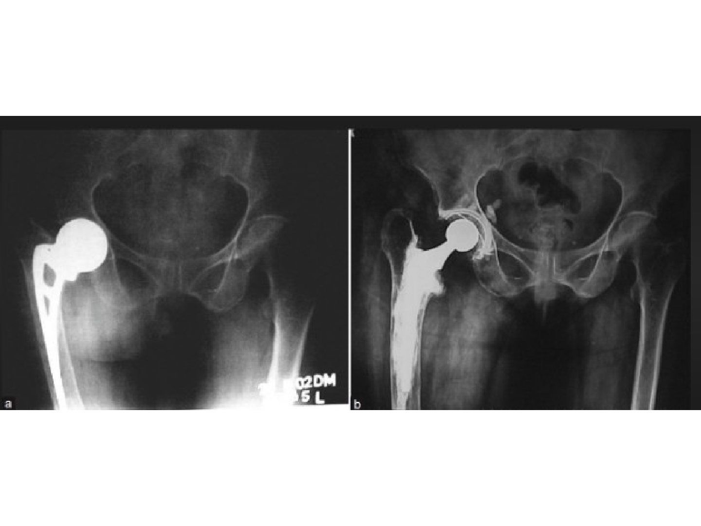
Q1-Q1212 questions — tap to reveal all answerslist
- What history and examination findings are important in a painful THR?
- What investigations are needed?
- What are the aims of revision THR?
- What pre-operative preparation is required?
- What are the key intra-operative steps for femoral implant removal?
- What are the principles of an extended trochanteric osteotomy (ETO)?
- What is a bone window and why may implant removal be difficult?
- How is a broken femoral stem removed?
- What ultrasonic and other methods are used to extract cement?
- How is the acetabular implant removed, including a ceramic liner?
- How is reconstruction planned for the femur and acetabulum?
- What are the principles and complications of femoral impaction bone grafting?
Q1.What history and examination findings are important in a painful THR?
- History: age and premorbid state, nature of pain, infection symptoms, current function (HHS), details of last operation, rule out spine, neuro, PVD as causes of pain
- PE: scar, deformity, sinus, ROM
- ROM clues: stiff - HO/impingement/contracture; end range pain - impingement; all range pain - infection; IR/ER pain - femoral stem loose
- Stinchfield test (resisted flexion ~SLR) suggests acetabular loosening; assess abductors
Q2.What investigations are needed?
- CT
- Scannogram + scoli series
Q3.What are the aims of revision THR?
- Restore offset and LLD
- Replenish bone stock
- Secure and accurate placement of components
Q4.What pre-operative preparation is required?
- Counsel patient regarding prognosis of revision THR; check previous implant and approach
- Prepare instruments for implant removal: AO large fragment, ex-plant, straight/curved osteotome, high speed burrs, cement splitters, US device, flexible light source, allograft, GTR plate and cables if planning ETO
- Inform pathology lab for intraop frozen section; prepare cement spacer in case frozen section is positive
- Prepare reconstruction options: long extended coated stem, jumbo cup/metal augment/cupcage/reinforcement rings, allograft
Q5.What are the key intra-operative steps for femoral implant removal?
- Intraop 1g tranexamic acid to reduce blood loss; old incision, protect sciatic nerve
- Remove bone/cement at Gruen zone 1; try hammering implant out with anchorage at collar/head/hole
- Remove cement mantle with osteotome, reamer, burr, K wires, cement splitter (split radially then longitudinally)
- +/- trochanteric osteotomy/trochanteric slide/ETO; if cement mantle is good, use tap in and tap out (cement on cement)
Q6.What are the principles of an extended trochanteric osteotomy (ETO)?
- Preserve abductor and vastus lateralis (blood supply to the flap); divide the lateralis insertion off the vastus tubercle and elevate vastus lateralis off the linea aspera
- Less than 1/3 of the femoral circumference; length determined by the length of the cement mantle
- Multiple drill holes then break with osteotome; fix with cables
Q7.What is a bone window and why may implant removal be difficult?
- Bone window by burr - create a trough over the posterior femur, remove the fenestrated part and dissect the bone block
- Difficulty in removal of an AMA is due to bone growth into the fenestration
Q8.How is a broken femoral stem removed?
- From above: trephine reamer
- From below: cortical window
- From the side: ETO
Q9.What ultrasonic and other methods are used to extract cement?
- Fish-bone like ultrasonic tip - melts the cement, sinks into it, hardens when USG is off, then hammered out
- Dish-like tip - just melts and lets cement leak through holes
- Drill and tap; segmental cement extraction (long screw with multiple nuts, pour in new cement, hammer out each nut sequentially - cement is weak in tension)
- Powered cement reamer - easy fracture especially over the lateral or anterior side
Q10.How is the acetabular implant removed, including a ceramic liner?
- Curved osteotome
- Explant
- Ceramic liner: disengage the smooth taperlock by a sharp tap into the liner, then use a suction cup with scallops that contact the peripheral rim of the metal shell to remove the liner
Q11.How is reconstruction planned for the femur and acetabulum?
- Prefer cementless components if previous cement was used - bone surface become sclerotic, making cement interdigitation difficult
- Femur: long cementless stem with diaphyseal fixation and adequate distal scratch fit
- Acetabulum: identify anatomy with landmarks, navigation, intraoperative imaging, robot
- Rim defect: metal augment, strut graft; medial wall defect: morcelised bone graft, anti-protrusion cage
- Pelvic discontinuity: cup cage construct; rim stable: line-to-line fit with screw fixation; standby dual mobility cup
- Ideally 36mm head
Q12.What are the principles and complications of femoral impaction bone grafting?
- Retrograde canal filling with impacted particulate graft
- Segmental defect -> contained defect -> allows subsidence + cement creep -> even load distribution -> bone remodelling
- Technique: mesh + wiring creates a neomedullary canal for a cemented femoral stem; advantage: preserves bone stock
- Complications: fracture during impaction, varus/valgus stem, incomplete cement mantle, subsidence (bone graft resorption)
▸ Slide 256 · Describe XrayHip · 28 questions expand

Q1-Q2828 questions — tap to reveal all answerslist
- Describe the X-ray findings of this Charnley cemented total hip replacement.
- How do you diagnose periprosthetic joint infection in this revision case?
- If infection is excluded, what causes aseptic loosening and what is your pre-operative assessment?
- What are the goals, principles and key surgical considerations of revision hip surgery?
- How do you manage acetabular bone defects in revision surgery?
- What are the reasons for a broken cement mantle and how is an extended trochanteric osteotomy (ETO) performed?
- What is the aim of assessment when a total hip replacement is loose?
- What history and examination findings are relevant in a loose total hip replacement?
- What investigations are required before revision hip surgery?
- What are the MSIS 2011 major criteria for periprosthetic joint infection?
- What are the MSIS 2011 minor criteria for periprosthetic joint infection?
- What are the chronic periprosthetic infection thresholds in the MSIS score?
- What are the acute periprosthetic infection thresholds in the MSIS score?
- What initial post-op X-ray findings suggest the aseptic loosening is technique-related?
- What surgical approach and instruments should be prepared for revision hip surgery?
- How are the sciatic nerve and gluteus medius protected during revision dissection?
- What is the role of frozen section during revision hip surgery?
- When can a cementless acetabular cup be used in revision?
- What is the AAOS classification of acetabular defects?
- Describe the Paprosky classification of acetabular defects.
- How is a contained acetabular defect managed in revision hip surgery?
- How is an uncontained acetabular defect managed?
- Which revision stem is chosen for a contained femoral defect?
- How are segmental or cortical femoral defects managed in revision hip surgery?
- Describe the technique of impaction bone grafting in the acetabulum.
- What is the role of cages in managing acetabular bone defects?
- What are the advantages of a jumbo cup?
- What are the disadvantages of a jumbo cup?
Q1.Describe the X-ray findings of this Charnley cemented total hip replacement.
- Stem with small head, round back, collared, tapered on AP; cemented PE cup; fixation of trochanteric osteotomy
- Femoral cement-bone interface radiolucency with absent cement at Gruen zones 5 and 6
- Stem in varus with lateral cortical reaction (Harris: probable loosening)
- >2mm radiolucency at DeLee and Charnley zones 1+2 (70% loosening)
- Bone defect both sides: femur Paprosky type 2 with intact diaphyseal bone stock
- Acetabulum likely AAOS type 3 - mixed segmental and cavitary defect
Q2.How do you diagnose periprosthetic joint infection in this revision case?
- Hip aspiration in 2 separate occasions for C/ST, cell count, alpha defensin
- MSIS major criteria: sinus tract, +ve C/ST in 2 separate tissue/fluid
- MSIS minor criteria: CRP/ESR, synovial WBC count, synovial neutrophil %, single +ve C/ST, >5 neutrophils in HPF
- Chronic: ESR >30, CRP >10mg/L, synovial WBC >3000 cell/uL, PMN >80%
- Acute: ESR not reliable, CRP >100mg/L, synovial WBC >10,000 cell/uL, PMN >90%
Q3.If infection is excluded, what causes aseptic loosening and what is your pre-operative assessment?
- Mild eccentric wear suggesting osteolysis
- Request initial post-op X-ray: poor cementation technique (<2mm cement mantle) and poor implant positioning (varus stem, increased cup open angle) cause abnormal stress
- Bloods for inflammatory markers and previous X-ray for comparison
- Pre-op CT for bone stock, column and rim integrity; CT angiogram for proximity of great vessels to the cup
- Trace old records for implant size, brand and removal instruments
- If fit for OT, offer operation irrespective of symptoms to prevent further bone stock loss
Q4.What are the goals, principles and key surgical considerations of revision hip surgery?
- Goal: painless, stable, mobile hip free of infection
- Principles: stable cup fixation with 70% host bone contact, replenish bone stock, restore hip centre, LLD and offset
- Use previous approach, dissect from virgin plane, protect sciatic nerve and gluteus medius
- Frozen section: >5 neutrophils/HPF means treat as infection
- Long stem with extended coating for distal fixation; prepare allograft/ trabecular metal/ jumbo cup/ ring; accept high hip centre in the elderly
- Prepare dual mobility cup and C/XLPE or M/XLPE articulation; slow post-op rehab due to higher dislocation rate
Q5.How do you manage acetabular bone defects in revision surgery?
- Cementless cup prerequisite: 70% cup coverage, 2/3 rim fit, intact columns
- Paprosky: 1 minimal lysis; 2 <3cm superior migration; 3 >3cm superior migration with column not intact
- Contained defect: avoid defect (high hip centre/ medialise), bone (autograft/allograft, morselized or structural, IBG), metal (wedge, jumbo/oblong cup, cage/ring), cement
- Uncontained defect: convert to contained with mesh, then treat as contained with bone graft +/- augment ring
- Impaction bone grafting: close segmental defects with flexible metal wire mesh, impact 8-10mm morselized trabecular graft, then cemented cup
- Cages offload the graft by bridging ilium or ischium and a liner is cemented in; anti-protrusio cage for pelvic discontinuity
- Jumbo cup >62mm women, >66mm men: simple with more host bone contact, but does not restore bone stock, may raise hip centre and has 10% dislocation rate
Q6.What are the reasons for a broken cement mantle and how is an extended trochanteric osteotomy (ETO) performed?
- Cement failure when stresses exceed the fatigue endurance limit of the stem-cement interface and the cement itself
- Mantle must be homogeneous and even (2-5mm) with proximal medial cancellous bone <2mm
- Stem must be smooth-contoured, not malaligned, with the tip filling >half of the distal medullary canal
- ETO: drill holes at least 12cm from GT tip, segment is 1/3 of femoral circumference, hinged anterolaterally on periosteum/muscle
- Apply cables distal to the osteotomy before reaming; fix with cerclage wires or cables
- Indications: well-fixed stem, difficult cement removal, varus remodelling of proximal femur, need for enhanced acetabular exposure
Q7.What is the aim of assessment when a total hip replacement is loose?
- Rule out infection
- Assess symptoms
Q8.What history and examination findings are relevant in a loose total hip replacement?
- Pain and constitutional symptoms
- Surgical details: timing of OT, initial post-op infection
- Recent infection, wound condition and any sinus
Q9.What investigations are required before revision hip surgery?
- Obtain previous X-ray for comparison
- Blood test for inflammatory markers
- Hip aspiration on 2 different occasions for C/ST, cell count and alpha defensin
- Pre-op CT for bone stock, column and rim integrity
- CT angiogram for proximity of great vessels to the acetabular cup
- Trace old records for implant size, brand and removal instruments
Q10.What are the MSIS 2011 major criteria for periprosthetic joint infection?
- Sinus tract communicating with the joint
- +ve C/ST in 2 separate tissue/fluid samples
Q11.What are the MSIS 2011 minor criteria for periprosthetic joint infection?
- CRP/ESR
- Synovial WBC count and synovial neutrophil %
- 1 C/ST +ve
- 5 neutrophils in HPF
Q12.What are the chronic periprosthetic infection thresholds in the MSIS score?
- ESR >30
- CRP >10mg/L
- Synovial fluid WBC >3000 cell/uL
- PMN >80%
Q13.What are the acute periprosthetic infection thresholds in the MSIS score?
- ESR not reliable
- CRP >100mg/L
- Synovial fluid WBC >10,000 cell/uL
- PMN >90%
Q14.What initial post-op X-ray findings suggest the aseptic loosening is technique-related?
- Poor cementation technique with <2mm cement mantle
- Poor positioning: varus stem position or increased open angle of the cup
- These cause abnormal stress onto the implant
Q15.What surgical approach and instruments should be prepared for revision hip surgery?
- Use the previous approach
- Prepare fibreoptic light source, XR, IM guide, osteotomes, ultrasonic cement removal device, cables, plates
Q16.How are the sciatic nerve and gluteus medius protected during revision dissection?
- Protect with hip extension and knee flexion
- Stick to bone during dissection
- Dissect from the virgin plane and extend the previous incision
Q17.What is the role of frozen section during revision hip surgery?
- Obtain intra-op samples before proceeding
- If >5 neutrophils under high power view -> treat as infection
Q18.When can a cementless acetabular cup be used in revision?
- Achieve 70% coverage of the cup
- 2/3 rim fit
- Intact columns
Q19.What is the AAOS classification of acetabular defects?
- Types 1-5: segmental, cavitary, mixed, discontinuity, ankylosed
Q20.Describe the Paprosky classification of acetabular defects.
- Type 1: minimal lysis
- Type 2: <3cm superior migration, column intact, rim distorted, >50% contact surface; cementless if >2/3 rim fit, otherwise augment ring
- Type 3: >3cm superior migration, column not intact
- X-ray features assessed: superior migration, tear drop, ilioischial line, ischial lysis
Q21.How is a contained acetabular defect managed in revision hip surgery?
- Avoid the defect: high hip centre or medialise
- Bone: autograft/allograft, structural or morselized, impaction bone grafting
- Metal: metal wedge, jumbo (60mm)/oblong cup, revision ring (Muller/Ganz) if host contact <70%, anti-protrusio cage for discontinuity, mesh for uncontained defect
- Cement
Q22.How is an uncontained acetabular defect managed?
- Convert to a contained defect using mesh
- Then treat as contained, usually with bone graft + augment ring
- Use cemented fixation as there is no bone ingrowth from bone graft
Q23.Which revision stem is chosen for a contained femoral defect?
- Cemented stem is poorly fixed due to poor interdigitation with lost cancellous bone and sclerosis
- Prefer extensively coated long stem in good bone stock
- Severe bone loss with a large canal cavity is difficult - needs an extra large implant with risk of fracture and stress shielding (extra large implant for initial stability --> easy fracture during OT, significant stress shielding)
- Alternative: impaction bone grafting + cemented stem
Q24.How are segmental or cortical femoral defects managed in revision hip surgery?
- Small segmental/cortical defect: cortical onlay strut graft (onlay struft graft)
- Impaction bone grafting + mesh
- Gross proximal bone defect: tumour prosthesis
Q25.Describe the technique of impaction bone grafting in the acetabulum.
- Close acetabular segmental defects with flexible metal wire mesh
- Fill the contained defect with a layer of morselized 8-10mm trabecular graft, impacted tightly with hammer and impactors
- Cement the cup
Q26.What is the role of cages in managing acetabular bone defects?
- Reconstruct the defect with allograft or trabecular metal
- Offload the graft by bridging the defect, anchoring to the ilium or ischium
- Liner cemented into the cage
Q27.What are the advantages of a jumbo cup?
- Defined as >62mm in women and >66mm in men
- Relatively simple
- Increased host bone-cup contact area maximising ingrowth
- Minimises migration by force dissipation over a large area
- Decreases the need for bone graft
Q28.What are the disadvantages of a jumbo cup?
- Bone stock is not restored
- Limited applicability in oblong defect - may ream away the posterior column or wall
- High dislocation rate due to acetabular size:femoral head ratio (10%) - may use dual mobility
- May raise the hip centre of rotation
▸ Slide 257 · Summary of revision reconHip · 16 questions expand
Q1-Q1616 questions — tap to reveal all answerslist
- How is a Paprosky type IIA (cavitary) acetabular defect reconstructed?
- How is a Paprosky type IIB (segmental defect <30% rim) acetabular defect reconstructed?
- How is a Paprosky type IIC (protrusion) acetabular defect reconstructed?
- How is a Paprosky type IIIA (spherical remodelling, up and out) acetabular defect reconstructed?
- How is a Paprosky type IIIB (oblong remodelling) acetabular defect reconstructed?
- How is a Paprosky IIIB acetabular defect without discontinuity managed?
- How is an acute pelvic discontinuity (Paprosky IIIB) managed?
- How is a chronic pelvic discontinuity (Paprosky IIIB) managed?
- Describe Paprosky femoral defect type I.
- Describe Paprosky femoral defect type II.
- Describe Paprosky femoral defect type IIIA.
- Describe Paprosky femoral defect type IIIB.
- Describe Paprosky femoral defect type IV.
- What cementless stem options are available for femoral revision?
- What cemented stem options are available for femoral revision?
- What other femoral reconstruction options exist for revision?
Q1.How is a Paprosky type IIA (cavitary) acetabular defect reconstructed?
- Hemispherical cup
Q2.How is a Paprosky type IIB (segmental defect <30% rim) acetabular defect reconstructed?
- Hemispherical cup or jumbo cup
Q3.How is a Paprosky type IIC (protrusion) acetabular defect reconstructed?
- Medial cancellous allograft
- Hemispherical cup
- Impaction bone grafting
Q4.How is a Paprosky type IIIA (spherical remodelling, up and out) acetabular defect reconstructed?
- Large hemispherical cup
Q5.How is a Paprosky type IIIB (oblong remodelling) acetabular defect reconstructed?
- Structural autograft/allograft with hemispherical cup
- Trabecular metal shell with superior augment
- High hip centre with hemispherical cup
Q6.How is a Paprosky IIIB acetabular defect without discontinuity managed?
- Non-biological: cancellous allograft with cage
- Structural allograft (posterior column with cage)
- Biological: trabecular metal with augments
- Triflange custom implant
Q7.How is an acute pelvic discontinuity (Paprosky IIIB) managed?
- Compression
- Plate with cage and allograft
- Internal plate with trabecular metal
Q8.How is a chronic pelvic discontinuity (Paprosky IIIB) managed?
- Distraction
- Acetabular transplant
- Trabecular metal with augments
- Triflange implant
Q9.Describe Paprosky femoral defect type I.
- Minimal metaphyseal bone loss
Q10.Describe Paprosky femoral defect type II.
- Extension of metaphyseal bone loss with an intact diaphysis
Q11.Describe Paprosky femoral defect type IIIA.
- Extensive metadiaphyseal bone loss
- At least 4cm of intact cortical bone in the diaphysis
Q12.Describe Paprosky femoral defect type IIIB.
- Extensive metadiaphyseal bone loss
- less than 4cm intact cortical bone in the diaphysis
Q13.Describe Paprosky femoral defect type IV.
- Extensive metadiaphyseal bone loss
- Non-supportive diaphysis
Q14.What cementless stem options are available for femoral revision?
- Primary cementless stem
- Diaphyseal fixation: extensively porous coated cylindrical stems
- Wagner conical stem
- Fluted tapered modular femoral stem
Q15.What cemented stem options are available for femoral revision?
- Primary cemented stem
- Long cemented stem
- Impaction bone grafting (IBG)
- Cement-in-cement revision
Q16.What other femoral reconstruction options exist for revision?
- Allograft prosthetic composite
- Tumour prosthesis
▸ Slide 258 · Failed fracture fixation in hip fractureHip · 8 questions expand
Q1-Q88 questions — tap to reveal all answerslist
- What mechanical and biological risk factors predispose to failed hip fracture fixation?
- What do you assess on history and examination of failed hip fracture fixation?
- What are the management options for failed fracture fixation in hip fracture?
- Outline the intraoperative steps of revision surgery for failed hip fracture fixation.
- What are the targets for stability and rehabilitation after revision for failed fracture fixation?
- What pre-operative optimisation is required before revision for failed hip fracture fixation?
- What are the general intra-operative considerations in revision for failed hip fracture fixation?
- To what endpoint is the acetabulum reamed and why is the cup medialised in revision?
Q1.What mechanical and biological risk factors predispose to failed hip fracture fixation?
- Mechanical: fracture location and pattern, reduction quality, fixation method and quality
- Biological patient factors: osteoporosis, smoking, compliance with rehab, reinjury
- Fracture factors and infection
Q2.What do you assess on history and examination of failed hip fracture fixation?
- Pain, functional demands, fitness for surgery, patient expectations
- Local: scars, sinus, sciatic nerve
- Systemic: LLD, limb deformity, muscle power, walking status
- XR: reason for failure, complications (AVN, arthritis, broken implant)
- Scannogram, bloods +/- joint aspiration; CT for bone stock, GT healing, screw cut-out
Q3.What are the management options for failed fracture fixation in hip fracture?
- Joint salvage: revision fixation (unreliable fixation due to osteoporosis, defeats goal of early weight bearing)
- Joint sacrifice: arthroplasty vs Girdlestone vs arthrodesis
- Hemiarthroplasty vs THR
Q4.Outline the intraoperative steps of revision surgery for failed hip fracture fixation.
- Old incision; watch the sciatic nerve; release contracted tissue (glut max, anterior capsule, iliopsoas)
- Remove implant after dislocation
- Take frozen section samples to rule out infection before proceeding
- Acetabulum: press-fit line to line, augment with screws, standby cemented cup; anatomical hip centre, medialise cup
- Femur: cemented if fracture healed; cementless diaphyseal fixation bypassing fracture if not healed +/- calcar replacement/GT reattachment
- 6. bearing articulation
Q5.What are the targets for stability and rehabilitation after revision for failed fracture fixation?
- Aim combined anteversion of 35 degrees
- Standby dual mobility cup
- +/- GT osteotomy to retension abductors
- Rehab: hip spica, HO prophylaxis
- Registry: no difference in 15-year cumulative survival between hybrid, cemented and cementless
Q6.What pre-operative optimisation is required before revision for failed hip fracture fixation?
- Preop anaesthetic and medical optimisation - revision OT is a long operation
- Ulcer prophylaxis
- DVT prophylaxis
Q7.What are the general intra-operative considerations in revision for failed hip fracture fixation?
- Osteopenic bone
- Careful manipulation of the limb
Q8.To what endpoint is the acetabulum reamed and why is the cup medialised in revision?
- Ream to the point of exposing bleeding subchondral bone
- Medialisation of the cup decreases joint reaction force (JRF)
▸ Slide 259 · Reinforcement ring/cageHip · 9 questions expand

Q1-Q99 questions — tap to reveal all answerslist
- What are the indications for a reinforcement ring or cage in acetabular revision?
- What are the advantages of reinforcement rings and cages?
- What does initial stability of a reinforcement ring or cage rely on?
- What are the types of reinforcement rings and cages?
- Describe the Muller ring.
- Describe the Ganz ring.
- Describe the Burch-Schneider antiprotrusio cage.
- What are the indications for a Burch-Schneider cage?
- What are the disadvantages of a Burch-Schneider cage?
Q1.What are the indications for a reinforcement ring or cage in acetabular revision?
- Rim defect preventing stable cementless cup fixation at the desirable position
- Pelvic discontinuity
Q2.What are the advantages of reinforcement rings and cages?
- Immediate post-op: span/ bypass defect (span/bypass the defect), support bone graft so it can heal
- Late post-op: incorporated bone graft confers long-term mechanical stability
Q3.What does initial stability of a reinforcement ring or cage rely on?
- Host bone factors: superior and inferior support
- Superolateral defect - strut bone graft or augment
- Fixation: supplementary screws
- Decreases the seesaw effect
Q4.What are the types of reinforcement rings and cages?
- Containing: convert uncontained to contained defect (acetabular rim/medial wall mesh cage)
- Lateralising: transfer load to roof and rim while decompressing bone graft (Muller ring)
- Bridging: span major defect (Ganz ring, Burch-Schneider antiprotrusio cage)
- All are used with a cemented PE cup
Q5.Describe the Muller ring.
- Provides anterior/medial wall support
- Advantage: simple and easy
- Disadvantage: cavitary defect only
Q6.Describe the Ganz ring.
- = Muller ring + inferior acetabular hook
- Restores an appropriate centre of rotation (COR)
- Decrease superior migration
Q7.Describe the Burch-Schneider antiprotrusio cage.
- Hemispherical shell with proximal flange + distal nose
- Bridges the acetabular defect by anchorage at the ilium and ischium
Q8.What are the indications for a Burch-Schneider cage?
- Combined segmental + cavitary defect
- Severe medial wall or posterior column defect
- Pelvic discontinuity
Q9.What are the disadvantages of a Burch-Schneider cage?
- Large field needed
- Lateralised hip centre
- Technically demanding
▸ Slide 260 · Knee society zone:Hip · 8 questions expand

Q1-Q88 questions — tap to reveal all answerslist
- Describe the Knee Society zones.
- What is the Anderson Orthopaedic Research Institute (AORI) classification of bone defects in TKR?
- How are tibial bone defects managed after the tibial cut?
- What are the problems of using a small femoral component with a thick insert for femoral deficiency?
- How do you increase exposure in a medial arthrotomy during TKR?
- What are the two technical problems with a segmental tibial defect?
- How is femoral bone deficiency managed in TKR?
- What is the alternative to a small femoral component with a thick insert for femoral deficiency?
Q1.Describe the Knee Society zones.
- AP tibia medial to lateral: 1234 base plate, 567 around stem
- Lateral tibia anterior to posterior: 12 base plate, 3 stem
- Femur: 12 anterior flange, 34 posterior condyle, 567 centre
- Patella skyline medial to lateral: 13452
Q2.What is the Anderson Orthopaedic Research Institute (AORI) classification of bone defects in TKR?
- 1: defect not affecting stability
- 2: defect affecting stability, needs reconstruction with cement/bone/metal
- 3: defect affecting collateral ligaments and patellar tendon, needs constrained implant or major reconstruction
Q3.How are tibial bone defects managed after the tibial cut?
- Cavitary (contained): cancellous bone grafting
- Segmental (peripheral, typically posteromedial in varus knee)
- <5mm: cement +/- screw
- 5-10mm: metal wedge
- >10mm: metal wedge or structural allograft
Q4.What are the problems of using a small femoral component with a thick insert for femoral deficiency?
- Elevated joint line - impinges on patella in early knee flexion
- Patella baja with ineffective quadriceps mechanism
- Exposed medial and lateral bone
Q5.How do you increase exposure in a medial arthrotomy during TKR?
- Rectus snip: proximal extension laterally across quadriceps tendon
- Preserves VL, no brace needed - faster rehab, less injury to lateral genicular artery, but less exposure
- VY plasty (need to cut vastus lateralis also need brace)
Q6.What are the two technical problems with a segmental tibial defect?
- Smaller tray away from the defect leads to worse load transfer
- Lower tibial cut is limited by Gerdy's tubercle
Q7.How is femoral bone deficiency managed in TKR?
- Cement
- Metal augment
- Allograft/bone block
Q8.What is the alternative to a small femoral component with a thick insert for femoral deficiency?
- More wedge + normal femoral component + thin insert