▸ Slide 159 · KneeKnee · 2 questions expand

Q1-Q22 questions — tap to reveal all answerslist
- What knee topic does this slide cover?
- What are the key learning points of this knee slide?
Q1.What knee topic does this slide cover?
- Not covered in the speaker notes
Q2.What are the key learning points of this knee slide?
- Not covered in the speaker notes
▸ Slide 160 · Goal and principle - TKRKnee · 2 questions expand

Q1-Q22 questions — tap to reveal all answerslist
- What is the goal of total knee replacement (TKR)?
- What are the principles of TKR?
Q1.What is the goal of total knee replacement (TKR)?
- Painless, stable, mobile joint free of infection
Q2.What are the principles of TKR?
- Restore mechanical axis
- Restore joint line
- Balanced F/E gaps
- Stable fixation
▸ Slide 161 · OA Knee non OPKnee · 6 questions expand

Q1-Q66 questions — tap to reveal all answerslist
- What does the AAOS 2021 guideline strongly recommend for knee OA?
- What does the AAOS 2021 guideline say about weight loss, HTO and IA steroid in knee OA?
- What does the AAOS 2021 guideline say CANNOT be recommended for knee OA?
- What is the guideline evidence on intra-articular hyaluronic acid injection for knee OA?
- What did the 2022 BMJ Pereira meta-analysis conclude about viscosupplementation?
- What is the evidence for oral viscosupplementation?
Q1.What does the AAOS 2021 guideline strongly recommend for knee OA?
- Rehabilitation, education and wellness activities (strong)
- NSAID (strong)
Q2.What does the AAOS 2021 guideline say about weight loss, HTO and IA steroid in knee OA?
- Weight loss - moderate
- HTO - limited
- IA steroid is listed on the slide (grade not clearly stated)
Q3.What does the AAOS 2021 guideline say CANNOT be recommended for knee OA?
- Acupuncture
- Viscosupplement
- Glucosamine
- Arthroscopic lavage
- IA HA
Q4.What is the guideline evidence on intra-articular hyaluronic acid injection for knee OA?
- AAOS 2021: cannot recommend
- NICE 2014: cannot recommend
- ACR 2012: may recommend (2020 ACR recommends against)
- EULAR 2020: may recommend
- AMSSM: can use in KL grade II or III
Q5.What did the 2022 BMJ Pereira meta-analysis conclude about viscosupplementation?
- Small reduction in knee OA pain compared with placebo
- Difference is less than the clinically important difference
- Does NOT support broad use of viscosupplementation
Q6.What is the evidence for oral viscosupplementation?
- BMJ 2010 Wandel meta-analysis: no evidence
▸ Slide 162 · Kellgren Lawrence Grading ScaleKnee · 4 questions 1 check expand

Q1-Q44 questions — tap to reveal all answerslist
- What are the Kellgren-Lawrence grades for knee OA?
- What are the **Xray** classification criteria for OA at each Kellgren-Lawrence grade, as printed on the slide?
- What radiographic finding defines Kellgren-Lawrence grade 4?
- How is joint space reduction used in Kellgren-Lawrence grading?
Q1.What are the Kellgren-Lawrence grades for knee OA?
- Grade 0: normal appearances
- Grade 1: osteophytes with normal joint space
- Grade 2: less than 50% joint space reduction
- Grade 3: more than 50% joint space reduction
- Grade 4: bone-on-bone contact
Q2.What are the Xray classification criteria for OA at each Kellgren-Lawrence grade, as printed on the slide?
- Grade1: doubtful narrowing and possible osteophytic lipping
- Grade2: definite osteophytes, definite narrowing
- Grade3: moderate multiple osteophytes (slide wording: 'osterophyte'), definite narrowing, some sclerosis and possible deformity
- Grade4: large osteophytes, marked narrowing, severe sclerosis and definite deformity
Q3.What radiographic finding defines Kellgren-Lawrence grade 4?
- Bone-on-bone contact
- Slide criteria: large osteophytes, marked narrowing, severe sclerosis and definite deformity
Q4.How is joint space reduction used in Kellgren-Lawrence grading?
- Grade 2 = less than 50% joint space reduction
- Grade 3 = more than 50% joint space reduction
Kellgren-Lawrence grade 1 = osteophytes with normal joint space; grades 2/3 defined by 50% joint space reduction — non-standard definitions — Original KL: grade 1 doubtful joint space narrowing with possible osteophytes; grade 2 definite osteophytes with possible narrowing; grade 3 definite narrowing with moderate osteophytes; grade 4 marked narrowing with sclerosis and bone end deformity — source
▸ Slide 163 · Choice of implant in TKRKnee · 12 questions 1 check expand
Q1-Q1212 questions — tap to reveal all answerslist
- What factors determine the choice of implant in TKR?
- How are TKR implants classified by constraint?
- What is a non-hinged constrained prosthesis?
- What are the pros and cons of a mobile bearing in TKR?
- What are the features of an all-polyethylene tibial component?
- What surgical approaches are used in TKR?
- How do you correct gap imbalance in TKR?
- What are the problems of patellar resurfacing versus non-resurfacing in TKR?
- What is the evidence on routine patellar resurfacing in TKR?
- How do you reduce transfusion in TKR?
- What alignment deviation is acceptable in TKR?
- What is the evidence on single stage versus sequential bilateral TKR?
Q1.What factors determine the choice of implant in TKR?
- Constraint
- Modularity of the tibial component
- Bearing fixation
- Type of PE insert
- Cemented vs cementless
- +/- patella
Q2.How are TKR implants classified by constraint?
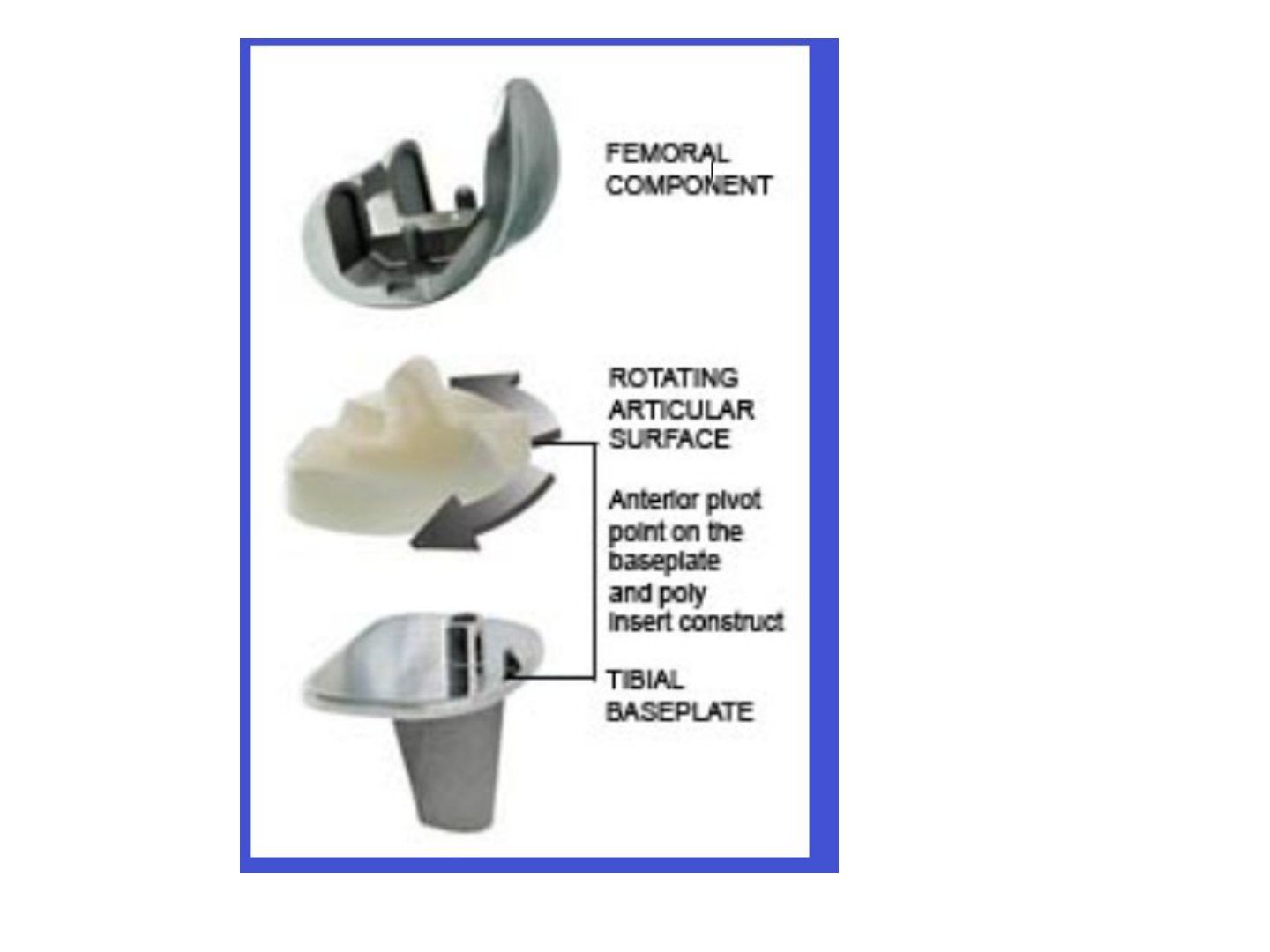
- Unconstrained: CR, P sacrificing, P stabilise (substitute)
- Constrained: non-hinged / hinged (or rotating hinge)
- Also fixed vs mobile bearing; metal back vs all PE
Q3.What is a non-hinged constrained prosthesis?
- A constrained prosthesis without an axle connecting the tibial and femoral components
- Used in soft tissue / bony defect
- Increased stability but more stress on the interface
Q4.What are the pros and cons of a mobile bearing in TKR?
- No benefit proven
- Pros: theoretically increased contact area leading to less wear; theoretically replicates native knee kinematics
- Cons: spinout, more back side wear
- Used in hinged implants
Q5.What are the features of an all-polyethylene tibial component?
- Cheaper
- Less modular
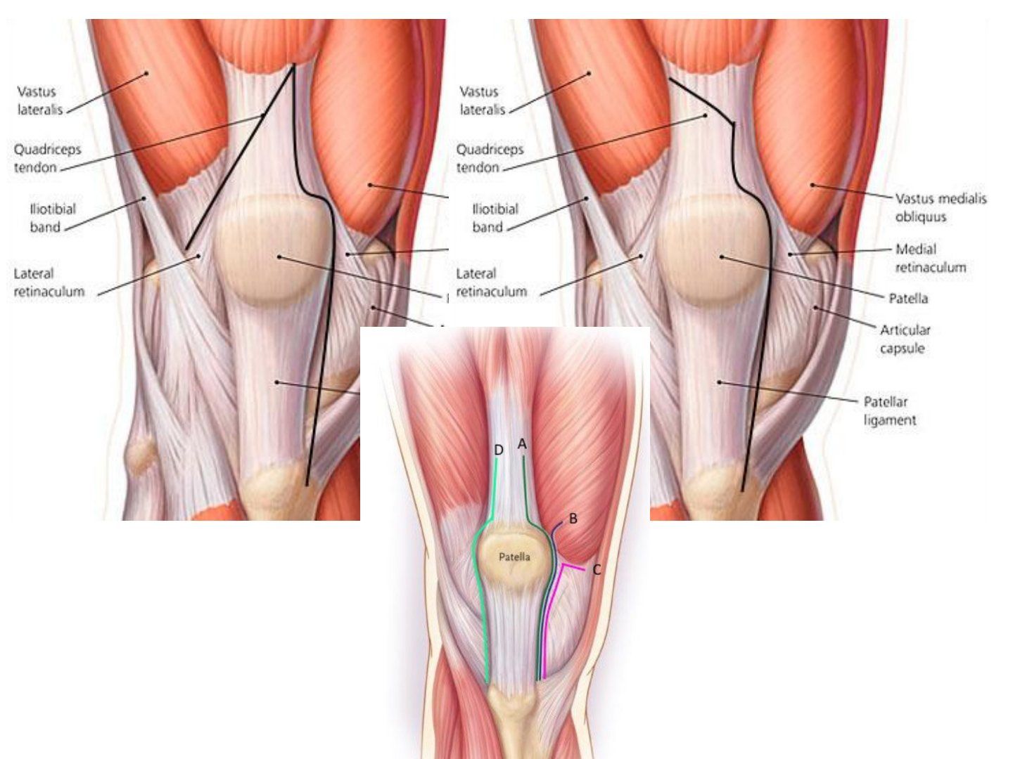
Q6.What surgical approaches are used in TKR?
- Medial parapatellar - extensile; patella perfusion impaired if lateral release needed
- Lateral parapatellar - direct access to lateral structures; difficulties in everting patella
- Smaller exposure: midvastus, subvastus
- Larger exposure: quadriceps snip, VY / osteotomy
Q7.How do you correct gap imbalance in TKR?
- Tight in flexion and extension: decrease PE insert size, resect more proximal tibia
- Tight in extension only: resect additional distal femur, posterior capsular release
- Tight in flexion only: downsize femoral component, recess/release PCL, increase posterior tibial slope, release posterior capsule
Q8.What are the problems of patellar resurfacing versus non-resurfacing in TKR?
- Resurfacing: fracture, loosening, clunk, AVN
- Not resurfacing: residual quads weakness (poor moment arm, patellar height not restored), maltracking, residual anterior knee pain
Q9.What is the evidence on routine patellar resurfacing in TKR?
- Zmistowski (Rothman) J Arthroplasty 2019: should NOT routinely replace the patella
- Teel J Arthroplasty 2019 meta-analysis: function no difference, non-resurfacing = more reoperations
Q10.How do you reduce transfusion in TKR?
- Pre-op: correct coagulopathy, stop medications, correct anaemia
- Intra-op: normovolaemic haemodilution, permissive hypotension, transamin, tourniquet, bone wax
- Cell saver for revision cases
- Post-op: transamin, no drain
Q11.What alignment deviation is acceptable in TKR?
- Accept 3 degrees deviation
- Outliers have poor results
Q12.What is the evidence on single stage versus sequential bilateral TKR?
- Local AHNH study: no outcome difference; backed by Denmark group 2015 KSSTA Lindberg-Larsen
- J Arthroplasty 2019 David Ward: simultaneous BTKA had significantly increased odds of multiple complications vs staged BTKA, but the absolute risk differences are minimal and complications are uncommon
- Stanford J Arthroplasty 2022: simultaneous TKR increases the risk of establishing the second knee outside the mechanical neutral
Zmistowski from Rothman Institute J Arthroplasty 2019 prospective RCT shows patella should not routinely be replaced — mischaracterised study design — It is a cost-utility analysis of 14 prospective RCTs, not itself an RCT; it concludes routine resurfacing of arthritis-free patellae is not cost-effective — source
▸ Slide 164 · CR vs PS TKRKnee · 7 questions expand

Q1-Q77 questions — tap to reveal all answerslist
- What is roll back in the knee?
- What are the prerequisites for CR TKR?
- What are the advantages of CR TKR?
- What are the disadvantages of CR TKR?
- What are the advantages of PS TKR?
- What are the disadvantages of PS TKR?
- What is the evidence comparing CR and PS TKR?
Q1.What is roll back in the knee?
- Posterior translation of the femur with progressive flexion
Q2.What are the prerequisites for CR TKR?
- PCL intact
- Varus/valgus <15 degrees
Q3.What are the advantages of CR TKR?
- PCL gives better proprioception
- Femoral roll back - better ROM and improved lever arm for the quadriceps
- PCL absorbs the AP and varus-valgus strain otherwise transferred to the bone-prosthesis interface
- Preserves bone stock
- Preserves the joint line
Q4.What are the disadvantages of CR TKR?
- Increased shearing + delamination leading to PE wear
- Technically more difficult (poorer exposure, gap balancing, see –saw effect if flexion and extension gaps are both too tight)
- Paradoxical roll forward if the PCL is defunctioned
Q5.What are the advantages of PS TKR?
- More conformity so less PE wear (delamination, pitting, fatigue)
- Does not rely on the PCL for roll back
- Technically easier
Q6.What are the disadvantages of PS TKR?
- More constraint - more stress at the bone-implant interface
- More bone loss
- Dislocation (cam jump)
- Patellar clunk - superior patellar scar caught in the box in extension
- Reduce ROM: cut PCL -> looser flexion gap -> thicker PE -> cut more distal femur -> elevate joint line -> patella baja
Q7.What is the evidence comparing CR and PS TKR?
- ROM similar
- Proprioception similar
- Australian joint registry 2021: PS 7.5% vs CR 6.1% revision at 15 years
▸ Slide 165 · Xray of patients knee early post op after total knee replacementKnee · 3 questions expand
Q1-Q33 questions — tap to reveal all answerslist
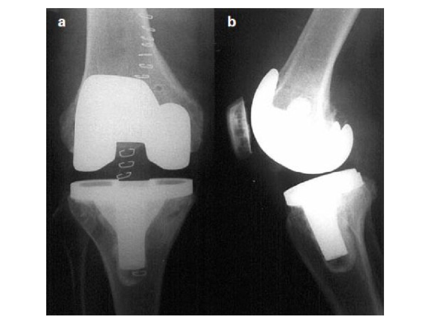
- Describe the radiographic findings in this early post op TKR X-ray.
- What further imaging or measurements would you like after TKR?
- What is checked on the lateral view of a post-TKR knee?
Q1.Describe the radiographic findings in this early post op TKR X-ray.
- This is an early post op film: both femoral and tibial implants are fixed with cement, no wedge/stem
- Patella replaced
- Femoral component size small, not flushed with the condyle, no notching on lateral view
- Tibial implant size optimal, no overhang
Q2.What further imaging or measurements would you like after TKR?
- Measure PE thickness
- Long standing film for alignment
Q3.What is checked on the lateral view of a post-TKR knee?
- Notching of the anterior femoral cortex
- Femoral component sizing and flushness
▸ Slide 166 · How are u going to perform TKR in varus knee ?Knee · 11 questions expand

Q1-Q1111 questions — tap to reveal all answerslist
- What does the pre-op X-ray show in this varus knee?
- What are the problems to plan for when performing TKR in a varus knee?
- What is the pre-op preparation for TKR in a varus knee?
- What is the sequence of medial release in a varus knee TKR?
- How do you manage a fixed flexion deformity in TKR?
- What is the Anderson classification of bone defects in TKR?
- How are contained and uncontained defects managed by size?
- How is a tibial bone defect managed during TKR?
- How is a femoral bone defect managed during TKR?
- What are the pros and cons of bone grafting and the role of a long stem in TKR?
- What are the post-op concerns and prophylaxis after TKR?
Q1.What does the pre-op X-ray show in this varus knee?
- Long standing and AP XR of the right knee showing a varus knee
- Tibiofemoral angle 45degs
- Medial tibia bone loss
- Widening of lateral joint space
- Tibia appears ER
Q2.What are the problems to plan for when performing TKR in a varus knee?
- Soft tissue deficiency - plan extensive soft tissue release, constrained implant
- Bony deficiency (contained/uncontained) - cement, cancellous graft, screw on cement, cortical graft, metal augment, tumour prosthesis
- Alignment - pre-op standing scannogram to plan tibial/femoral cuts and guides (intramedullary, extramedullary or navigation)
Q3.What is the pre-op preparation for TKR in a varus knee?
- Optimize patient fitness for operation
- Prophylactic antibiotic
- Same day admission to a clean ward
- The most important pre-op part is planning (soft tissue, bony, alignment)
Q4.What is the sequence of medial release in a varus knee TKR?
- Osteophyte
- Medial joint capsule and deep MCL
- PMC (capsule, semimembranosus, posterior oblique ligament)
- Superficial MCL (subperiosteal release / pie crusting)
- Pes anserius (pes anserinus)
- Downsizing tibial side + removing uncapped part; if not balanced use a semi constrained implant (lateral epicondyle advancement / distalize fibular head)
Q5.How do you manage a fixed flexion deformity in TKR?
- Extension gap tight, flexion gap normal
- Post osteophyte --> post capsule
- May cut more distal femur initially (e.g. 10mm vs normal 8mm)
Q6.What is the Anderson classification of bone defects in TKR?
- 1: does not affect stability
- 2: affects stability (metaphyseal damage) - cement, bone graft, metal wedge
- 3: deficient metaphysis affecting collateral/patellar tendon attachment - struct allograft, IBG, trabecular metal augment, tumour prosthesis
Q7.How are contained and uncontained defects managed by size?
- Contained: <5mm cement; large defect cancellous bone graft
- Uncontained: <5mm cement; 5mm-1cm bone graft/screw on cement; >1cm augment/strut graft + screw
- Cannot use cement alone in a large defect (may crack)
Q8.How is a tibial bone defect managed during TKR?
- Translate the tibial tray away from the defect
- Resect the tibia at a lower level
- Reassess the defect after the tibial cut: <5mm cement, 6-10mm bone graft, >10mm metal wedge
Q9.How is a femoral bone defect managed during TKR?
- Affecting the chamfer cut - cement
- Affecting the distal femur cut - metal augment
- Massive bone loss - bone block / allograft
Q10.What are the pros and cons of bone grafting and the role of a long stem in TKR?
- Bone graft replenishes bone stock but has donor morbidity
- Young age tends to preserve bone stock
- Use a long stem tibial component to share stress over the diaphysis (20% stress)
Q11.What are the post-op concerns and prophylaxis after TKR?
- Chest infection, wound infection, UTI, DVT, knee stiffness
- Post op: Early mobilization (early mobilisation), prophylactic antibiotics, wean off foley as early as possible
- DVT: mechanical +/- medical prophylaxis (enoxaparin, pradaxa) in high risk group
▸ Slide 167Knee · 2 questions expand
Q1-Q22 questions — tap to reveal all answerslist
- What topic is covered on this slide?
- What assessment steps would you take for the knee condition shown on this slide?
Q1.What topic is covered on this slide?
- Not covered in the speaker notes
Q2.What assessment steps would you take for the knee condition shown on this slide?
- Not covered in the speaker notes
▸ Slide 168 · Consent for TKRKnee · 5 questions expand
Q1-Q55 questions — tap to reveal all answerslist
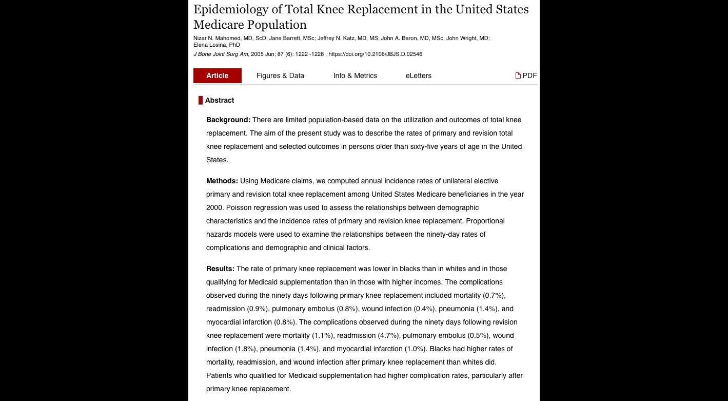
- What are the 90-day complication rates after primary TKR in the US Medicare population?
- What are the 90-day complication rates after revision TKR?
- What are the risks in the 90 days after discharge following TKR (JBJS 2006)?
- What risks do you discuss when consenting for TKR?
- Would you consider bilateral TKR in the same OT and why?
Q1.What are the 90-day complication rates after primary TKR in the US Medicare population?
- Source: JBJS 2005 (US Medicare population)
- Mortality 0.7%
- Readmission 0.9%
- PE 0.8%
- Wound infection 0.4%
- Pneumonia 1.4%
- MI 0.8%
Q2.What are the 90-day complication rates after revision TKR?
- Mortality 1.1%
- Readmission 4.7%
- PE 0.5%
- Wound infection 1.8%
- Pneumonia 1.4%
- MI 1.0%
Q3.What are the risks in the 90 days after discharge following TKR (JBJS 2006)?
- Study of 222,684 patients
- Mortality 0.53%
- Infection 0.71%
- PE 0.41%
Q4.What risks do you discuss when consenting for TKR?
- Anaesthetic risk
- Intra-op: bleeding, nerve/vascular injury, fracture
- Post-op: infection (wound, deep, urine), DVT/PE, pneumonia, aseptic loosening
Q5.Would you consider bilateral TKR in the same OT and why?
- Yes if the patient is fit
- Overall less complications
- Post-op may need ICU/HDU care
- More difficult to rehab
- Not everyone progresses to a 2nd TKR
▸ Slide 169 · Status post oxford mobile bearing UKAKnee · 13 questions 1 check expand
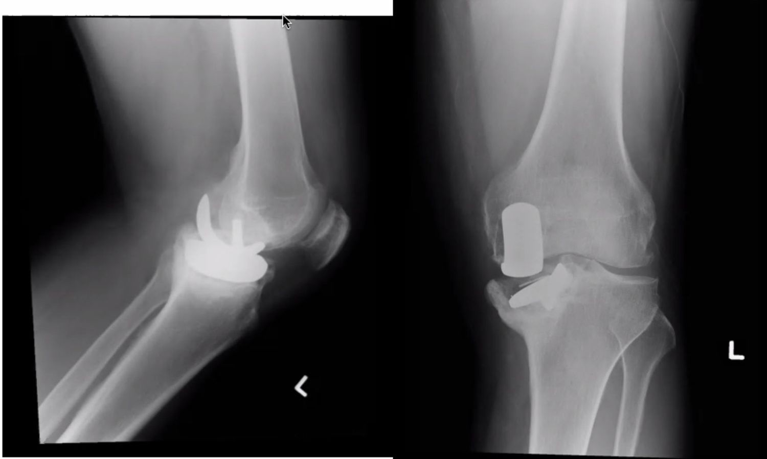
Q1-Q1313 questions — tap to reveal all answerslist
- What are the X-ray findings in this failed Oxford mobile bearing UKA?
- How do you recognise the PE marker on X-ray?
- What history do you take in a failed UKA?
- What do you examine in a failed UKA?
- What investigations are needed for a failed UKA?
- How would you revise a failed UKA?
- What are the anticipated difficulties and solutions when revising a UKA?
- What are the risk factors for bearing dislocation after mobile bearing UKA?
- What is the dislocation rate of the Oxford mobile bearing UKA?
- What is the soft tissue tension principle for bearing stability in UKA?
- What causes impingement of the mobile bearing?
- What happens to the MCL in UKA?
- Why is ACL deficiency a contraindication for UKA?
Q1.What are the X-ray findings in this failed Oxford mobile bearing UKA?
- Loosening of the tibial baseplate with subsidence into varus
- Bearing still on top of the tibial baseplate
- No obvious femoral component loosening
- No lucency or periosteal reaction suggestive of infection
Q2.How do you recognise the PE marker on X-ray?
- Normal PE marker: line in front, point at back
Q3.What history do you take in a failed UKA?
- Current symptoms: onset of pain, rest pain, fever or constitutional symptoms
- Functional limitations: OKS
- Details of the previous operation: pre-op indications, intra-op problems and implant sizing, post-op compliance and delayed wound healing
- Other risk factors: obesity, inflammatory joint disease; premorbid state and PMHx
Q4.What do you examine in a failed UKA?
- Local: sinus, varus/valgus laxity, ROM
- Systemic: is the patient fit for surgery
Q5.What investigations are needed for a failed UKA?
- WCC, CRP +/- knee aspiration
- Scannogram to check overall alignment
- CT for bone stock
Q6.How would you revise a failed UKA?
- Pre-op CT to measure bone defect, prepare bone and wedge
- Same incision, raise a full thickness skin flap below the fascial layer; extensile approach (quadriceps snip) if needed
- Remove the implant first (tibia usually easier; femur with ossilating saw (oscillating saw) + flexible osteotomy)
- Take at least 5 intra-op samples at the bone-implant junction for frozen section
Q7.What are the anticipated difficulties and solutions when revising a UKA?
- Soft tissue: valgus-varus balance - standby constraint; gap balancing, build up bone defect with augment/cones
- Bone: bone defect - use cone or wedge to restore bone stock; implant fixation by zonal fixation
Q8.What are the risk factors for bearing dislocation after mobile bearing UKA?
- Patient: compliance, infection
- Implant/surgeon: poor tension (MCL, ACL), poor alignment (varus/valgus, rotational), impingement, implant too small/deep flexion
- Too loose spins out; too tight impinges and rotates out
Q9.What is the dislocation rate of the Oxford mobile bearing UKA?
- 1 in 200 (0.5%) in medial UKA
- 10% in lateral UKA
Q10.What is the soft tissue tension principle for bearing stability in UKA?
- Too loose -> spin out
- Too tight -> impinge and rotate out
- When there is a discrepancy between 90 degree flexion and deep flexion, use a larger femur implant with a thinner insert
Q11.What causes impingement of the mobile bearing?
- Cam impingement - the insert hits the medial side of the tibial implant
- Cement, meniscus or osteophytes
- Undersized femur implant due to retained cartilage / osteophytes over the posterior condyle
Q12.What happens to the MCL in UKA?
- Acute MCL and chronic MCL (overstuff) problems
- The MCL elongates and loosens; the lateral compartment is under stress and develops OA
Q13.Why is ACL deficiency a contraindication for UKA?
- ACL deficiency causes posteromedial OA from anterior subluxation of the tibia
- In an ACL intact knee the posteromedial cartilage retensions the MCL
- In posteromedial OA the MCL cannot regain tension and becomes contracted
Bearing dislocation is 1 in 200 (0.5%) in medial UKA and 10% in lateral UKA — lateral figure outdated/context-specific — 10% applies to the original flat lateral Oxford bearing; the modern domed lateral design has a lower rate of about 1-6% (systematic review 3.7%) — medium confidence — source
▸ Slide 170 · SA vs GA in TKRKnee · 3 questions expand
Q1-Q33 questions — tap to reveal all answerslist
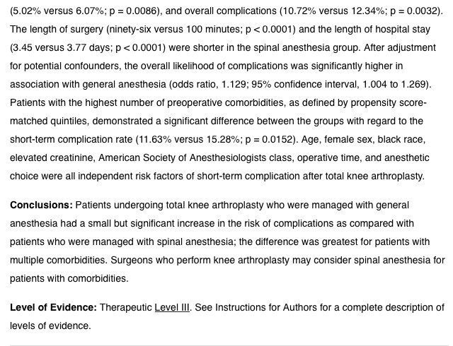
- What are the short-term (30-day) outcomes of spinal versus general anaesthesia in TKR?
- Which anaesthetic technique is preferred for total knee replacement?
- In which patient group is the benefit of spinal anaesthesia greatest?
Q1.What are the short-term (30-day) outcomes of spinal versus general anaesthesia in TKR?
- JBJS 2012 evidence
- Less superficial infection
- Less transfusion
- Shorter OT time
- Shorter length of stay
- Especially in those with comorbidities
Q2.Which anaesthetic technique is preferred for total knee replacement?
- Spinal anaesthesia - less superficial infection, less transfusion, shorter OT time and shorter length of stay at short term (30 days)
- Based on JBJS 2012
Q3.In which patient group is the benefit of spinal anaesthesia greatest?
- Especially in those with comorbidities
▸ Slide 171 · Valgus kneeKnee · 14 questions expand
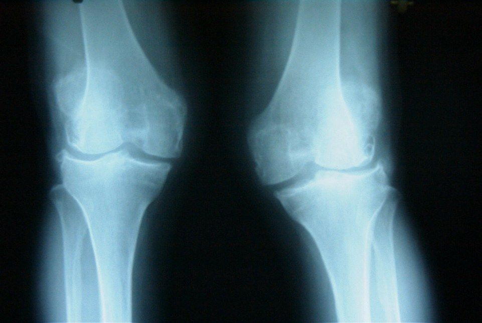
Q1-Q1414 questions — tap to reveal all answerslist
- What are the X-ray findings in this valgus knee?
- What are the causes of a valgus knee?
- What history do you take in a valgus knee?
- What do you examine in a valgus knee?
- What do you say if a patient with a valgus knee fails conservative management?
- What is the aim of TKR in a valgus knee?
- What are the approach options and pitfalls in valgus knee TKR?
- What bone problems are encountered in valgus knee TKR?
- How is coronal imbalance managed in valgus knee TKR?
- How is sagittal balance managed in valgus knee TKR?
- How is patella tracking managed in valgus knee TKR?
- What are the risk factors for peroneal nerve palsy after TKR?
- How do you manage acute peroneal nerve palsy after TKR?
- How do you manage late peroneal nerve palsy after TKR?
Q1.What are the X-ray findings in this valgus knee?
- Bilateral valgus knee, no obvious fracture
- Bone quality good, joint space preserved - no evidence of RA
- Hypoplastic lateral femoral condyle with increased lateral distal femur angle
- Patella lateral subluxation
Q2.What are the causes of a valgus knee?
- Rheumatoid arthritis
- Post-traumatic
- Iatrogenic (HTO)
- OA with hypoplastic lateral femoral condyle / constitutional valgus
- Haemophilia (Hemophilia), polio, HME
Q3.What history do you take in a valgus knee?
- Enquire causes (RA, post-traumatic, iatrogenic HTO, OA with hypoplastic lateral femoral condyle/constitutional valgus, haemophilia, polio, HME)
- Disability: KFS
Q4.What do you examine in a valgus knee?
- Assess patella tracking
- Document peroneal nerve status
- ROM, any FFC
- Collateral laxity
Q5.What do you say if a patient with a valgus knee fails conservative management?
- "This is a complex primary knee and I would refer to an arthroplasty surgeon"
Q6.What is the aim of TKR in a valgus knee?
- Stable painless mobile knee
- Restore joint line and mechanical axis
- Restore sagittal and coronal gap balance
- Choose cemented PS knee, standby constraint implant
Q7.What are the approach options and pitfalls in valgus knee TKR?
- Medial parapatellar: familiar and good exposure, but poor PL exposure, disrupt VMO cause patellar tracking problem, devascularises skin/patella if lateral release needed
- Lateral parapatellar: for a large lateral bone defect needing lateral exposure for augment/cone
- Need a coronal Z plasty
- Prepare distal femur augment for the hypoplastic lateral condyle
Q8.What bone problems are encountered in valgus knee TKR?
- Hypoplastic lateral femoral condyle / peripheral uncontained defect - use TEA + Whiteside as reference, prepare distal femur augment
- Lateral tibial plateau defect - usually central contained: minimal bone cut, reference off the medial side 5mm
Q9.How is coronal imbalance managed in valgus knee TKR?
- Contracted lateral soft tissue with attenuation of medial soft tissue
- LFE sliding osteotomy or MCL advancement
- Semiconstraint implant standby
Q10.How is sagittal balance managed in valgus knee TKR?
- Release osteophyte, PL capsule first
- Then depends on tightness: F: popliteus; E: ITB
- Both: piecrust LCL
Q11.How is patella tracking managed in valgus knee TKR?
- Lateral retinacular release (1cm from the border)
- Medial imbrication
- ER femoral and tibial implants
Q12.What are the risk factors for peroneal nerve palsy after TKR?
- Fixed flexion contracture + valgus
- Valgus deformity >12 degrees
- Epidural anaesthesia for pain control
- Previous laminectomy
- Overall 2% esp pre op valgus >12deg; suggest intra-op avoid excessive release; post op recovery room asssessment; prophylactic exploration is not beneficial
Q13.How do you manage acute peroneal nerve palsy after TKR?
- Remove the compressive dressing
- Check for haematoma, check the drain
- Flex the knee
- Check anaesthetic notes for blocks used
- Check XR: lateral overhang, cement, sawblade cut at fibula
Q14.How do you manage late peroneal nerve palsy after TKR?
- AFO to prevent foot drop
- Regular follow-up, NCT to document recovery
- +/- exploration if no improvement (controversial)
▸ Slide 172 · Tell me the pros and cons of one versus two stageKnee · 7 questions expand

Q1-Q77 questions — tap to reveal all answerslist
- What is the main concern in deciding one versus two stage revision?
- What are the advantages of one stage revision for a failed HTO?
- What are the anticipated difficulties with approach and exposure in one stage revision?
- What are the key steps of reconstruction in one stage revision?
- How do you choose between osteotomy and TKR?
- What are the advantages of UKA over HTO?
- What are the survival figures for UKA and HTO?
Q1.What is the main concern in deciding one versus two stage revision?
- Whether 1. bone stock 2. soft tissue difficulties can be dealt with in one go
Q2.What are the advantages of one stage revision for a failed HTO?
- One GA session
- Shorter rehab
- Only one extra-articular deformity; CORA is close to the joint
- Long stem bypasses the screw hole - less stress riser
- Decreased infection and wound problems
Q3.What are the anticipated difficulties with approach and exposure in one stage revision?
- Use previous/most lateral incision, maintain a thick fasciocutaneous flap
- Medial parapatellar approach (give reason and cons)
- Removal of implant - prepare screwdrivers/broken screw set, protect the MCL
- Inspect osteotomy healing/infection, take samples for c/st
- Retropatellar scar: prepare rectus snip, quadricepsplasty, TT osteotomy
Q4.What are the key steps of reconstruction in one stage revision?
- Femur: TEA + Whiteside line, expect lateral condyle erosion, less VCA
- Tibia: extramedullary guide, reference off lateral 2mm or medial 5mm, Beware of large tibial slope from index operation
- Obtain balance of the extension gap; PS implant, standby constraint; 2/3 zones of fixation; cemented fixation for poor bone
- Tibia stem bypasses screw hole by 2 cortical widths, prepare offset stem
- Patellar tracking: lateral release, medialised button; ensure no femoral/tibial ER; monitor peroneal nerve
Q5.How do you choose between osteotomy and TKR?
- TKR ideal: old, less active, tricompartmental, more soft tissue imbalance, larger deformity
- Osteotomy ideal: young fit non-smoker, good bone stock/range/skin, isolated compartment disease, deformity not severe
Q6.What are the advantages of UKA over HTO?
- Faster recovery, no reliance on bone healing, early weight bearing
- Smaller incision, less pain, shorter length of stay
- Easier conversion; less blood loss and morbidity
- HTO: preserves bone stock, wider indication (ACL deficiency), Better kinematics, ?ROM better; Cao 2018 - HTO for higher activity
Q7.What are the survival figures for UKA and HTO?
- UKA: 21.4% revision rate at 15 years (Australian joint registry)
- HTO: Kunze AJSM 2020 86% at 6 years; Insall series 70% at 10 years
▸ Slide 173 · Standing XR right knee showing osteoarthritis change mainly over medial compartmKnee · 15 questions expand

Q1-Q1515 questions — tap to reveal all answerslist
- What does the standing X-ray of the right knee show?
- How would you assess a patient with medial compartment OA of the knee?
- What is the management algorithm for isolated medial OA of the knee?
- What are the indications for HTO?
- What are the indications for UKA and TKA in isolated medial OA?
- What are the pros and cons of UKA versus HTO?
- What are the options for HTO?
- What are the contraindications and advantages of medial open wedge HTO?
- What are the disadvantages of medial open wedge HTO?
- What is the Nakamura classification of lateral cortex fracture in open wedge HTO?
- What are the pros and cons of lateral closing wedge HTO?
- What did the JBJS 2014 RCT show about open versus closing wedge HTO?
- How do you differentiate the HTO types on X-ray?
- What are the outcomes of HTO?
- How does UKA compare with TKR?
Q1.What does the standing X-ray of the right knee show?
- Osteoarthritis mainly over the medial compartment
- Lateral compartment preserved
- Slightly varus alignment
Q2.How would you assess a patient with medial compartment OA of the knee?
- Hx: age, functional demand, occupation, symptom onset/progression, previous treatment, Inflammatory arthritis, infection, trauma
- P/E: BMI, FFC, deformity, range, ACL, instability, NV, varus thrust, skin
- X-ray: scannogram, lateral, skyline
Q3.What is the management algorithm for isolated medial OA of the knee?
- Conservative management according to AAOS guideline
- Consider HTO, UKR, TKR
- Needs an individualized approach: age, functional demands, bone and soft tissue integrity, ACL status
Q4.What are the indications for HTO?
- Patient: <60 years old, active
- Joint: unicompartmental OA, FFC < 5 degree, ROM > 90, correctable varus <5 degrees, no instability
- Aim: unload medial compartment, shift mechanical axis, overcorrection 3-6deg
Q5.What are the indications for UKA and TKA in isolated medial OA?
- UKA: Kozin and Scott criteria, aim to undercorrect
- TKA: absolute indication if ACL insufficient, deformity too large, or poor range
- TKA may consider CR for theoretical advantages
Q6.What are the pros and cons of UKA versus HTO?
- HTO pros: preserve bone stock, wider indication (ACL deficiency), Better kinematics and ROM
- HTO cons: union problems, cosmesis (valgus knee), longer rehab
- Cao 2018 meta-analysis: UKA less revision/complications/post-op pain, HTO better ROM; HTO may be better for high activity level
Q7.What are the options for HTO?
- Medial open wedge
- Lateral closing wedge
- Dome osteotomy
Q8.What are the contraindications and advantages of medial open wedge HTO?
- C/I if patella baja as it will elevate the joint line
- Advantages: higher corrective power, no fibular osteotomy with decreased peroneal nerve injury, less tibial offset, less scarring leading to less patella baja
Q9.What are the disadvantages of medial open wedge HTO?
- Non-union leading to implant failure (plate and screw)
- Lateral cortex fracture
- Increase posterior slope
- Bone graft donor site morbidity (may use calcium phosphate wedge graft)
Q10.What is the Nakamura classification of lateral cortex fracture in open wedge HTO?
- Type1, along osteotomy
- Type2, go down
- Type3, go intraarticular region
Q11.What are the pros and cons of lateral closing wedge HTO?
- Advantage: fewer non-unions
- Disadvantages: peroneal nerve palsy, short limb, Distort anatomy, truncated making TKR harder, scarring leading to patella baja
Q12.What did the JBJS 2014 RCT show about open versus closing wedge HTO?
- No clinical or radiological difference between open and closing wedge
Q13.How do you differentiate the HTO types on X-ray?
- Fibular osteotomy = closing wedge or dome (closing wedge shows a step on the lateral side)
- Open wedge usually has a plate; dome may have 2 staples
Q14.What are the outcomes of HTO?
- 65% at 10 years (Insall); most HTOs eventually require TKR
- Best results: younger, moderate varus; obesity, undercorrection and excessive overcorrection are adverse
- Ideal correction: hip-knee-ankle angle between 2° & 6° (overcorrection); plan with Miniaci method, Fujisawa point; the overall preoperative state of the knee is the most important determinant
Q15.How does UKA compare with TKR?
- UKA preserves both cruciates: better knee function, better joint forgettability, quicker rehab, less blood loss and morbidity
- Good survival data come from high volume centres; overall survival 90% at 10 years (Swedish joint registry)
- Long term survival still inferior to TKR: 3.2x higher revision rate in the UK joint registry (possible selection bias)
- BMJ 2019 Andrew Price meta-analysis: both viable for isolated unicompartmental OA; UKA better in several outcome domains but TKA has lower revision risk
▸ Slide 174 · The indication by ScottKnee · 3 questions expand
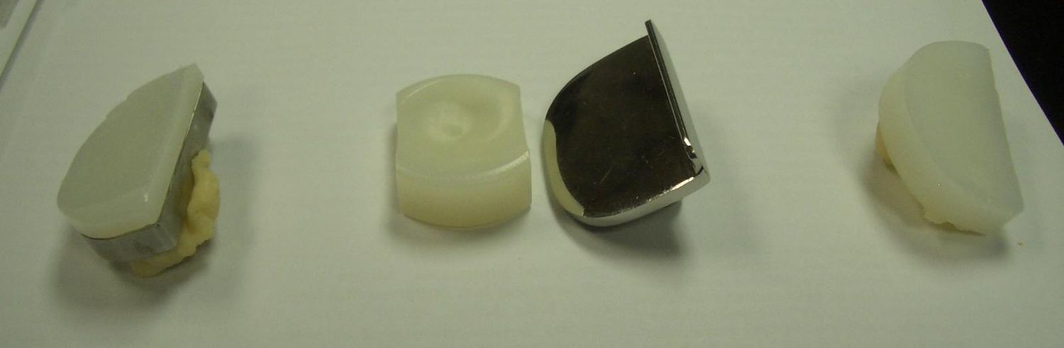
Q1-Q33 questions — tap to reveal all answerslist
- What are Scott's indications for UKA?
- What are the contraindications to UKA?
- What are the complications and failure mechanisms of UKA?
Q1.What are Scott's indications for UKA?
- Intact ACL
- Flexion arc >90
- Contracture <5
- Varus deformity <10, passively correctable to neutral
- Generally age >60, weight <82kg, not a manual worker
Q2.What are the contraindications to UKA?
- Multicompartmental involvement
- Inflammatory arthritis
- Significant fixed deformity (FFC >10 degrees)
- Previous meniscectomy on the other side
- ACL deficiency (in mobile bearing UKA)
Q3.What are the complications and failure mechanisms of UKA?
- Stress fracture (always tibial) with heavy weight/activity
- Fixed-bearing implants: fail with mechanical loosening
- Mobile-bearing implants: fail from disease progression
- Patella impingement may need revision to TKR
▸ Slide 175 · Lateral dislocation of patellar in a case after TKRKnee · 4 questions expand
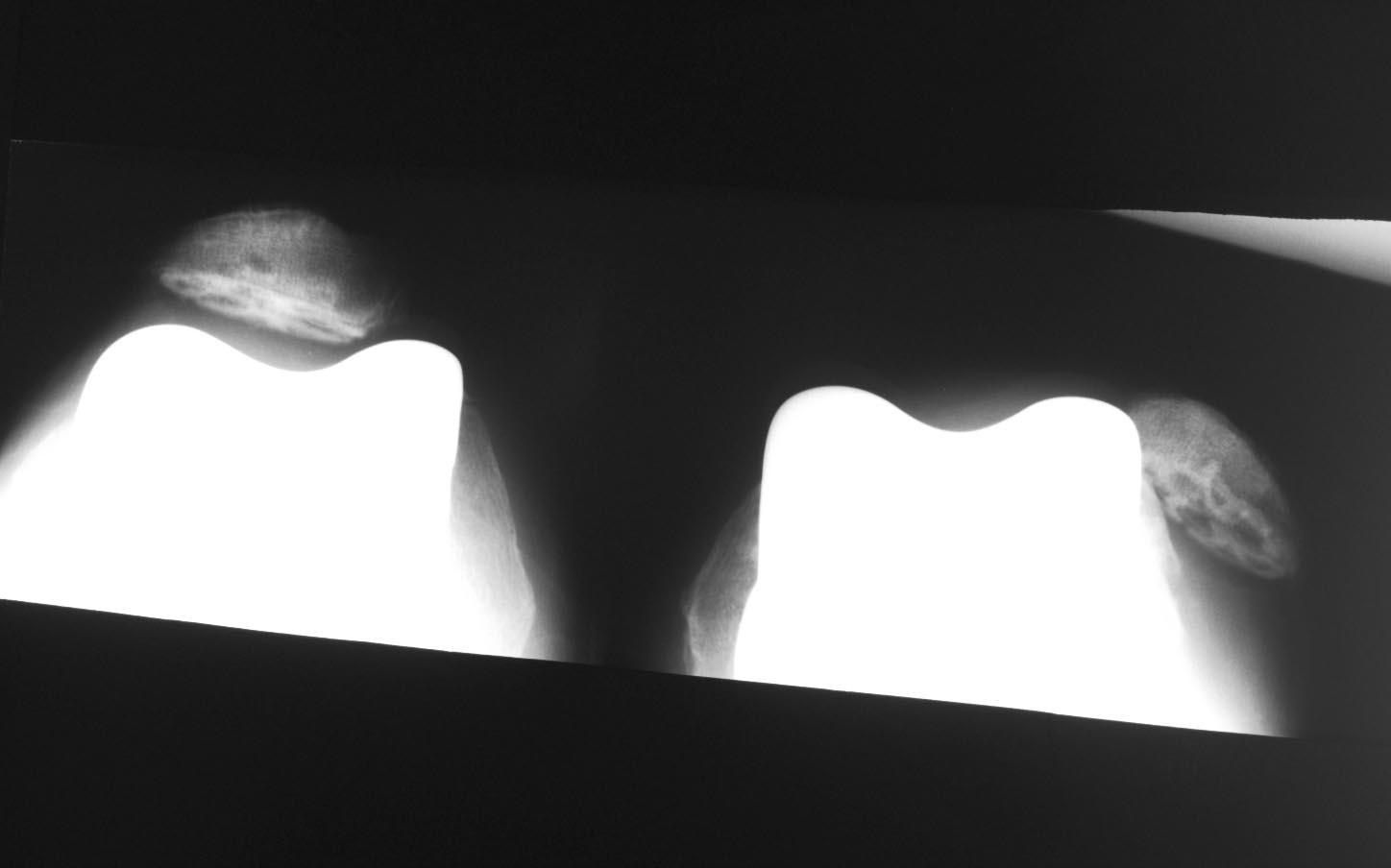
Q1-Q44 questions — tap to reveal all answerslist
- What factors affect patellar dislocation after TKR?
- How do you assess patellar tracking intra-operatively?
- How do you investigate post-operative patellar dislocation?
- What are the treatment options for post-TKR patellar dislocation?
Q1.What factors affect patellar dislocation after TKR?
- Pre op: valgus knee with lateral tightness
- Implant alignment/position increasing the Q angle
- Patella component: laterally placed, overstuff
- Femoral component: medially placed, IR, overstuff
- Tibial component: medially placed, IR; overall excessive valgus
Q2.How do you assess patellar tracking intra-operatively?
- No thumb technique
- If failed, release the tourniquet and try again
- If still failed, release the lateral retinaculum 1cm from the patella
Q3.How do you investigate post-operative patellar dislocation?
- Rule out infection, loosening, fracture
- Check alignment with CT (axial cuts)
- Check rotation: TEA vs posterior femoral component edge, perpendicular line of posterior tibial plane through stem centre, TT to stem line for IR
Q4.What are the treatment options for post-TKR patellar dislocation?
- Correct the underlying causes
- If no alignment problem: Insall procedure (lateralize and distalise VM)
- Lateral retinaculum release, vastus medialis advancement, medial plication
- Bony: TT osteotomy
▸ Slide 176 · Post TKR knee painKnee · 6 questions expand
Q1-Q66 questions — tap to reveal all answerslist
- What is your history in a patient with post-TKR knee pain?
- What do you examine in a patient with post-TKR knee pain?
- What investigations are needed for post-TKR knee pain?
- What are the intrinsic causes of post-TKR knee pain?
- What causes posterolateral knee pain after TKR?
- What causes posteromedial pain and what are the extrinsic causes of post-TKR pain?
Q1.What is your history in a patient with post-TKR knee pain?
- Rule out infection: risk factors, delayed wound healing, constitutional symptoms
- Nature of pain: referred, triphasic, resting
- Current functional limitations
Q2.What do you examine in a patient with post-TKR knee pain?
- Local: soft tissue, sinus, ROM, FFC, instability
- Systemic: hygiene
Q3.What investigations are needed for post-TKR knee pain?
- X-ray: early loosening, sizing, overhang, PFJ overstuffing, HO
- Scannogram for mechanical alignment
- Bloods: ESR, CRP
- Aspiration: C/ST + WBC + alpha defensin (MSIS score)
- CT for malrotation
Q4.What are the intrinsic causes of post-TKR knee pain?
- Infection, loosening, malalignment, instability
- Loose bodies (cement, osteophyte)
- PFJ problem, impingement
Q5.What causes posterolateral knee pain after TKR?
- Tibial tray overhang
- Retained PL osteophyte/cementophyte - biceps tendonitis, popliteus impingement
- ITB dysfunction - lateral pain with ROM 20-80 degrees
Q6.What causes posteromedial pain and what are the extrinsic causes of post-TKR pain?
- Semimembranosus tendonitis
- Dual pathology (hip, foot, PVD)
- CRPS, cutaneous neuromas
▸ Slide 177 · Post TKR instabilityKnee · 12 questions expand
Q1-Q1212 questions — tap to reveal all answerslist
- What are the 4 Ps to mention when assessing instability after TKR?
- What history is important in post-TKR instability?
- What are the components of the Knee Society knee score?
- What are the components of the Knee Society function score?
- What local examination findings do you look for in post-TKR instability?
- What systemic examination and gait findings are relevant in post-TKR instability?
- How do you work up and manage post-TKR instability?
- What are the types of instability after TKR?
- What causes flexion (AP) instability after TKR and how is it treated?
- How do you differentiate symmetrical from asymmetrical extension (ML) instability?
- What is mid-flexion instability and what are its potential causes?
- How are genu recurvatum and global instability after TKR managed?
Q1.What are the 4 Ps to mention when assessing instability after TKR?
- Patient
- Prosthesis
- Procedure
- Present status
Q2.What history is important in post-TKR instability?
- Initial indication and diagnosis
- PMHx: connective tissue disease, inflammatory joint disease, Charcot arthropathy
- Preop ROM and pre-existing laxity
- Intraoperative complications
- Postop compliance and recent trauma
- Current function
Q3.What are the components of the Knee Society knee score?
- Pain
- FFC (fixed flexion contracture)
- Extensor lag
- Total ROM
- Alignment
- Stability (AP and ML)
Q4.What are the components of the Knee Society function score?
- Walking aids
- Stairs
- Walking tolerance
Q5.What local examination findings do you look for in post-TKR instability?
- Soft tissue envelope, ROM, any recurvatum
- Quads power
- Collateral ligament testing, A and P drawer
- Patellar tracking
Q6.What systemic examination and gait findings are relevant in post-TKR instability?
- generalized ligamentous laxity (generalised)
- Check proprioception
- Gait: varus or valgus thrusting gait
- CT for implant position
Q7.How do you work up and manage post-TKR instability?
- Determine the cause
- Bone: X-ray and CT
- Soft tissue: examination
- Rule out infection: bloods +/- aspiration
Q8.What are the types of instability after TKR?
- Flexion (AP)
- Extension (ML)
- Mid-flexion
- Genu recurvatum
- Global
Q9.What causes flexion (AP) instability after TKR and how is it treated?
- Knee dislocation (PS knees can jump the post)
- Undersized femoral component --> upsize
- Overresection of posterior condyle --> upsize or posterior femoral augments
- Too much tibial slope --> reduce slope + use PS prosthesis
- PCL insufficiency in a CR --> revision to PS knee
Q10.How do you differentiate symmetrical from asymmetrical extension (ML) instability?
- Symmetrical = bony problem (too much distal femur cut or oversized femoral implant)
- Asymmetrical = ligament problem
Q11.What is mid-flexion instability and what are its potential causes?
- Malrotation when the knee is flexed between 45-90 degrees
- Poorly understood
- Femoral component design in the sagittal plane
- Attenuation of anterior MCL
- Overall geometry of the tibiofemoral joint
Q12.How are genu recurvatum and global instability after TKR managed?
- Recurvatum: fixed valgus deformity and IT band contracture; causes polio, RA, Charcot
- Recurvatum: long-stemmed PS or ML constrained implant; rotating hinge for salvage as residual hyperextension may occur
- Global: due to severe bone loss
- Global: varus-valgus constrained prosthesis at minimum, hinged prosthesis +/- augments, megaprosthesis if severe bone loss
▸ Slide 178 · Post TKR stiffnessKnee · 5 questions expand

Q1-Q55 questions — tap to reveal all answerslist
- What preoperative factors predispose to stiffness after TKR?
- What intraoperative causes produce poor flexion after TKR?
- What intraoperative causes produce poor extension after TKR?
- What intraoperative causes produce poor global ROM after TKR?
- What postoperative complications cause stiffness after TKR?
Q1.What preoperative factors predispose to stiffness after TKR?
- Poor preoperative range of movement
- RA
- Post traumatic (post-traumatic) knee
- Obesity
Q2.What intraoperative causes produce poor flexion after TKR?
- Tight flexion gap: inadequate tibial slope, oversized femur, posteriorized femur
- Overstuffed PFJ
- Raised joint line (patella baja)
Q3.What intraoperative causes produce poor extension after TKR?
- Flexed PS component
- Posterior osteophytes
- Inadequate distal femur cut
Q4.What intraoperative causes produce poor global ROM after TKR?
- Big polyethylene insert
- Inadequate tibial cut
Q5.What postoperative complications cause stiffness after TKR?
- Arthrofibrosis
- Infection
- CRPS
- HO
▸ Slide 179 · Patellar clunk syndromeKnee · 5 questions expand
Q1-Q55 questions — tap to reveal all answerslist
- What is patellar clunk syndrome?
- How does patellar clunk differ from patellar crunch and dislocation?
- What preoperative risk factors predispose to patellar clunk?
- Which patellar implant factors cause patellar clunk?
- Which femoral and insert factors cause patellar clunk?
Q1.What is patellar clunk syndrome?
- Painful, palpable clunk at the PFJ of a PS TKR
- Fibrous nodular scar at the posterior surface of the distal quads tendon / superior patellar pole
- Catches on the box of the femoral component during knee extension
- Flexion, then catch during extension
Q2.How does patellar clunk differ from patellar crunch and dislocation?
- Crunch: scar around the patellar implant, crunch sensation moving from extension to flexion, catch during flexion
- Clunk: catch near extension
- Dislocation: clunk near flexion
Q3.What preoperative risk factors predispose to patellar clunk?
- Patella baja
- Previous knee surgery
- Valgus knee
- Extensive synovitis
Q4.Which patellar implant factors cause patellar clunk?
- Small patellar implant --> decreased quads tendon contact forces against the superior intercondylar box --> fibrosynovial proliferation
- Overresection / thin implant --> reduces the offset of the quad tendon
- Inferior placement --> bare bone at superior pole exposed
Q5.Which femoral and insert factors cause patellar clunk?
- PS femur (IB2 with short, abrupt flange)
- Increased posterior femoral condylar offset
- Small femoral component
- Femoral component in flexion
- Thick PE insert causing raised joint line and patella baja
▸ Slide 180 · Indications for knee arthrodesisKnee · 6 questions expand

Q1-Q66 questions — tap to reveal all answerslist
- What are the indications for knee arthrodesis?
- What are the contraindications to knee fusion?
- What alignment should a knee arthrodesis be fixed in?
- Compare plate, nail and external fixator for knee arthrodesis.
- What are the complications of knee arthrodesis?
- What are the key considerations when converting a fused knee to TKA?
Q1.What are the indications for knee arthrodesis?
- Loss of extensor mechanism
- Charcot arthropathy
- Tumour resection
- Unsalvageable TKR
Q2.What are the contraindications to knee fusion?
- Absolute: active infection
- Relative: bilateral knees involved
- Relative: ipsilateral hip fusion
- Relative: contralateral amputation
- Relative: significant bone loss
Q3.What alignment should a knee arthrodesis be fixed in?
- 7 degrees valgus
- 15 degrees flexion
- 0-10 degrees external rotation (match the other leg)
- On examination, tell pathological from surgical fusion by looking at alignment
Q4.Compare plate, nail and external fixator for knee arthrodesis.
- Plate: advantage allows compression; disadvantage extensive dissection
- Nail: advantage load transmitting (increases healing and early WB); disadvantage difficult insertion
- Ex Fix: advantage usable in poor soft tissue envelope; disadvantage pin tract infection
Q5.What are the complications of knee arthrodesis?
- Nonunion
- Infection
- Adjacent joint disease
- Supracondylar and proximal tibia fracture
Q6.What are the key considerations when converting a fused knee to TKA?
- Preop: extensor mechanism (clinical/EMG/MRI), contracted soft tissue envelope (expander), CT for bone stock, instruments (navigation, BG/augment, semiconstraint + stem)
- Skin: choose most lateral wound, mainly full thickness fasciocutaneous flap
- Bone stock: osteoporotic, hard to evaluate with hardware; +/- staged implant removal + bone graft; prepare BG/augment/stem
- Soft tissue: shortened extensor mechanism --> quadriceps VY plasty; deficient collaterals --> semiconstraint; rotational flap for closure
- Previous deformity
▸ Slide 181 · hemophiliaKnee · 10 questions expand
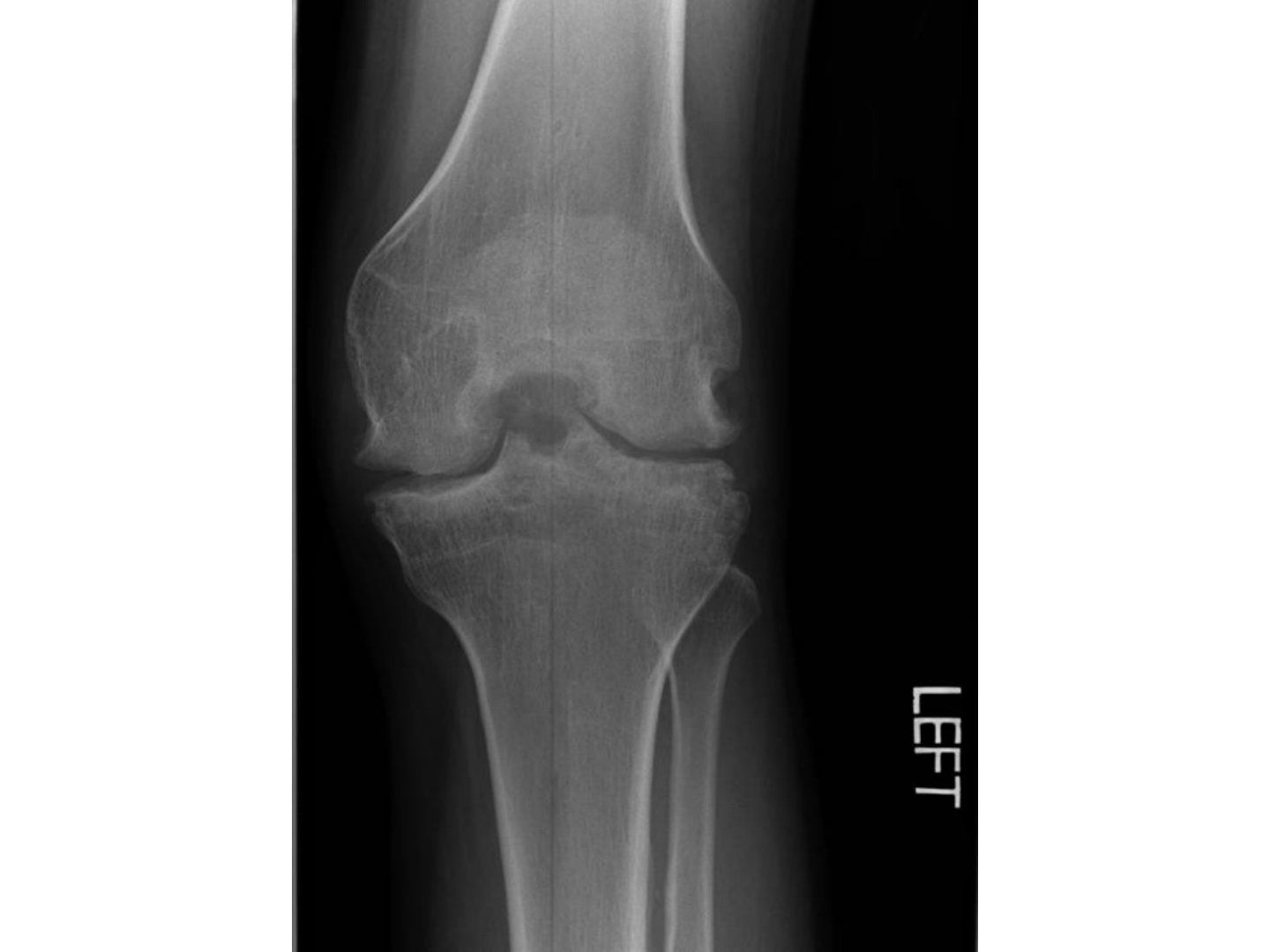
Q1-Q1010 questions — tap to reveal all answerslist
- Describe the X-ray features of haemophilic arthropathy.
- What is the Arnold-Hilgartner classification?
- Tell me about haemophilia.
- What is the pathology of haemophilic arthropathy?
- What history and examination findings are important in haemophilia?
- What investigations are needed in haemophilia?
- What is the aim and conservative management of haemophilic joint disease?
- How are bleeding episodes and surgery managed in haemophilia?
- What are the difficulties of TKR in haemophilia?
- What is the prognosis of TKR in haemophilia?
Q1.Describe the X-ray features of haemophilic arthropathy.
- Decreased joint space, OA change, osteopenia
- Ballooning of distal femur (paediatric epiphyseal overgrowth), wide notch, squaring of femoral condyle
- Squaring of patella (Jordan sign), no fracture
- Large subchondral cyst
- Pseudotumour
- Ddx: OA, post-traumatic OA, post-infectious OA, inflammatory arthritis
Q2.What is the Arnold-Hilgartner classification?
- 0 normal
- I soft tissue synovitis, bone normal
- II epiphyseal hypertrophy, osteopenia
- III patellar squaring, subchondral cyst, widened notch
- IV joint space narrowing
- V ankylosis
Q3.Tell me about haemophilia.
- Inherited disease of clotting dysfunction; most common type A (factor 8) and type B (factor 9), both X-linked recessive
- Childhood onset of easy bleeding, usually at joints and intramuscular regions; mucosa less likely; affects hinged joints (knee, elbow, ankle)
- Mild 5-25% / moderate 1-5% / severe <1% factor concentration determines bleeding risk
- Recombinant factor transfusion in bleeding episodes (no blood-borne disease now)
- Factor VIII inhibitors (including IgG antibodies) in 5-25% - inhibit response to therapeutic factor (monoclonal recombinant factor VIII); relative contraindication to surgery, screen preoperatively
- Factor <1% --> spontaneous bleeding
Q4.What is the pathology of haemophilic arthropathy?
- Bleeding --> haemosiderin deposition --> synovitis --> bleeding
- Repeated haemarthrosis --> capsular fibrosis
- Articular damage (lyzosomal enzyme from synoviocytes) (lysosomal enzymes from synoviocytes)
- Hyperemia --> LLD/ deformity (+/- overgrowth) (hyperaemia --> LLD/deformity)
- Disuse: atrophy, contracture, osteopenia --> arthropathy
Q5.What history and examination findings are important in haemophilia?
- Hx: symptoms of bleeding/effusion, male or female, onset, previous medical follow-up, investigation, factor transfusion, complications of treatment
- PE: joint effusion, ROM, deformity, gait (function)
- Numbness in the L4 distribution caused by iliacus haematoma compressing the femoral nerve
Q6.What investigations are needed in haemophilia?
- Blood test: Hb, aPTT prolonged, PT normal
- Bethesda assay measures factor 8/9 antibody (megadose/ neutralize)
- Preop workup: factor transfusion, cell saver, check inhibitor IgG, HIV testing
Q7.What is the aim and conservative management of haemophilic joint disease?
- Systemic: refer haematologist for bleeding control (regular/symptomatic transfusion)
- Aim: preventive - decrease bleeding episodes, prevent soft tissue contracture, deformity and ankylosis
- Conservative: analgesics + physiotherapy
- Prophylactic factor replacement >2%
Q8.How are bleeding episodes and surgery managed in haemophilia?
- Acute haemarthrosis: aspiration, factor infusion aim 40-50%, splint/compressive dressing, +/- steroid, early rehab
- Recurrent: aspiration, factor VIII >20% for 6 weeks, IA steroid, cast; desmopressin
- Chronic stage 1-3 (joint space preserved): synovectomy, radioactive synoviorthesis with colloidal 32P chromic phosphate
- Chronic stage 4-5: arthrodesis / TKR (higher infection rate)
- Skeletal surgery: factor to 100% in 1st week, keep >50% in 2nd week
- Targets: vigorous physiotherapy 20%; acute haematoma 30%; acute haemarthrosis / soft tissue surgery 40-50%
Q9.What are the difficulties of TKR in haemophilia?
- Pre-op: factor transfusion, cell saver, check inhibitor IgG, HIV testing
- Intra-op soft tissue: contracture, easy bleeding
- Intra-op bone: soft bone easy fracture, bone graft cysts, large femoral epiphysis (difficult sizing), small IM canal, valgus knee, tibia posterolateral subluxation + decreased slope, thin patella
- Post-op: factor transfusion to cover (1 week 100%, 2 weeks >75%), haematoma formation
Q10.What is the prognosis of TKR in haemophilia?
- J Arthroplasty meta-analysis 2022 Fenelon: overall complication rate 28.7%
- Similar survivorship to the general population
▸ Slide 182 · TKR in polio patientsKnee · 5 questions expand

Q1-Q55 questions — tap to reveal all answerslist
- What is the pathogenesis of knee deformity in polio?
- What bone problems complicate TKR in polio patients?
- What soft tissue problems complicate TKR in polio patients?
- How is recurvatum addressed during TKR in polio?
- What is the reported outcome of TKR in polio patients?
Q1.What is the pathogenesis of knee deformity in polio?
- Weak quads with failed knee extension in stance phase
- Hyperextension of the knee to bring the ITB anterior to the knee centre of rotation, acting as an extensor
- Gradual stretching of the posterior capsule
- Weight bearing and gravity elongate the tibial condyles posteriorly
- Articular surface angle to the tibial axis becomes more acute; proximal third of tibia bows posteriorly
Q2.What bone problems complicate TKR in polio patients?
- Genu recurvatum and genu valgum
- Osseous abnormalities: narrow canal, metaphyseal and articular deformities
- Osteoporosis
- ER of tibia with subluxed patella, patella baja
- Bone loss
Q3.What soft tissue problems complicate TKR in polio patients?
- Lax medial and posterior structures
- Tight lateral structures
- Weak quads
- Fixed flexion contracture
Q4.How is recurvatum addressed during TKR in polio?
- Exposure limited by patella baja: lateral retinacular release, medial soft tissue release, quads snip / TTO
- Extension gap: cut less distal femur
- Flexion gap: small component to increase flexion gap
- Constraint
Q5.What is the reported outcome of TKR in polio patients?
- 7% revision rate at 6 years (systematic review, EFORT 2018)
▸ Slide 183 · Bilateral knee XRKnee · 7 questions expand

Q1-Q77 questions — tap to reveal all answerslist
- What is a Charcot joint and what are the X-ray findings in the knee?
- What are the differential diagnoses for a destroyed knee?
- What history and examination findings are relevant in a Charcot knee?
- How do you rule out infection in a Charcot knee?
- What underlying causes of a Charcot joint should be sought?
- What is the management of a Charcot knee?
- What are the issues with TKR in a Charcot knee and the reported outcomes?
Q1.What is a Charcot joint and what are the X-ray findings in the knee?
- Progressive, noninfectious, destructive disorder of bone and joint with underlying sensory neuropathy
- Destruction of the joint with subluxation
- Periarticular bone debris
- Increased bone density
Q2.What are the differential diagnoses for a destroyed knee?
- Infection
- Advanced OA knee
- RA
Q3.What history and examination findings are relevant in a Charcot knee?
- Hx: premorbid function, pain, instability, functional limitations, expectations
- PE: sensation and proprioception
- Difficult to differentiate from osteomyelitis
Q4.How do you rule out infection in a Charcot knee?
- Difficult to differentiate from osteomyelitis
- Blood tests, WBC scan, aspiration, synovial biopsy
- Bone scan both hot; WBC scan may be cold in Charcot
Q5.What underlying causes of a Charcot joint should be sought?
- DM
- Syphilis (Argyle-Robertson pupil)
- Hansen disease
- Syringomyelia
Q6.What is the management of a Charcot knee?
- Limitation of activity and bracing
- Surgery: key is to intervene at the consolidation phase
- Fusion is classical: resect sclerotic bone (dead bone), apposition of bleeding bone; prolonged immobilisation >12 weeks
- Recent trend towards TKR
Q7.What are the issues with TKR in a Charcot knee and the reported outcomes?
- Poor bone stock; soft tissue laxity --> instability; poor implant fixation --> 2/3 zonal fixation
- TKR reported to give good pain relief; some say relatively contraindicated (high complication rate)
- EFORT meta-analysis 2021: survivorship 85.4% (mid >5years to long term >10years)
- Complication rate 26.4%: instability 24.0%, periprosthetic fracture 17.4%, infection 13.0%, ligament injury 10.9%, aseptic loosening 10.9%
▸ Slide 184 · Key numbers:Knee · 3 questions expand

Q1-Q33 questions — tap to reveal all answerslist
- What coronal and sagittal deformity thresholds are quoted for osteotomy vs TKA?
- What are the prerequisites, advantages and disadvantages of TKA with intraarticular correction?
- What are the advantages and disadvantages of extraarticular correction then TKR?
Q1.What coronal and sagittal deformity thresholds are quoted for osteotomy vs TKA?
- Coronal: femur >20, tibia >30
- or >20 in the sagittal plane
- Then consider severity --> soft tissue --> LLD --> nail/plate
Q2.What are the prerequisites, advantages and disadvantages of TKA with intraarticular correction?
- Prerequisite: not to violate the collateral ligament (+ minimal malrotation)
- Advantages: less extensive, easy and fast rehab, avoid osteotomy
- Disadvantages: soft tissue imbalance, malrotation of the ankle joint
Q3.What are the advantages and disadvantages of extraarticular correction then TKR?
- Greater surgical flexibility (open vs closing wedge osteotomy, technically easier)
- Allows healing of the osteotomy (no risk of cement extrusion from TKR)
- Improve knee alignment – might alleviate symptoms and delay TKA
- Disadvantages: 2 surgeries, 2 anaesthetics, longer rehab
▸ Slide 185 · Revision TKRKnee · 7 questions expand

Q1-Q77 questions — tap to reveal all answerslist
- What history and examination findings are important before revision TKR?
- What investigations are required before revision TKR?
- What are the aims of revision TKR and when is a constraint implant indicated?
- What must be prepared preoperatively for revision TKR?
- Describe the intraoperative approach and implant removal in revision TKR.
- What is the bone reconstruction ladder and AORI classification in revision TKR?
- What are the postoperative instructions after revision TKR?
Q1.What history and examination findings are important before revision TKR?
- Hx: preop, intraop (trace OT record) and postop course
- Recent infection (skin, dental, UTI); nature of pain
- Local PE: soft tissue envelope, ligamentous laxity, ROM, pulses
- Systemic PE: signs of active infection e.g. ulcer, dirty toes
Q2.What investigations are required before revision TKR?
- Bloods - inflammatory markers
- Joint aspirate
- XR + scannogram
- CT
Q3.What are the aims of revision TKR and when is a constraint implant indicated?
- General: stable, painless, mobile knee
- Specific: restore joint line and mechanical axis, balance coronal and sagittal gaps
- Constraint because of loss of collateral function
- Indications: flexion instability with largest femoral component + thickest insert
- Extension instability (recurvatum)
- ML instability upon full knee extension
Q4.What must be prepared preoperatively for revision TKR?
- Counsel patient re survival and complication rate; know previous implant, contact sales on standby
- Implant removal devices: oscillating saw, flexible osteotome, burr, Gigli saw
- Inform pathology lab for intraop frozen section; prepare cement spacer in case of infection
- Bone loss: prepare augment, wedge, cone, long stem
- Varus-valgus constrain, rotating hinge prosthesis
Q5.Describe the intraoperative approach and implant removal in revision TKR.
- Previous approach, most lateral incision (6 cm skin bridge; blood supply to skin from medial side); full thickness flap
- Evert patella, partial lateral release to lateral gutter; careful not to disrupt extensor mechanism
- Prepare extensile approach: quadricepsplasty, TT osteotomy, rectus snip
- Remove femur first, then tibia using stacked osteotome or K wire technique
- Remove all cement, take representative sample x5 for c/st, also frozen section (x5 samples for C/S); assess bone loss (AORI)
Q6.What is the bone reconstruction ladder and AORI classification in revision TKR?
- Ladder: cement +/- screw, bone graft (structural or morselized), modular augmentation (wedges/blocks), custom made/tumour/hinge prosthesis
- Identify joint line (meniscal scar, fibular head, epicondyles, tibial tuberosity); do tibia first to restore gaps; aim 2/3 zones fixation (long cementless stem = short cemented stem, Jones BJJ 2012)
- AORI 1: contained minor defects, intact metaphyseal bone, implant stable
- AORI 2: uncontained defect, damaged metaphyseal bone requiring reconstruction for stability
- AORI 3: deficient metaphyseal segment compromising major portion of femoral condyle/tibial plateau, +/- collateral or patellar tendon detachment
Q7.What are the postoperative instructions after revision TKR?
- Tell the patient to maximise rehab to avoid stiffness
- Rehab: weight bear as tolerated
- Insert drain
- Antibiotics til c/st is back