▸ Slide 100 · statisticsStatistics · 2 questions expand

Q1-Q22 questions — tap to reveal all answerslist
- What statistics topic is covered on this slide?
- What are the key statistical concepts tested in orthopaedic exams?
Q1.What statistics topic is covered on this slide?
- Not covered in the speaker notes
Q2.What are the key statistical concepts tested in orthopaedic exams?
- Not covered in the speaker notes
▸ Slide 101 · Wilsons criteria for screening programStatistics · 7 questions 1 check expand
Q1-Q77 questions — tap to reveal all answerslist
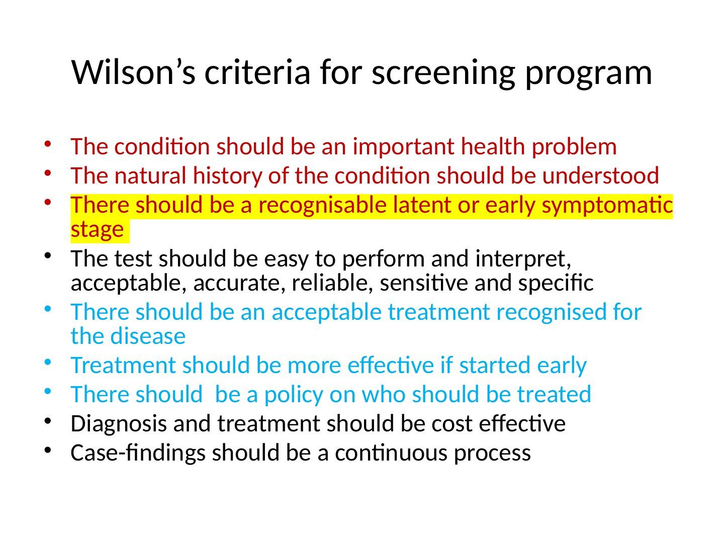
- What are **Wilson's criteria** for a screening program?
- What features should the screening test and the screened population have?
- Give two examples of screening programmes discussed.
- What is the incidence of DDH and how do the Barlow/Ortolani test and ultrasound compare?
- What did the 2009 International Hip Dysplasia Institute study show?
- Does DDH screening fulfil **Wilson's criteria**?
- Describe the Hong Kong scoliosis screening programme.
Q1.What are Wilson's criteria for a screening program?
- Condition: an important health problem; natural history understood; recognisable latent or early symptomatic stage
- Test: easy to perform and interpret, acceptable, accurate, reliable, sensitive and specific
- Treatment: acceptable treatment recognised, more effective if started early, policy on who should be treated
- Diagnosis and treatment should be cost effective; case-findings should be a continuous process
Q2.What features should the screening test and the screened population have?
- Test: acceptable and tolerated by patients; high sensitivity to detect disease before the critical point; high specificity to reduce false positives; cost effective
- Population: disease should have high prevalence to allow screening; accepted and effective treatment is available; treatment and further evaluation acceptable to the patient
- Disease: significant impact on community; recognisable latent or early symptomatic phase; natural history understood
Q3.Give two examples of screening programmes discussed.
- DDH screening
- Scoliosis screening in Hong Kong (P5, F1 and F3)
Q4.What is the incidence of DDH and how do the Barlow/Ortolani test and ultrasound compare?
- DDH incidence 1 in 1000
- B and O test: sensitivity 67%, specificity 96%
- Ultrasound: sensitivity 89%, specificity 97%, PPV 62%
Q5.What did the 2009 International Hip Dysplasia Institute study show?
- Compared no screening, selective screening and universal screening
- Selective screening most cost effective
Q6.Does DDH screening fulfil Wilson's criteria?
- Fulfils most criteria
- BUT there is no universal agreement on who should receive treatment
- Cost of the screening program versus the full cost of delayed detection has not been fully established
Q7.Describe the Hong Kong scoliosis screening programme.
- 3-tier system at P5, F1 and F3
- FBT sensitivity 84%; ATR sensitivity 83% (>=15 degrees refers to specialist hospital; 5-14 degrees goes to Moire topography)
- Moire topography >=2 lines leads to XR; Cobb >20 degrees refers to specialist hospital
- Overall sensitivity 88%
Selective screening is the most cost-effective DDH screening strategy (International Hip Dysplasia Institute 2009 JBJS) — imprecise — The 2009 JBJS study was a decision analysis of hip outcomes (not costs) that favoured physical examination for all with selective ultrasonography; a later systematic review found screening cost evidence inconclusive/heterogeneous — medium confidence — source
▸ Slide 102 · Kaplan meier curveStatistics · 6 questions 1 check expand
Q1-Q66 questions — tap to reveal all answerslist
- What is a Kaplan-Meier curve?
- What is censoring in a Kaplan-Meier analysis?
- How are survival probabilities calculated in a Kaplan-Meier curve?
- What is the difference between a life table and a Kaplan-Meier curve?
- How do you compare survival between studies?
- Define conditional survival, median survival time and the limitations of KM curves.
Q1.What is a Kaplan-Meier curve?
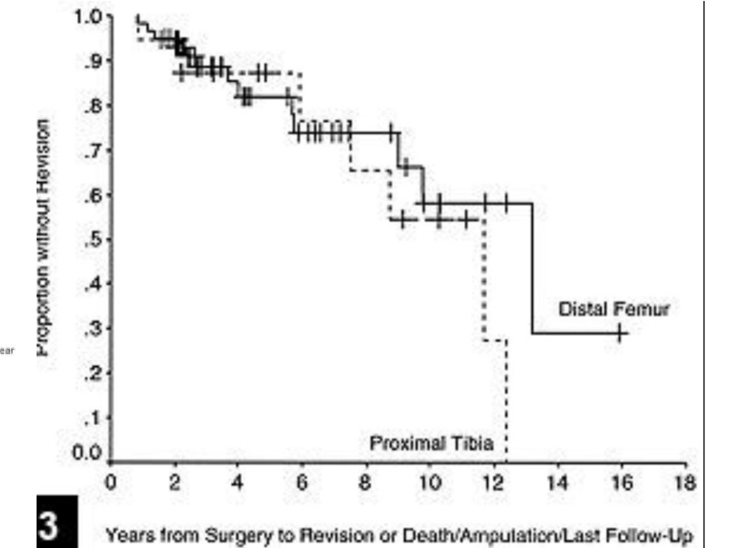
- Non-parametric estimate of the survival function
- Visual representation of the cumulative probability of an event at a specific time (a type of cohort study with outcome plotted over time)
- Commonly used in joint registry data for implant survivorship; X axis is time, Y axis is cumulative survival probability
- The event is represented by a downward step in the curve
Q2.What is censoring in a Kaplan-Meier analysis?
- Subjects who drop out of the study for reasons other than failure
- Right censored: items that have not yet failed
- Left censored: items that failed before the start of the test
- Interval censored: items that have failed but the interval is uncertain
Q3.How are survival probabilities calculated in a Kaplan-Meier curve?
- For each interval, survival = number of patients surviving / number at risk
- Censored patients are not included in the denominator
- Successive conditional probabilities are multiplied to give the cumulative probability
- Large vertical step down = many deaths/failures; large horizontal step = few deaths
Q4.What is the difference between a life table and a Kaplan-Meier curve?
- Life table divides time into regular intervals and calculates survival at each interval (actuarial method)
- Kaplan-Meier recalculates the survival rate each time a failure occurs (product limit method)
Q5.How do you compare survival between studies?
- Need 95% confidence interval (1.96 SD from the mean)
- Upper line represents censored data if they survived; lower line assumes all censored data died
- If CI overlap >25%, not statistically significant; use the Cox proportional hazard test or log rank test
- CI widens on the right side as population size decreases
Q6.Define conditional survival, median survival time and the limitations of KM curves.
- Conditional survival probability: chance of surviving a specific time frame between failures
- Unconditional survival probability: chance of surviving from the beginning of the study
- Median survival time: time until 50% of the population survive
- Do not extrapolate beyond the defined time frame, and only specific hard endpoints should be used
If 95% confidence intervals overlap by more than 25%, the difference is not statistically significant — misleading — Overlapping confidence intervals do not determine significance; formal testing (log-rank/Cox for survival data, or a CI for the difference) is required. The >25% overlap rule is not a standard statistical criterion — source
▸ Slide 103 · Sensitivity: probability of the test result being positive in patients with the Statistics · 6 questions expand
Q1-Q66 questions — tap to reveal all answerslist
- Define sensitivity and specificity.
- Define false positive rate, false negative rate and accuracy.
- What are the positive and negative likelihood ratios?
- What are PPV and NPV and how are they affected by prevalence?
- What is an ROC curve?
- How do you choose a cut-off for a screening versus a confirmation test?
Q1.Define sensitivity and specificity.
- Sensitivity: probability of a positive test in patients with the disease (true positive / disease positive)
- Specificity: probability of a negative test in patients without the disease (true negative / disease negative)
Q2.Define false positive rate, false negative rate and accuracy.
- False positive rate = 1 - specificity
- False negative rate = 1 - sensitivity
- Accuracy: probability of correct identification of a disease = (true positive + true negative) / total number
Q3.What are the positive and negative likelihood ratios?
- Positive LR = sensitivity / (1 - specificity)
- Negative LR = (1 - sensitivity) / specificity
- LR is the likelihood of correctly predicting disease versus the probability of incorrectly predicting it
Q4.What are PPV and NPV and how are they affected by prevalence?
- PPV: probability of disease when the test is positive (true positive / test positive)
- NPV: probability of no disease when the test is negative (true negative / test negative)
- Sensitivity and specificity are intrinsic to the test itself, but PPV/NPV change with prevalence
- prevalence affects the pretest probability
- PPV is higher if prevalence is higher
Q5.What is an ROC curve?
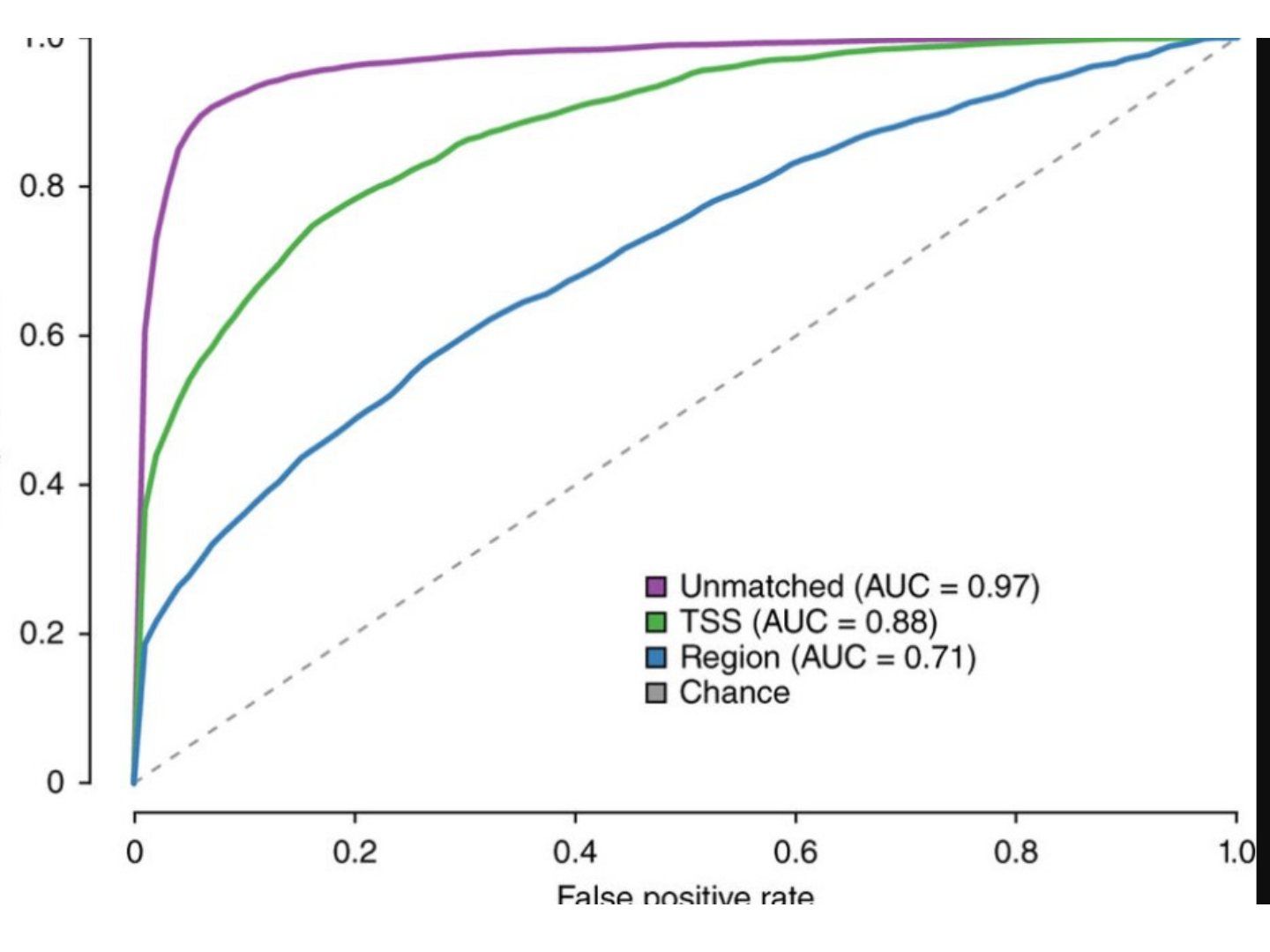
- Plot of the true positive rate against the false positive rate (sensitivity / 1 - specificity) at different cut-off points
- Shows the tradeoff between sensitivity and specificity with different cut off values: any increase in sensitivity is accompanied by a decrease in specificity
- Area 0.5 = useless test (complete overlap); 1.0 = perfect test (no overlap)
- AUC 0.5-0.7 marginally useful tests, 0.7-0.9 good, >0.9 excellent
Q6.How do you choose a cut-off for a screening versus a confirmation test?
- Screening test: choose high sensitivity (right side of the curve)
- Confirmation test: choose high specificity (left side of the curve)
▸ Slide 104Statistics · 3 questions expand

Q1-Q33 questions — tap to reveal all answerslist
- What distinguishes the good test from the bad test on the upper ROC curves?
- What do the axes of these ROC curves represent?
- Which three test scenarios are marked at cut-offs on the lower ROC curve?
Q1.What distinguishes the good test from the bad test on the upper ROC curves?
- Good test: curve rises steeply towards the top-left corner
- Bad test: curve lies close to the diagonal (close to no better than chance)
Q2.What do the axes of these ROC curves represent?
- X axis: 1 - specificity (false positive rate)
- Y axis: sensitivity (true positive rate)
Q3.Which three test scenarios are marked at cut-offs on the lower ROC curve?
- Confirmation test, balanced test and screening test
▸ Slide 105 · Interpretation of result in cohortStatistics · 3 questions expand
Q1-Q33 questions — tap to reveal all answerslist
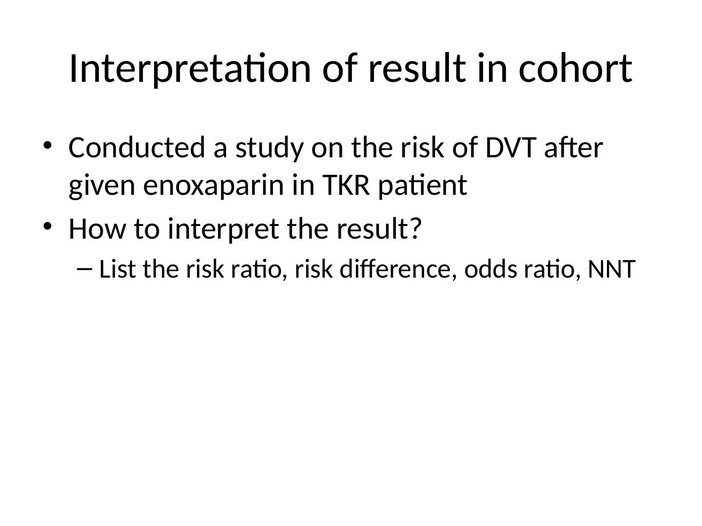
- What cohort study example is used to interpret results?
- What are the exposure, outcome and population in this study?
- How is a cohort study result interpreted?
Q1.What cohort study example is used to interpret results?
- A study on the risk of DVT after given enoxaparin in TKR patients
- The question asked is: how to interpret the result?
Q2.What are the exposure, outcome and population in this study?
- Exposure: enoxaparin
- Outcome: DVT
- Population: TKR patients
Q3.How is a cohort study result interpreted?
- List the risk ratio, risk difference, odds ratio and NNT
▸ Slide 106 · Control group risk: 3/45 = 0.067Statistics · 7 questions expand

Q1-Q77 questions — tap to reveal all answerslist
- Given a control risk of 3/45 and experimental risk of 1/50, calculate the risk difference and NNT.
- Calculate the relative risk from these data and state when it can be used.
- Calculate the odds ratio and state when odds can be calculated.
- Can causality be established from these results?
- List the Bradford Hill criteria.
- How is a chi-squared test calculated?
- Calculate the expected values for the male/female 2x2 example.
Q1.Given a control risk of 3/45 and experimental risk of 1/50, calculate the risk difference and NNT.
- 2x2 table, outcome DVT: exposure enoxaparin +ve 1 DVT+ / 49 DVT- / 50; exposure -ve 3 DVT+ / 42 DVT- / 45; column totals 4 DVT+ / 91 DVT-
- Control group risk 3/45 = 0.067; experimental group risk 1/50 = 0.02
- Risk difference (absolute risk reduction) = 0.067 - 0.02 = 0.047
- NNT = 1/ARR = 1/0.047 = 21.3
Q2.Calculate the relative risk from these data and state when it can be used.
- RR = 0.02/0.067 = 0.3
- Risk of developing the disease with the exposure compared with the risk without the exposure
- Relative risk can be calculated in a cohort study
Q3.Calculate the odds ratio and state when odds can be calculated.
- Odds in exposed group 1/49 = 0.02; odds in unexposed group 3/42 = 0.07
- Odds ratio = 0.29
- Odds can only be calculated in a case control study
Q4.Can causality be established from these results?
- No - these measures cannot establish causality
- Causality is established through the Bradford Hill criteria
Q5.List the Bradford Hill criteria.
- Strength (effect size), consistency (reproducibility), specificity
- Temporality, biological gradient (dose-response), plausibility, coherence
- Experiment, analogy
Q6.How is a chi-squared test calculated?
- Chi2 = sum of (observed - expected)^2 / expected
- Expected value = (row sum x column sum) / total
Q7.Calculate the expected values for the male/female 2x2 example.
- Disease male = 10x14/21 = 6.67; disease female = 10x7/21 = 3.33
- Control male = 14x11/21 = 7.33; control female = 7x11/21 = 3.67
- Expected value = (row sum x column sum) / total number
▸ Slide 107 · What is a hypothesis?Statistics · 3 questions expand

Q1-Q33 questions — tap to reveal all answerslist
- What is a hypothesis?
- What is a null hypothesis?
- What are type 1 and 2 errors?
Q1.What is a hypothesis?
- A proposition that serves as a starting point for further investigation
Q2.What is a null hypothesis?
- A primary assumption that any differences between groups occurred purely by chance
Q3.What are type 1 and 2 errors?
- Type 1 (alpha) error: rejecting the null hypothesis when it is true - no true difference but a difference is found
- Type 2 (beta) error: failing to reject a false null hypothesis - a true difference exists but is not detected
▸ Slide 108 · How do you conduct a RCT?Statistics · 14 questions expand

Q1-Q1414 questions — tap to reveal all answerslist
- How do you conduct a randomised controlled trial?
- What types of analysis can be used for deviation from the study protocol?
- What factors determine sample size in a power analysis?
- Define power and power analysis.
- What are the steps of a power analysis?
- What is effect size?
- What is bias and how is it reduced?
- What is confounding and how does randomisation address it?
- What types of randomisation are used and what are their drawbacks?
- List the types of bias in clinical research.
- Differentiate type 1 and type 2 errors and how they are reduced.
- What is a confidence interval and how is it calculated?
- What is a p value?
- What is the Bonferroni correction and what is its drawback?
Q1.How do you conduct a randomised controlled trial?
- Identify the problem, define the research question, set null and alternate hypotheses
- Literature review using the PICO principle (patients, intervention, comparison, outcome) to identify gaps; ethics committee
- Study design (PROSD): population (control and treatment groups, inclusion/exclusion criteria), methodology (randomisation/blinding/stratification for confounding), outcome measures, sample size by power analysis
- Register and conduct the trial, recruit patients, collect and analyse data, interpret and publish
Q2.What types of analysis can be used for deviation from the study protocol?
- Intention to treat
- Per protocol
- As treated
Q3.What factors determine sample size in a power analysis?
- Power of study 0.8 and P value 0.05 (predetermined)
- Variability of the result (standard deviation)
- Chosen clinically important difference in the primary outcome
Q4.Define power and power analysis.
- Power = the ability of a study to detect a difference between 2 interventions if one in fact exists = 1 - beta (1 - type II error)
- Power analysis = the process of determining the sample size needed to reject the null hypothesis
- Conventional power 80% = 80% chance of finding a statistical difference if there is one (probability of a type II error is <20%)
- Cut-off determined from: pilot study, literature research, minimum clinically important difference (MCID)
Q5.What are the steps of a power analysis?
- Set the smallest meaningful outcome difference and effect size
- Set alpha and beta (0.05 / 80%)
- Find the variance
Q6.What is effect size?
- The magnitude of the difference in the means of the control and experimental groups
- Expressed with respect to the pooled standard deviation
Q7.What is bias and how is it reduced?
- A systematic error, conscious or unconscious, that leads to a false representation of the true state of affairs
- Reduced by randomisation, masking/blinding and meticulous attention to the study protocol
Q8.What is confounding and how does randomisation address it?
- Confounding occurs when factors not under study affect the results
- Randomisation reduces it by distributing independent variables equally among the treatment arms
Q9.What types of randomisation are used and what are their drawbacks?
- Simple (by computer): easy to implement but may result in unequal groups
- Stratified: separate randomisation procedures within subgroups defined according to the predefined characteristics (e.g. smoking)
- Block: addresses imbalance but the executer can predict the next assignment
Q10.List the types of bias in clinical research.
- Questions bias (study design), sampling bias (inclusion/exclusion criteria)
- Selection bias (randomisation), information bias (recall, workup, interview)
- Windowing (analysis), publication bias
Q11.Differentiate type 1 and type 2 errors and how they are reduced.
- Type 1 (alpha): no true difference but a difference is found - falsely rejecting the null hypothesis; reduce by decreasing the p value (Bonferroni correction)
- Type 2 (beta): a true difference exists but is not detected - falsely accepting the null hypothesis
- Reduce type 2 error by increasing p value, sample size, effect size or outcome variability
Q12.What is a confidence interval and how is it calculated?
- The range in which the true effect lies on either side of the mean; refers to the uncertainty of the study
- 95% CI = mean +/- 1.96 SD
- 99% CI = mean +/- 2.58 SD
Q13.What is a p value?
- The probability of an observed difference occurring by chance; p < 0.05 is taken as statistically significant
Q14.What is the Bonferroni correction and what is its drawback?
- A post-hoc statistical correction made to P values when several dependent or independent statistical tests are performed simultaneously on a single data set
- May increase a type 2 error
▸ Slide 109 · What is the level of study of a RCT?Statistics · 6 questions expand
Q1-Q66 questions — tap to reveal all answerslist
- What determines the level of study of a randomised controlled trial?
- Outline the levels of evidence.
- What are the types of study designs?
- What is the difference between a cohort and a case control study?
- What is the difference between a systematic review and a meta-analysis?
- What is heterogeneity and how is it measured?
Q1.What determines the level of study of a randomised controlled trial?
- Depends on the quality of the RCT: confidence interval, percentage of follow-up, blinding
- Can be level 1 or 2
Q2.Outline the levels of evidence.
- Level 1: RCT with narrow CI; systematic review of RCTs with homogeneous findings
- Level 2: cohort study, RCT with <80% follow-up; systematic review of cohort studies with homogeneous findings
- Level 3: case control study; systematic review of case control studies
- Level 4: case series; Level 5: expert opinion
Q3.What are the types of study designs?
- Descriptive: cross sectional, case reports, correlational studies
- Analytical: cohort, case control, RCT, survival analysis
Q4.What is the difference between a cohort and a case control study?
- Cohort study: observational design where patients are selected on the basis of an exposure variable
- Case control study: observational design where patients are selected on the basis of an outcome variable
Q5.What is the difference between a systematic review and a meta-analysis?
- Systematic review: combines the information of several studies on the same topic
- Meta-analysis: collects the results of several studies and analyses them again with a statistical technique
- Meta-analysis is a type of systematic review
Q6.What is heterogeneity and how is it measured?
- Variation between the studies' design (clinical, methodology)
- Chi-squared heterogeneity test (Q test): large value = heterogeneity
- I2: large value = heterogeneity
▸ Slide 110 · What types of data you know?Statistics · 6 questions expand

Q1-Q66 questions — tap to reveal all answerslist
- What types of data do you know?
- What is a normal (parametric) distribution?
- How do you formally test for normality and homogeneity of variance?
- How can non-parametric data be transformed to parametric?
- How do you measure variability?
- What are the measures of central tendency?
Q1.What types of data do you know?
- Categorical: nominal (e.g. eye colour) or ordinal (e.g. mild/moderate/severe)
- Numerical: discrete or continuous
Q2.What is a normal (parametric) distribution?
- Continuous data that is symmetrical
- Mean = median = mode
- 1SD = 68%, 2SD = 95%, 3SD = 99.7%
Q3.How do you formally test for normality and homogeneity of variance?
- Kolmogorov-Smirnov test or Shapiro-Wilki's test for normality
- Levene's test for homogeneity of variance
Q4.How can non-parametric data be transformed to parametric?
- How to transform nonparametric to parametric?
- Logarithm
- Square
- Square root
Q5.How do you measure variability?
- Variance = sum of squares of difference about the mean / number of subjects
- SD = square root of variance
- Standard error = SD / square root of n
Q6.What are the measures of central tendency?
- Mean: sum of all observations divided by number of subjects
- Median: central value of the data
- Mode: most frequent value
▸ Slide 111 · Statistical inference used to test specific hypothesis about associations or dStatistics · 1 question expand

Q1-Q11 questions — tap to reveal all answerslist
- What is statistical inference used for?
Q1.What is statistical inference used for?
- Used to test specific hypotheses about associations or differences among groups of subjects
- Applied to sample data
▸ Slide 112 · Forrest plot numerical and graphical representation of a metanalysisStatistics · 8 questions expand

Q1-Q88 questions — tap to reveal all answerslist
- What is a forest plot?
- What is a meta-analysis and how does it differ from a systematic review?
- What information is shown in the columns of a forest plot?
- What do the symbols in a forest plot represent?
- How is heterogeneity assessed and graded in a meta-analysis?
- What do 'fixed' and 'M-H' mean and how is heterogeneity managed?
- What is heterogeneity per the lecture definition?
- What is meta-regression and what is it used for?
Q1.What is a forest plot?
- Numerical and graphical representation of a meta-analysis
- Displays individual study results together with the combined overall effect
Q2.What is a meta-analysis and how does it differ from a systematic review?
- Meta-analysis = quantitative systematic review combining results of relevant studies to produce an estimation of the overall effect of interest
- Systematic review = overview of primary studies using explicit and reproducible methods
Q3.What information is shown in the columns of a forest plot?
- 1st column: name of study in alphabetical order
- 2nd-5th columns: incidence of event in control and experimental group
- 6th column: weight - reflects precision of the study (size and CI)
- 7th column: RR and CI interval
Q4.What do the symbols in a forest plot represent?
- Square = weight of study
- Horizontal line = confidence interval
- Central line = no-effect line; if the CI crosses it the result is not statistically significant
- Diamond at the bottom = combined effect of the studies
Q5.How is heterogeneity assessed and graded in a meta-analysis?
- Chi-squared heterogeneity test (Q test) or I2
- I2 <50 = low, 50-75% = moderate, >75% = high heterogeneity
- I2 = the proportion of variation that is due to heterogeneity rather than chance
- p<0.05 rejects the null hypothesis of no statistical heterogeneity
Q6.What do 'fixed' and 'M-H' mean and how is heterogeneity managed?
- Mantel-Haenszel (M-H) test used to combine studies (Review Manager, Cochrane Library)
- Fixed-effects model: one true effect underlies all studies
- Random-effects model: normal distribution of true effect sizes; mean and variance estimated
- Management: subgroup analysis, random-effects model (may report plot without combined estimate), meta-regression for confounders
Q7.What is heterogeneity per the lecture definition?
- Variability in patient characteristics within each study
Q8.What is meta-regression and what is it used for?
- An extension of subgroup analysis allowing the effects of multiple factors to be investigated simultaneously
- Used to look for confounders
- Similar to simple regression: an outcome variable predicted from one or more independent variables
▸ Slide 113 · Inverted funnel plotStatistics · 4 questions expand
Q1-Q44 questions — tap to reveal all answerslist
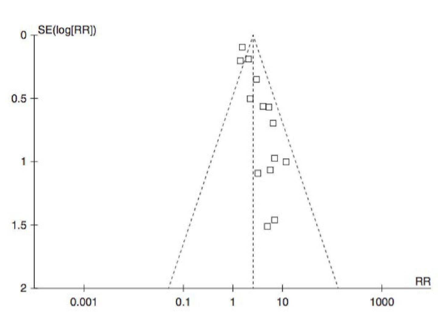
- What is an inverted funnel plot?
- What is an inverted funnel plot used for?
- What are the axes and reference lines of an inverted funnel plot?
- How should studies be distributed and what does asymmetry indicate?
Q1.What is an inverted funnel plot?
- Scatter plot of the intervention effect estimates from individual studies
- Plotted against some measure of each study's size or precision
Q2.What is an inverted funnel plot used for?
- Helps detect publication bias in a meta-analysis
- Small size study with lower methodology quality tends to overestimate true effect
Q3.What are the axes and reference lines of an inverted funnel plot?
- Y axis: precision of study (standard error, or standard deviation/square of sample size)
- X axis: effect of treatment (relative risk)
- Vertical dotted line: estimated combined RR of the meta-analysis
- Diagonal dotted lines form the funnel
Q4.How should studies be distributed and what does asymmetry indicate?
- Upper dots: larger sample size/more precise, lie closer to the vertical dotted line
- Lower dots: smaller/less precise studies, more scattered and symmetrically distributed
- Asymmetry suggests publication bias, or small low-quality studies overestimating the true effect
▸ Slide 114 · Outcome measurementStatistics · 4 questions expand

Q1-Q44 questions — tap to reveal all answerslist
- The Harris hip score is shown - what type of outcome measurement is it?
- What is a PROM?
- What is a CBOM (clinician-based outcome measure)?
- How do you choose a scoring system?
Q1.The Harris hip score is shown - what type of outcome measurement is it?
- A PROM (patient-reported outcome measure)
- A self-completed questionnaire assessing symptoms and function
Q2.What is a PROM?
- Self-completed questionnaires
- Assess symptoms and functional disability
- Examples: Oxford hip/knee score
Q3.What is a CBOM (clinician-based outcome measure)?
- Objective measurement
- Dependent on the reliability/reproducibility of the clinician's assessment
Q4.How do you choose a scoring system?
- Reliability: consistency of results on repeated measurement (Kappa for categorical data), internal consistency, reproducibility (intra/inter-observer)
- Validity: does it measure what it is supposed to - construct/content/criterion/concurrent
- Clinical utility: patient and clinician friendliness
▸ Slide 115 · What is incidence and prevalence?Statistics · 5 questions expand

Q1-Q55 questions — tap to reveal all answerslist
- Define prevalence and incidence.
- What population and disease factors make a good screening test (Wilson's criteria)?
- What test and economic factors make a good screening test?
- Why is scoliosis suitable for screening?
- What are the four categories of Wilson's criteria for a screening test?
Q1.Define prevalence and incidence.
- Prevalence: cross section measure of the people having the event / whole population
- Incidence: number of new case in a period of time/ population at risk
Q2.What population and disease factors make a good screening test (Wilson's criteria)?
- Condition is an important health problem with high enough prevalence
- Well-understood natural history
- Presence of an early symptomatic/recognizable latent stage
- Accepted treatment exists and an agreed policy on whom to treat
Q3.What test and economic factors make a good screening test?
- A well-accepted test
- High sensitivity essential, high specificity desirable
- Cost of case-finding (diagnosis + treatment) economically balanced against overall medical care expenditure
Q4.Why is scoliosis suitable for screening?
- Known natural history with an early detectable phase (>20 degrees in Risser stage 1 or before high chance of progress)
- Early treatment (bracing) may alter the course of disease
- Test: Adam forward bending with scoliometer - well accepted
- Sensitivity uncertain
Q5.What are the four categories of Wilson's criteria for a screening test?
- Population factor: important health problem with high enough prevalence
- Disease factor: well-understood natural history, early/latent stage, accepted treatment, agreed policy
- Test factor: well-accepted test - high sensitivity essential, high specificity desirable
- Economic factor: cost of case-finding balanced against overall medical care expenditure
▸ Slide 116 · TumourStatistics · 1 question expand
Q1-Q11 questions — tap to reveal all answerslist
- What does the lecturer advise about these tumour revision notes?
Q1.What does the lecturer advise about these tumour revision notes?
- Refer to Orthobullets for more details
- These notes are not comprehensive
- Remembering all Orthobullets tumours may only gain 1-2 more MCQs
▸ Slide 117 · Classification of staging: enneking/AJCCStatistics · 4 questions expand

Q1-Q44 questions — tap to reveal all answerslist
- Which staging classification is used for bone and soft tissue tumours?
- What do AJCC stages I-IV mean?
- What are the Enneking stages for malignant bone tumours?
- What is the Enneking classification for benign bone tumours?
Q1.Which staging classification is used for bone and soft tissue tumours?
- Bone benign/malignant: Enneking
- Soft tissue sarcoma: AJCC
- STC: AJCC
Q2.What do AJCC stages I-IV mean?
- I: low grade
- II: high grade
- III: deep to fascia
- IV: lymph node/metastasis
Q3.What are the Enneking stages for malignant bone tumours?
- I: low grade
- II: high grade
- III: metastasis
- A: intra-compartmental, B: extra-compartmental
Q4.What is the Enneking classification for benign bone tumours?
- 1: latent
- 2: active
- 3: aggressive
▸ Slide 118 · DescribeStatistics · 16 questions expand

Q1-Q1616 questions — tap to reveal all answerslist
- Describe the X-ray findings of this skeletally immature knee.
- What is your impression and what X-ray do you request next?
- What are the poor prognostic factors in osteosarcoma?
- How would you investigate this lesion?
- What is the management principle and neoadjuvant chemotherapy plan?
- What are the relative contraindications to limb salvage and the allograft 'terrible triad'?
- What history and examination findings are relevant in suspected osteosarcoma?
- What are the prerequisites for limb salvage and the principle of reconstruction?
- What are the options for reconstruction after tumour resection?
- How is a biopsy taken and how does percutaneous compare with incisional biopsy?
- What are the intra-operative and post-operative principles of biopsy?
- When is an excisional biopsy appropriate?
- What histological features suggest osteosarcoma on H&E?
- How is limb length discrepancy approached in tumour patients?
- What novel treatments are being studied for metastatic osteosarcoma?
- What are the properties of allograft reconstruction?
Q1.Describe the X-ray findings of this skeletally immature knee.
- Lytic lesion in the metaphyseal region of the distal femur with destruction of the lateral cortex
- Wide zone of transition with periosteal reaction medially
- Matrix is osseous and does not cross the physis
- Associated soft tissue swelling
Q2.What is your impression and what X-ray do you request next?
- Aggressive left distal femoral lesion; concerned about osteosarcoma
- I want XR of the whole femur to look for obvious skip lesions
- Other DDx: Ewing sarcoma, haematological (e.g. lymphoma), infection/osteomyelitis
Q3.What are the poor prognostic factors in osteosarcoma?
- Advanced stage of disease (most predictive)
- Age >40 at diagnosis; male sex
- <90% necrotic index; positive surgical margins
- Tumour site and size; vascular involvement
- Expression of p glycoprotein (P-glycoprotein); high ALP and LDH
Q4.How would you investigate this lesion?
- Aims: establish the diagnosis and stage the disease
- Bloods: Hb, WBC, platelets, clotting, ALP (bone turnover), LDH (cell turnover), ESR, CRP
- Local imaging: whole-bone X-ray 2 views, MRI with contrast (skip lesions, joint/physis involvement, NV bundle proximity)
- Systemic staging: CT thorax + bone scan
- Image-guided biopsy at a tertiary tumour centre - representative tissue with minimal contamination
- Discuss the case at the MSK oncology MDT
Q5.What is the management principle and neoadjuvant chemotherapy plan?
- Oncologic clearance with preservation of the limb and function if possible: save life (systemic control), save limb (local control)
- Principle: Margin free excision (resect all inflammatory zone and go through cuff of normal tissue)
- Neoadjuvant chemotherapy (4 reasons; no proven survival effect vs post-op chemo alone)
- 2 cycles pre-op MAC: methotrexate, adriamycin, cisplatin; 4-week cycles
- Restaging MRI, then OT; continue adjuvant chemo and amend by necrotic index
- Good responder (90-100%): 3 more cycles; poor responder: VP16 + ifosfamide with 5 cycles (EURAMOS1 2016 does not support this - toxicity without EFS benefit)
Q6.What are the relative contraindications to limb salvage and the allograft 'terrible triad'?
- Major NV encasement
- Pathological fracture with haematoma violating compartment boundary
- Inappropriately performed biopsy
- Severe infection in surgical field; immature skeleton with predicted LLD >8cm
- Extensive muscle/soft tissue involvement; poor response to chemo
- Allograft terrible triad: fracture, infection, non-union (15% each); host bone ingrowth only 1-2mm/year
Q7.What history and examination findings are relevant in suspected osteosarcoma?
- History: PMHx, constitutional symptoms
- Local PE: soft tissue status, NV status, LLD, any knee effusion
- Systemic PE: listen to lungs
Q8.What are the prerequisites for limb salvage and the principle of reconstruction?
- Prerequisites: clear margins, durable and useful limb, limited morbidity, equal oncological outcome
- Reconstruction principle: stable, painless, durable, mobile limb
Q9.What are the options for reconstruction after tumour resection?
- Biological: autograft or allograft
- Metallic implant: modular or custom-made
- Combined reconstruction
Q10.How is a biopsy taken and how does percutaneous compare with incisional biopsy?
- Options: percutaneous image-guided, incisional, excisional; most people now do percutaneous image-guided biopsy
- True-cut biopsy x3 vs FNAC: architecture obtained 70% vs 9%
- Incisional: GA, bigger/deeper wound with more bleeding, but a better chunk of tissue
- Percutaneous may be inconclusive (reactive zone, central necrosis) - radiologically representative tissue may not be histologically representative
- Better performed by the surgeon who will do the final excision
Q11.What are the intra-operative and post-operative principles of biopsy?
- Pre-op: bloods + imaging
- Intra-op: tourniquet without exsanguination, longitudinal incision, cross the least compartments, avoid NV structures, aim at representative tissue
- Oval shape if bone; send for frozen section + microbiology
- Careful haemostasis, drain in line, suture with small bites
- Post-op: bed rest +/- protection, Abx (antibiotics)
Q12.When is an excisional biopsy appropriate?
- Clinically benign lesion: small, superficial to fascia
- Incisional and excisional biopsy margins are similar to the definitive surgery (1-2cm)
Q13.What histological features suggest osteosarcoma on H&E?
- Pleomorphic spindle cells with lacey osteoid
- high nuclear cytoplasmic ratio (high nuclear-to-cytoplasmic ratio), cellular atypia
- Multiple mitotic bodies
Q14.How is limb length discrepancy approached in tumour patients?
- Key considerations: growth potential and articular involvement
- Options: growing prosthesis, lengthening, growth plate transfer, amputation, Van Nes rotationoplasty
- Van Nes indication: major bone/soft tissue involvement without NV involvement
- Used to change a high AKA to a BKA
Q15.What novel treatments are being studied for metastatic osteosarcoma?
- Almost half of current trials evaluate immunotherapies such as mifamurtide, IL-2, PD-1 inhibitors
- Tyrosine kinase inhibitors including regorafenib
Q16.What are the properties of allograft reconstruction?
- Host bone grows into donor bone slowly: 1-2mm/year
- Strength decreases by 50% at 10 years
- Worse with allograft arthrodesis
- APC usu in prox humerus and proximal tibia (tendon attachment to prosthesis difficult)
▸ Slide 119 · Pathological # in OSStatistics · 4 questions expand
Q1-Q44 questions — tap to reveal all answerslist
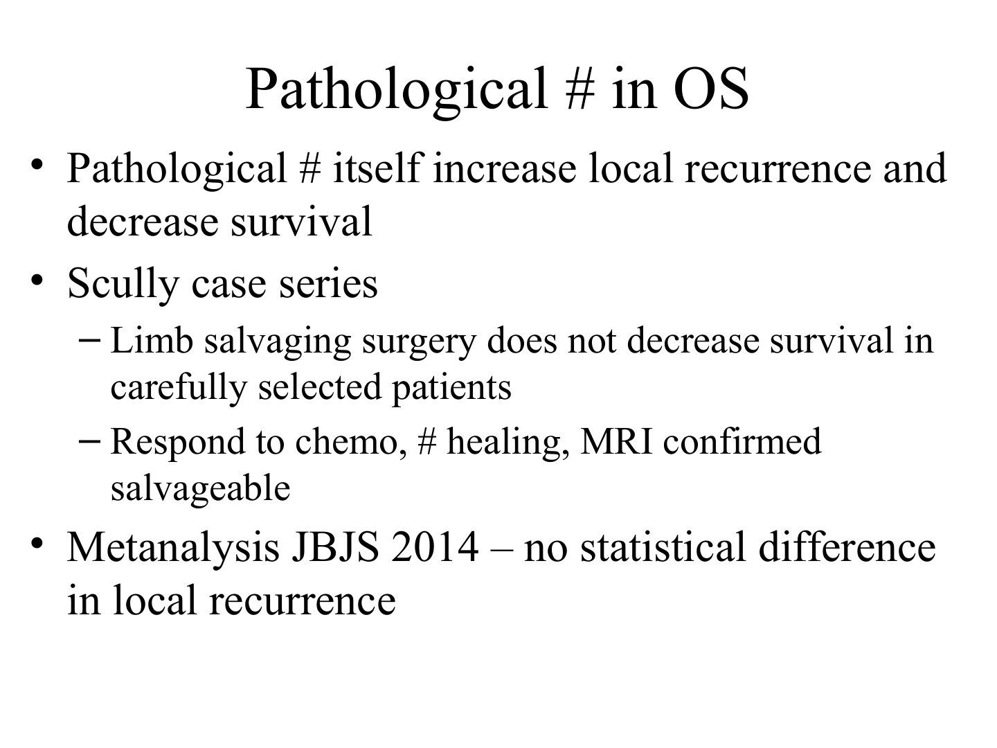
- What is the prognostic significance of a pathological fracture in osteosarcoma?
- What does the Scully case series show regarding limb salvage in osteosarcoma with pathological fracture?
- What does the JBJS 2014 meta-analysis show regarding local recurrence after pathological fracture in osteosarcoma?
- What makes a pathological fracture in osteosarcoma amenable to limb salvage?
Q1.What is the prognostic significance of a pathological fracture in osteosarcoma?
- Pathological fracture itself increases local recurrence and decreases survival
Q2.What does the Scully case series show regarding limb salvage in osteosarcoma with pathological fracture?
- Limb salvaging surgery does not decrease survival in carefully selected patients
Q3.What does the JBJS 2014 meta-analysis show regarding local recurrence after pathological fracture in osteosarcoma?
- Metanalysis JBJS 2014 - no statistical difference in local recurrence
Q4.What makes a pathological fracture in osteosarcoma amenable to limb salvage?
- Respond to chemo
- # healing
- MRI confirmed salvageable
▸ Slide 120 · 10/MStatistics · 8 questions 1 check expand

Q1-Q88 questions — tap to reveal all answerslist
- What are the types of osteosarcoma?
- What is the classical X-ray appearance and DDx of osteosarcoma?
- What is the management plan and chemotherapy regime for osteosarcoma?
- What are the indications for amputation in osteosarcoma?
- What is the necrotic index and its prognostic value?
- How is a pathological fracture in osteosarcoma managed?
- What are the options for managing a bone defect after tumour resection?
- What determines the choice between limb salvage and limb sacrifice for local control?
Q1.What are the types of osteosarcoma?
- Primary: intramedullary, telangiectatic, parosteal, periosteal
- Secondary: Paget's disease, fibrous dysplasia, post-irradiation
Q2.What is the classical X-ray appearance and DDx of osteosarcoma?
- Distal femur medullary region, wide transitional zone with bone formation and excessive periosteal reaction
- Codman's triangle and sunburst appearance
- DDx: infection, Ewing (young), metastasis (old), haematological malignancy
Q3.What is the management plan and chemotherapy regime for osteosarcoma?
- Principle: oncological clearance with limb preservation if possible, without sacrificing tumour margin
- Systemic control (chemo) + local control (surgery); regime neoadjuvant chemo / OT / chemo
- 2 cycles pre-op (MTX, vincristine + cisplatin); reassessment (clinical, MRI, PET-CT); OT at week 10
- Good response: 4 more cycles; poor response: change regime, 3w x 8 cycles
Q4.What are the indications for amputation in osteosarcoma?
- Surgeon factor: not competent for limb reconstruction surgery
- Patient factor: not fit for prolonged/multiple surgery or not fit for rehab
- Disease factor: cannot achieve margin-free excision, or infection
Q5.What is the necrotic index and its prognostic value?
- Percentage of necrosis in the excised specimen
- Good response >90%: 80% of patients, 5-year survival 70-80%
- Bad response: 5-year survival 40-50%
- Guides the adjuvant chemotherapy regime
Q6.How is a pathological fracture in osteosarcoma managed?
- Amputation is safe but not a must
- Reassess with MRI to see whether margin-free wide local excision is achievable
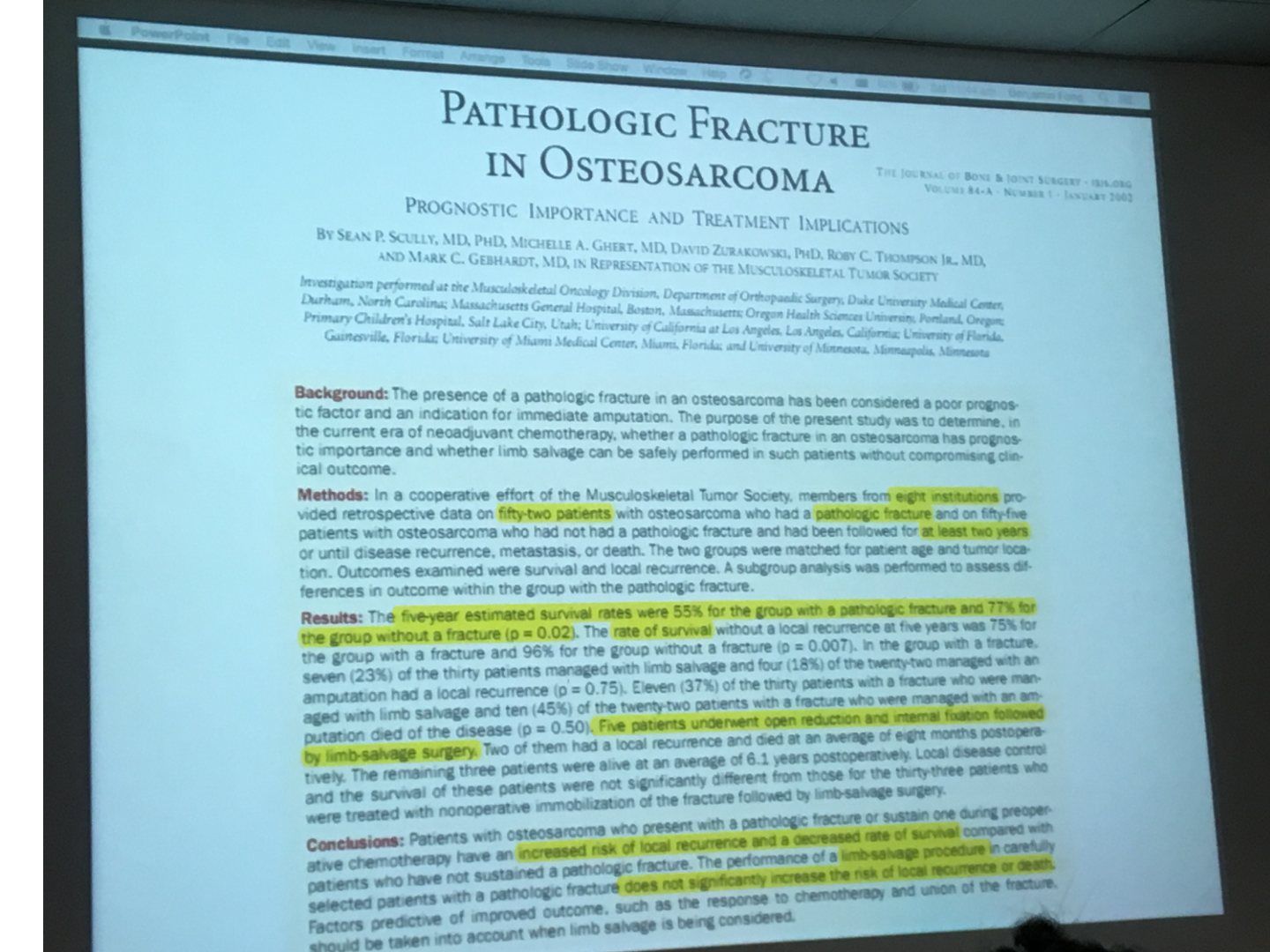
- Scully case series: pathological fracture has higher local recurrence and decreased survival, but limb salvage does not increase recurrence/death in carefully selected patients
- Prognosis better if healing present
Q7.What are the options for managing a bone defect after tumour resection?
- Amputation (soft tissue + bone + NV involvement)
- Van Nes rotationoplasty (soft tissue + bone involved, NV intact)
- Reconstruction: biological (autograft/allograft), metallic implant (modular/custom made), or combined
Q8.What determines the choice between limb salvage and limb sacrifice for local control?
- Limb salvage possible if it does not affect oncological outcomes, there is a functional limb, and the risk of surgery is acceptable
- Options include joint/physis-preserving surgery
- Local control is achieved by surgery: limb salvaging vs limb sacrificing
Neoadjuvant chemotherapy for osteosarcoma is MTX + vincristine + cisplatin (2 cycles pre-op) — erroneous regimen — Standard neoadjuvant regimen is MAP: high-dose methotrexate, doxorubicin (Adriamycin) and cisplatin; vincristine is not part of standard osteosarcoma regimens (slide 118 correctly lists methotrexate, adriamycin, cisplatin) — source
▸ Slide 121 · Disease factor: stage, location, size, grade, type, pathological #Statistics · 2 questions expand
Q1-Q22 questions — tap to reveal all answerslist
- What disease factors determine prognosis?
- What biochemical and treatment-response factors determine prognosis?
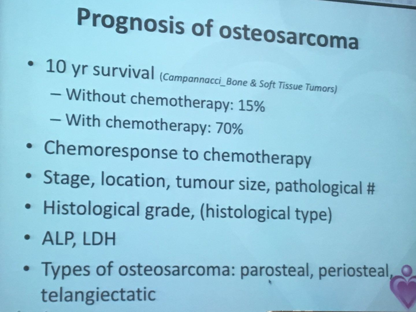
Q1.What disease factors determine prognosis?
- Stage, location, size, grade, type, pathological fracture
- Assessed alongside biochemical and treatment-response factors
Q2.What biochemical and treatment-response factors determine prognosis?
- Biochemical: ALP, LDH
- Response to treatment: chemoresponse
▸ Slide 122 · Summary:Statistics · 2 questions expand
Q1-Q22 questions — tap to reveal all answerslist
- What is the effect of a pathological fracture on recurrence in osteosarcoma?
- Does limb salvage affect survivorship after a pathological fracture?
Q1.What is the effect of a pathological fracture on recurrence in osteosarcoma?
- Associated with an increased recurrence rate
- Also associated with worse prognosis
Q2.Does limb salvage affect survivorship after a pathological fracture?
- No - limb salvage surgery does not affect survivorship
- Therefore pathological fracture is not an absolute contraindication to limb salvage
▸ Slide 123 · 50/ MStatistics · 8 questions expand

Q1-Q88 questions — tap to reveal all answerslist
- What radiographic features of this pelvic lesion suggest a chondroid origin?
- What is the DDx for a chondral lesion at the pubic rami?
- How do you differentiate a benign from a malignant chondral lesion?
- How is treatment of chondrosarcoma decided?
- What is the prognosis of chondrosarcoma by grade?
- Which benign lesions can progress to chondrosarcoma?
- What history, examination and investigations are needed for a chondral lesion of the pubic rami?
- Why are PE and X-ray important in enchondroma?
Q1.What radiographic features of this pelvic lesion suggest a chondroid origin?
- Sclerotic, well-defined lesion adjacent to the pubic symphysis
- Associated flocculent calcification (chondroid matrix)
- No cortical erosion, scalloping or periosteal reaction
- Overall suspicious for chondrosarcoma
Q2.What is the DDx for a chondral lesion at the pubic rami?
- Chondroblastic osteosarcoma
- Enchondroma
- Fracture callus
- Synovial chondromatosis
- Fibrous dysplasia with cartilaginous differentiation
Q3.How do you differentiate a benign from a malignant chondral lesion?
- Clinical: growth after puberty, new symptoms e.g. pain
- Imaging: cartilage cap >2cm, soft tissue component, cortical breakage/erosion
- Histology: enlarged chondrocytes with multinucleated lacunae, high N:C ratio, disorganization, hypercellularity
- Diagnosis requires clinical + histological correlation (same histology may be benign in the hand but malignant in long bones)
Q4.How is treatment of chondrosarcoma decided?
- Treatment depends on grading
- Grade 1 in pelvis: intralesional curettage or wide excision
- Grade 2-3: wide local excision
- Chemotherapy is useful only in mesenchymal chondrosarcoma
Q5.What is the prognosis of chondrosarcoma by grade?
- Grade 1: 90%
- Grade 2: 70%
- Grade 3: 30%
- Undifferentiated: 10%
Q6.Which benign lesions can progress to chondrosarcoma?
- Exostosis/enchondroma: 1%
- MHE: 10%
- Ollier's disease: 25-40%
- Maffucci: 100%
Q7.What history, examination and investigations are needed for a chondral lesion of the pubic rami?
- Hx/PE: other masses, duration and progress of symptoms
- Ix: blood test, MRI with contrast (soft tissue extension, cartilage)
Q8.Why are PE and X-ray important in enchondroma?
- Enchondroma: PE is important to differentiate - the difference between PE and X-ray is cartilage cap size
- Important to X-ray the pelvis (cannot see otherwise)
▸ Slide 124 · XR of this skeletally mature patients right knee showing a large juxtaarticularStatistics · 7 questions 1 check expand
Q1-Q77 questions — tap to reveal all answerslist
- What is the likely diagnosis and which conditions must be excluded?
- What history, examination and investigations are needed?
- What is the histological appearance and the Campanacci classification?
- What are the management principles and recurrence rates?
- Why is cement inserted after extended curettage and what does extended curettage mean?
- What adjunct medical treatments are used and what are their caveats?
- What other surgical option exists for GCT besides extended curettage?
Q1.What is the likely diagnosis and which conditions must be excluded?
- Large juxtaarticular (juxta-articular), eccentric, lytic expansile lesion with geographic destruction of the lateral condyle
- No cortical break, no matrix, no sclerotic margins, narrow zone of transition
- Likely an aggressive benign lesion such as GCT
- Tumourous DDx: ABC, clear cell chondrosarcoma, telangiectatic osteosarcoma
- Non-tumourous DDx: brown’s tumor (brown's tumour), infection
Q2.What history, examination and investigations are needed?
- History: onset, progression, pain, constitutional symptoms
- PE: knee tenderness, AROM, effusion; systemic exam for lymph nodes/masses
- Bloods to r/o infection/metabolic cause: CBC, CRP, ESR, Ca/PO4 (brown's tumour), bone profile
- MRI (T1 hypointense lesion); confirm with image-guided biopsy
- Systemic staging: CT thorax (2% pulmonary met) + bone scan
Q3.What is the histological appearance and the Campanacci classification?
- Mononuclear cells (monocytes), spindle cells and multinucleated giant cells
- Campanacci 1-3: cortex undisrupted, thinning, disrupted
- Guides recurrence rate and local progressiveness
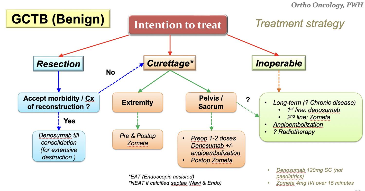
Q4.What are the management principles and recurrence rates?
- Reduce local recurrence and preserve function
- Discuss pros and cons of resection vs curettage with the patient
- Resection 5% recurrence, extended curettage 10%, curettage 20-50%
- Overall 5-year disease-free survival 75%, mortality 15%
Q5.Why is cement inserted after extended curettage and what does extended curettage mean?
- Cement provides thermal ablation of tumour, immediate mechanical support and easy detection of recurrence
- Recurrence decreased from 10% to 5% with adjuvant therapy
- Extended curettage = curettage (extensive exterioration) + adjuncts: mechanical burring, thermal cement, cryo nitrogen, chemical phenol, osmosis water
- 2014 paper: curettage + nitrogen + cement recurrence rate 6%, similar to resection
Q6.What adjunct medical treatments are used and what are their caveats?
- Bisphosphonate (Shi et al 2019 meta-analysis: reduces local recurrence)
- Denosumab 120mg once/week x3, then once/2 weeks, then monthly (Lancet 2019)
- Denosumab is very potent at stopping all osteoclastic activity but has no anti-tumour effect; stopping may cause 44% recurrence (JBJS 2021) and it makes curettage harder
- Zoledronic acid has dose-dependent cell inhibition and apoptosis in stromal cells
- Denosumab may be neoadjuvant in extensive bone destruction to consolidate tumour before resection; shown to reduce angiogenesis and microvessel density
- RT for unfit/multiple recurrence: 15% malignant transformation risk
Q7.What other surgical option exists for GCT besides extended curettage?
- Resection + reconstruction with graft / prosthesis / both
Denosumab for GCT is 120mg once/week x 3 then once/2 weeks then monthly — dosing schedule imprecise — Approved GCTB dosing is 120 mg SC every 4 weeks with additional 120 mg loading doses on days 8 and 15 of the first month (weekly for the first 3 doses, then every 4 weeks - not 2-weekly) — source
▸ Slide 125 · XR left humerus showing a metaphyseal intramedullary centrally located bone lesiStatistics · 6 questions expand

Q1-Q66 questions — tap to reveal all answerslist
- Describe the X-ray findings and impression.
- What is the DDx and what is the role of MRI?
- What is the management of enchondroma and the indications for surgery?
- Immediate vs delayed surgery for a pathological fracture through an enchondroma?
- What is the pathogenesis and biopsy appearance of enchondroma?
- What are the XR differences between enchondroma and chondrosarcoma?
Q1.Describe the X-ray findings and impression.
- Left humerus: metaphyseal, intramedullary, centrally located bone lesion with flocculent calcification
- Narrow zone of transition; no cortical erosion, no periosteal reaction, no soft tissue swelling
- Impression: enchondroma
Q2.What is the DDx and what is the role of MRI?
- Bone infarct
- Chondrosarcoma
- MRI to rule out chondrosarcoma: increase in T2 signal, no marrow oedema/periosteal reaction
Q3.What is the management of enchondroma and the indications for surgery?
- Observe if asymptomatic or static in size
- Curettage + bone graft + fixation
- Indications: symptoms, increase in size/>5cm, pathological fracture
Q4.Immediate vs delayed surgery for a pathological fracture through an enchondroma?
- Immediate: one stage, immediate rehab; disadvantage - difficult to maintain stability
- Delayed: bone healing increases stability; disadvantage - more stiffness
- Outcomes in terms of union are equal, therefore immediate is preferred
Q5.What is the pathogenesis and biopsy appearance of enchondroma?
- Incomplete enchondroma ossification
- Escape of chondroblasts and epiphyseal cartilage proliferation at the metaphysis
- Beware chondrosarcoma if in pelvis, rib or scapula
- Biopsy: hypocellular, mature hyaline cartilage, chondrocytes with small single nuclei in lacunar space
Q6.What are the XR differences between enchondroma and chondrosarcoma?
- Enchondroma: chondroid/lytic, <5cm, cortical expansion, <50% canal width
- Chondrosarcoma: lytic + blastic, >5cm, cortical erosion + reactive thickening, >50% canal width
▸ Slide 126 · Ewings sarcomaStatistics · 7 questions expand

Q1-Q77 questions — tap to reveal all answerslist
- Where does Ewing sarcoma occur and what are the X-ray features?
- What is the clinical presentation and DDx of Ewing sarcoma?
- What investigations confirm Ewing sarcoma?
- What is the treatment principle in Ewing sarcoma?
- What is the local control strategy and when is radiotherapy used?
- What are the RT complications, poor prognostic factors and survival?
- What is the epidemiology and origin of Ewing sarcoma?
Q1.Where does Ewing sarcoma occur and what are the X-ray features?
- Typically lower limb, pelvis, proximal humerus
- Destructive permeative, moth eaten, lytic lesion in the metaphysis and diaphysis with variable new bone formation
- Onion-skin periosteal lifting (characteristic but uncommon)
- Soft tissue component is often large
Q2.What is the clinical presentation and DDx of Ewing sarcoma?
- Fever + pain - thus mimics infection
- DDx: osteosarcoma, haematological, osteomyelitis, eosinophilic granuloma
Q3.What investigations confirm Ewing sarcoma?
- Bloods: raised ESR, anaemia, increased WBC, LDH (prognostic >200IUml / >200 IU/mL)
- MRI: soft tissue component, reduced T1 due to marrow invasion, increased with contrast
- Biopsy: monotonous small blue round cells, high N:C ratio, CD99+
- Bone marrow biopsy to rule out metastasis to the marrow: consistent chromosomal translocation (11;22) with EWS-FLI 1 fusion protein
- CT/CXR: metastases in 30% at presentation
Q4.What is the treatment principle in Ewing sarcoma?
- Oncologic clearance with preservation of limb and function: save life (systemic control), save limb (local control)
- Multimodality approach: multiagent chemotherapy, irradiation and surgical resection
- Pre-op chemotherapy 2-3 months, then resection, maintenance chemo 6-12 months
Q5.What is the local control strategy and when is radiotherapy used?
- Trend towards surgical resection (wide margin excision) for all surgically accessible sites; assess tumour necrosis
- RT indications: positive margin, metastasis, poor response to chemo
- Chemo + OT +/- adjuvant RT = chemo + RT
Q6.What are the RT complications, poor prognostic factors and survival?
- RT complications: 10-20% secondary sarcoma, fracture, physeal damage/LLD, osteonecrosis, muscle atrophy, stiffness, Joint: contracture
- Poor prognostic factors: metastases (1/4), >14yo, male, spine/pelvic tumours, >100cm3, poor chemo response (<90% necrosis), raised LDH/WCC, low Hb, p53 mutation
- 5-year disease-free survival ~65% (~half with mets); lung mets 30% cure, marrow 30% cure, both 10-15% cure
Q7.What is the epidemiology and origin of Ewing sarcoma?
- Distinctive small round cell sarcoma occurring most often in children and young adults (5-25)
- 2nd most common malignant tumour in children
- Neuroectodermal cells arise from the medullary canal and invade the haversian system
▸ Slide 127 · 70/y Known Ca liver with bone metStatistics · 7 questions expand

Q1-Q77 questions — tap to reveal all answerslist
- What is the diagnosis and what differentials must be considered for this humeral lesion?
- What history and examination findings are important in this patient with a suspected bone metastasis?
- How would you investigate a suspected bone metastasis?
- What is the management aim and plan for bone metastasis?
- What is the Mirel's score and how does it guide management?
- Describe the pathogenesis of bone metastasis and osteolysis.
- What newer radiological and oncological assessment scores exist for bone metastases?
Q1.What is the diagnosis and what differentials must be considered for this humeral lesion?
- Dx: bone metastasis - poorly defined lytic lesion over the distal 1/3 of the humerus with endosteal scalloping; no fracture
- Request whole-length X-ray to screen the rest of the bone
- DDx: haematological (myeloma), infection, brown's tumour
Q2.What history and examination findings are important in this patient with a suspected bone metastasis?
- Premorbid status + ECOG (0 fully active, 1 strenuous activity restricted, 2 self care, 3 limited self care, 4 completely disabled)
- Pain; Sx of hypercalcemia (moans, groans, stones etc); LL weakness or numbness; constitutional symptoms
- Local: NV status, soft tissue
- Systems examination: thyroid, breast, PR
Q3.How would you investigate a suspected bone metastasis?
- Acute: hypercalcaemia; inflammatory markers (ESR - MM), LDH (lymphoma), tumour markers
- Haematology: serum electrophoresis, A/G ratio (normal 1.1-2.5)
- Find primary: bone scan or PET-CT
- No primary found -> biopsy to rule out primary bone tumour
- Image the whole bone
Q4.What is the management aim and plan for bone metastasis?
- MDT approach; Take into account patient factors, and also disease factors (primary prognosis <3 months -> conservative; local lesion Mirel's score)
- Aim: single operation to restore function, improve quality of life/pain and facilitate nursing care
- Construct should allow immediate full weight bearing and be durable enough to outlast the patient's remaining lifespan
- Non-op: RT, bisphosphonate, denosumab (superior effect decreasing fractures)
- Peri-op: embolization to reduce bleeding (RCC, thyroid, HCC), consent: perioperative mortality >10% (1/2 due to tumor emboli), antibiotics/cement, post-op RT + bisphosphonate
Q5.What is the Mirel's score and how does it guide management?
- Prognostic tool predicting risk of pathological fracture (retrospective study of fractures 6 months post RT)
- Components: site (UL, LL, peritrochanteric), size (<1/3, 1/3-2/3, >2/3), lesion (blastic, mixed, lytic), pain (mild, moderate, severe)
- <=7: 4% risk (RT); 8: 15%; >=9: 33% (OT)
- Sensitivity 91%, specificity 35%
- Rule of thumb: >50% of a single cortex of a long bone = significant fracture risk; avulsion of the lesser trochanter = imminent hip fracture
Q6.Describe the pathogenesis of bone metastasis and osteolysis.
- Metastasis: intravasation (E cadherin, PDGF), immunological evasion, location (integrins), extravasation (matrix metalloproteinase (MMP)), proliferation (VEGF), reduced apoptosis (decrease thrombospondin)
- Osteolysis: tumour cytokines (IL-6, IL-11, PTHrP, TGFbeta / TGF-beta) activate osteoblasts
- Osteoblasts secrete RANKL, which binds RANK and activates osteoclasts
- Endothelin-1: osteoblast activation (osteoblastic lesions of breast, prostate)
Q7.What newer radiological and oncological assessment scores exist for bone metastases?
- Tatar (J Radiol Oncol 2014): circumferential involvement >30% was the only predictive factor for pathological fracture
- Scandinavian Sarcoma Group score (2013) predicts survival to tailor treatment
- Takes into account: number of skeletal mets, presence of organ mets, type of cancer, Karnofsky score
▸ Slide 128 · XR of proximal humeral head flattening + surgical clips in axillaStatistics · 4 questions expand
Q1-Q44 questions — tap to reveal all answerslist
- What is the DDx for proximal humeral head flattening with surgical clips in the axilla?
- What history is relevant in this patient?
- What are the problems with using a nail in this case?
- What conservative options may be tried?
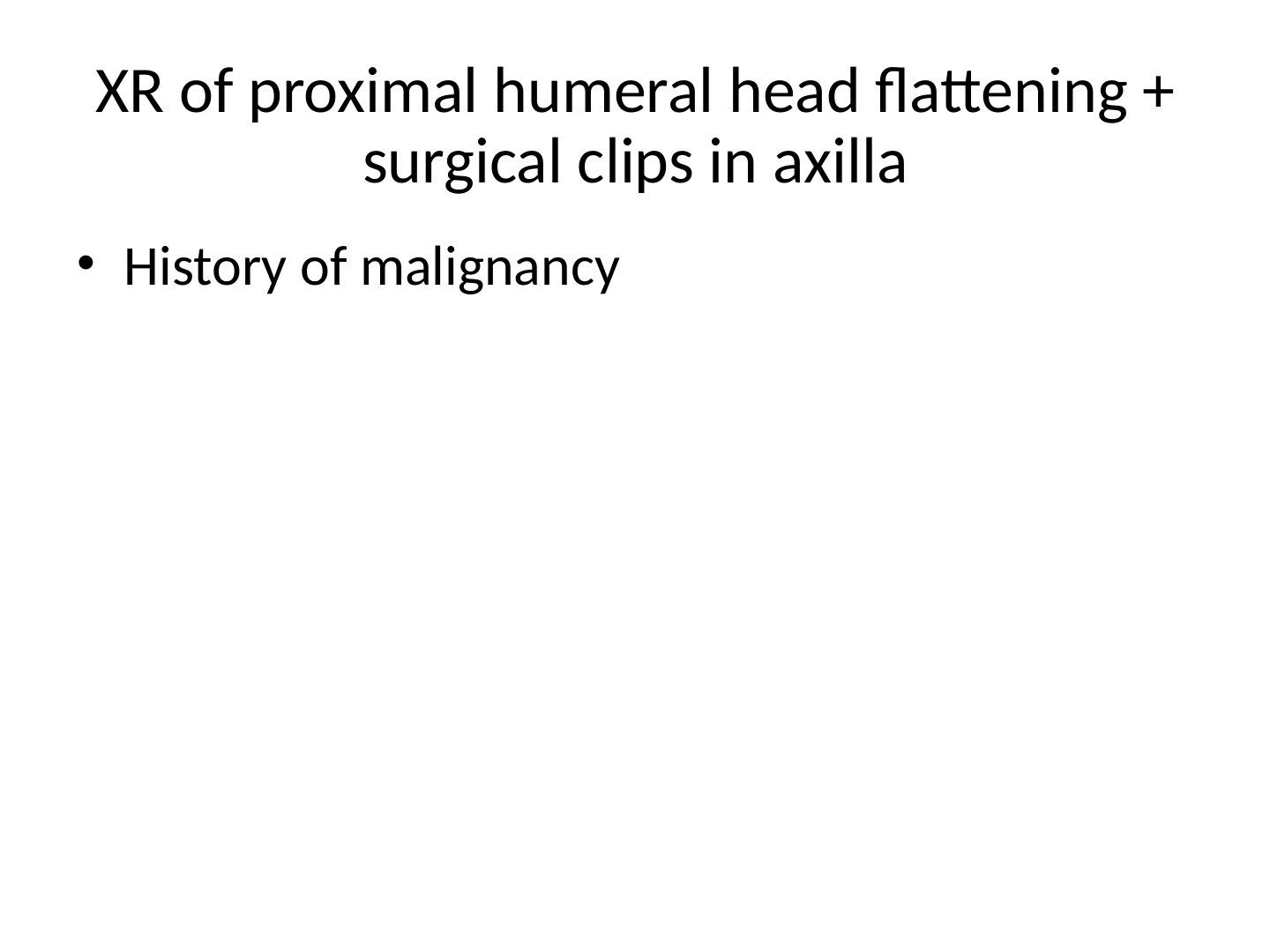
Q1.What is the DDx for proximal humeral head flattening with surgical clips in the axilla?
- Post-radiotherapy AVN
- Tumour recurrence with metastasis
- Post RT osteosarcoma (post-radiotherapy osteosarcoma)
- Came back to be metastasis
Q2.What history is relevant in this patient?
- History of malignancy (surgical clips in axilla)
Q3.What are the problems with using a nail in this case?
- Humeral head already collapsed - potential pain generator
- Difficulty in getting the entry site in a stiff shoulder
Q4.What conservative options may be tried?
- Bisphosphonates
- Radiotherapy
▸ Slide 129 · liposarcomaStatistics · 6 questions expand

Q1-Q66 questions — tap to reveal all answerslist
- Describe the clinical photo and how you assess the mass.
- What features suggest malignancy in a soft tissue tumour?
- How are soft tissue sarcomas classified and staged?
- How would you investigate a suspected soft tissue sarcoma?
- What is the management and when is pre-operative radiotherapy given?
- What are the features of MFH, synovial sarcoma and liposarcoma?
Q1.Describe the clinical photo and how you assess the mass.
- Large swelling over the left back with some overlying skin bruises
- Assess for inflammation, nature of mass (consistency), position and attachment, regional lymph nodes
- Large soft intramuscular swelling
Q2.What features suggest malignancy in a soft tissue tumour?
- >5cm, deep to fascia
- Progressive in size, onset of pain
Q3.How are soft tissue sarcomas classified and staged?
- AJCC: 1. size (>5cm), 2. deep to fascia, 3. node, 4. metastasis, 5. grade
- Stage 1 = low grade, 2 = high grade, 3 = deep to fascia, 4 = metastasis
- Grading: low (well differentiated), intermediate (myxoid), high (de-differentiated)
- Stage 1-2: 80% 5-year survival; stage 3-4: 50% 5-year survival
Q4.How would you investigate a suspected soft tissue sarcoma?
- X-ray to see bony destruction
- MRI: extent, invasion, NV involvement (increase T2, decrease T1; gadolinium - increased T1 peripherally, decreased centrally in cystic/necrotic areas)
- Bloods to rule out infection and check INR
- Image-guided biopsy (histology + MDM2 genetic marker)
- At least CT thorax to rule out metastasis
Q5.What is the management and when is pre-operative radiotherapy given?
- Wide local excision +/- radiotherapy
- May consider marginal excision in low grade (well-differentiated liposarcoma)
- Pre-op RT depends on histological grade, cell type (myxoid), and proximity to NV bundle
- Myxoid type: CT TAP for abnormal sites of metastasis; preop radiotherapy as vascular
Q6.What are the features of MFH, synovial sarcoma and liposarcoma?
- MFH (= undifferentiated pleomorphic sarcoma): most common soft tissue sarcoma; slow growing, no ecchymosis, lung mets; iso T1, increased T2; pleomorphic (>80%) > giant cell (10%) >infl (<10%), most high grade
- Synovial sarcoma: not synovial origin; commonest sarcoma in young adults (15-40) in the foot; LN and lung mets; calcification 25%; gene (X;18); biphasic epithelial + spindle cells; always high grade
- Liposarcoma: 2nd most common soft tissue sarcoma, not from lipoma; lipoblasts; types well differentiated/sclerosing/myxoid; high grade round cell/pleomorphic/dedifferentiated
▸ Slide 130 · Neural tumorStatistics · 7 questions expand

Q1-Q77 questions — tap to reveal all answerslist
- What is a neurilemmoma and what are its histological features?
- What is the treatment of neurilemmoma?
- How do neurofibroma and schwannoma differ in relation to nerve fibres?
- Compare NF type I and type II.
- Compare dermal and plexiform neurofibromas.
- What is MPNST and neuroblastoma?
- What are the types of Schwann cells and their significance?
Q1.What is a neurilemmoma and what are its histological features?
- Benign schwannoma
- Antoni A: spindle cells in intersecting bundles (battle formation); Antoni B: less cellular, loosely arranged cells
- Verocay bodies: pathognomonic - two rows of aligned nuclei in palisading formation
- S100 positive; MRI string sign; paraesthesia in distribution of the peripheral nerve
Q2.What is the treatment of neurilemmoma?
- Observe
- Excision carries a high rate of sensory deficit
- Careful dissection, excise lesion parallel to nerve fascicles
Q3.How do neurofibroma and schwannoma differ in relation to nerve fibres?
- Schwannoma is eccentric to nerve fibres
- Solitary neurofibroma is central to nerve fibres
- Neurofibroma involves non-myelinating Schwann cells and other perineural cells
- Excision with nerve graft if symptomatic
Q4.Compare NF type I and type II.
- Type I (von Recklinghausen): chromosome 17, tibia pseudoarthrosis, spine most common skeletal involvement - dystrophic scoliosis and dural ectasia
- Type II: chromosome 22, bilateral vestibular schwannoma, central meningioma, no scoliosis
- Autosomal dominant disorder of neural crest
- Skeletal: scoliosis, kyphosis, atlantoaxial instability; congenital anterolateral bowing of tibia/forearm; hemi-hypertrophy
Q5.Compare dermal and plexiform neurofibromas.
- Dermal: single peripheral nerve, fusiform swelling; 90% sporadic, 10% in NF1; at puberty; do not become malignant
- Plexiform: multiple nerve bundles, bag of worms; pathognomonic of NF1; early childhood; 10% become malignant (MPNST)
Q6.What is MPNST and neuroblastoma?
- MPNST (malignant schwannoma): arises from large nerves (sciatic, brachial plexus), motor and sensory deficit; treat with wide surgical resection + radiotherapy
- Neuroblastoma: malignant from sympathetic neural tissue; most common solid tumour of childhood
- Adrenal involvement causes tachycardia and abdominal mass; bone mets common and poor prognostic; lecturer describes overall good prognosis
- Treatment: chemo + stem cell transplant + surgical excision
Q7.What are the types of Schwann cells and their significance?
- Myelinating Schwann cells - found in large nerves
- Non-myelinating Schwann cells - found in small nerves, responsible for neurofibromatosis
▸ Slide 131 · other Soft tissue tumorStatistics · 9 questions expand

Q1-Q99 questions — tap to reveal all answerslist
- What is an intramuscular myxoma?
- What is rhabdomyosarcoma?
- What is leiomyosarcoma?
- What is dermatofibrosarcoma protuberans?
- What is malignant fibrous histiocytoma (undifferentiated pleomorphic sarcoma)?
- What is extra-abdominal desmoid tumour and its associations?
- What is nodular fasciitis?
- What is plantar fibromatosis (Ledderhose disease)?
- What is calcifying aponeurotic fibroma?
Q1.What is an intramuscular myxoma?
- Benign, homogeneous
- Excise if symptomatic
- MRI: bright T2, dark T1
Q2.What is rhabdomyosarcoma?
- Malignant; most common soft tissue sarcoma in children
- Rapidly growing painless mass
- Treatment: chemo for metastasis, radiotherapy for inoperable, excision
Q3.What is leiomyosarcoma?
- Aggressive malignant tumour from smooth muscle cells lining small blood vessels
- Osteolytic, moth-eaten lesions in the metaphysis of long bones
- Treatment: chemotherapy + wide surgical resection
Q4.What is dermatofibrosarcoma protuberans?
- Fibrogenic cutaneous sarcoma
- Can result from neurofibromatosis
- t(17;22) encodes PDGFB/COL1A1 fusion protein
Q5.What is malignant fibrous histiocytoma (undifferentiated pleomorphic sarcoma)?
- Most common soft tissue sarcoma in adults
- Pain and swelling; high grade - looks like osteosarcoma
- Types: pleomorphic 80-85%, giant cell 10%, inflammatory <10%
- Treatment: same as osteosarcoma - wide local resection and radiation
Q6.What is extra-abdominal desmoid tumour and its associations?
- Aggressive fibromatosis; most invasive of benign soft tissue tumours
- Associations: Dupuytren's disease, Ledderhose disease, FAP
- Distinctive rock hard mass; oestrogen receptor positive
- Treatment: tamoxifen (oestrogen receptor blocker) and radiotherapy
Q7.What is nodular fasciitis?
- A reactive lesion
Q8.What is plantar fibromatosis (Ledderhose disease)?
- Myofibroblast and collagen proliferation
- Similar process to Dupuytren's fibromatosis
- Ask about hand involvement and Peyronie's disease in men
Q9.What is calcifying aponeurotic fibroma?
- Painless mass in the hands and feet of children and young adults
▸ Slide 132 · Multiple myelomaStatistics · 9 questions 1 check expand
Q1-Q99 questions — tap to reveal all answerslist
- What is the epidemiology and immunoglobulin profile of multiple myeloma?
- Why are osteolytic lesions seen in multiple myeloma?
- What are the diagnostic criteria and CRAB features of multiple myeloma?
- What are the myeloma-defining events (MDEs) and ISS staging?
- What are the XR and imaging findings in myeloma and the role of bone marrow aspirate?
- What are the types, treatment and prognosis of plasma cell neoplasms?
- What histological features are seen in myeloma plasma cells?
- What are the diagnostic criteria for solitary plasmacytoma?
- What are the poor prognostic factors in multiple myeloma?
Q1.What is the epidemiology and immunoglobulin profile of multiple myeloma?
- Most common primary bone malignancy; 1% of all malignancies; males > females
- Neoplastic plasma cells produce immunoglobulins
- Heavy chains: IgG 52%, IgA 21%, IgM 12%
- Light chains: kappa and lambda (Bence Jones proteins)
Q2.Why are osteolytic lesions seen in multiple myeloma?
- Osteoclastic stimulation via RANKL and pro-osteoclastic mediators (M-CSF, IL-6, IL-11)
- Osteoprotegerin (OPG) synthesis is suppressed, causing further osteoclast activation
- Osteoblastic differentiation inhibited by TNF and Dickkopf-1 (DKK-1)
Q3.What are the diagnostic criteria and CRAB features of multiple myeloma?
- >=10% monoclonal plasma cells on bone marrow biopsy (or biopsy-proven plasmacytoma) plus >=1 CRAB feature/MDE
- Calcium: >0.25 mmol/L above upper limit of normal or >2.75 mmol/L (>11mg/dL)
- Renal: creatinine clearance <40mL/min or creatinine >177umol/L (>2mg/dL)
- Anaemia: Hb <100g/L or >20g/L below the lowest limit of normal
- Bone: >=1 osteolytic lesion on radiographs, CT or PET/CT
- If marrow has <10% clonal plasma cells, >1 bone lesion is required to distinguish from solitary plasmacytoma with minimal marrow involvement
Q4.What are the myeloma-defining events (MDEs) and ISS staging?
- MDE: >=60% clonal plasma cells on bone marrow biopsy
- MDE: involved:uninvolved free light chain ratio >=100 (involved light chain >=100mg/L)
- MDE: >=1 focal lesion on MRI >=5mm
- ISS stage I: beta-2 microglobulin <3.5mg/L, albumin >=3.5g/dL (62 months); II: 3.5-5.4mg/L (44 months); III: >=5.5mg/L (29 months)
Q5.What are the XR and imaging findings in myeloma and the role of bone marrow aspirate?
- Punched-out lytic lesions, only visible once >50% destruction has occurred
- No sclerotic border - due to lack of osteoblastic activity
- Bone scans are cold in 30%; skeletal survey is recommended
- FDG-PET 93% sensitivity; may uncover additional sites in 'solitary' plasmacytoma
- Marrow aspirate: plasmacytoma 10-30% vs myeloma >30% plasma cells (normal <2%); CD138+, Hoffa clear zone
Q6.What are the types, treatment and prognosis of plasma cell neoplasms?
- Solitary plasmacytoma: single skeletal site, radiosensitive, progresses to myeloma in >50%; MRI/FDG-PET finds additional lesions in 33%
- Osteosclerotic myeloma: rare POEMS - polyneuropathy, organomegaly, endocrinopathy, sclerotic bone lesions, M protein, skin changes
- Treatment: asymptomatic/MGUS - annual surveillance; solitary plasmacytoma - external beam irradiation alone; myeloma - multiagent chemo +/- stem cell transplant +/- bisphosphonates
- Fractures: surgical stabilisation + RT (life expectancy >3 months); kyphoplasty for vertebral compression with instability/neural compression resistant to radiation
- Prognosis: 5-year survival 30%, 10-year 11%, median 3 years; shortest survival with renal failure; solitary plasmacytoma has the best prognosis
Q7.What histological features are seen in myeloma plasma cells?
- Round plasma cells with eccentric nucleus and prominent nucleolus
- Clock-face organisation of chromatin
Q8.What are the diagnostic criteria for solitary plasmacytoma?
- Solitary lesion on skeletal survey
- Histologic biopsy confirmation of plasmacytoma
- Negative bone marrow biopsy (no plasma cells in bone marrow)
Q9.What are the poor prognostic factors in multiple myeloma?
- Chromosome 13 deletion or translocations t(4;14), t(14;16)
- Circulating plasma cells
- Increased beta-2 microglobulin (elevated tumour burden); decreased serum albumin
- Increased marrow microvessels
- Overall survival relates to stage and secondary factors like renal failure or hypercalcaemia
Multiple myeloma 5-year survival is 30%, 10-year survival 11%, median survival 3 years — outdated — With modern therapy, SEER data (2016-2022) report 5-year relative survival around 62-64%; the 30%/11% figures are historical and should not be quoted as current — source
▸ Slide 133 · XR of patients right hip showing a centrally located lytic lesion over metaphysStatistics · 15 questions expand
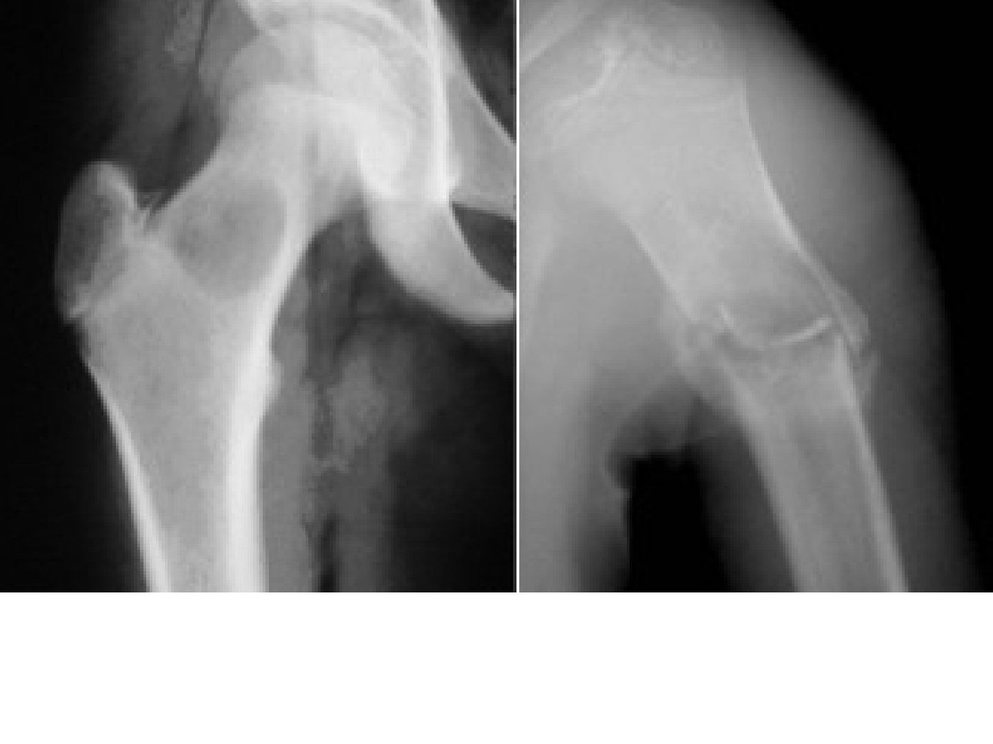
Q1-Q1515 questions — tap to reveal all answerslist
- Describe the X-ray findings of a unicameral bone cyst in this patient.
- What is a unicameral (simple) bone cyst?
- What is the modified Neer classification of a UBC?
- What are the risk factors for pathological fracture in a UBC?
- Describe the Scaglietti injection technique for a UBC.
- What are the management options for a UBC?
- What are the differentials for a centrally located metaphyseal lytic lesion in a child?
- What history should be elicited in a suspected UBC?
- What is the histology of a UBC?
- What is the role of MRI in a UBC?
- What factors determine the management of a UBC?
- What is the difference between an active and a latent UBC?
- What is the fallen leaf sign?
- What are the common sites and natural history of a UBC?
- What is the combined technique for a UBC and its healing rate?
Q1.Describe the X-ray findings of a unicameral bone cyst in this patient.
- Centrally located lytic lesion in the metaphysis, abutting the physis
- Narrow zone of transition - favours a nonaggressive lesion
- Fallen leaf sign+ with pathological fracture
- Dx: UBC with pathological fracture
Q2.What is a unicameral (simple) bone cyst?
- Benign serous fluid-filled bone lesion with a fibrous lining
- Temporary failure of medullary bone formation near the physis
- Theories: synovial remnant, traumatic haematoma, venous occlusion
- Common in children <20 years; central metaphyseal lesion abutting the physis
- Often presents with pathological fracture (50%)
Q3.What is the modified Neer classification of a UBC?
- I healed: cyst filled with new bone, radiolucent area <1cm
- II healed with defect: radiolucent area <50% of the bone diameter
- III persistent cyst: radiolucent area >50% of the bone diameter with a thin cortical rim
- IV recurrent cyst: reappearance or increase in size of the radiolucent area
Q4.What are the risk factors for pathological fracture in a UBC?
- Peritrochanteric region
- Cyst transverse diameter ballooning >85% of the affected bone
- Cyst wall <0.5mm thick
- Active phase, male, multilocuated
- Curettage + bone grafting +/- internal fixation for fracture/AVN (structural concern)
Q5.Describe the Scaglietti injection technique for a UBC.
- Technique: (Scaglietti method)
- Insert the needle and aspirate
- Yellow serous fluid + complete contrast filling -> second needle for irrigation; no suction (increases bleeding), then steroid injection
- Bloody or incomplete contrast filling -> biopsy
- Mechanism of steroid: exerts a destructive action on pathological tissue and thus favoured a progressive process of repair and avoid surgery (80-200mg)
- Inhibit prostagalndin E
Q6.What are the management options for a UBC?
- Conservative first; if 6/52 not heal then likely ABC - go for MRI
- Intervene if high risk of fracture regardless of symptoms, or symptomatic + active cyst
- Injection (steroid, autogenous bone marrow aspirate +/- DBM/calcium phosphate), decompression, combined techniques
- Decompression had better healing than steroid or bone marrow aspirate (JPO 2011)
- Calcium sulfate pellets 66%; allograft only 25-36%; combined healing >95%
- Humerus fracture: immobilise; femur fracture: curettage + BG + ORIF
Q7.What are the differentials for a centrally located metaphyseal lytic lesion in a child?
- Aneurysmal bone cyst (ABC)
- Fibrous dysplasia
- Non-ossifying fibroma (NOF)
- Telangiectatic osteosarcoma
Q8.What history should be elicited in a suspected UBC?
- Pain?
- Previous fracture?
- Infection?
Q9.What is the histology of a UBC?
- Thin, fibrous lining containing fibrous tissue
- Giant cells
- Haemosiderin pigment
- A few chronic inflammatory cells
Q10.What is the role of MRI in a UBC?
- Not necessary for diagnosis
- T1 dark, T2 bright
- Gadolinium shows classic rim enhancement of a cystic lesion
- May look for a solid component
Q11.What factors determine the management of a UBC?
- 1. Symptoms? 2. Active? 3. Location? 4. Fracture?
- Aim: promote healing and prevent complications
- Conservative first
Q12.What is the difference between an active and a latent UBC?
- Active: touching the physis, increasing in size, thin cortex
- Latent: grows with increasing distance between the lesion and physis, intact cortex
- UBC and ABC abut the physeal scar; GCT can cross the physis
Q13.What is the fallen leaf sign?
- Pathological fracture with a fallen cortical fragment in the base of a fluid-filled cyst
- Pathognomonic of a unicameral bone cyst
Q14.What are the common sites and natural history of a UBC?
- Children <20 years; proximal humerus (also proximal femur, distal tibia, ilium, calcaneum)
- Symmetric cystic expansion with thinning of the cortices; becomes Trabaculated after multiple # (fractures)
- Often decrease/heal as the patient approaches skeletal maturity
- Fracture healing leads to cyst resolution in 15%
- If active -> close follow-up for recurrence, fracture and growth arrest
Q15.What is the combined technique for a UBC and its healing rate?
- 1. Cystography with aspiration of cyst fluid + injection of dye
- 2. Percutaneous curettage of the cyst lining
- 3. IM decompression with angle curet or flexible IM nail
- 4. Calcium sulfate pellets
- Healing rate >95%
▸ Slide 134Statistics · 2 questions expand

Q1-Q22 questions — tap to reveal all answerslist
- What topic does this slide cover?
- What are the key learning points from this slide?
Q1.What topic does this slide cover?
- Not covered in the speaker notes
Q2.What are the key learning points from this slide?
- Not covered in the speaker notes
▸ Slide 135 · Lytic expansile lesion with internal septae and sclerotic rim in metaphyseal regStatistics · 9 questions expand
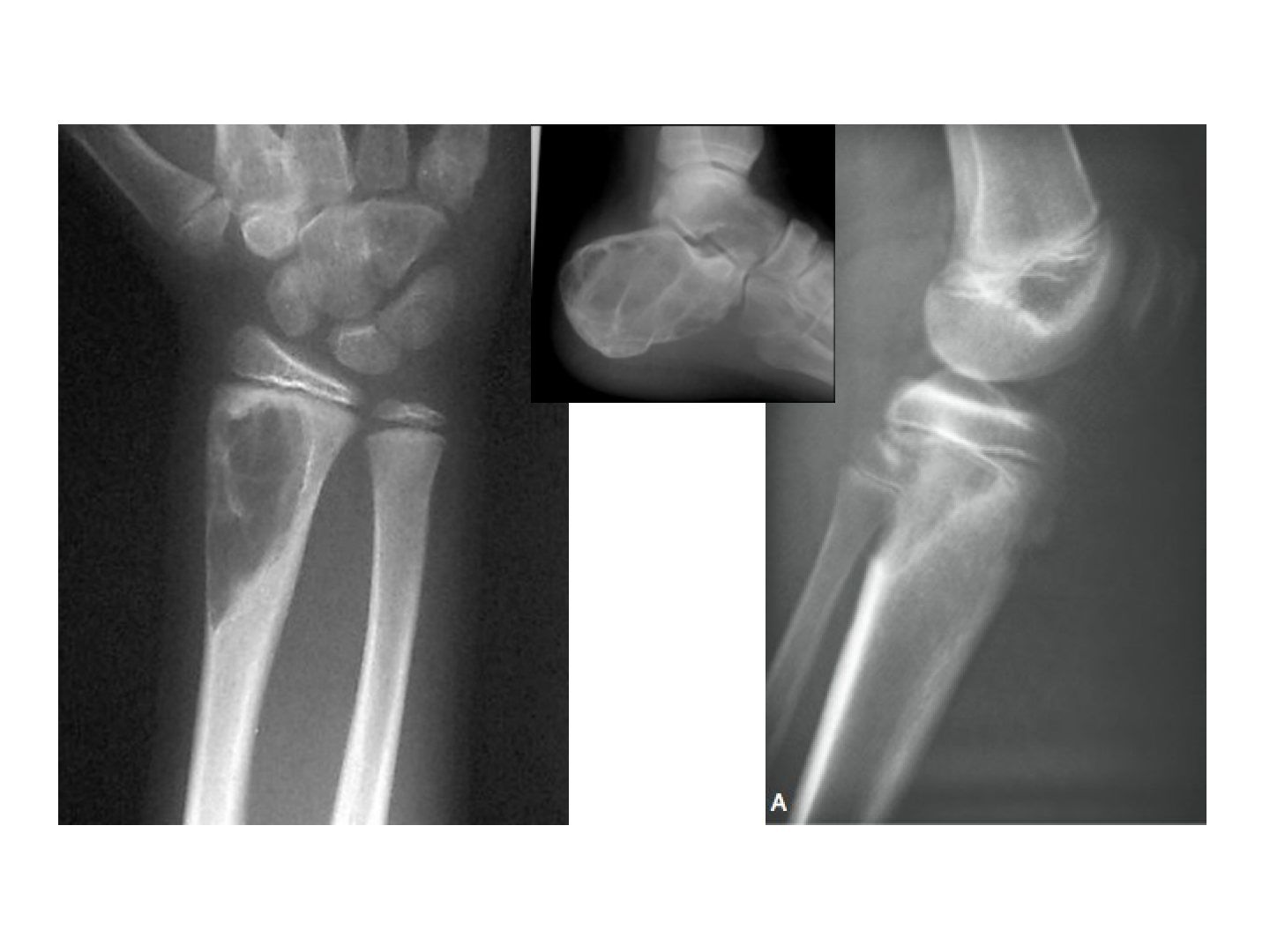
Q1-Q99 questions — tap to reveal all answerslist
- Describe the X-ray findings of an aneurysmal bone cyst.
- What are the differentials for an expansile metaphyseal lytic lesion in a young patient?
- What are the key features of an aneurysmal bone cyst?
- What are the histology and MRI findings in an ABC?
- What is the management of an aneurysmal bone cyst?
- What are the clinical features and complications of an aneurysmal bone cyst?
- What laboratory tests are done in an aneurysmal bone cyst?
- Why must every aneurysmal bone cyst be biopsied?
- What are the typical sites of an aneurysmal bone cyst?
Q1.Describe the X-ray findings of an aneurysmal bone cyst.
- Lytic expansile lesion with internal septae and a sclerotic rim in the metaphyseal region abutting the physis
- Sclerotic rim; narrow zone of transition
- No periosteal reaction
- Eccentric metaphyseal lesion with bone destruction, a classic rim of new bone and bony septae (bubbly appearance)
Q2.What are the differentials for an expansile metaphyseal lytic lesion in a young patient?
- Chondromyxoid fibroma (if young), UBC, GCT
- Telangiectatic osteosarcoma
- Brown tumour
Q3.What are the key features of an aneurysmal bone cyst?
- Non-neoplastic reactive lesion able to destroy local bone and extend into soft tissue
- Multiple blood-filled cavities
- 75% <20 years; long bone >60% (usually metaphysis), spine 15%
- Gene: USP-6 ubiquitin specific protease (Paget = UBPS/sequestosome)
Q4.What are the histology and MRI findings in an ABC?
- Cavernous blood-filled spaces without an endothelial lining (essential)
- Thin bony septa with numerous giant cells
- MRI: T2 fluid-fluid levels (characteristic) +/- periosteal layer surrounding the lesion
Q5.What is the management of an aneurysmal bone cyst?
- Rule out a secondary cause, control symptoms, prevent progression
- Always look for a primary tumour (30%): GCT, chondroblastoma, FD, CMF, NOF
- Fracture: immobilise and let the fracture heal first, then treat as no fracture
- No fracture: curettage +/- adjunct (phenol, argon beam, liquid nitrogen) + bone graft +/- OIRF if high stress area
- 25% recurrence if the physis is still open
Q6.What are the clinical features and complications of an aneurysmal bone cyst?
- Pain from expansion
- Pathological fracture
- May be secondary to a primary tumour
Q7.What laboratory tests are done in an aneurysmal bone cyst?
- Inflammatory markers
- CaPO4, ALP, LDH
Q8.Why must every aneurysmal bone cyst be biopsied?
- It can be secondary to GCT, chondroblastoma, chondromyxoid fibroma or fibrous dysplasia
- Always look for a primary tumour (30%)
Q9.What are the typical sites of an aneurysmal bone cyst?
- Spine 15%
- Long bones >60% (usually metaphysis)
- Posterior pelvis; calcaneum
▸ Slide 136 · Telangiectatic OSStatistics · 5 questions expand
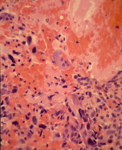
Q1-Q55 questions — tap to reveal all answerslist
- Describe the X-ray findings of telangiectatic osteosarcoma.
- What is the typical presentation of telangiectatic osteosarcoma?
- How does the MRI of telangiectatic osteosarcoma differ from ABC?
- What are the differentials and management of telangiectatic osteosarcoma?
- What features favour telangiectatic osteosarcoma over an aneurysmal bone cyst?
Q1.Describe the X-ray findings of telangiectatic osteosarcoma.
- Skeletally mature patient
- Eccentric lytic lesion in the metaphyseal region of the medial distal femoral metaphysis
- Cortical destruction; no extension into the joint
- Periosteal reaction with Codman triangle; matrix is osseous
- Significant soft tissue component
Q2.What is the typical presentation of telangiectatic osteosarcoma?
- Age 15-25, male
- Previous radiation
- Pain; pathological fracture in 25%
Q3.How does the MRI of telangiectatic osteosarcoma differ from ABC?
- Less defined fluid levels compared with ABC
- Extensive soft tissue oedema
Q4.What are the differentials and management of telangiectatic osteosarcoma?
- Ddx: GCT, ABC
- Mx as per conventional osteosarcoma
Q5.What features favour telangiectatic osteosarcoma over an aneurysmal bone cyst?
- Codman triangle and periosteal reaction
- Osseous matrix
- Significant soft tissue component
- Less defined fluid levels and extensive soft tissue oedema on MRI
▸ Slide 137 · AP Xray pelvis and bilateral proximal femurStatistics · 10 questions expand

Q1-Q1010 questions — tap to reveal all answerslist
- Describe the X-ray findings in polyostotic Paget's disease.
- What are the causes of pain in Paget's disease?
- What is the risk and prognosis of malignant transformation in Paget's disease?
- What investigations are used in Paget's disease?
- When is medical treatment indicated in Paget's disease?
- What are the operative concerns in Paget's disease?
- What is Paget's disease?
- What is the histology and what are the phases of Paget's disease?
- What deformities and gait changes occur in Paget's disease?
- What is the role of NSAIDs and monitoring in Paget's disease?
Q1.Describe the X-ray findings in polyostotic Paget's disease.
- Asymmetry of the pelvis; left hemipelvis larger with diffuse osteosclerosis and coarse trabeculae
- Right proximal femur: thickened cortex, loss of corticomedullary junction, coxa vara
- Brim sign: thickened iliopectineal line
- Dx: Polyosteotic Paget's disease
- Ddx: bone metastases, fibrous dysplasia, myelofibrosis
Q2.What are the causes of pain in Paget's disease?
- Compression neuropathy (spinal stenosis, hearing loss, basilar invagination)
- 2.Arthritis; 3.Blood flow/bone enlargement
- Pathological fracture (fissure or chalk-stick transverse fracture)
- Malignant transformation
Q3.What is the risk and prognosis of malignant transformation in Paget's disease?
- Paget's osteosarcoma <1%, usually >70 years
- Poor prognosis: 5% survival at 5 years
Q4.What investigations are used in Paget's disease?
- Raised ALP (osteoblast activity), raised serum acid phosphatase (osteoclast activity); calcium normal
- Raised urine hydroxyproline and collagen cross-linked peptides (collagen turnover)
- X-ray: deformity/enlarged bone, coarse trabeculae, loss of corticomedullary differentiation
- Osteoporosis circumscripta; flame sign (advancing V-shaped lytic lesion)
- Bone scan (80% polyostotic); echo
Q5.When is medical treatment indicated in Paget's disease?
- Pain, before operation, high-risk lesion, or medical complication (high-output failure)
- Aim: retard osteoclast activity
- Bisphosphonate (oral/IV) first line; calcitonin (SC/IM)
- Teriparatide contraindicated (increases Paget's sarcoma)
Q6.What are the operative concerns in Paget's disease?
- General: high bleeding risk, post-op HO, hypercalcaemia with immobilisation (hypercalcemia with immobilization)
- Fracture: flexible or stiff locked IM nail with osteotomy; post-op bracing due to slow healing
- TJR: malalignment from very soft and patchy very hard bone
- Causes of hypercalcaemia: secondary hyperPTH, immobilisation, neoplastic change
Q7.What is Paget's disease?
- Disorder of high bone turnover causing distortion of architecture and abnormal lamellar bone formation
- Increased osteoclastic bone resorption is the primary cellular abnormality
- Mono-ostotic or poly-ostotic (80%)
- Most are sporadic; some AD inheritance; paramyxovirus infection implicated
Q8.What is the histology and what are the phases of Paget's disease?
- Histology: mosaic pattern of disorganized lamellar bone
- Phases: lytic, mixed, blastic - can co-exist in the same bone
Q9.What deformities and gait changes occur in Paget's disease?
- Saber tibia; LLD (leg length discrepancy)
- Gait: stooped posture, lumbar kyphosis, hips in fixed flexion contracture (FFC)
- History should include premorbid status and family history
Q10.What is the role of NSAIDs and monitoring in Paget's disease?
- NSAIDs for symptom control
- Monitor for complications
▸ Slide 138 · #:Statistics · 3 questions expand

Q1-Q33 questions — tap to reveal all answerslist
- How is a fracture in Paget's disease managed?
- Why is immobilisation detrimental in Paget's disease?
- What are the pre-, intra- and post-operative considerations?
Q1.How is a fracture in Paget's disease managed?
- Avoid immobilisation; prefer flexible IM fixation with bracing afterwards
Q2.Why is immobilisation detrimental in Paget's disease?
- Causes osteopenia
- Causes hypercalcaemia
Q3.What are the pre-, intra- and post-operative considerations?
- Pre-op: bisphosphonate, cardiac condition optimisation
- Intra-op: deformity, bleeding
- Post-op: heterotopic ossification (HO), slow healing
▸ Slide 139 · Fibrous dysplasiaStatistics · 12 questions 1 check expand
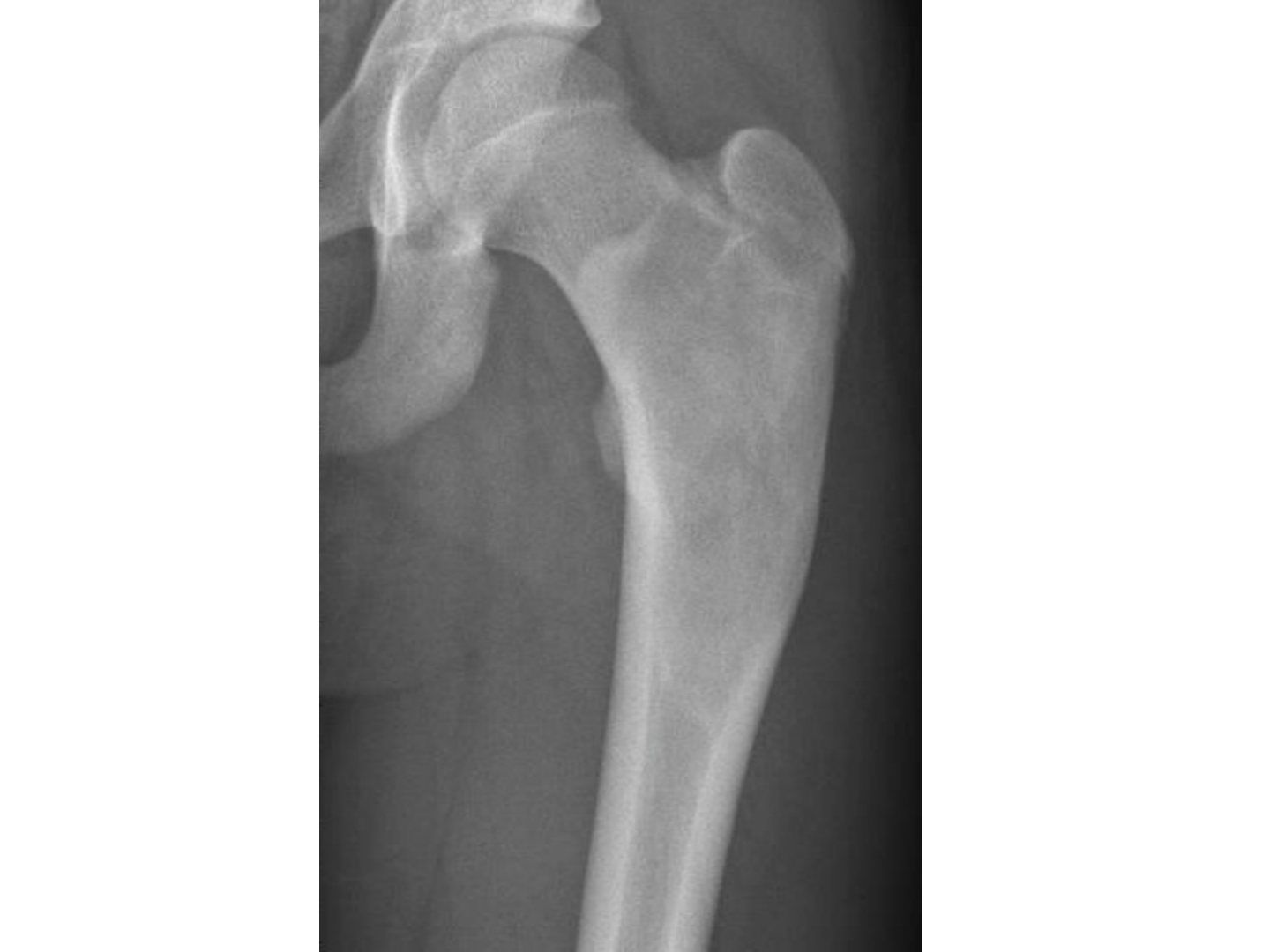
Q1-Q1212 questions — tap to reveal all answerslist
- Describe the X-ray findings in fibrous dysplasia.
- What is the pathophysiology of fibrous dysplasia?
- What syndromes are associated with fibrous dysplasia?
- What is the histology and risk of malignant transformation in fibrous dysplasia?
- What are the indications and principles of surgery in fibrous dysplasia?
- What medical treatment is used in fibrous dysplasia?
- Which patients and bones are affected by fibrous dysplasia?
- What deformities can fibrous dysplasia cause?
- What natural history of fibrous dysplasia must patients be counselled about?
- What investigations are performed in fibrous dysplasia?
- What are the aims of treatment in fibrous dysplasia?
- What intraoperative difficulties are anticipated in fibrous dysplasia surgery?
Q1.Describe the X-ray findings in fibrous dysplasia.
- Expansile lesion at the metaphyseal region with cortical thinning
- Intramedullary with a narrow transition zone
- Ground-glass matrix, no periosteal reaction
- Mild varus deformity of the proximal femur (shepherd crook deformity)
- Ddx: bone cyst/ enchondroma (if matrix less ground glass), infection, brown tumour, haematological, metastasis in older patients
Q2.What is the pathophysiology of fibrous dysplasia?
- Localised dysfunction of osteoblast differentiation (via increased cAMP signalling) and maturation
- Failure of lamellar bone formation (failure of lamella bone formation); bone is replaced by fibrous tissue
- Mutation of Ch 20q23 (GNAS), not inherited
Q3.What syndromes are associated with fibrous dysplasia?
- McCune-Albright syndrome (McCune Albright syndrome): polyostotic FD, cafe au lait spots (coast of Maine), hyperfunctioning endocrine disease (precocious puberty, hyperT4, acromegaly)
- Scoliosis; GNAS mutation
- Mazabraud syndrome: polyostotic FD + soft tissue intramuscular myxoma
Q4.What is the histology and risk of malignant transformation in fibrous dysplasia?
- Histology: alphabet soup appearance - trabeculae of osteoid within fibrous stroma
- Malignant transformation 1% (osteosarcoma/fibrosarcoma); 5-10% in polyostotic disease (per notes)
- Nocturnal pain suggests malignant transformation
Q5.What are the indications and principles of surgery in fibrous dysplasia?
- Indications: pathological fracture, enlarging lesion, recurrent fracture/deformity, high stress area (femoral neck)
- Polyostotic: mainly deformity correction and fracture prevention
- Monostotic: curettage; monostotic -> aim to remove tumor (curative)
- Curettage, allograft (autograft transforms into fibrous tissue), stable titanium IM nail +/- osteotomy
Q6.What medical treatment is used in fibrous dysplasia?
- Bisphosphonate for symptomatic polyostotic patients: pain relief, decrease fracture, partial resolution of lesion
- Denosumab - ongoing study (J Bone Miner Res 2021)
Q7.Which patients and bones are affected by fibrous dysplasia?
- Can involve any bone
- More common in females
Q8.What deformities can fibrous dysplasia cause?
- Mono-ostotic vs polyostotic disease
- Coxa vara with Trendelenburg gait
- Blindness with skull involvement
Q9.What natural history of fibrous dysplasia must patients be counselled about?
- Progression of the existing lesion + appearance of new lesions
- Old lesions will not mature - problem of remodelling: decreased cellularity, increased collagen deposition, persistent woven bone, no compressive/tensile trabeculae -> progressive deformity
- Progressive deformity until maturity, but may progress again with maturity
Q10.What investigations are performed in fibrous dysplasia?
- Inflammatory markers, ALP, LDH - increase suggests malignant transformation
- Skeletal survey to rule out polyostotic disease (if polyostotic, screen for syndromes)
- If mono-ostotic: consider biopsy for diagnosis (monoostotic: consider biopsy for Dx)
Q11.What are the aims of treatment in fibrous dysplasia?
- Decrease pain, prevent fracture +/- correct deformity
Q12.What intraoperative difficulties are anticipated in fibrous dysplasia surgery?
- Lesions can be very vascular - prepare blood
- Choose the correct implant with the right NSA
Malignant transformation in fibrous dysplasia is 1%, rising to 5-10% in polyostotic disease — Overstated for polyostotic disease — Published series report ~0.4-1% overall and up to ~4% in McCune-Albright/familial cases; 5-10% is higher than most sources — medium confidence — source
▸ Slide 140 · XR: well defined intramedullary lytic lesion over the diaphyseal region of the rStatistics · 11 questions expand
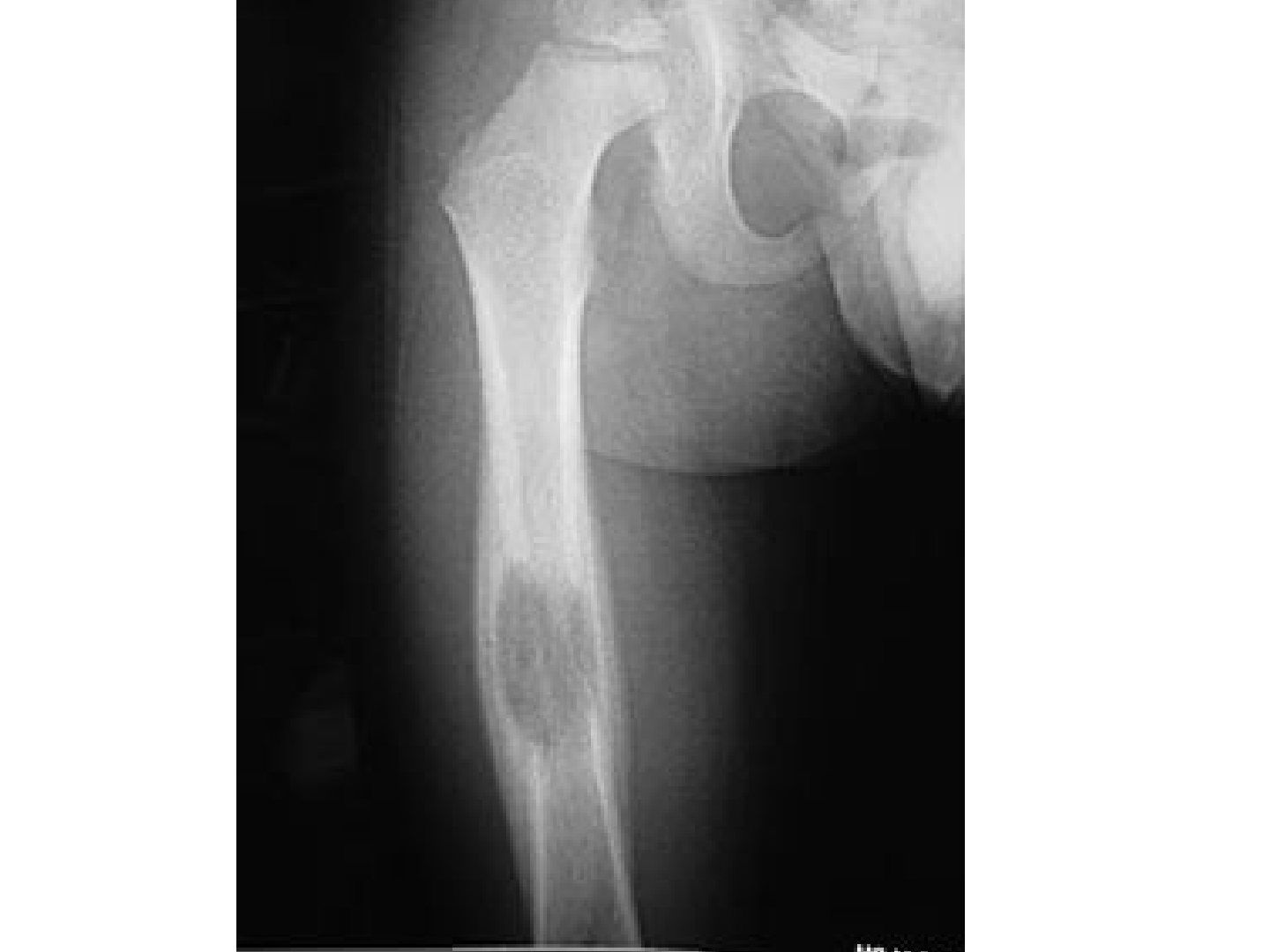
Q1-Q1111 questions — tap to reveal all answerslist
- Describe the X-ray findings in eosinophilic granuloma.
- What is Hand-Schuller-Christian disease?
- What is the histology of eosinophilic granuloma?
- What investigations are needed in eosinophilic granuloma?
- How is spinal deformity in eosinophilic granuloma managed?
- What are the clinical features of eosinophilic granuloma?
- What is the natural history of eosinophilic granuloma?
- What are the sites of involvement in eosinophilic granuloma?
- What is the spectrum of histiocytosis X (Langerhans cell histiocytosis)?
- What is vertebra plana and what is its differential?
- What is the great mimicker differential of eosinophilic granuloma?
Q1.Describe the X-ray findings in eosinophilic granuloma.
- Well-defined intramedullary lytic lesion in the diaphysis of the right femur
- Laminated periosteal reaction with thinning of the cortex
- Highly destructive with a well-defined border
- Ddx: Ewing sarcoma, infection, haematological
Q2.What is Hand-Schuller-Christian disease?
- Triad: multiple lytic lesions, diabetes insipidus, exophthalmos
- Chronic disseminated Langerhans histiocytosis with visceral involvement, >3 years old
Q3.What is the histology of eosinophilic granuloma?
- Langerhans cells (antigen-presenting): pale staining, coffee bean nuclei, eosinophilic, Birbeck granules
- Giant cells present
- No atypia; normal nuclear/cytoplasmic ratio
Q4.What investigations are needed in eosinophilic granuloma?
- Bloods; local imaging with MRI
- Systemic staging: CT thorax + bone scan
- Tissue diagnosis (biopsy)
Q5.How is spinal deformity in eosinophilic granuloma managed?
- Bracing first line: corrects 90% of deformity and regains 50% of vertebral height
- Failed (10%) -> low-dose radiotherapy
- Curettage + BG +/- steroid if nonoperative treatment fails or for a solitary lesion close to an articular surface
- HSC: vinblastine + steroid
Q6.What are the clinical features of eosinophilic granuloma?
- Pain + limp
- Progressive kyphotic posture
Q7.What is the natural history of eosinophilic granuloma?
- Self-limiting benign histiocytic lesion
- Monostotic bone disease
- Highly destructive with a well-defined border
- Biopsy alone may be sufficient treatment
Q8.What are the sites of involvement in eosinophilic granuloma?
- Skull, ribs, clavicle, scapula, mandible
- Spine especially thoracic (vertebral plana)
- Diaphyseal regions of long bones and the pelvis
Q9.What is the spectrum of histiocytosis X (Langerhans cell histiocytosis)?
- A reticuloendothelial system pathology
- Eosinophilic granuloma (monostotic)
- Hand-Schuller-Christian disease: chronic disseminated with visceral involvement, >3 years old
- Letterer-Siwe disease: <3 years old, fatal
Q10.What is vertebra plana and what is its differential?
- Single level vertebral collapse
- Ddx: post-TB, post-fracture, haematogenous
- Compare with platyspondyly (diffuse) in SED/osteomalacia
Q11.What is the great mimicker differential of eosinophilic granuloma?
- Osteomyelitis, leukaemia, lymphoma
- Fibrous dysplasia, Ewing sarcoma
▸ Slide 141 · Describe Xray: osteolytic lesion over intertrochanteric region with surrounding Statistics · 9 questions expand
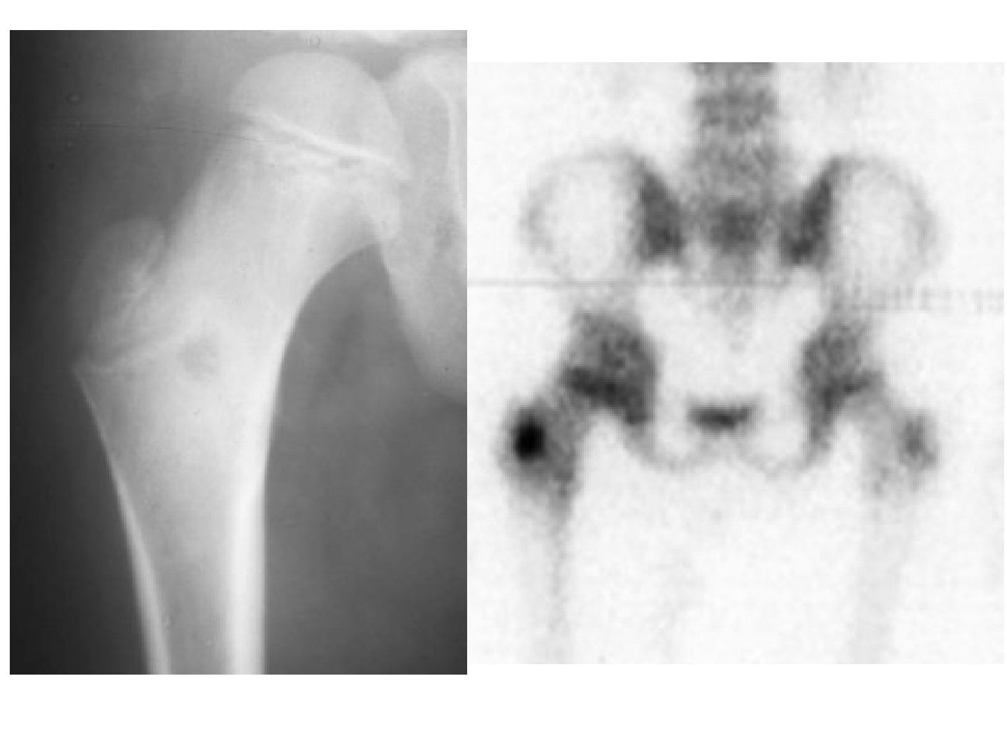
Q1-Q99 questions — tap to reveal all answerslist
- Describe the X-ray findings of osteoid osteoma.
- What is the classic clinical presentation of osteoid osteoma?
- What is the double-density sign on bone scan?
- How is nidus size used to distinguish osteoid osteoma from osteoblastoma?
- What are the management options for osteoid osteoma?
- What are the differences between osteoid osteoma and osteoblastoma?
- What is the pathophysiology of pain in osteoid osteoma?
- What is the histology of osteoid osteoma?
- What investigations are performed for a suspected osteoid osteoma?
Q1.Describe the X-ray findings of osteoid osteoma.
- Osteolytic lesion over the intertrochanteric region with surrounding sclerosis
- +/- homogenous cortical thickening
- +/- radiolucent nidus
- +/- solid periosteal reaction
- Ddx: Brodie's abscess, stress/healing fracture, fibrous cortical defect, Ewing sarcoma
Q2.What is the classic clinical presentation of osteoid osteoma?
- Night pain, worse after alcohol, relieved by NSAIDs
- Flexion contracture of the knee
- Painful scoliosis
Q3.What is the double-density sign on bone scan?
- A small focus of intense radioactivity in the nidus
- Superimposed on a larger area of lower, but still increased, radioactivity
Q4.How is nidus size used to distinguish osteoid osteoma from osteoblastoma?
- CT delineates the nidus: <1.5cm = osteoid osteoma
- >1.5cm = osteoblastoma (typically 3-4cm)
Q5.What are the management options for osteoid osteoma?
- Conservative NSAIDs: 50% treated with NSAID alone; sole treatment in spinal involvement without scoliosis; self-resolves in 3 years
- CT-guided radiofrequency ablation: failed conservative or juxta-articular lesions with contracture; controversial in spine as close to neural elements, need 1cm safe zone in spine; 85% success
- RFA complications: skin burn, stress riser at long bone; allows concomitant biopsy
- Surgery (marginal excision): failed conservative when RFA not possible, painful scoliosis; CT navigation adjunct
Q6.What are the differences between osteoid osteoma and osteoblastoma?
- Nidus size: <1.5cm (osteoid osteoma) vs >1.5cm/typically 3-4cm (osteoblastoma)
- Location: proximal femur > tibia > spine vs posterior elements of the spine > proximal femur
- Course: self-limiting vs progressive; NSAID relief yes vs no
- Surgery: +/- vs yes; osteoblastoma has more giant cells histologically
Q7.What is the pathophysiology of pain in osteoid osteoma?
- Nidus of central woven bone with osteoblastic rimming surrounded by a reactive zone
- Pain due to prostaglandin E2 concentration and COX1/2 expression
- Increased unmyelinated nerve endings within
- Spontaneous bleeding within
Q8.What is the histology of osteoid osteoma?
- Distinct border between the osteoid nidus and reactive bone
- Nidus of immature osteoid + osteoblastic rim
- Increased mitotic figures with normal NC ratio
- Osteoblastoma = more giant cells
Q9.What investigations are performed for a suspected osteoid osteoma?
- Blood test to rule out infection
- X-ray: reactive bone with a radiolucent nidus
- CT to delineate the size of the nidus
- Bone scan: double-density sign
▸ Slide 142 · Describe CT:Statistics · 4 questions expand

Q1-Q44 questions — tap to reveal all answerslist
- Describe the CT findings in this patient.
- What is the differential diagnosis of an anterior spinal tumour?
- What is the assessment of osteoblastoma?
- What is the histology and management of osteoblastoma?
Q1.Describe the CT findings in this patient.
- Axial cut showing a lytic lesion over the facet
- Expansile, with no cortical breakage and no periosteal reaction
- No definite narrowing of the spinal canal
- Ddx: osteoblastoma, ABC, GCT, exostosis, chondrosarcoma
Q2.What is the differential diagnosis of an anterior spinal tumour?
- Metastasis, haemangioma, multiple myeloma
- Eosinophilic granuloma, chordoma
- Osteosarcoma, chondrosarcoma, Ewing sarcoma
Q3.What is the assessment of osteoblastoma?
- Hx/PE: dull pain not relieved by NSAIDs, neurological symptoms
- Tumour markers including SPE
- X-ray: expansile lytic lesion with reactive bone; CT to delineate nidus
- Bone scan hot; MRI if neurological complication
Q4.What is the histology and management of osteoblastoma?
- Similar to osteoid osteoma but with more giant cells
- Nidus of immature osteoid surrounded by osteoblasts, increased mitotic figures but no atypia
- Marginal excision; recurrence 10-20%
▸ Slide 143 · Clinical photo of patient with upper and lower limb deformitiesStatistics · 3 questions expand

Q1-Q33 questions — tap to reveal all answerslist
- Describe the deformities shown in the clinical photo.
- What negative findings are documented?
- What upper limb features are present?
Q1.Describe the deformities shown in the clinical photo.
- Genu valgum; right knee flexed and internally rotated posture
- No gross leg length discrepancy
- Forearm radial bowing + ulnar deviation of the hand
- Prominent radial styloid; ulnar shortened; +/- radial head subluxation
Q2.What negative findings are documented?
- No absent ray
- No gross joint swelling
- No skin changes or stigmata
Q3.What upper limb features are present?
- Radial bowing with ulnar deviation of the hand
- Prominent radial styloid
- Ulnar shortening
- +/- radial head subluxation
▸ Slide 144 · adamantinomaStatistics · 5 questions expand
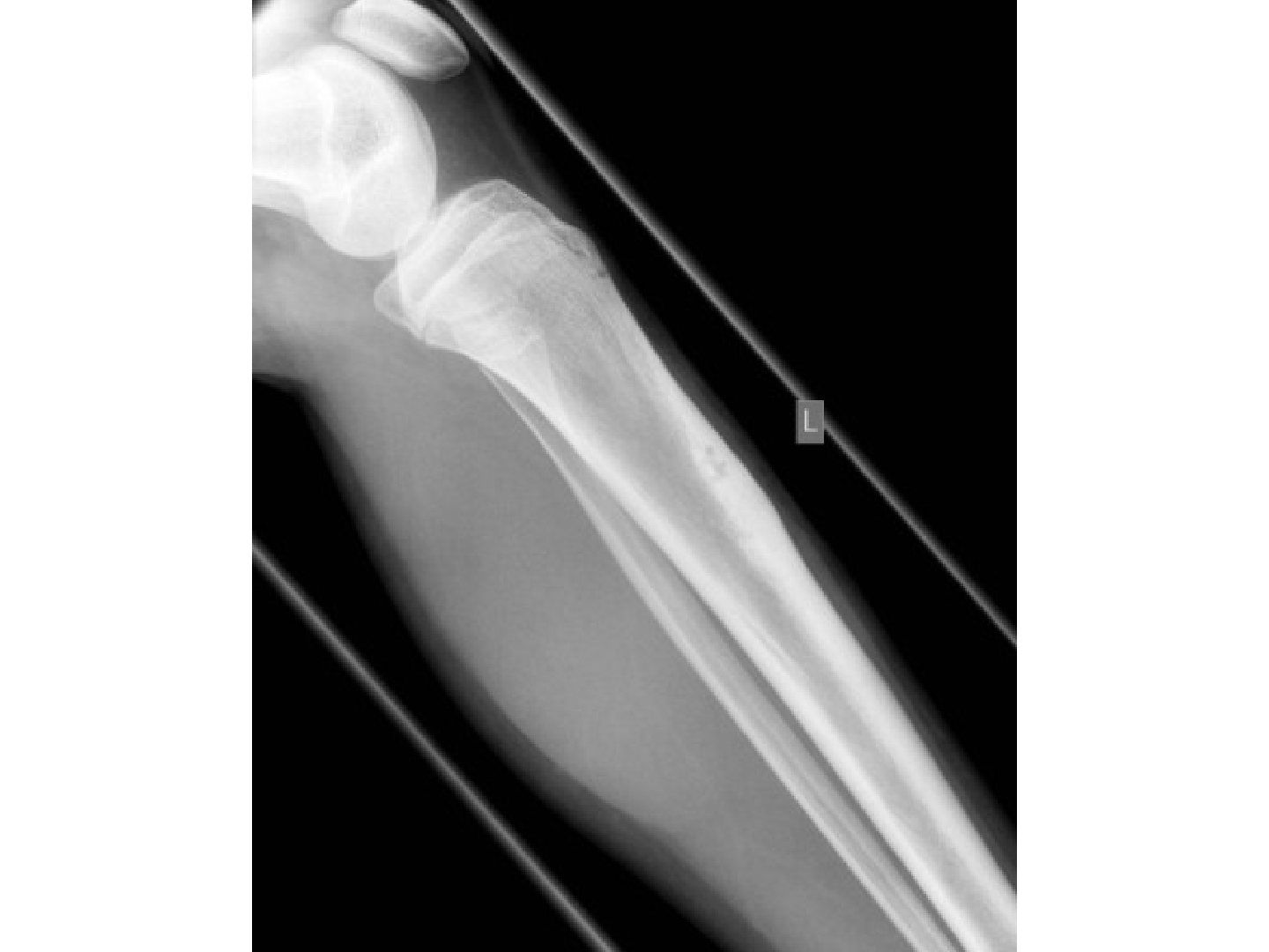
Q1-Q55 questions — tap to reveal all answerslist
- Describe the X-ray findings in adamantinoma.
- What is adamantinoma?
- What are the differentials for adamantinoma?
- What is the assessment and biopsy appearance in adamantinoma?
- What is the management of adamantinoma?
Q1.Describe the X-ray findings in adamantinoma.
- Skeletally mature patient
- Multiple lucent lesions separated by septa of bone with a soap bubble appearance
- Cortical lesion at the anterior cortex of the proximal tibia with cortical thickening
- Wide transition zone, no increase in anterior bowing, no obvious periosteal reaction
Q2.What is adamantinoma?
- Low grade malignancy of unknown origin
- Usually 20-40 years old, located nearly always at the anterior tibia
- May metastasize to lung (25%)
Q3.What are the differentials for adamantinoma?
- Osteofibrous dysplasia, ossifying fibroma, osteoid osteoma
- Brodie's abscess, fracture callus
- Haematological malignancy
Q4.What is the assessment and biopsy appearance in adamantinoma?
- Hx: insidious pain; PE: anterior bowing deformity, palpable mass
- CXR: lung mets 25%; MRI: decrease in T1 signal
- Biopsy: fibrous and epithelial +/- glandular tissue in a gland-like pattern (glandular tissue in gland like pattern)
Q5.What is the management of adamantinoma?
- Wide margin excision + reconstruction
- RT/chemotherapy not used due to slow growth
▸ Slide 145 · Describe Xray:Statistics · 5 questions expand
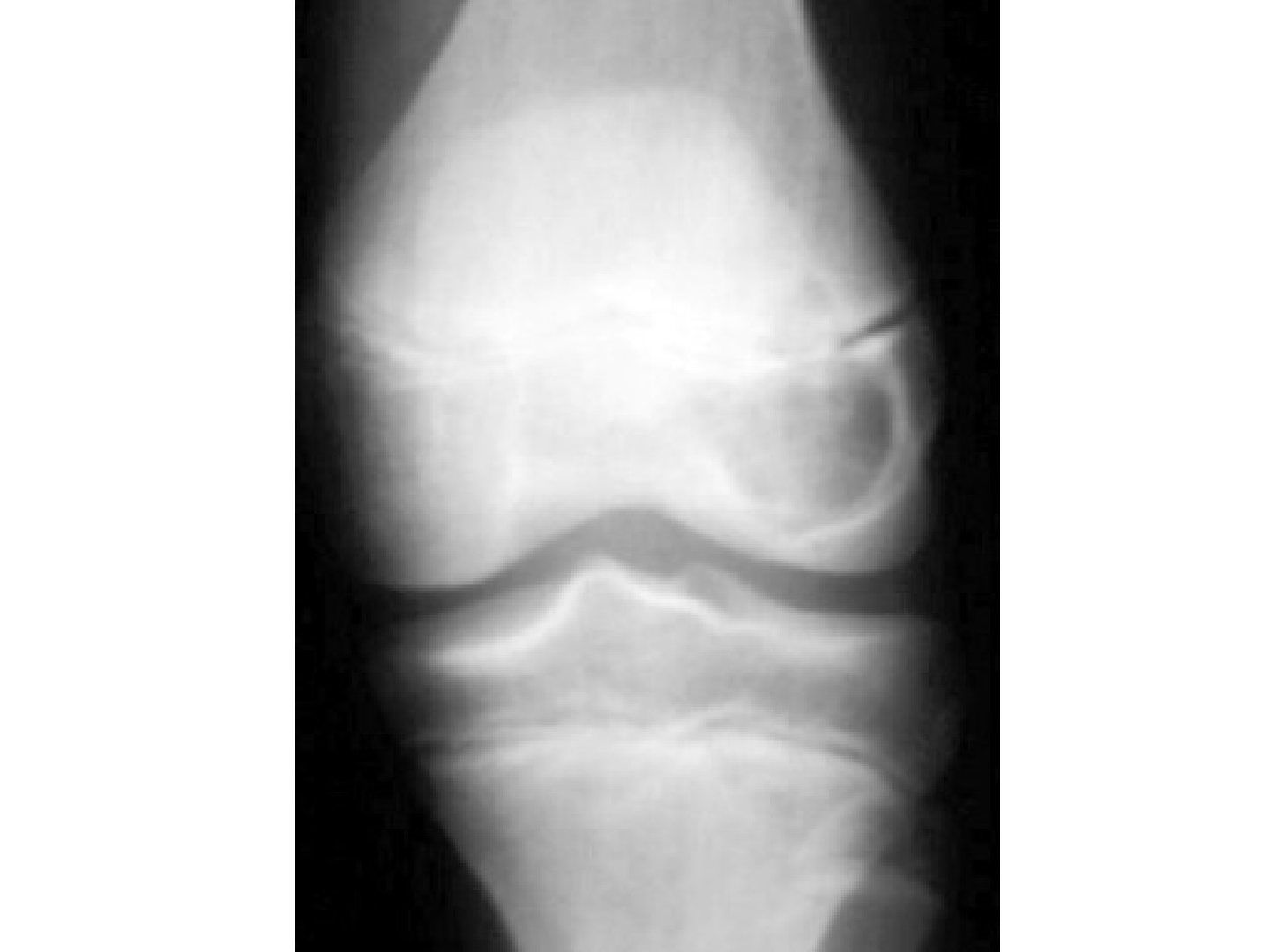
Q1-Q55 questions — tap to reveal all answerslist
- Describe the X-ray findings in chondroblastoma.
- What are the differentials for an epiphyseal lytic lesion?
- What is the natural history of chondroblastoma?
- What is the biopsy appearance and treatment of chondroblastoma?
- What assessment is performed for chondroblastoma?
Q1.Describe the X-ray findings in chondroblastoma.
- Lytic lesion in the epiphysis of a skeletally immature patient
- no obvious matrix seen; narrow transition zone with sclerotic border
- Not expansile; no periosteal reaction; the physis has been breached
Q2.What are the differentials for an epiphyseal lytic lesion?
- Telangiectatic osteosarcoma, infection, metabolic disease
- GCT and clear cell chondrosarcoma in older patients
- Can have a secondary ABC
Q3.What is the natural history of chondroblastoma?
- Majority <25 years, common around 12
- Usually epiphyseal, can cross the physis; most common around the knee, also proximal humerus/femur
- <1% benign metastasis to lung; CXR shows lung mets in 2% (per notes)
Q4.What is the biopsy appearance and treatment of chondroblastoma?
- Biopsy: cobblestone, chickenwire appearance, scattered multinucleated giant cells, chondroid matrix
- Intralesional curettage +/- adjuvant therapy (phenol or cryotherapy); 10-15% recurrence
- Excision in patients with lung metastasis
Q5.What assessment is performed for chondroblastoma?
- Hx/PE: pain and tenderness
- CXR for lung mets (2%)
- Blood test; MRI
- Biopsy
▸ Slide 146 · Non ossifying fibromaStatistics · 4 questions expand
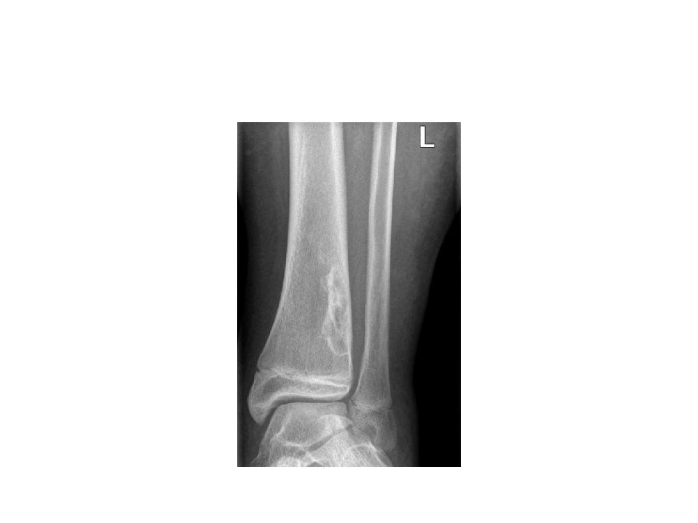
Q1-Q44 questions — tap to reveal all answerslist
- What is a non-ossifying fibroma and who does it affect?
- Describe the X-ray findings in non-ossifying fibroma.
- What conditions are associated with non-ossifying fibroma?
- What is the histology and treatment of non-ossifying fibroma?
Q1.What is a non-ossifying fibroma and who does it affect?
- Most common benign bone tumour in childhood (5-15 years)
- Metaphysis/meta-diaphyseal junction
- Pathogenesis: abnormal osteoclastic resorption at the subperiosteal level during metaphyseal remodelling
Q2.Describe the X-ray findings in non-ossifying fibroma.
- Meta-diaphyseal junction
- Eccentric
- Bubbly lytic lesion surrounded by a sclerotic rim
Q3.What conditions are associated with non-ossifying fibroma?
- ABC, neurofibromatosis
- Jaffe-Campanacci syndrome: multiple NOF, cafe au lait pigmentation, mental retardation, heart/eyes/gonads involvement
Q4.What is the histology and treatment of non-ossifying fibroma?
- Fibroblastic spindle cells in a storiform pattern (whorls of elongated spindle cells)
- Treatment: always conservative
- Ddx: UBC, ABC, fibrous dysplasia, infection
▸ Slide 147 · XR:Statistics · 4 questions expand
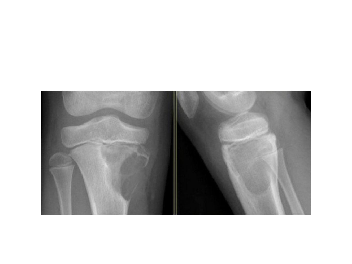
Q1-Q44 questions — tap to reveal all answerslist
- What are the differentials and MRI findings in CMF?
- What is the biopsy appearance of chondromyxoid fibroma?
- What is the management and recurrence rate of CMF?
- Describe the X-ray findings in chondromyxoid fibroma.
Q1.What are the differentials and MRI findings in CMF?
- Ddx: NOF, ABC, chondroblastoma with breaching of the physis
- MRI: T1 hypointense, T2 hyperintense
Q2.What is the biopsy appearance of chondromyxoid fibroma?
- Hypercellular fibrous matrix
- Hypocellular chondroid matrix
- Stellate spindle cells in a myxoid matrix
Q3.What is the management and recurrence rate of CMF?
- Intra-lesional curettage + bone graft/cement
- Recurrence up to 30%
Q4.Describe the X-ray findings in chondromyxoid fibroma.
- Skeletally immature patient
- Lytic lesion at the proximal tibial metaphysis
- Eccentrically located with cortical expansion and destruction
- Narrow zone of transition
- No periosteal reaction or soft tissue swelling
- Dx: CMF
▸ Slide 148 · Clinical photo showing patients nail with partial bluish discolorationStatistics · 5 questions expand

Q1-Q55 questions — tap to reveal all answerslist
- What is a glomus tumour and where does it occur?
- What is the classic clinical triad of a glomus tumour?
- What are the Love and Hildreth tests?
- What imaging and treatment are used for a glomus tumour?
- Describe the clinical findings in this patient with a glomus tumour.
Q1.What is a glomus tumour and where does it occur?
- Benign subungual tumour arising from the glomus body, a perivascular temperature-regulating structure
- Frequently at the tip of a digit or beneath the nail
- Usually young adults 20-40 years; 3/4 in hand, 1/2 in finger, 1/2 with distal phalanx erosion
- Ddx: melanoma
Q2.What is the classic clinical triad of a glomus tumour?
- Paroxysmal pain
- Exquisite tenderness to touch
- Cold intolerance
Q3.What are the Love and Hildreth tests?
- Love test: pinhead -> pain
- Hildreth test: tourniquet -> less pain
Q4.What imaging and treatment are used for a glomus tumour?
- X-ray: pressure erosion; MRI: low T1, high T2
- Marginal excision +/- reconstruct nailbed contour with autogenous fat graft
- Recurrence 20%
Q5.Describe the clinical findings in this patient with a glomus tumour.
- Partial bluish discoloration of the nail
- No nail ridging
- Ddx: melanoma
- Also known as paraganglioma
▸ Slide 149 · melenomaStatistics · 6 questions expand

Q1-Q66 questions — tap to reveal all answerslist
- What is Hutchinson sign and what does it imply in this patient?
- What are the classical ABCDE features of melanoma?
- What are the risk factors for melanoma?
- How is melanoma worked up?
- What are the Breslow and Clark classifications of melanoma?
- What determines the treatment of melanoma?
Q1.What is Hutchinson sign and what does it imply in this patient?
- Subungual discolouration extending to involve the eponychium
- Hutchinson sign - treat as melanoma until proven otherwise
Q2.What are the classical ABCDE features of melanoma?
- Asymmetry
- Border
- Color
- Diameter (<6mm likely benign)
- Elevation/enlargement
Q3.What are the risk factors for melanoma?
- Fair skin, multiple nevi, family history
- Sun exposure, immunosuppression
Q4.How is melanoma worked up?
- Hx: recent change, itch, bleeding
- PE: ulceration, satellite lesions, regional lymph nodes
- Imaging: CT thorax and abdomen to rule out metastasis (lung/liver); USG for lymph nodes
- Dx: incisional biopsy, LN biopsy
Q5.What are the Breslow and Clark classifications of melanoma?
- Breslow: depth in mm - <0.75 / 1.5 / 4 / >4; survival 96% if <0.7mm, 47% if >4mm
- Clark I: epidermis (in situ), no invasion (normal melanocytes at deepest epidermal layer)
- Clark II: papillary dermis; III: papillary dermis up to papillary-reticular interface
- Clark IV: reticular dermis; V: subcutaneous tissue
Q6.What determines the treatment of melanoma?
- Depends on grading, lymph nodes, metastasis, location of lesion
- 1mm -> 1cm resection margin
- >1mm -> 2cm margin + sentinel LN biopsy (+/- radical LN dissection)
- Subungual melanoma -> distal amputation + sentinel LN biopsy
- Positive LN/metastasis: local resection, LN dissection, chemotherapy
- Poor prognostic factors: depth, male, neck/scalp site, ulceration, LN or metastasis
▸ Slide 150 · Mass with +ve tinel signStatistics · 4 questions expand

Q1-Q44 questions — tap to reveal all answerslist
- What is a schwannoma (neurilemma)?
- What are the clinical and MRI findings in a schwannoma?
- What is the histology of a schwannoma?
- What is the treatment of a schwannoma?
Q1.What is a schwannoma (neurilemma)?
- Encapsulated tumour on the surface of a peripheral nerve
- Associated with mutation affecting the NF2 gene
Q2.What are the clinical and MRI findings in a schwannoma?
- Hx: paraesthesia in the distribution of the peripheral nerve
- PE: positive Tinel sign
- MRI: T1 low, T2 high, enhances with contrast; string sign (Ddx: neurofibroma)
Q3.What is the histology of a schwannoma?
- Antoni A: spindle cells arranged in intersecting bundles
- Antoni B: loosely arranged cells
- Verocay bodies are pathognomonic
- Uniform S100 antibody staining
Q4.What is the treatment of a schwannoma?
- Marginal excision with careful dissection
- Excise the mass parallel to the nerve
▸ Slide 151 · osteopetrosisStatistics · 6 questions 2 check expand
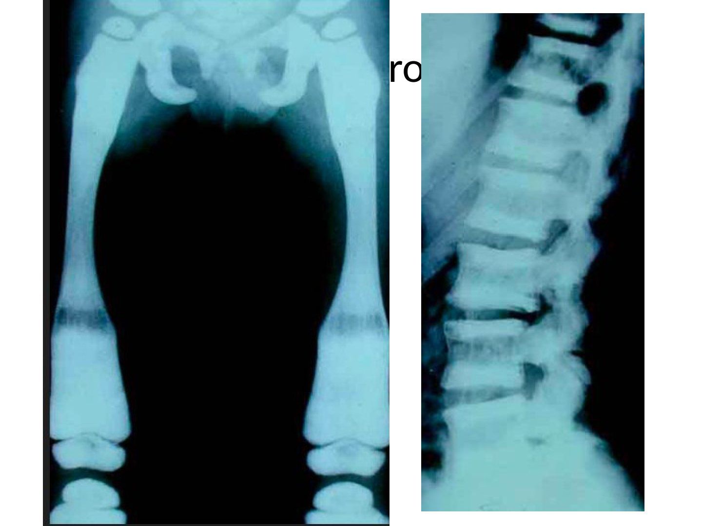
Q1-Q66 questions — tap to reveal all answerslist
- Describe the X-ray findings in osteopetrosis.
- What is the pathophysiology of osteopetrosis?
- What are the orthopaedic and non-orthopaedic features of osteopetrosis?
- What is the treatment of osteopetrosis?
- What is the histology of osteopetrosis?
- What are the types of osteopetrosis?
Q1.Describe the X-ray findings in osteopetrosis.
- Skeletally immature patient with generalised increased bone density
- Canal not well seen; radiolucency at the distal femur
- Erlenmeyer flask appearance over the distal femur
- Dense endplate with rugger jersey appearance
- Wormian bones; Ddx: hypoparathyroidism, pyknodysostosis, lead poisoning
Q2.What is the pathophysiology of osteopetrosis?
- Congenital disease causing dysfunction of the osteoclast
- Defect in proton pump, carbonic anhydrase 2, or chloride channel
Q3.What are the orthopaedic and non-orthopaedic features of osteopetrosis?
- Ortho: usually asymptomatic in AD type except fracture, OA, higher risk of osteomyelitis
- X-ray: coxa vara due to repeated fracture, bone-in-bone appearance, Erlenmeyer flask, rugger jersey spine
- Non ortho: blindness/ deafness in AR type, anemia, cranial nerve dysfunction
- Bloods usually normal (AR type may show increased acid phosphatase/PTH)
Q4.What is the treatment of osteopetrosis?
- AR: bone marrow transplant, high-dose vitamin D
- AD: interferon gamma-1b (interferon gamma -1 beta) (Key et al NEJM 1995)
- Surgery for proximal femur fracture: use a plate (marrow space obliterated)
Q5.What is the histology of osteopetrosis?
- Osteoclasts lack a ruffle border and clear zone
- Empty lacunae
- Plugging of the haversian canals
Q6.What are the types of osteopetrosis?
- Malignant (severe): AR, may be fatal in childhood without transfusion
- Intermediate: AR, can survive to adulthood
- Benign (mild): AD, most common, 2 types
- Type I: no increased fracture risk
- Type II (Albers-Schonberg disease): anaemia, pathological fractures, premature osteoarthritis; first presentation is fracture
Intermediate osteopetrosis is autosomal recessive and is called Albers-Schonberg disease — Misleading / internally contradictory — Albers-Schonberg disease is the autosomal dominant type II (mild) form, as the same slide later states; intermediate osteopetrosis can be AR or AD — source
Autosomal dominant osteopetrosis is treated with interferon gamma-1b (Key et al NEJM 1995) — Misleading - Key 1995 studied severe infantile (recessive) disease — IFN-gamma-1b is FDA approved for severe malignant osteopetrosis (autosomal recessive), often as a bridge to HSCT; evidence in ADO is insufficient and it was poorly tolerated — source
▸ Slide 152 · HyperparathyroidismStatistics · 5 questions expand

Q1-Q55 questions — tap to reveal all answerslist
- What are the X-ray features of hyperparathyroidism?
- What is the clinical presentation of hyperparathyroidism?
- What is a brown tumour?
- What is the histology and management of hyperparathyroidism?
- What is metastatic calcification in hyperparathyroidism?
Q1.What are the X-ray features of hyperparathyroidism?
- Metastatic calcification: nephrocalcinosis, lungs, blood vessels, synovium/chondrocalcinosis, calcinosis cutis
- Subperiosteal resorption: salt and pepper skull, middle phalanx and ulna (increased concavity)
- Brown tumour (osteitis fibrosa cystica): lytic and expansile
- Rugger jersey spine with sclerotic endplates
Q2.What is the clinical presentation of hyperparathyroidism?
- Bones, groans, stones, psychic moans
- PE: signs of CKD (oedema, cafe au lait appearance), vitamin D deficiency, renal transplant scar/RLQ mass
Q3.What is a brown tumour?
- Osteitis fibrosa cystica
- Lytic and expansile; rapid bone loss -> hypervascular granulation -> fibrous tissue
- Histology = GCT
- Other Ddx: metastasis, multiple myeloma, GCT/ABC
Q4.What is the histology and management of hyperparathyroidism?
- Focal area of increased osteoclasts amidst fibrous stroma
- Peripheral rim of osteoid
- Treat the underlying cause (parathyroidectomy / renal replacement therapy)
Q5.What is metastatic calcification in hyperparathyroidism?
- Calcification in normal soft tissue due to a local increase in calcium -> calcium phosphate deposition
- Sites: nephrocalcinosis, lungs, blood vessels, synovium/chondrocalcinosis, calcinosis cutis
▸ Slide 153 · Renal osteodystrophyStatistics · 4 questions expand
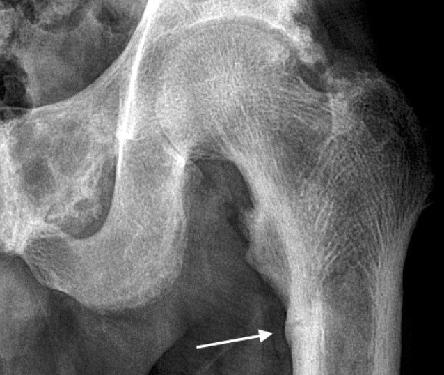
Q1-Q44 questions — tap to reveal all answerslist
- What is renal osteodystrophy and what are the XR findings?
- What is the pathophysiology of low-turnover renal osteodystrophy?
- What other bone disease occurs in chronic renal failure?
- What is the pathophysiology of high-turnover renal osteodystrophy?
Q1.What is renal osteodystrophy and what are the XR findings?
- Failure of bone mineralisation due to chronic renal failure
- XR: brown tumour and looser zone at the compression side
Q2.What is the pathophysiology of low-turnover renal osteodystrophy?
- Aluminium toxicity
- Decreases PTH release
- Decreases osteoblast differentiation and proliferation
- Decreased mineralisation -> osteomalacia (adynamic bone secondary to Al toxicity)
Q3.What other bone disease occurs in chronic renal failure?
- Osteoporosis
- Amyloid bone disease: B2 macroglobulin deposits in the juxta-articular region, especially ball-and-socket joints
- XR: bone cyst with sclerotic edge causing erosive arthritis
- Other sites: carpal tunnel syndrome + carpal cyst
Q4.What is the pathophysiology of high-turnover renal osteodystrophy?
- Phosphate retention increases PTH by 3 mechanisms
- 1. Direct decrease in serum calcium, stimulating PTH
- 2. Decrease in renal 1-alpha hydroxylation -> less serum calcium
- 3. Direct increase in synthesis of PTH
- Secondary hyperPTH raises serum Ca, which binds phosphate -> ectopic calcification
▸ Slide 154 · Well circumscribed radioopacityStatistics · 6 questions expand
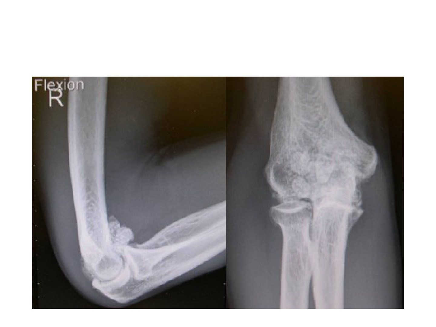
Q1-Q66 questions — tap to reveal all answerslist
- Describe the imaging findings of loose bodies in the elbow.
- What is the clinical presentation and what is it associated with?
- What is the pathogenesis of loose bodies?
- What is the classification of loose bodies?
- What are the phases of synovial osteochondromatosis?
- What are the MRI findings and management of loose bodies?
Q1.Describe the imaging findings of loose bodies in the elbow.
- Well-circumscribed radio-opacity
- Clustered ossification/calcification
- Mainly over the anterior elbow, also around the trochlear groove
- Minimal OA; no varus/valgus malalignment; no stippled calcification
Q2.What is the clinical presentation and what is it associated with?
- Age 30-40 years
- Pain, loose body sensation, locking
- Association: OA, OCD, osteochondroma
Q3.What is the pathogenesis of loose bodies?
- Synovial cell metaplasia into chondrocytes (primary spontaneous; secondary incited by loose bodies)
- Nutrition from synovial fluid
- Cartilage shell: increased nourishment -> increase in size
- Calcified core: decreased nourishment, apoptosis, calcium salt remains
Q4.What is the classification of loose bodies?
- Primary: synovial metaplasia; XR smaller, similar in size
- Secondary: OCD, OA, AVN, osteochondroma; XR larger, varied
Q5.What are the phases of synovial osteochondromatosis?
- Phase 1: active synovitis without loose bodies
- Phase 2: nodular synovitis along with loose bodies
- Phase 3: loose bodies present with resolution of synovitis
Q6.What are the MRI findings and management of loose bodies?
- Cartilage shell: decrease T1, increase T2
- No cartilage shell: decrease in both T1 and T2
- Mx: symptom relief, prevent secondary OA
- Synovectomy + removal of loose bodies
▸ Slide 155 · Soft tissue calcificationStatistics · 7 questions expand
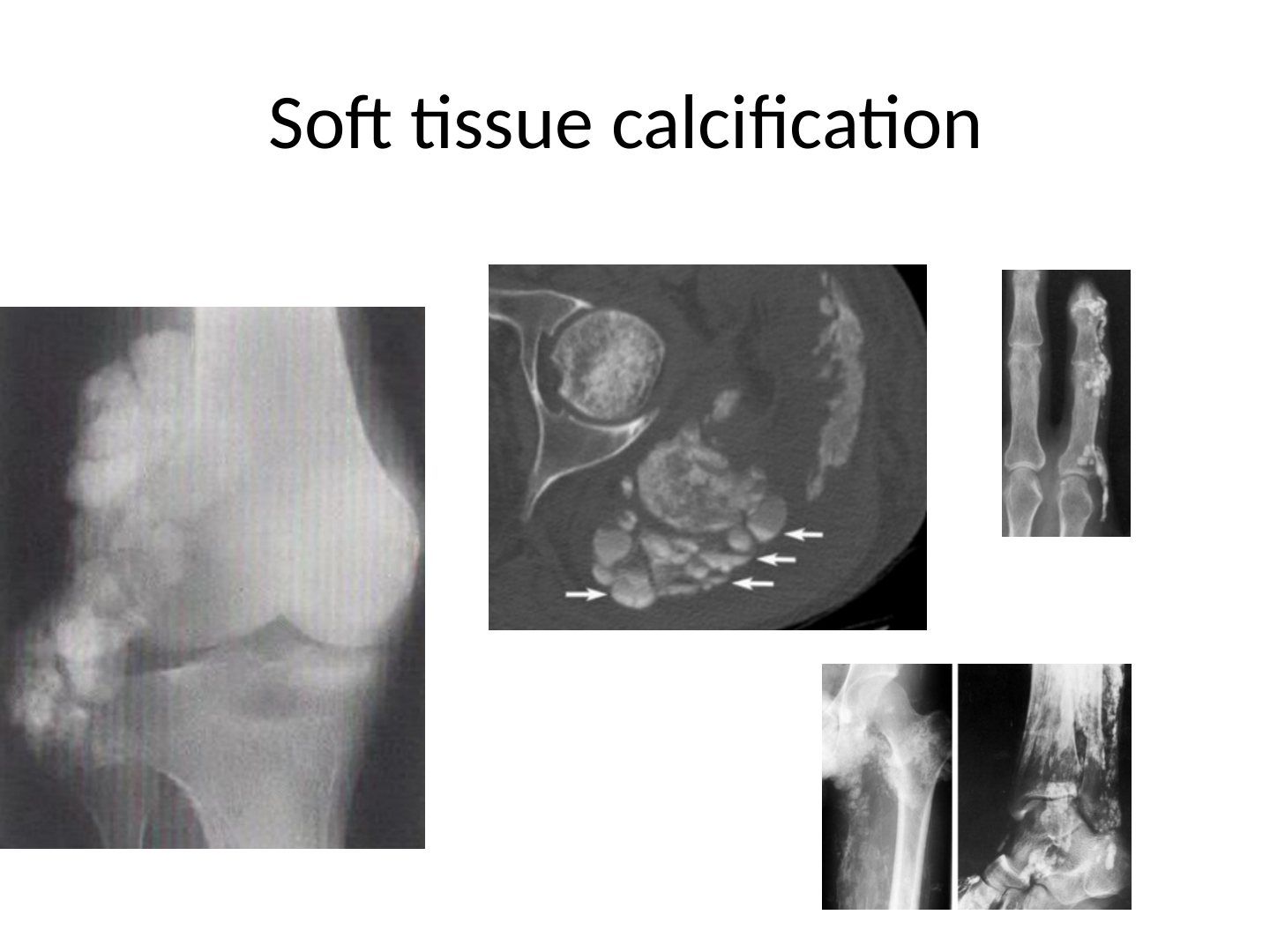
Q1-Q77 questions — tap to reveal all answerslist
- What are the three main categories of soft tissue calcification?
- What are the causes of dystrophic soft tissue calcification?
- What are the types and causes of metastatic soft tissue calcification?
- How is calcinosis classified by extent?
- Where can calcinosis be deposited anatomically?
- What are the radiological features of tumoral calcinosis?
- What is calcinosis circumscripta and how does it present clinically?
Q1.What are the three main categories of soft tissue calcification?
- Dystrophic
- Metastatic
- Calcinosis (cutaneous, subcutaneous, deep connective tissue)
Q2.What are the causes of dystrophic soft tissue calcification?
- Vascular: phlebolith
- Infection: TB
- Tumour: synovial sarcoma, osteosarcoma, chondrosarcoma, synovial chondromatosis, haemangioma
- Haematoma
- Calcific tendinitis
Q3.What are the types and causes of metastatic soft tissue calcification?
- Increased calcium - hyperPTH
- Increased phosphate - CKD
- Gout
- May resemble tumoral calcinosis
Q4.How is calcinosis classified by extent?
- Focal: hand - calcinosis circumscripta; joint - chondrocalcinosis, tumoral calcinosis
- Diffuse: calcinosis universalis, myositis ossificans progressiva
Q5.Where can calcinosis be deposited anatomically?
- Cutaneous
- Subcutaneous
- Deep connective tissue
Q6.What are the radiological features of tumoral calcinosis?
- Amorphous, lobulated
- Chicken-wire lucencies (thin fibrous septae)
- Cobblestone appearance
- Cystic lesions with fluid level = calcium hydroxyapatite precipitation
Q7.What is calcinosis circumscripta and how does it present clinically?
- Well defined subcutaneous nodules
- Ulcerating with chalky hydroxyapatite discharge