▸ Slide 73 · Basic Science - TraumaBasic Science Trauma · 2 questions expand
Q1-Q22 questions — tap to reveal all answerslist
- What does the Basic Science - Trauma topic cover?
- What are the key biomechanical principles of fracture fixation?
Q1.What does the Basic Science - Trauma topic cover?
- Not covered in the speaker notes - the slide only carries the section title
Q2.What are the key biomechanical principles of fracture fixation?
- Not covered in the speaker notes
▸ Slide 74 · ScrewBasic Science Trauma · 5 questions expand
Q1-Q55 questions — tap to reveal all answerslist
- What is a cable and when is it used?
- What materials are used for cables?
- What neurovascular and reduction complications can occur with cables, and how are they prevented?
- What mechanical complications can occur with cables, and how are they prevented?
- Which implants and complications are listed on the slide?
Q1.What is a cable and when is it used?
- Device used to exert a circumferential force
- For long spiral or oblique fractures
Q2.What materials are used for cables?
- Stainless steel / vitallium alloy
- Vitallium alloy = 65% cobalt, 30% chromium, 5% molybdenum
Q3.What neurovascular and reduction complications can occur with cables, and how are they prevented?
- Neurovascular injury - keep the cable passer close to bone
- Loss of reduction intraoperatively or postoperatively - avoid weight bearing / use external bracing
Q4.What mechanical complications can occur with cables, and how are they prevented?
- Loosening of cable - tension each cable before locking
- Iatrogenic fracture - do not over tension, use a plate to support the cable, gentle reduction especially with rotation
Q5.Which implants and complications are listed on the slide?
- Implants: screw, plate, locking plate, DHS, nail, CMN, external fixator
- Considerations: fat embolism, compartment syndrome and removal of screws
▸ Slide 75 · K-wire (Kirschner wire)Basic Science Trauma · 5 questions expand
Q1-Q55 questions — tap to reveal all answerslist
- What are K-wires made of and what tip/end designs exist?
- What are the functions of a K-wire?
- What are the features of a threaded K-wire tip?
- What are the features of a trocar K-wire tip?
- What are the features of a diamond K-wire tip?
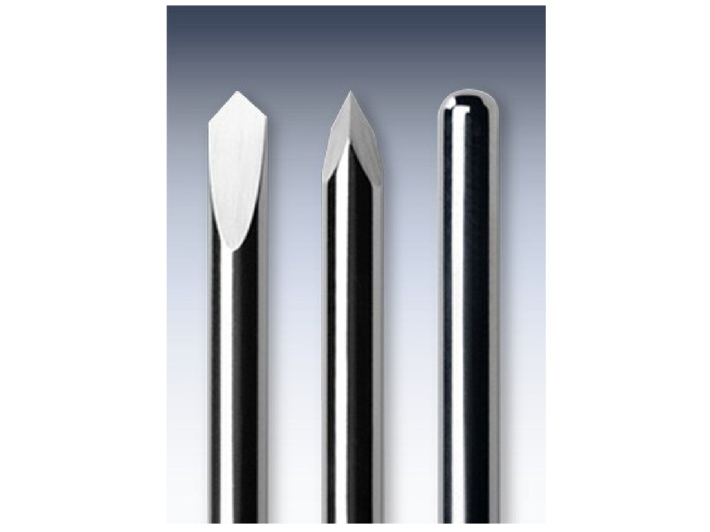
Q1.What are K-wires made of and what tip/end designs exist?
- Materials: stainless steel or nitinol (nickel titanium)
- Tips: threaded or plain
- Ends: trocar / diamond / round
Q2.What are the functions of a K-wire?
- Fracture fixation: IM splintage / temporary reduction
- Used as a guide wire
Q3.What are the features of a threaded K-wire tip?
- Minimises migration
- Higher chance of breakage
- Better holding than a trocar tip
Q4.What are the features of a trocar K-wire tip?
- Worse holding power than threaded
- More head generated than threaded (generates more heat than threaded)
Q5.What are the features of a diamond K-wire tip?
- Creates larger holes with worse circumferential fit and holding to the bone
- Requires less thrust force for bone penetration and generates less heat
▸ Slide 76 · These are screwsBasic Science Trauma · 19 questions 1 check expand
Q1-Q1919 questions — tap to reveal all answerslist
- What is a screw and what is the mechanism of action?
- What are the functions of a screw?
- What are the features of the head of a screw?
- What other head features are described?
- What are the features of the shaft of a screw?
- Define lead and pitch.
- What are the described thread designs?
- State the relationships for screw bending and pullout strength.
- What tip designs are available for screws?
- Compare cortical and cancellous screws.
- What is a lag screw?
- Describe the design of a cannulated screw.
- What are the advantages and disadvantages of a cannulated screw?
- Describe the malleolar screw and its pros and cons.
- What happens when bone is heated during drilling?
- What drill design and drilling technique are recommended?
- What is tapping and what are its pros and cons?
- How can screw head pressure be reduced?
- What factors affect pull out resistance of a screw?
Q1.What is a screw and what is the mechanism of action?
- A device which converts rotational motion into linear motion (torque -> linear force)
Q2.What are the functions of a screw?
- Compression (interfragmentary / plate to bone)
- Positional
- Buttress
- Polar
- Locking bolt / screw
Q3.What are the features of the head of a screw?
- (1) Recess engages the screwdriver: hexagonal / star shape / cruciate / Philips / slotted
- (2) Arrest forward motion
- Threaded (for locking)
Q4.What other head features are described?
- Countersink
- Runoff
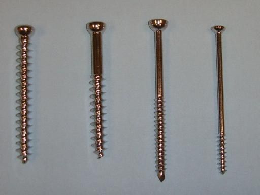
Q5.What are the features of the shaft of a screw?
- A core with a spiral surface wrapping around it
- Thread may be partial or full
- May be cannulated
- Root/crest
Q6.Define lead and pitch.
- Lead = length the screw travels with a 360 degree turn
- Pitch = distance between adjacent threads
Q7.What are the described thread designs?
- V-shaped - with shear + compression force
- Buttress - resists pull out
- Reversed buttress - resists cut out
- Thread angle is also described
Q8.State the relationships for screw bending and pullout strength.
- Bending strength is proportional to inner (minor) radius ^4
- Pullout strength is proportional to outer (major) diameter ^2
- Major (outer) diameter determines resistance to pull out
Q9.What tip designs are available for screws?
- Standard round tipped
- Self tap (fluted)
- Self tap self drill
- Corkscrew
- Trocar
Q10.Compare cortical and cancellous screws.
- Cortical: increased core to thread ratio; fine pitch, more threads engaged in cortex, greater pullout resistance (needs more turns)
- Cancellous: self-tapping corkscrew tip pushes spongy bone aside to thread its way into bone
- Cancellous: increased pitch increases torque needed for insertion; fully or partially threaded
Q11.What is a lag screw?
- A screw that gains purchase only over the far cortex
Q12.Describe the design of a cannulated screw.
- Replaced the malleolar screw
- Low-profile head; Rev-cutting flute (reverse-cutting flute); cancellous thread + self tap; full or half threaded
Q13.What are the advantages and disadvantages of a cannulated screw?
- Advantage: precise insertion
- Disadvantages: decrease bending and torsional rigidity. Decrease pull out strength (core to thread ratio increased as the core accommodates the guide pin)
Q14.Describe the malleolar screw and its pros and cons.
- Trephine tip, smooth shaft and partially threaded
- Advantage: trephine tip means no need for tapping
- Disadvantages: V shaped screw thread profile (poor holding power), large head and diameter
Q15.What happens when bone is heated during drilling?
- Once heat reaches 45 degrees, the mechanical property of bone is altered irreversibly - osteocyte necrosis, ALP deactivation, collagen-HA bone degradation
Q16.What drill design and drilling technique are recommended?
- Drill: sharp, straight, with three flutes at 70 degrees (70deg)
- Technique: clean tip, drill sleeve, start gradually, maintain angle, use water
Q17.What is tapping and what are its pros and cons?
- Tapping: create threads in bone that the screw can enter
- Pros: decreases torque to overcome friction, increases torque for holding +/- axial load -> increases pull out strength; less toggling, more precision
- Cons: takes longer
Q18.How can screw head pressure be reduced?
- Washer
- Countersink
- Insertion through plate
Q19.What factors affect pull out resistance of a screw?
- Screw design: thread diameter (proportional to square of thread diameter) and thread profile
- Technique of placement: number of threads engaged (>5 in cortical bone), bicortical purchase, placement parallel to trabeculae pattern
- Augmentation with PMMA
- Shear load on bone (most important factor)
Bone heated to 45 degrees undergoes irreversible alteration of its mechanical properties — imprecise — The widely accepted threshold for thermal osteonecrosis is 47 degrees Celsius for 1 minute (or 50 degrees for 30 s); 45 degrees is used in some studies only as a conservative safety margin — medium confidence — source
▸ Slide 77Basic Science Trauma · 2 questions expand

Q1-Q22 questions — tap to reveal all answerslist
- What topic does this slide belong to?
- What implants are used in basic science trauma?
Q1.What topic does this slide belong to?
- Not covered in the speaker notes - the slide has no notes or slide content
Q2.What implants are used in basic science trauma?
- Not covered in the speaker notes
▸ Slide 78 · These are biodegradable interference screwsBasic Science Trauma · 10 questions expand
Q1-Q1010 questions — tap to reveal all answerslist
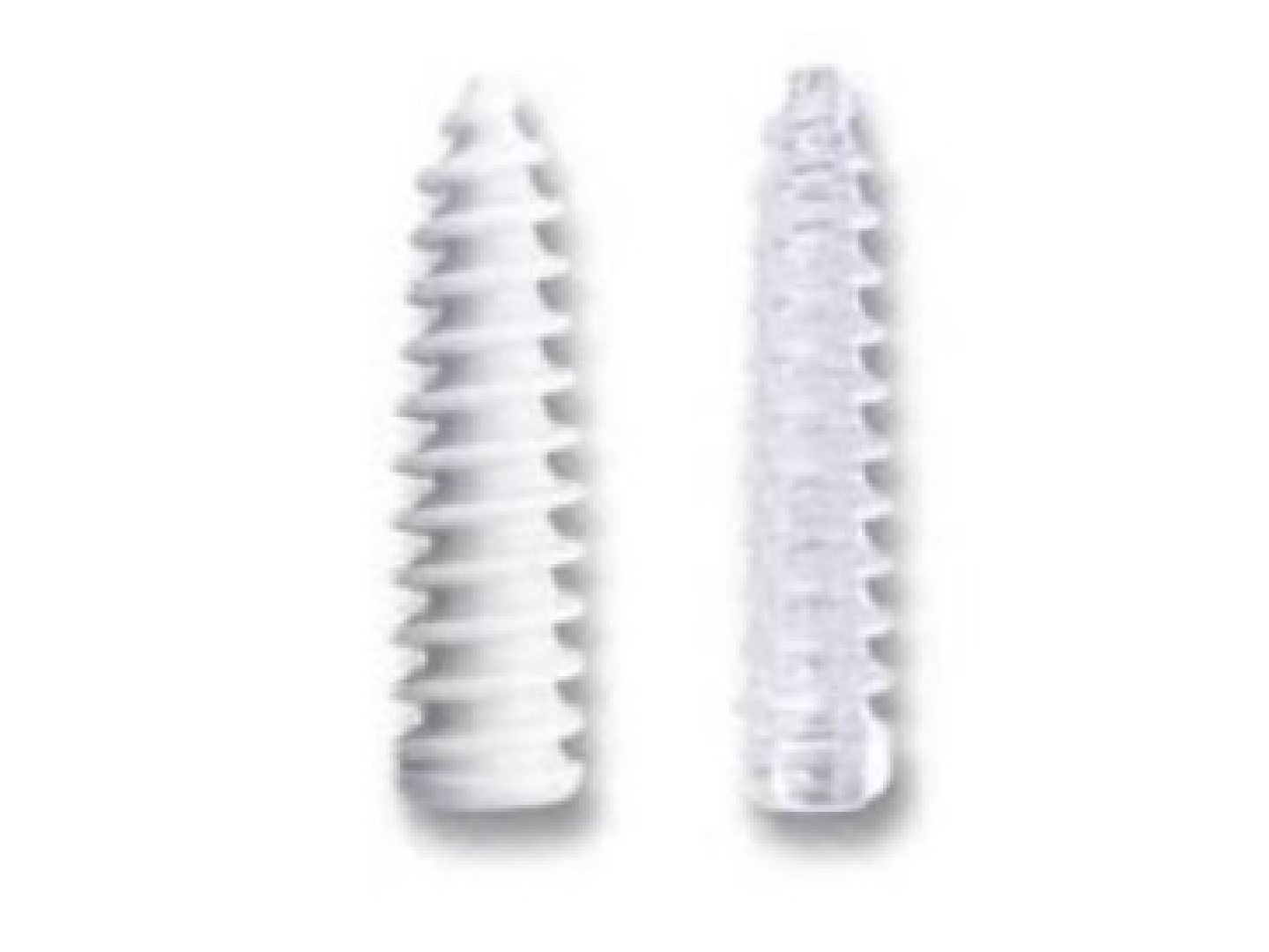
- What is an interference fit and what are biodegradable interference screws made of?
- Compare the properties of PLA and PGA.
- What is the glass transition temperature of PLA and what does it mean?
- What are the advantages of biodegradable interference screws?
- What load-to-failure values were quoted for bioabsorbable screws versus stainless steel?
- What are the disadvantages of biodegradable screws?
- Describe the delayed sterile inflammatory foreign body reaction to biodegradable screws.
- How do biodegradable screws degrade?
- What factors affect the degradation of biodegradable implants?
- What is the composition of Biosure Regenesorb and the role of each component?
Q1.What is an interference fit and what are biodegradable interference screws made of?
- An interference fit is achieved by shaping the two mating parts so one or both slightly deviate in size from the nominal dimension
- Usually made of polylactic acid (PLA) and polyglycolic acid (PGA)
- PLA mainly uses the L isomer as it is more stable
Q2.Compare the properties of PLA and PGA.
- PLA is hydrophobic and more crystallic (crystalline)
- PLA has a longer degradation time (6 months vs 6 weeks for PGA)
Q3.What is the glass transition temperature of PLA and what does it mean?
- 57 degrees
- Below it PLA is rigid and brittle; above it PLA is malleable and rubber-like
Q4.What are the advantages of biodegradable interference screws?
- Load to failure similar to stainless steel
- No need for removal of implant (ROI)
- No artifacts on MRI
- Paediatric fracture - can be used as a transphyseal screw
- Antibiotics eluting; carrier for growth factors (BMP2/7)
Q5.What load-to-failure values were quoted for bioabsorbable screws versus stainless steel?
- PGA/PLLA: 320/300 vs SS 280N (PGA/PLLA 320/300N vs stainless steel 280N)
Q6.What are the disadvantages of biodegradable screws?
- Delayed sterile inflammatory foreign body reaction
- Expensive
Q7.Describe the delayed sterile inflammatory foreign body reaction to biodegradable screws.
- Occurs ~months afterwards, presenting with fluid accumulation / sinus
- Culture negative; X-ray shows osteolysis
- Consider removal if infected or implant failure
Q8.How do biodegradable screws degrade?
- Hydrolysis of ester bonds into small particles, phagocytosed by macrophages and polymorphs
- Polymers to monomers, entering the Krebs cycle to CO2 and H2O
- Lost mechanical property
Q9.What factors affect the degradation of biodegradable implants?
- Crystalline / hydrophobic polymers degrade slower than amorphous / hydrophilic ones (less contact with water)
- Starting molecular weight
Q10.What is the composition of Biosure Regenesorb and the role of each component?
- 65% PLGA
- 15% beta tricalcium phosphate - sustained bone formation
- 20% calcium sulfate - early healing
▸ Slide 79 · Tibial nailBasic Science Trauma · 4 questions expand

Q1-Q44 questions — tap to reveal all answerslist
- Describe the AO UTN tibial nail.
- What are the special features of the AO UTN tibial nail?
- Describe the Stryker GK tibial nail.
- Compare the AO UTN and Stryker GK tibial nails.
Q1.Describe the AO UTN tibial nail.
- Slot: anterior
- Herzog bend: 11 degree, proximal 1/3
- Entry: just distal to the tibial articular surface
- Cross-section: cloverleaf; material: stainless steel
- Proximal: 3 slots - middle dynamic; distal: transverse x2, AP x1
Q2.What are the special features of the AO UTN tibial nail?
- Dovetail proximal - increases torsional rigidity
- Beveled proximal anterior - decreases irritation to the patellar tendon
- Beveled distal posterior - slides smoothly along the dorsal tibial canal, prevents jamming
Q3.Describe the Stryker GK tibial nail.
- Slot: posterior
- Herzog 15 degrees, proximal 1/5
- Entry: just proximal to the tibial tuberosity (TT)
- Cross-section: cloverleaf; material: stainless steel
- Proximal: transverse x1, AP x1; distal: transverse x2
Q4.Compare the AO UTN and Stryker GK tibial nails.
- Slot: AO UTN anterior vs Stryker GK posterior
- Herzog bend: AO UTN 11 degrees (proximal 1/3) vs Stryker GK 15 degrees (proximal 1/5)
- Entry: AO UTN just distal to tibial articular surface vs Stryker GK just proximal to TT
- Both are cloverleaf, stainless steel; AO UTN proximal 3 slots (middle dynamic), distal transverse x2 + AP x1; Stryker GK proximal transverse x1 + AP x1, distal transverse x2
▸ Slide 80 · Describe the nailBasic Science Trauma · 9 questions expand
Q1-Q99 questions — tap to reveal all answerslist
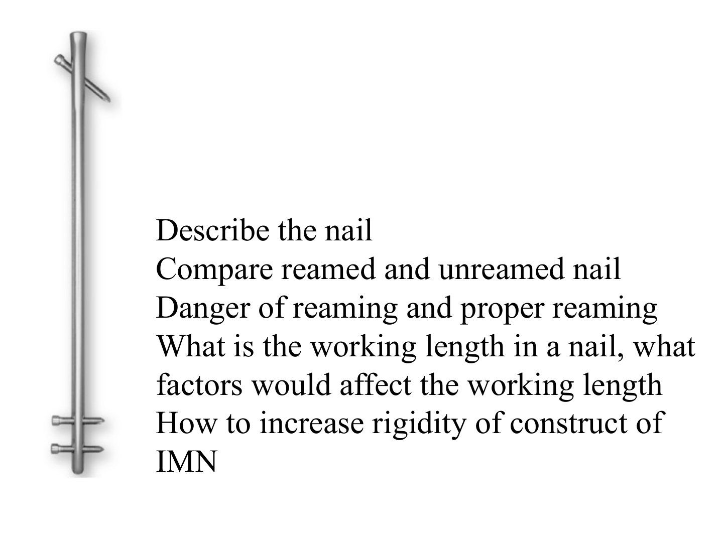
- What is the framework for describing an intramedullary nail?
- What are the pros of reaming?
- What are the dangers/cons of reaming?
- What constitutes proper reaming technique?
- What is the working length in a nail and what factors affect it?
- Compare bending and torsional working length.
- What are the three generations of intramedullary nails?
- What are the advantages of interlocking?
- How can the rigidity of an intramedullary nail construct be increased?
Q1.What is the framework for describing an intramedullary nail?
- Intramedullary nail used for fracture fixation of the femur
- Biological reduction + relative stability and secondary bone healing
- Describe: longitudinal, transverse profile, material, interlocking, reamed/unreamed
- Example given: Stryker GK nail
Q2.What are the pros of reaming?
- Decrease working length
- Increased width of nail = more rigid
- Provides autogenous bone graft
- Decreases hoop stress generated - less fracture and breakage of implant than an unreamed nail
Q3.What are the dangers/cons of reaming?
- Disrupted blood supply
- Thermal necrosis
- Pressure -> fat embolism
Q4.What constitutes proper reaming technique?
- Patient selection
- Equipment: sharp flute with bixcut tip, good clearance by small diameter of flexible shaft keeps pressure low
- Technique: slow advancement, high reaming speed, serial increment, venting
Q5.What is the working length in a nail and what factors affect it?
- Length of implant spanning the fracture that remains unsupported
- Factors: fracture pattern; construct (nail size, bone contact, locking); type of force
Q6.Compare bending and torsional working length.
- Bending working length = unsupported area; bending stiffness inversely proportional to square of working length
- Torsional working length = distance between locking bolts; torsional stiffness inversely proportional to working length
Q7.What are the three generations of intramedullary nails?
- 1st gen: internal splint, minimal rotational stability, close fit (e.g. Kuntscher nail / V nail)
- 2nd gen: locking screw, increased rotational stability (e.g. Russell Taylor nail)
- 3rd gen: anatomical design for a specific bone, increased stability and easier insertion
Q8.What are the advantages of interlocking?
- Increase torsional rigidity
- Increase bending rigidity
- Control length in comminuted fracture
Q9.How can the rigidity of an intramedullary nail construct be increased?
- Reaming to allow a wider nail = more rigid
- Interlocking increases torsional and bending rigidity
- Decrease working length (more bone contact)
▸ Slide 81 · Kuntscher nailBasic Science Trauma · 6 questions expand

Q1-Q66 questions — tap to reveal all answerslist
- Describe the general design of the Kuntscher nail.
- What are the effects of the cloverleaf cross-section of the Kuntscher nail?
- What are the effects of the slot on the Kuntscher nail and where is it placed?
- What is the principle of fixation of the Kuntscher nail?
- What are the advantages and disadvantages of the Kuntscher nail?
- What is the indication for a Kuntscher nail?
Q1.Describe the general design of the Kuntscher nail.
- Stainless steel
- Straight (3-point fixation)
- No interlocking
- Unreamed
Q2.What are the effects of the cloverleaf cross-section of the Kuntscher nail?
- Increases bending stiffness (higher 2nd moment of inertia)
- Increases rotational control of the fracture
Q3.What are the effects of the slot on the Kuntscher nail and where is it placed?
- Radial compliance -> better interference fit
- decrease torsional strength
- Place over the tension side
Q4.What is the principle of fixation of the Kuntscher nail?
- Tight fitting between the elastic nail and stiff bone
- The elastic nail is compressed upon insertion and returns to its original shape, eliminating lateral translation
Q5.What are the advantages and disadvantages of the Kuntscher nail?
- Advantage: increased callus (elastic)
- Disadvantage - slotted: poor torsional strength
- Disadvantage - telescoping: loose axial coupling between nail and bone if not a tight fit
Q6.What is the indication for a Kuntscher nail?
- Transverse / short oblique fracture
- Minimal shortening
- Fracture ends interdigitate - rotation instability
▸ Slide 82 · Centromedullary nailBasic Science Trauma · 5 questions expand
Q1-Q55 questions — tap to reveal all answerslist
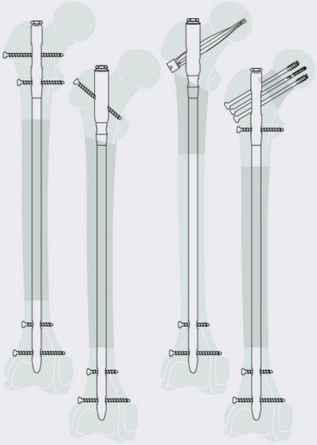
- Describe the Stryker GK centromedullary nail.
- Describe the old AO UFN design.
- Describe the new AO UFN design.
- What is the purpose of the dovetail proximal feature on the AO UFN?
- Compare slotted and non-slotted nails.
Q1.Describe the Stryker GK centromedullary nail.
- Straight nail
- GT entry
- Cloverleaf cross-section, posterior slot
- Proximal: diagonal locking; distal: transverse locking
Q2.Describe the old AO UFN design.
- Slot on the anterior side
- Entry: piriformis entry
- Material: stainless steel
- Anterior curve 1.5m
- Proximal transverse x2; distal transverse x2
- Cross-section: cloverleaf
- Special feature: dovetail proximal
Q3.Describe the new AO UFN design.
- Unslotted
- Entry: piriformis entry
- Material: titanium alloy
- Anterior curve 1.5/2.0m
- Proximal: transverse x2, 130 degree antegrade, spiral blade; distal: transverse x2
- Cross-section: grooved
- Special: solid, unreamed
Q4.What is the purpose of the dovetail proximal feature on the AO UFN?
- Prevents spreading of the proximal end when connected to instruments
Q5.Compare slotted and non-slotted nails.
- Slotted nails allow more flexible insertion (at the site and during insertion)
- Cloverleaf is usually combined with slotting - radial compliance during insertion allows a bigger nail with maximum 3-point endosteal bone contact, decreasing working length -> increasing bending stiffness
- Cons of slotting: poor torsional stiffness (40x), difficult targeting during distal locking
▸ Slide 83 · Cephalomedullary nailBasic Science Trauma · 7 questions expand

Q1-Q77 questions — tap to reveal all answerslist
- What type of nail is the Gamma nail and what is its indication and material?
- Describe the frontal, side and cross-sectional geometry of the Gamma nail.
- What are the locking features and technique for the Gamma nail?
- Describe the design features of the sliding hip screw.
- What is the role of the set screw in a cephalomedullary nail?
- What type of nail is the Russell Taylor nail and what are its general features?
- What are the screw holes and entry technique for the Russell Taylor nail?
Q1.What type of nail is the Gamma nail and what is its indication and material?
- Third generation anatomical nail
- Indication: femur fracture
- Material: titanium
Q2.Describe the frontal, side and cross-sectional geometry of the Gamma nail.
- Frontal view: tapered distal to the lag screw hole (15.5 diameter, allowing a small incision) and also distally
- Frontal view: ML curve 4 degrees
- Side view: anterior curve 3.0mm, radius 1.5m
- Cross-section: unslotted; cannulated
Q3.What are the locking features and technique for the Gamma nail?
- One lag screw proximally
- 2 distal locking transverse screws (oblong x1 for axial dynamization)
- Technique: GT entry, reamed/unreamed
Q4.Describe the design features of the sliding hip screw.
- Load transfer with a large thread area
- Reverse buttress thread to reduce cut out
- Asymm depth of groove (asymmetric depth of groove) -> allows lateral slide only
Q5.What is the role of the set screw in a cephalomedullary nail?
- Decreases sliding hip screw rotation
- Decreases hip screw medialization
Q6.What type of nail is the Russell Taylor nail and what are its general features?
- 2nd generation interlocked nail
- Indication: femur fracture
- Material: stainless steel
- Frontal view: straight nail; side view: anterior curve 2.3m
- Cross-section: unslotted, cannulated, fluted
Q7.What are the screw holes and entry technique for the Russell Taylor nail?
- 2 proximal sliding hip screw holes
- 4 distal locking screw holes
- Technique: piriformis fossa entry
▸ Slide 84 · Rush pin on right, enders nail on leftBasic Science Trauma · 8 questions expand

Q1-Q88 questions — tap to reveal all answerslist
- What are the features of the Enders nail?
- What is the indication for an Enders nail?
- How is an Enders nail inserted in the femur?
- What are the complications of Enders nails?
- What are the features of the Rush pin?
- What is the indication for a Rush pin?
- How is a Rush pin inserted in the femur?
- What are the complications of Rush pins?
Q1.What are the features of the Enders nail?
- Body: flat, C-shaped
- Lead end: blunt and bevelled
- Drive end: flattened eyelet, perpendicular to the curve
Q2.What is the indication for an Enders nail?
- Initially: TOF fracture
- Later: narrow canal diaphyseal fracture
Q3.How is an Enders nail inserted in the femur?
- MFC entry point, through the medial femoral cortex
- To within 1.5cm subchondral to the femoral head
- Entry point roof tile
- Insert >3 nails (3-point fixation)
- Tips fan out in AP and lateral
Q4.What are the complications of Enders nails?
- Fall into bone
- Back out -> impinge on soft tissue
- Joint penetration
Q5.What are the features of the Rush pin?
- Circular and straight
- Lead end: sharp and bevelled
- Drive end: hooked
Q6.What is the indication for a Rush pin?
- Narrow canal diaphyseal fracture
Q7.How is a Rush pin inserted in the femur?
- +/- prebend if 3-point fixation is needed
- Open cortex with a reamer (diameter < nail)
- Entry angle <40 degrees
- Driven into bone by a driver extractor
- Snugly down so the hook grasps the cortex firmly
Q8.What are the complications of Rush pins?
- Iatrogenic fracture
- Fracture distraction
- Loss of reduction
▸ Slide 85 · Plating effect on fracture healingBasic Science Trauma · 3 questions expand
Q1-Q33 questions — tap to reveal all answerslist
- What is the effect of a plate on cortical bone?
- Describe the remodelling sequence under a plate.
- Compare titanium and stainless steel plates for fracture fixation.
Q1.What is the effect of a plate on cortical bone?
- Stress shielding with transient osteopenia under the plate
- Implant contact -> cortical bone blood supply affected
- First: necrosis upon contact
Q2.Describe the remodelling sequence under a plate.
- Second phase: remodelling
- At 1 week (1/52): transient osteopenia under the plate (Ocl)
- Intramembranous ossification from the plate ends towards the centre
Q3.Compare titanium and stainless steel plates for fracture fixation.
- Titanium: less stress shielding, less infection, less stiff, less MRI artifact
- Stainless steel: high yield strength, high ultimate tensile strength, high toughness, cheaper
▸ Slide 86 · D1 post femoral # fixationBasic Science Trauma · 11 questions 1 check expand
Q1-Q1111 questions — tap to reveal all answerslist
- What is the clinical picture and concern on this day 1 post femoral fracture fixation?
- What is the quoted incidence and mortality of fat embolism?
- What is fat embolism syndrome?
- How is fat embolism syndrome diagnosed?
- What are the MAJOR Gurd's criteria and their timing?
- What are the MINOR Gurd's criteria?
- What is the mechanical pathophysiology of fat embolism?
- What is the metabolic pathophysiology of fat embolism?
- What laboratory finding is described in fat embolism?
- How is fat embolism syndrome managed?
- How is fat embolism prevented?
Q1.What is the clinical picture and concern on this day 1 post femoral fracture fixation?
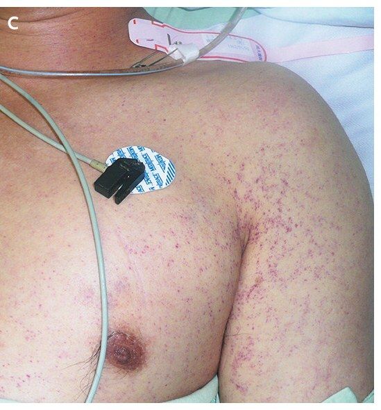
- Clinical photo showing petechiae over the axillary region
- History of recent femoral fracture fixation, likely nailing with reaming done
- Worry: fat embolism
Q2.What is the quoted incidence and mortality of fat embolism?
- 30% of polytrauma patients
- 10% mortality
Q3.What is fat embolism syndrome?
- A systemic inflammatory response to embolised fat
Q4.How is fat embolism syndrome diagnosed?
- Gurd's criteria: 2 major criteria, or 1 major + 4 minor criteria
Q5.What are the MAJOR Gurd's criteria and their timing?
- Cerebral involvement - 6-12 hrs before respiratory signs
- Respiratory compromise - 12-72 hrs
- Petechiae - 24-36 hrs, non-blanchable, over the upper trunk
Q6.What are the MINOR Gurd's criteria?
- Fever
- Tachycardia
- Retinal involvement
- Jaundice
- Renal involvement
- Fat macroglobulinaemia
Q7.What is the mechanical pathophysiology of fat embolism?
- Fat from disrupted bone marrow or adipose tissue is forced into the torn venule in the area of trauma
Q8.What is the metabolic pathophysiology of fat embolism?
- Stress -> fat hydrolysed -> fatty acids
- Fatty acids cause endothelial damage
- Fatty acids are directly toxic to pneumocytes
Q9.What laboratory finding is described in fat embolism?
- Chyle in urine
Q10.How is fat embolism syndrome managed?
- Preventive and supportive
- MV + high PEEP +/- steroid
Q11.How is fat embolism prevented?
- Early long bone stabilisation
- Reaming precautions
Fat embolism occurs in 30% of polytrauma patients with 10% mortality — misleading/outdated — Rates as high as 30% reflect historic fat embolism (fat globules) rather than the clinical syndrome; with early fracture fixation the incidence of fat embolism syndrome is now ~1-11%, while mortality of ~10% remains accepted — medium confidence — source
▸ Slide 87 · Plate is a a load-bearing device that is most effective when placed on the tensiBasic Science Trauma · 8 questions expand

Q1-Q88 questions — tap to reveal all answerslist
- Where is a plate most effective and what is the working length of a plate?
- How are plates classified by function?
- How are plates classified by design?
- How do you exert compression with a plate?
- Describe how compression is exerted by a 'compression plate'.
- What affects the rigidity of a plating construct?
- What are the beam formulas relevant to plate constructs?
- What are the rod formulas relevant to plate constructs?
Q1.Where is a plate most effective and what is the working length of a plate?
- A plate is a load-bearing device most effective when placed on the tension side
- Working length = distance between the two screws closest to the fracture on each end
Q2.How are plates classified by function?
- Neutralization plate - neutralizes bending and rotational force
- Compression plate
- Bridging
- Buttress - placed at 90 degrees against the deforming force
- Tension band
Q3.How are plates classified by design?
- Tubular
- Reconstruction plate
- DCP
- LC DCP
- Locking
Q4.How do you exert compression with a plate?
- Lag screw through plate
- Compression plate
- Prebend plate - screws from central to peripheral for straight/convex bone, and peripheral to central for concave
- Tensioning device
Q5.Describe how compression is exerted by a 'compression plate'.
- One screw inserted in neutral, so plate and bone become one unit
- Hole drilled eccentrically on the other side of the fracture
- Screw inserted, head slides down the edge of the screw hole like an inclined cylinder
- Relative movement of bone on this side towards the fracture; gives ~1mm compression
Q6.What affects the rigidity of a plating construct?
- Material (stainless steel vs titanium)
- SMA (bH3/12)
- Mode of application (principles of plating, quality of reduction, working length)
- Design of plate and screws (locking, design of screw)
Q7.What are the beam formulas relevant to plate constructs?
- Bending rigidity = BH3/12 x Young's modulus
- Deflection (bending strain) proportional to length^3
- Simple terms: plate construct rigidity is proportional to the 3rd power of thickness; the amount of bending is proportional to length^3
Q8.What are the rod formulas relevant to plate constructs?
- Bending rigidity = pi r4/4 x Young's modulus
- Torsional rigidity = pi r4/2 x shear modulus
▸ Slide 88 · These are DCP and LC DCPBasic Science Trauma · 7 questions expand
Q1-Q77 questions — tap to reveal all answerslist
- What are the advantages of the LC DCP?
- What is a locking plate?
- What are the biological and mechanical pros of locking plates?
- What are the cons of locking plates?
- What are the contraindications to locking plates?
- What is the mode of failure of a locking plate?
- How does conventional plating fail compared with LCP?
Q1.What are the advantages of the LC DCP?
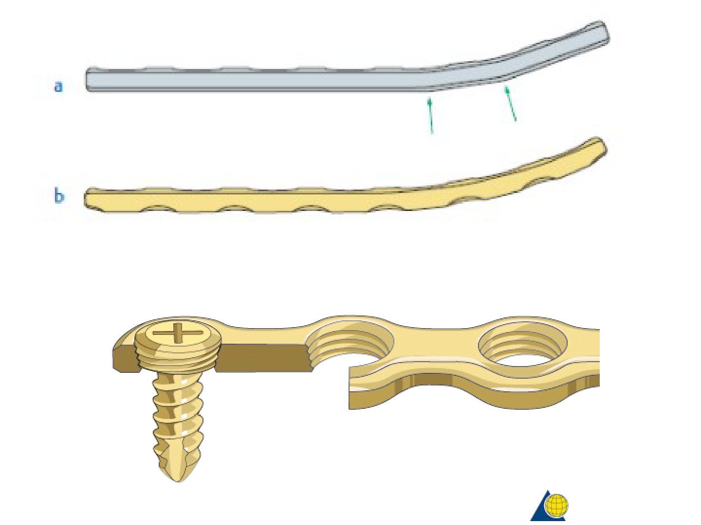
- Less contact (smaller footprint) with bone - preserves blood supply, less necrosis, less bone resorption and stress shielding
- Stiffness evenly distributed (DCP tends to bend at the level of the screw hole)
- Trapezoid cross section leaves an area for bone growth (broad based bone bridge after removal of plate, less prone to refracture)
- 'Combi hole' symmetrical on both sides allows compression in both directions
Q2.What is a locking plate?
- A plate with a threaded screw hole used with screws with a threaded screw head
- Becomes a fixed angle device with angular stability, compared with conventional plating which depends on friction between plate and bone
- Can be used in compression mode, buttress mode, or bridging mode as an internal external fixation
Q3.What are the biological and mechanical pros of locking plates?
- Biologically: less disruption to blood supply, less dissection, can be MISS
- Does not rely on direct contact/friction of the plate to bone
- Mechanically less likely to fail by screw pullout (bending and torsional stress shared amongst the entire single beam construct)
- Larger polar and 2nd moment of inertia when considering the whole construct; fixed angle device especially useful for osteoporotic bone
Q4.What are the cons of locking plates?
- Do not aid reduction
- Technically demanding
- Expensive
- Construct can become too rigid if improperly applied
- Less tactile feedback when inserting screws
Q5.What are the contraindications to locking plates?
- Simple fracture
- Unreduced intra-articular fracture
Q6.What is the mode of failure of a locking plate?
- The shearing/ bending/ torsional load is shared amongst the screws. The implant will fail in one unit rather than sequential failure
- The implant fails as one unit rather than by sequential failure
- LCP is a single beam construct - screws must shear through bone simultaneously
Q7.How does conventional plating fail compared with LCP?
- Conventional plating: when subjected to bending force, strength depends on the pull out strength of the individual screw closest to the fracture site
- LCP: single beam construct, screws must shear through bone simultaneously
▸ Slide 89 · Pedicle screwBasic Science Trauma · 7 questions expand
Q1-Q77 questions — tap to reveal all answerslist
- What design features give a pedicle screw high pull-out resistance?
- What type of screw head does a pedicle screw have?
- Describe the proximal (pedicle) portion of a pedicle screw.
- Describe the distal (vertebral body) portion of a pedicle screw.
- What is meant by a double lead (double start) design in a pedicle screw?
- What is the transition zone between the proximal and distal screw designs?
- What are the handling features of a pedicle screw?
Q1.What design features give a pedicle screw high pull-out resistance?
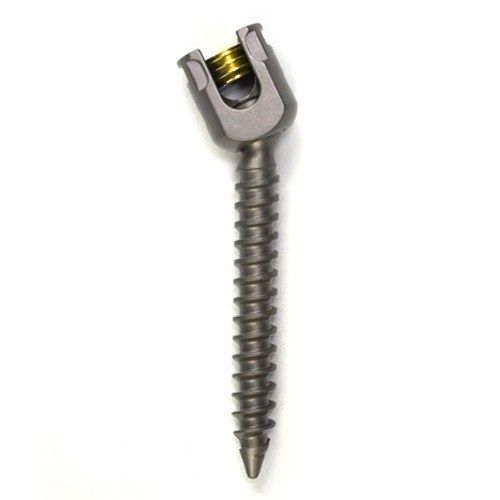
- Dual core design with bone specific anchorage
- Double lead (lead = 2 x pitch)
- Proximal cortical and distal cancellous thread designs
Q2.What type of screw head does a pedicle screw have?
- Polyaxial screw head
Q3.Describe the proximal (pedicle) portion of a pedicle screw.
- Cortical screw design: large core, low and broad thread
- Gives high strength and tight grip in dense cortical bone
Q4.Describe the distal (vertebral body) portion of a pedicle screw.
- Cancellous screw design: small core, high and thin thread
- Large surface area allows even load distribution
Q5.What is meant by a double lead (double start) design in a pedicle screw?
- Double lead (double start) = lead is 2 x pitch
- This contributes to the screw's high pull-out resistance
Q6.What is the transition zone between the proximal and distal screw designs?
- Short transition zones between the two designs
- A smooth link between the cortical and cancellous portions
Q7.What are the handling features of a pedicle screw?
- Self-tapping and self-centering
- Blunt tip without a sharp cutting edge for safe handling
▸ Slide 90 · Ideal working length of a bridge plate = at least twice that of the fracture lenBasic Science Trauma · 1 question expand

Q1-Q11 questions — tap to reveal all answerslist
- What is the ideal working length of a bridge plate?
Q1.What is the ideal working length of a bridge plate?
- At least twice the length of the fracture
- Working length : fracture length ratio of at least 2:1
▸ Slide 91 · Tension band (shifting the fulcrum so the system is shifting from a 1st class leBasic Science Trauma · 4 questions expand

Q1-Q44 questions — tap to reveal all answerslist
- What is a tension band?
- List the prerequisites for a tension band construct.
- Which implants can act as a tension band?
- How does a tension band alter loading across the fracture?
Q1.What is a tension band?
- A device that converts a tensile force into a compression force
- Shifts the fulcrum so the system changes from a 1st class lever to a 2nd class lever
Q2.List the prerequisites for a tension band construct.
- Bone must be eccentrically loaded
- Opposite cortex intact to withstand compressive forces
- Fracture pattern must be transverse and simple
- Construct applied on the tension side
- Implant must be able to withstand tensile force
Q3.Which implants can act as a tension band?
- Tension band wiring is only one type of construct
- A plate can also act as a tension band when the criteria are met
Q4.How does a tension band alter loading across the fracture?
- Shifts the neutral axis from the mid axis to the plate-bone junction
- Axial loading produces pure tension in the plate and pure compression across the fracture line
- This bending configuration is used to produce bending close situation to reduce the working length of the plate
▸ Slide 92 · This is a knee spanning external fixatorBasic Science Trauma · 11 questions expand
Q1-Q1111 questions — tap to reveal all answerslist
- What is an external fixator and what are its advantages and disadvantages?
- What is the goal of external fixation?
- What are the four ways to improve the stability of an external fixation?
- How can pins improve the stability of an external fixator?
- Explain the near-near far-far principle.
- How can rods improve the stability of an external fixator?
- How does the construct improve the stability of an external fixator?
- How does reduction improve the stability of an external fixator?
- How is stability improved in a ring external fixator?
- What are the ways to decrease infection in external fixation?
- What are the safety corridors for external fixation pins (right side)?
Q1.What is an external fixator and what are its advantages and disadvantages?
- External device stabilising the fracture with pins and rods coupled by clamps
- Can be used for temporary or definitive fixation
- Advantages: minimally invasive, quick application, flexible during construction
- Disadvantages: pin tract infection and neurovascular injury during insertion
Q2.What is the goal of external fixation?
- Create a stable construct with low shear and torque
- High micromotion at the level of the fracture
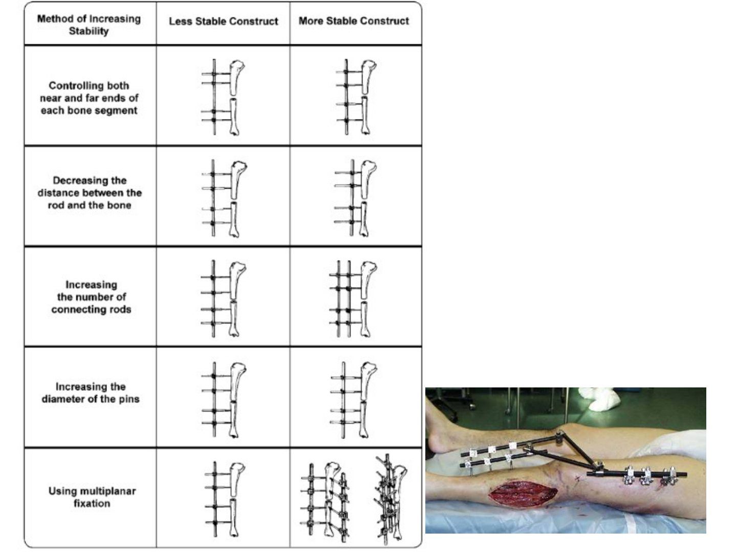
Q3.What are the four ways to improve the stability of an external fixation?
- Pins
- Rods
- Construct
- Reduction
Q4.How can pins improve the stability of an external fixator?
- Place pins close to the fracture (near-near) to decrease working length
- Span a longer length (far-far) to decrease the moment arm of bending force and bending stress
- More pins; larger pins (not to exceed 1/3 of cortical diameter)
- Radial preload decreases micromotion-induced loosening and bone resorption
- Bicortical purchase, Material (HA coated), reduce thermal necrosis (predrill, cooling)
Q5.Explain the near-near far-far principle.
- 2 pins are placed close to the fracture to achieve a short working length and better stability
- 2 far pins over both fragments share out the stress and increase bending and torsional stability
Q6.How can rods improve the stability of an external fixator?
- More rods (increase 2SMA) - double stacking
- Thicker rods (increase 2SMA)
- Rods close to bone decrease the working length of the pins
- Material choice
Q7.How does the construct improve the stability of an external fixator?
- More planes
- More columns
- Ring
Q8.How does reduction improve the stability of an external fixator?
- Reduction is listed as one of the four ways to improve stability of the external fixation
Q9.How is stability improved in a ring external fixator?
- Decrease ring diameter, increase number of rings, decrease ring spacing, inner rings closer to fracture
- Increase wire diameter and pretension (up to 130kg); add wires or half pins
- Wire crossing at 90 degrees to bone surface (at least >30 degrees)
Q10.What are the ways to decrease infection in external fixation?
- Adequate pin tract release
- Pin tract care
- Sharp drill, H2O (cooling)
Q11.What are the safety corridors for external fixation pins (right side)?
- Femur: proximal 7-10 o'clock, distal 7-10 o'clock
- Tibia: proximal 10-11 and 1-3 o'clock, middle 12-3 o'clock, distal 1-3 o'clock
- Humerus: proximal 9-11 o'clock, distal 7-8:30 o'clock (Distal: 7-830 OC)
▸ Slide 93 · Skin tractionBasic Science Trauma · 2 questions expand

Q1-Q22 questions — tap to reveal all answerslist
- What is skin traction?
- What are the indications, weight limits and complications of skin traction?
Q1.What is skin traction?
- Not covered in the speaker notes
Q2.What are the indications, weight limits and complications of skin traction?
- Not covered in the speaker notes
▸ Slide 94 · Skeletal tractionBasic Science Trauma · 3 questions expand

Q1-Q33 questions — tap to reveal all answerslist
- Describe the entry point and trajectory for a distal femoral traction pin.
- Describe the entry point and trajectory for a proximal tibial traction pin.
- What is balanced traction?
Q1.Describe the entry point and trajectory for a distal femoral traction pin.
- Entry just proximal to the MFE (too proximal risks Hunter canal, too distal risks intercondylar notch)
- 2-3 fingerbreadths above the superior pole of the patella with the knee extended
- Trajectory parallel to the knee joint, medial to lateral
- Insert with the knee flexed, then release the ITB to prevent tethering
Q2.Describe the entry point and trajectory for a proximal tibial traction pin.
- Entry 2cm distal and posterior to the tibial tuberosity
- Too proximal enters cancellous bone; too distal risks CPN injury
- Trajectory parallel to the knee joint, lateral to medial
Q3.What is balanced traction?
- Traction in line with the femoral shaft
- Thigh sling just distal to the fracture to decrease extension by gastro, allow proximal fragment to reduce by gravity
- Counterweight is body weight, with a Pearson attachment
- End sling allows assisted knee flexion when prolonged traction is expected; prerequisite is stable callus
▸ Slide 95 · Indication for removal of implantBasic Science Trauma · 4 questions expand
Q1-Q44 questions — tap to reveal all answerslist
- What are the indications for removal of an implant?
- How do you remove a broken screw?
- How do you remove a locking screw cold-welded to a plate?
- How are cannulated and solid nails removed?
Q1.What are the indications for removal of an implant?
- Infection
- Cross joint (implant crossing a joint)
Q2.How do you remove a broken screw?
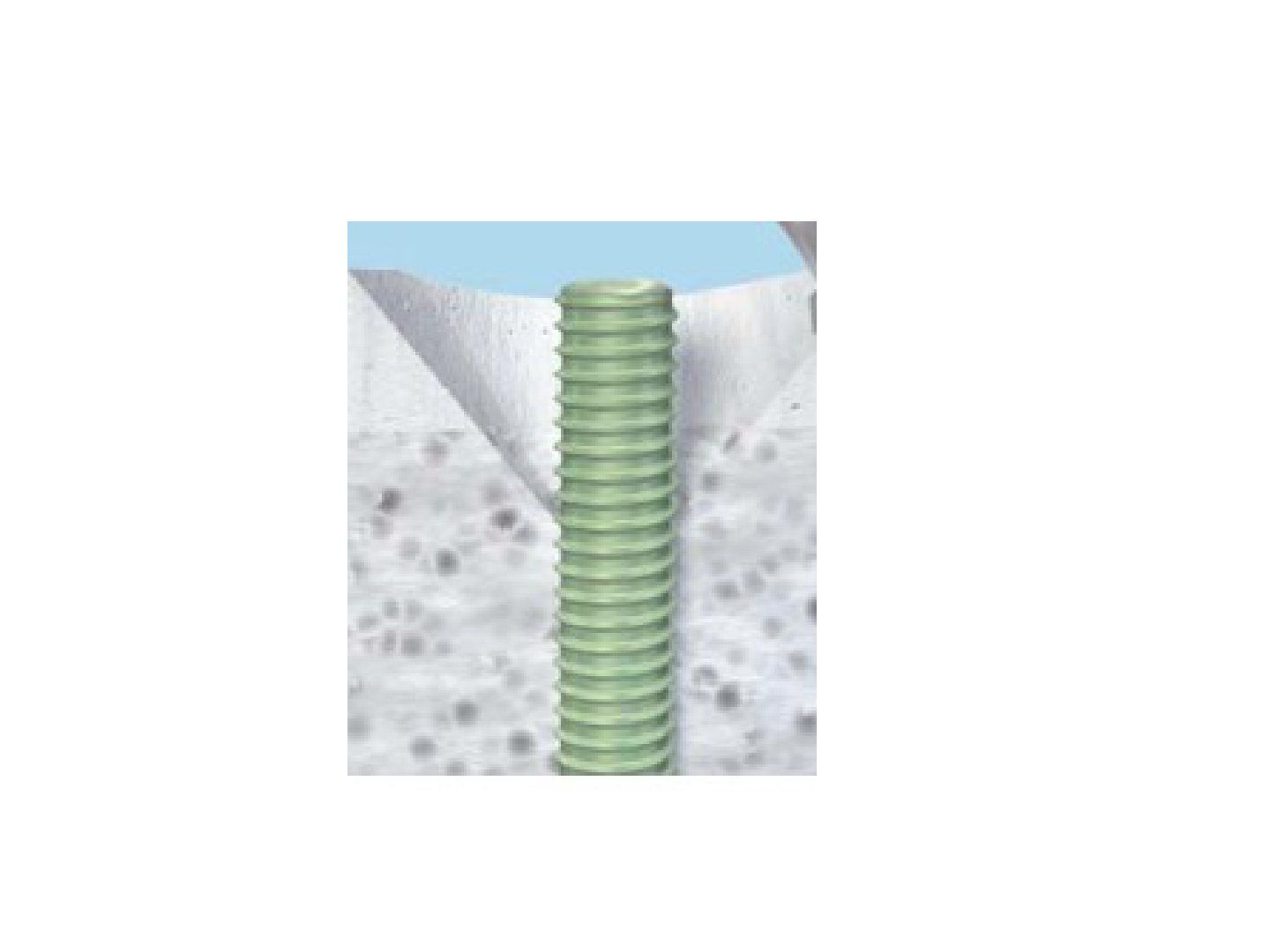
- Vice grip plier if the shaft is exposed
- Hollow reamer with a reverse thread conical extraction device
Q3.How do you remove a locking screw cold-welded to a plate?
- Use a carbide drill
- Separate the head from the shaft
- Separate the plate at the level of the screw
- Separate the plate slightly away from the screw and use the plate as a T handle
Q4.How are cannulated and solid nails removed?
- Cannulated nail: Winquist technique
- Solid nail: antegrade, metaphyseal window or retrograde
▸ Slide 96 · What is compartment syndromeBasic Science Trauma · 10 questions 1 check expand
Q1-Q1010 questions — tap to reveal all answerslist
- What is compartment syndrome?
- What are the first sign and the late signs of compartment syndrome?
- What is the ischaemia time window in compartment syndrome and how is it managed?
- What pressure measurements indicate compartment syndrome?
- Describe the fasciotomy for thigh compartment syndrome.
- Describe the double incision fasciotomy of the leg.
- How many compartments are in the foot and how are they decompressed?
- How is fasciotomy performed in the arm and forearm?
- How many compartments are in the hand and how are they decompressed?
- What is the post-fasciotomy management?
Q1.What is compartment syndrome?
- A state of reduction in perfusion to tissue contained within a confined space
- Can be due to intra-compartmental or extra-compartmental causes
Q2.What are the first sign and the late signs of compartment syndrome?
- First sign: pain disproportional to the condition, exacerbated by passive stretching of muscles in that compartment
- Late signs are the 6 Ps: pain, paresthesia, paralysis, pale, pulselessness, perinsishingly cold
Q3.What is the ischaemia time window in compartment syndrome and how is it managed?
- Orthopaedic emergency - fasciotomy is indicated
- >8 hours of ischaemia causes irreversible skeletal muscle damage and nerve axonotmesis
Q4.What pressure measurements indicate compartment syndrome?
- Within 30mmHg of diastolic pressure, or 33mmHg absolute pressure
- 30mmHg as an absolute cutoff, beyond which nerve will have ischaemia
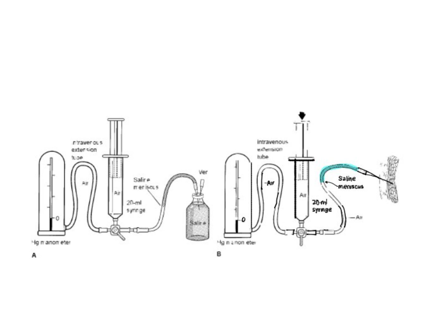
- Whiteside method, indicated when the patient is unconscious
- Need manometer, syringe and water tube connected by a 3-way stopcock; measure at the level of the compartment, look at the meniscus, measure all compartments
Q5.Describe the fasciotomy for thigh compartment syndrome.
- Lateral incision +/- medial (adductor compartment seldom involved)
- Incise the fascia lata to decompress the anterior compartment, then retract vastus lateralis
- Incise the lateral intermuscular septum to decompress the posterior compartment
Q6.Describe the double incision fasciotomy of the leg.
- Medial incision 2cm medial to the tibia decompresses the posterior compartments (saphenous vein and nerve at risk)
- Enter superficial posterior compartment, reflect soleus posteriorly to enter the deep posterior compartment
- Lateral incision midway between tibia and fibula decompresses anterior and lateral compartments (superficial peroneal nerve at risk)
- Look for the anterior intermuscular septum, transverse incision, enter lateral and anterior compartments 1cm anterior and posterior to the septum
- Extensive longitudinal incision of the whole leg length
Q7.How many compartments are in the foot and how are they decompressed?
- 9 compartments: 4 interosseous, lateral, medial, central deep, middle and superficial
- 3 incisions: 2 dorsal along the 2nd and 4th metatarsals, plus one medial
Q8.How is fasciotomy performed in the arm and forearm?
- Arm: lateral incision from the deltoid insertion to the lateral epicondyle
- Forearm: decompress the mobile wad, anterior and posterior compartments
- Anterior wound: lateral proximally to expose the mobile wad, curved medially to allow more skin coverage for median nerve and tendons
- Release the carpal tunnel at wrist level without crossing the skin crease perpendicularly
- Posterior wound: from the lateral epicondyle to the DRUJ
Q9.How many compartments are in the hand and how are they decompressed?
- 10 compartments: 4 dorsal interosseous, 3 volar interosseous, adductor, thenar and hypothenar
- Incisions: hypothenar, thenar, and dorsal 2nd and 4th metacarpals, plus mid-axial blind side of the finger
Q10.What is the post-fasciotomy management?
- Assess muscle viability
- Leave open
- Monitor circulation and rhabdomyolysis
- Plan wound coverage
An absolute compartment pressure of 33mmHg (or 30mmHg) diagnoses compartment syndrome — contested/imprecise — Contemporary guidance uses the delta pressure (diastolic minus compartment pressure) of 30mmHg or less; absolute pressure thresholds alone are not recommended and may cause unnecessary fasciotomies — source
▸ Slide 97 · Classical sequela of untreated acute compartment syndrome developing from advancBasic Science Trauma · 3 questions expand
Q1-Q33 questions — tap to reveal all answerslist
- What is the classical sequela of untreated acute compartment syndrome of the forearm?
- Describe the classical posture of the forearm sequela of compartment syndrome.
- How is this sequela classified and treated (Tsuge classification)?
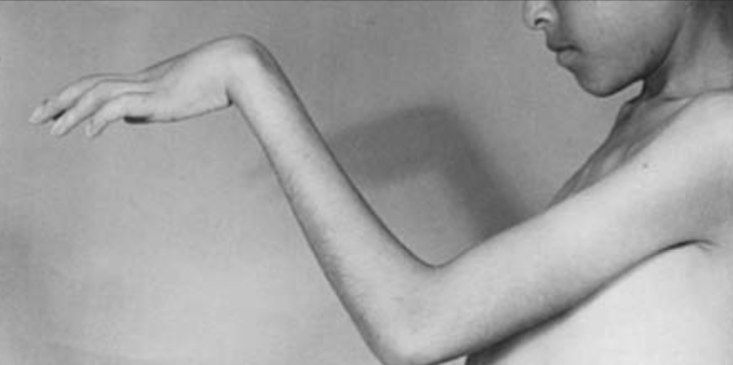
Q1.What is the classical sequela of untreated acute compartment syndrome of the forearm?
- Develops from advanced myonecrosis and muscle fibrosis of the forearm
- FDP and FPL muscles are most vulnerable
Q2.Describe the classical posture of the forearm sequela of compartment syndrome.
- Insensate hand
- Flexed elbow, pronated forearm, flexed wrist, adducted thumb
- Extended MCPJ with flexed IPJ
- Intrinsic minus hand
Q3.How is this sequela classified and treated (Tsuge classification)?
- Mild (finger flexor): dynamic splint, tendon lengthening
- Moderate (finger and wrist flexor): excision of necrotic muscle, neurolysis, tendon transfer (BR to FPL, ECRL to FDP)
- Severe (extensor): free muscle transfer
▸ Slide 98 · DHSBasic Science Trauma · 7 questions expand
Q1-Q77 questions — tap to reveal all answerslist
- What is a DHS and when is it used?
- Describe the components and key dimensions of a DHS.
- Why is the DHS plate screw angle 135 degrees?
- What are the contraindications to a DHS?
- What factors affect sliding of the DHS?
- What are the 4 modes of failure of a DHS?
- What are the advantages of a cephalomedullary nail and the registry evidence?
Q1.What is a DHS and when is it used?
- Sliding screw plate device for fixation of stable trochanteric fractures or fracture neck of femur with a high Pauwels angle
- Material: 316L stainless steel
Q2.Describe the components and key dimensions of a DHS.
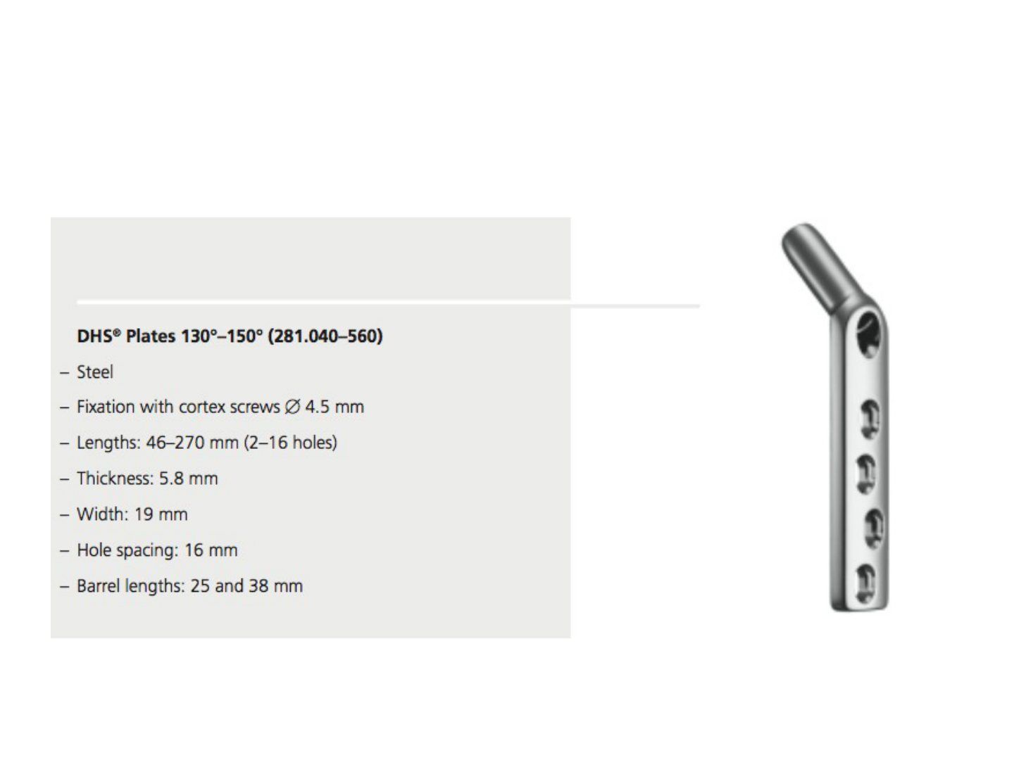
- Lag screw 12.5, 8; long barrel 38mm, short barrel 25mm, thread 22mm
- Minimum sliding 10mm; recommended sliding 25mm (38+22+10=70, so 85mm is the cut-off; Gundel et al. Injury 1995)
- Plate has a staggered array of holes to avoid stress concentration and bone splitting
Q3.Why is the DHS plate screw angle 135 degrees?
- 130 degrees causes too much bending
- 150 degrees is difficult to insert and risks screw cut out
Q4.What are the contraindications to a DHS?
- No posteromedial support
- Reverse oblique fracture pattern
- No lateral buttress
- Subtrochanteric extension
Q5.What factors affect sliding of the DHS?
- Plate barrel angle: decreasing the angle makes it less collinear with the joint reaction force, increasing friction at the screw-barrel junction and the bending moment
- Screw length: a class 1 lever, so more screw protruding from the barrel generates more friction at the barrel, increasing bending and fatigue failure
Q6.What are the 4 modes of failure of a DHS?
- Cutting out of the compression screw from the femoral head
- Pulling off of the side plate from the femoral shaft
- Dissociation of the sliding compression hip screw from the barrel
- Failure of the hip screw itself
Q7.What are the advantages of a cephalomedullary nail and the registry evidence?
- Biological: minimally invasive, does not disrupt the haematoma or rely on reaming graft
- Mechanical: load sharing, more efficient load transfer, prevents uncontrolled collapse, intramedullary buttress for reverse oblique fractures
- Norwegian hip registry (Injury 2013, 2000+ patients): DHS increase reop rate; cephalomedullary nail gives better pain, satisfaction, QoL and mobility
- Long vs short nail: similar outcome but short nail has a higher peri-implant fracture rate
▸ Slide 99 · Dynamic condylar screwBasic Science Trauma · 3 questions expand

Q1-Q33 questions — tap to reveal all answerslist
- What are the features of the dynamic condylar screw?
- What is the principle behind the dynamic condylar screw?
- Why was the dynamic condylar screw abandoned?
Q1.What are the features of the dynamic condylar screw?
- 95 degree plate barrel angle
- 2 proximal round plate holes for independent screws to the calcar (6.5mm cancellous screw)
Q2.What is the principle behind the dynamic condylar screw?
- Tension band plating
- Prerequisite: intact or well reduced calcar acting as a medial buttress
- Need to lag the fracture through the plate for absolute stability
Q3.Why was the dynamic condylar screw abandoned?
- High rate of failure
- Difficult to reduce the calcar and lesser trochanter from a lateral approach
- Non locking, no angular stability
- Calcar malreduction and loss of medial buttress: compression side of the tension band not restored, extra bending moment causing fatigue failure