▸ Slide 56 · ArthritisArthritis · 2 questions expand

Q1-Q22 questions — tap to reveal all answerslist
- What is the named condition on this slide?
- What are the key features of arthritis shown on this slide?
Q1.What is the named condition on this slide?
- Arthritis
- No speaker notes were provided, so further details are not in the notes
Q2.What are the key features of arthritis shown on this slide?
- Not covered in the speaker notes
- Only the slide image is available as a source
▸ Slide 57 · Systemic metabolic disease of monosodium urate crystal depositionArthritis · 8 questions expand

Q1-Q88 questions — tap to reveal all answerslist
- What is the definition and metabolic pathway of gout?
- What is primary gout?
- What is secondary gout and what causes it?
- How is gout diagnosed?
- What is the mechanism of colchicine in gout?
- What is the mechanism of NSAIDs in gout?
- Which drugs inhibit xanthine oxidase in gout?
- What is the role of probenecid in gout?
Q1.What is the definition and metabolic pathway of gout?
- Systemic metabolic disease of monosodium urate crystal deposition
- Purine -> hypoxanthine -> xanthine -> uric acid
- Xanthine oxidase acts in the 2nd and 3rd steps
Q2.What is primary gout?
- Idiopathic disorder of nucleic acid metabolism
- Leads to hyperuricaemia and crystal deposition in joints
Q3.What is secondary gout and what causes it?
- Secondary to increased purine production (high metabolic turnover) or decreased excretion
- Causes: psoriasis, haemolytic anaemia, leukaemia, chemotherapy, renal impairment
Q4.How is gout diagnosed?
- Joint aspiration with examination of the fluid
- Strongly negatively birefringent, thin, tapered, needle-shaped intracellular crystals
Q5.What is the mechanism of colchicine in gout?
- Affects microtubule formation
- Inhibits neutrophils
Q6.What is the mechanism of NSAIDs in gout?
- Inhibit COX
- Decrease prostaglandin formation and inflammation
Q7.Which drugs inhibit xanthine oxidase in gout?
- Allopurinol
- Febuxostat
Q8.What is the role of probenecid in gout?
- Probenecid is a uricosuric agent
▸ Slide 58 · Compartment syndromeArthritis · 6 questions expand

Q1-Q66 questions — tap to reveal all answerslist
- Define compartment syndrome and give its causes.
- How is compartment syndrome diagnosed clinically?
- When and how is compartment pressure measured?
- What is the timing and incision for fasciotomy?
- What are the principles of fasciotomy?
- How many compartments are in each region?
Q1.Define compartment syndrome and give its causes.
- Impaired perfusion in an enclosed fascio-osseous compartment due to increased compartmental pressure
- Intracompartmental: haematoma (fracture, bleeding disorder), muscle oedema (crush injury)
- Extracompartmental: tight dressing or cast preventing the compartment from expanding
Q2.How is compartment syndrome diagnosed clinically?
- It is a clinical diagnosis
- Early: pain out of proportion, pain on stretching the compartment
- Late: the 5 Ps
Q3.When and how is compartment pressure measured?
- In unconscious patients or children
- Absolute pressure 30 mmHg, or measured pressure minus DBP <30 mmHg
- Measure all compartments within 5 cm of the fracture
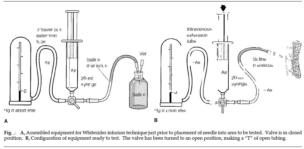
- Commercial kit or the Whiteside method
Q4.What is the timing and incision for fasciotomy?
- <8 hours to prevent irreversible muscle ischaemia
- Delayed fasciotomy is not advisable - high infection rate
- Expansile longitudinal incision
Q5.What are the principles of fasciotomy?
- Longitudinal extensile approach; release all compartments
- Avoid injury to the NV bundle
- Assess muscle viability - colour, contraction, consistency
- Debridement and delayed closure
Q6.How many compartments are in each region?
- Arm 2, forearm 3, thigh 3, leg 4
- Hand 10, foot 9
▸ Slide 59 · Whiteside Method:Arthritis · 4 questions 1 check expand
Q1-Q44 questions — tap to reveal all answerslist
- What is the pathophysiology of compartment syndrome?
- What are the steps of fasciotomy?
- What are the consequences of delaying fasciotomy beyond 12 hours?
- What equipment and technique does the Whiteside method use to measure compartment pressure?
Q1.What is the pathophysiology of compartment syndrome?
- Increased tissue pressure within an enclosed anatomical space
- Causes can be extra-compartmental or intra-compartmental
Q2.What are the steps of fasciotomy?
- Extensile longitudinal full-thickness skin incision
- Release all compartments, protect NV bundles
- Debride necrotic tissue
- Wound coverage at 7-10 days
Q3.What are the consequences of delaying fasciotomy beyond 12 hours?
- Infection 50%
- Amputation 20%
- Supportive care for acute renal failure plus late reconstruction
Q4.What equipment and technique does the Whiteside method use to measure compartment pressure?
- 18-gauge needles and a 20 ml syringe
- The portion of the tube containing the top of the column of saline to be observed is placed carefully at the same level as the tip of the needle in the patient
- Record measurements from the manometer when the saline meniscus is flat - that is, when the pressure in the tissue at the tip of the needle and the pressure in the column of air behind the saline are equal
Delayed fasciotomy beyond 12 hours carries a 50% infection rate and 20% amputation rate — specific figures not supported by current literature — Published rates after fasciotomy are infection up to about 30% and amputation 15-31% in delayed groups; the 50%/20% figures lack a clear source — medium confidence — source
▸ Slide 60Arthritis · 2 questions expand
Q1-Q22 questions — tap to reveal all answerslist
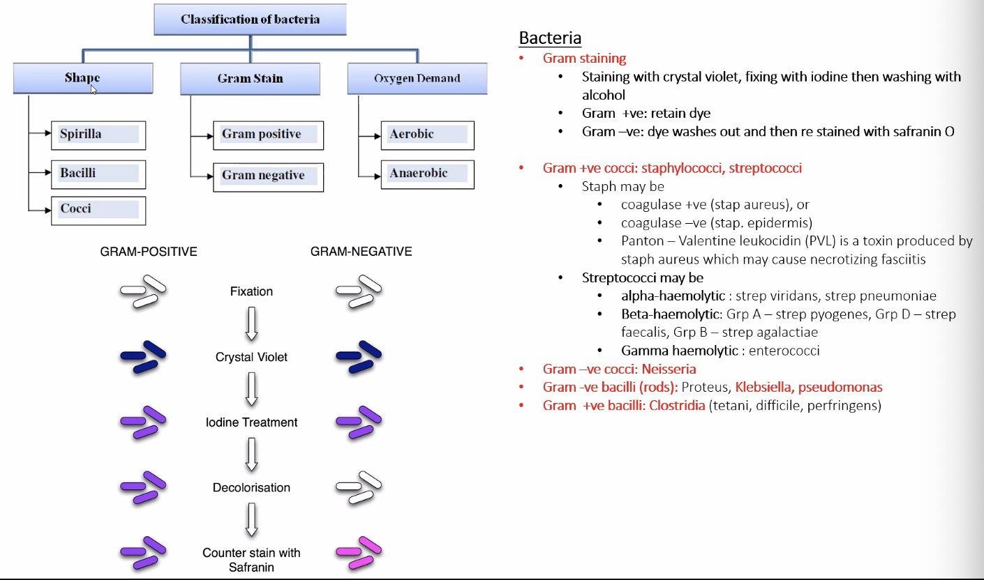
- Which topic area does this untitled slide belong to?
- Why can the content of this slide not be examined in detail?
Q1.Which topic area does this untitled slide belong to?
- Arthritis
- No speaker notes were provided for this slide
Q2.Why can the content of this slide not be examined in detail?
- The speaker notes contain no learning points for this slide
- Only the slide image is available as a source
▸ Slide 61 · BacteriaArthritis · 5 questions expand
Q1-Q55 questions — tap to reveal all answerslist
- List the structures of a bacterium.
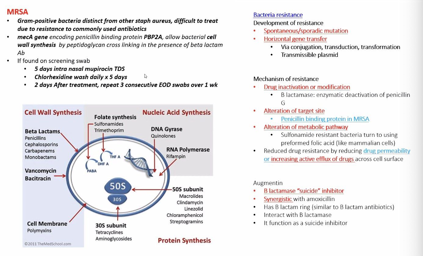
- What is the mechanism of MRSA resistance to beta-lactams?
- When is MRSA classified as community-acquired?
- What are the implications of community-acquired MRSA?
- How is MRSA decolonised?
Q1.List the structures of a bacterium.
- Cell wall, cell membrane
- Pili, flagella
- Plasmid, nucleoid, ribosome
Q2.What is the mechanism of MRSA resistance to beta-lactams?
- MRSA thrives despite penicillin-like (beta-lactam) antibiotics
- Beta-lactams normally prevent bacterial growth by inhibiting cell wall synthesis - inactivating transpeptidases
- Resistance gene mecA, which encodes the Penicillin binding Protein 2 alpha, a tanspeptidase for cell wall synthesis, but has a LOW affinity for β-lactam to bind to; this protein is also known as PBP2a
Q3.When is MRSA classified as community-acquired?
- Picked up within 48 hours of admission
Q4.What are the implications of community-acquired MRSA?
- Contains the PVL locus -> more severe soft tissue infection
- Causes neutrophil lysis
Q5.How is MRSA decolonised?
- Mupirocin 2% applied to the anterior nares 3 times/day for 5 days
- Chlorhexidine 4% shampoo and skin cleanser for 5 days
- Daily change of sheets and clothes
▸ Slide 62 · Theatre designArthritis · 8 questions 1 check expand
Q1-Q88 questions — tap to reveal all answerslist
- What is the aim and zoning of theatre design?
- What defines each theatre zone?
- What temperature, humidity and lighting are used in theatre?
- How is theatre ventilation designed to reduce infection?
- How is theatre air quality monitored?
- What does the literature show for laminar flow?
- How should theatre doors be designed?
- Compare betadine and chlorhexidine skin preparation.
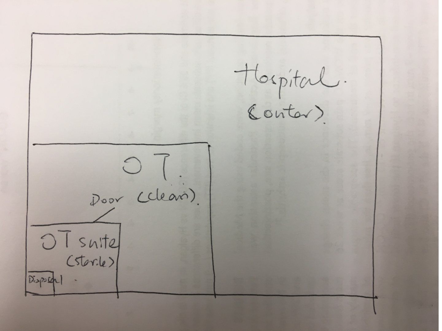
Q1.What is the aim and zoning of theatre design?
- Aim: decrease infection rate and provide optimal conditions for patient and staff
- 4 zones: outer, clean, aseptic, disposal
Q2.What defines each theatre zone?
- Outer zone - the rest of the hospital
- Clean zone - reception to the theatre door
- Aseptic zone - theatre suite
- Disposal zone
Q3.What temperature, humidity and lighting are used in theatre?
- Temperature 22 degrees; radiant warming blankets
- Humidity 40-60%
- Light without shadow, minimum 40000 lux at the incision site, avoid heat
Q4.How is theatre ventilation designed to reduce infection?
- Filter air; positive pressure displaces contaminated air away from the operation site
- HEPA (high efficiency particulate air) filters particles >0.5 microns with 99.97% efficiency
- Laminar flow: air moves at uniform speed and direction - horizontal (more effective in hip than knee arthroplasty), vertical, exponential (less entrainment)
Q5.How is theatre air quality monitored?
- Colony forming units (CFU)/m3 measured by slit sampler (draws 30 L/min for 2 days)
- Sample every 3 months
- <20 CFU/m3 in the theatre periphery; <10 CFU/m3 in the centre
Q6.What does the literature show for laminar flow?
- Charnley 1972: deep infection reduced from 9% to 1.5%
- Lidwell 1982: clean air 1.7%; clean air + perioperative antibiotic 0.4%; unclean air 3.4%
Q7.How should theatre doors be designed?
- Air cutting doors
- Door direction in to out
Q8.Compare betadine and chlorhexidine skin preparation.
- Betadine: inactivated by organic material, skin sensitivity, avoid in pregnancy/breastfeeding; fast onset, bactericidal and works against spores
- Chlorhexidine: less sensitivity, longer duration of action, non-toxic in pregnancy/breastfeeding; not effective against viruses and spores
- Cochrane 2013 suggests use of chlorhexidine
HEPA filters remove particles larger than 0.5 microns with 99.97% efficiency — incorrect particle size threshold — HEPA filters are defined as removing at least 99.97% of particles of 0.3 microns, the most penetrating particle size — source
▸ Slide 63 · Multiple trigger fingerArthritis · 15 questions expand

Q1-Q1515 questions — tap to reveal all answerslist
- What is a tourniquet and what are its contraindications?
- How is a tourniquet applied?
- What inflation pressure and duration are used for a tourniquet?
- What are the complications of tourniquet use?
- What is Bier's block and what are its indications and contraindications?
- How is a Bier's block performed?
- What is the maximum dose of local anaesthetic in a Bier's block?
- What is the mechanism of action of local anaesthetics?
- Compare amide and ester local anaesthetics.
- What is the order of nerve fibre blockade by local anaesthetics?
- What are the properties of lignocaine?
- Which steroid preparation and dose are used for trigger finger/1st CMCJ OA and what are the side effects?
- What is the paediatric dose of midazolam (benzodiazepam)?
- What is the dose of chloral hydrate sedation?
- What aspects of multiple trigger finger does the slide ask about?
Q1.What is a tourniquet and what are its contraindications?
- A temporary constrictive device to occlude blood flow to the extremities and create a bloodless field
- Systemic contraindications: sickle cell anaemia, hypercoagulation
- Local contraindications: skin problems, PVD (peripheral vascular disease), vascular graft
Q2.How is a tourniquet applied?
- Choose a site with no bony prominence; adequate padding
- Width: widest possible but with sufficient distance from the joint (usually the diameter of the limb)
- Length with 3-inch overlap (x1.5 circumference of the limb)
- Tension: 2 fingers fit underneath
Q3.What inflation pressure and duration are used for a tourniquet?
- Upper limb: sBP + 50 mmHg
- Lower limb: sBP + 100 mmHg
- Latest concept: limb occlusion pressure (LOP) - the minimal pressure to completely occlude the artery
- Duration <90 min, maximum 2 hours
Q4.What are the complications of tourniquet use?
- Local: skin, muscle, nerve, vessels
- Systemic: cardiovascular, rhabdomyolysis
Q5.What is Bier's block and what are its indications and contraindications?
- LA injected into the local circulation/perivenously plus a tourniquet to prevent entry into the systemic circulation
- Contraindicated if unable to perform Esmarch, LA allergy, severe HT, compartment syndrome, uncooperative or obese patient
- Good in tenolysis
Q6.How is a Bier's block performed?
- Preparation: emergency trolley, IV access at the contralateral limb, BP/cardiac/SpO2 monitoring
- Double-cuff tourniquet of appropriate size with padding; IV access to the operative limb
- Elevation + Esmarch for exsanguination
- Inflate the proximal cuff to 100 mmHg above sBP, then inject LA slowly
- Change cuff at 30 minutes or when tourniquet pain occurs - inflate distal and deflate proximal
- Deflate after >=20mins, max <90mins
Q7.What is the maximum dose of local anaesthetic in a Bier's block?
- LA maximum 3 mg/kg (with adrenaline 7 mg/kg)
- 1% lignocaine, slow injection, onset <=5 minutes
- Dilution example: 1% lignocaine diluted to 10 ml = 0.5% lignocaine, x4 =40ml; 40 ml 0.5% lignocaine = 200 mg
Q8.What is the mechanism of action of local anaesthetics?
- Most are sodium-gated voltage channel blockers
- Prevent depolarisation of the nerve cell and propagation of the action potential
Q9.Compare amide and ester local anaesthetics.
- Amide: metabolised by the liver, excreted in urine; longer acting (e.g. lignocaine, bupivacaine)
- Ester: metabolised by plasma and tissue fluid cholinesterase, excreted in urine (e.g. cocaine)
Q10.What is the order of nerve fibre blockade by local anaesthetics?
- Small myelinated axons (A delta) - sharp pain
- Unmyelinated axons (C fibres) - dull pain
- Large myelinated axons last
Q11.What are the properties of lignocaine?
- Short-acting drug
- 65% protein bound
- Onset 5-15 minutes
- Duration 40-120 minutes
Q12.Which steroid preparation and dose are used for trigger finger/1st CMCJ OA and what are the side effects?
- Usual preparation: methylprednisolone 20 mg/40 mg per ml
- Trigger finger / 1st CMCJ OA: 20 mg methylprednisolone + 5 mg lignocaine
- Side effects: fat atrophy, pigmentation change, tendon/ligament damage
Q13.What is the paediatric dose of midazolam (benzodiazepam)?
- Child dose 0.05mg/Kg
- No maximum dose but not >5 mg
- Stepwise dosage and monitor SaO2
- Antidote: flumazenil
Q14.What is the dose of chloral hydrate sedation?
- 50mg/Kg 30 minutes before the procedure
- Single dose <1 g in infants, <2 g in children
Q15.What aspects of multiple trigger finger does the slide ask about?
- Initial management?
- Which type of steroid and dosage?
- Side effects of steroid injection?
- What is Bier's block?
- What is the dosage of LA?
- What is a tourniquet - how to apply (indication and CI)?
▸ Slide 64 · DiathermyArthritis · 6 questions expand
Q1-Q66 questions — tap to reveal all answerslist
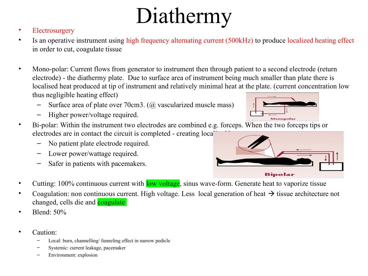
- What is diathermy (electrosurgery)?
- What pad preparation is required before diathermy?
- Where should the diathermy pad be placed?
- How does mono-polar diathermy work?
- How does bi-polar diathermy work and what are its advantages?
- Compare cutting, coagulation and blend diathermy modes and state the cautions.
Q1.What is diathermy (electrosurgery)?
- An operative instrument using high frequency alternating current (500 kHz) to produce a localised heating effect
- Used to cut and coagulate tissue
Q2.What pad preparation is required before diathermy?
- Use a pad of 70 cm2
- Ensure the skin is clean
- Apply conduction gel under the pad
Q3.Where should the diathermy pad be placed?
- Same side as the operative site
- Away from bony prominences
- Away from metal implants
Q4.How does mono-polar diathermy work?
- Current flows from the generator to the instrument, then through the patient to a second (return) electrode - the diathermy plate
- Instrument surface area is much smaller than the plate, so heat is produced at the tip with minimal heat at the plate (low current concentration -> negligible heating effect)
- Plate surface area over 70 cm2 (at vascularised muscle mass)
- Higher power/voltage required
Q5.How does bi-polar diathermy work and what are its advantages?
- Two electrodes are combined within the instrument, e.g. forceps
- The circuit is completed when the two tips/electrodes are in contact - creating localised heat
- No patient plate electrode required
- Lower power/wattage required
- Safer in patients with pacemakers
Q6.Compare cutting, coagulation and blend diathermy modes and state the cautions.
- Cutting: 100% continuous current, low voltage, sinus wave-form - heat vaporises tissue
- Coagulation: non-continuous current, high voltage - less local heat so tissue architecture is not changed; cells die and coagulate
- Blend: 50%
- Local caution: burn, channelling/funnelling effect in narrow pedicle
- Systemic caution: current leakage, pacemaker; environment: explosion
▸ Slide 65 · Plaster of Paris/ dynacastArthritis · 4 questions expand

Q1-Q44 questions — tap to reveal all answerslist
- What is plaster of Paris made of and what does it become when water is added?
- What type of reaction occurs when plaster of Paris sets?
- What question does the slide pose about plaster of Paris?
- What is fibreglass (dynacast) made of and what are its pros and cons?
Q1.What is plaster of Paris made of and what does it become when water is added?
- POP = anhydrated calcium sulphate + cotton fibre
- +H2O -> becomes the crystalline form of gypsum
Q2.What type of reaction occurs when plaster of Paris sets?
- An exothermic reaction - one of the different types of exothermic reaction
- POP sets by crystallisation
Q3.What question does the slide pose about plaster of Paris?
- How long does it need to set?
- The speaker notes record the question but not its answer - details must come from the slide image
Q4.What is fibreglass (dynacast) made of and what are its pros and cons?
- Synthetic polymer reinforced by glass fibres with polyurethane resin
- Pros: light, strong, short drying time, radiolucent
- Cons: expensive, difficult to mould, irritation, sharp turns
▸ Slide 66 · Sickle cell diseaseArthritis · 4 questions expand

Q1-Q44 questions — tap to reveal all answerslist
- What is sickle cell disease and how is it inherited?
- Describe the pathophysiology of sickling in sickle cell disease.
- How is sickle cell disease diagnosed and managed?
- What are the orthopaedic implications of sickle cell disease?
Q1.What is sickle cell disease and how is it inherited?
- A haemoglobinopathy producing HbS
- Autosomal codominant inheritance (slide wording: 'codorminant')
Q2.Describe the pathophysiology of sickling in sickle cell disease.
- Low oxygen tension -> RBC sickling
- Cell membrane damaged -> decreased elasticity -> cannot 'deform' when passing through vessels
- -> occlusion/ischaemia
Q3.How is sickle cell disease diagnosed and managed?
- Dx: haemoglobin electrophoresis
- Prevent infection: vaccination/antibiotics
- Prevent stress: high fluid intake
- Folic acid, pain control, hydroxyurea
- Bone marrow transplant
Q4.What are the orthopaedic implications of sickle cell disease?
- NO TOURNIQUET
- Osteomyelitis: salmonella (spleen damage, encapsulated organism)
- AVN
- Dactylitis
- Shorter life expectancy in general
▸ Slide 67 · Free body diagramArthritis · 9 questions expand

Q1-Q99 questions — tap to reveal all answerslist
- Define force.
- What is a free body diagram?
- What are the assumptions of a free body diagram?
- Define moment.
- Define a lever.
- What are the joint reaction forces in normal walking, running and straight leg raise?
- How can the hip joint reaction force be reduced non-surgically?
- How can the hip joint reaction force be reduced surgically?
- Which joints and applications are listed on the free body diagram slide?
Q1.Define force.
- Load acting on a subject, with magnitude and direction - thus a vector
Q2.What is a free body diagram?
- A diagram to illustrate the various forces acting on an object, e.g. a joint
Q3.What are the assumptions of a free body diagram?
- The system is in equilibrium
- Bones are rigid levers
- Joints are (1) frictionless hinges and (2) joint reaction forces are assumed to be only compressive
- Muscles only act in tension, there is no antagonistic muscle action, and they act along the centre of muscle mass
- Weight of the body is concentrated at the exact centre of body mass (leg in single leg stance)
Q4.Define moment.
- Force applied at a distance from the pivot
Q5.Define a lever.
- A rigid bar that turns about an axis of rotation or fulcrum
Q6.What are the joint reaction forces in normal walking, running and straight leg raise?
- Normal walking = 3 times body weight
- Running = 7 times body weight
- SLR = twice body weight
Q7.How can the hip joint reaction force be reduced non-surgically?
- Decrease weight
- Walk in Trendelenburg position
- Hold a stick on the contralateral side (helps the moment of the abductor)
Q8.How can the hip joint reaction force be reduced surgically?
- Increased offset
- Medialize cup
Q9.Which joints and applications are listed on the free body diagram slide?
- Hip - how to decrease JRF
- Knee - tibiofemoral joint and PFJ
- Ankle - how to decrease ankle joint reaction force after subtalar fusion
- Shoulder - reverse shoulder arthroplasty
▸ Slide 68 · Clinical photo of patients right upper limb showing patches of mottled looking Arthritis · 12 questions expand

Q1-Q1212 questions — tap to reveal all answerslist
- What does this clinical photo show and what is the concern?
- What history is important in suspected necrotising fasciitis?
- How is the patient with necrotising fasciitis and septic shock resuscitated?
- What antibiotics are used for necrotising fasciitis per the IMPACT guidelines?
- What is the role of the LRINEC score in NF?
- What are the operative principles in necrotising fasciitis?
- What is the firebreak theory for debridement in necrotising fasciitis?
- When is amputation considered in necrotising fasciitis?
- What other adjunctive therapy is mentioned for necrotising fasciitis?
- What are the four types of necrotising fasciitis infection?
- Describe the pathophysiology of necrotising fasciitis.
- What are the predictors of mortality in upper limb NF?
Q1.What does this clinical photo show and what is the concern?
- Patches of mottled-looking skin along the right upper limb
- Serosanguinous discharge over the dorsum of the hand
- Early formation of haemorrhagic blisters
- Worry: necrotising fasciitis (NF)
Q2.What history is important in suspected necrotising fasciitis?
- Timing
- Any previous treatment
Q3.How is the patient with necrotising fasciitis and septic shock resuscitated?
- Resuscitate with the sepsis 6 protocol
- Inform ICU for inotropic support
Q4.What antibiotics are used for necrotising fasciitis per the IMPACT guidelines?
- Polymicrobial: imipenem
- Monomicrobial: clinda + linezolid
- Sea (marine): fluoro + amoxi
- +/- IVIG for STSS
Q5.What is the role of the LRINEC score in NF?
- Adjunct if there is diagnostic doubt
- Parameters: WCC, Hb, Cr, Na, Glu, CRP
- >8 is strongly predictive
- Finger probe test if diagnostic doubt
Q6.What are the operative principles in necrotising fasciitis?
- Save life, then limb, infection control
- Urgent surgery for dermatofasciectomy +/- amputation
- Longitudinal extensile incision
Q7.What is the firebreak theory for debridement in necrotising fasciitis?
- Divide skin and fascia into 3 zones
- Skin and subcutaneous tissue: excise zone I and II, leave III
- Fascia: excise zone I, II and III
Q8.When is amputation considered in necrotising fasciitis?
- Systemic factors: poor premorbid, refractory shock (need >1 inotropes)
- Local factors: large area of myonecrosis, extensive involvement especially the plantar region, significant PVD
- Reference: JBJS Tang 2001 HKU
Q9.What other adjunctive therapy is mentioned for necrotising fasciitis?
- Hyperbaric oxygen
Q10.What are the four types of necrotising fasciitis infection?
- I: polymicrobial - at least one anaerobe +/- facultative anaerobes (enterobacteriaceae, non-group A strep)
- II: monomicrobial - group A strep
- III: marine vibrio
- IV: MRSA
Q11.Describe the pathophysiology of necrotising fasciitis.
- Stage 1: spread along fascia
- Stage 2: thrombosis of perforators
- Stage 3: extensive tissue necrosis
Q12.What are the predictors of mortality in upper limb NF?
- Shock
- Involvement of elbow or above
- Platelet count <150
- Creatinine >=1.5x normal
- Deranged LFT
- Overall mortality ~27%
▸ Slide 69 · This is a magnetic resonance imaging scannerArthritis · 11 questions expand

Q1-Q1111 questions — tap to reveal all answerslist
- What are the components of an MRI scanner?
- How does an MRI scanner generate an image?
- Why are hydrogen protons used for MRI?
- What happens to protons when a patient first enters the MRI scanner?
- What happens when the radiofrequency pulse is applied?
- Describe T2 relaxation in MRI.
- Describe T1 relaxation in MRI.
- What is TR and what contrast does it optimise?
- What is TE and what contrast does it optimise?
- What are STIR, FLAIR and MARS used for?
- How does gadolinium contrast work in MRI?
Q1.What are the components of an MRI scanner?
- A superconducting electromagnet made of niobium-titanium, cooled by liquid helium
- A radiofrequency coil system: RF synthesizer, power amplifier and transmitting coil
- Gradient coils - determine the positions of protons in the scanning field
Q2.How does an MRI scanner generate an image?
- The superconducting electromagnet and radiofrequency coil impart energy on hydrogen protons, shifting them to a higher energy level by spinning and wobbling in phase
- Upon removal of the RF, the released energy is converted into a signal represented in an image
Q3.Why are hydrogen protons used for MRI?
- Hydrogen protons are abundant in human tissue, in differing densities in different tissues
- Each hydrogen proton has its own spin and wobble
Q4.What happens to protons when a patient first enters the MRI scanner?
- The vast majority align along the net longitudinal magnetization vector of the superconducting magnet
- The spin is still out of phase
Q5.What happens when the radiofrequency pulse is applied?
- RF imparts energy to lower energy protons, flipping their axis to a higher energy state - half point one way, half the other - cancelling the longitudinal magnetization vector
- The protons precess, so their spins synchronise in phase and the net magnetization vector becomes transverse/horizontal
Q6.Describe T2 relaxation in MRI.
- T2 (spin-spin) relaxation: spins previously in phase become out of phase - loss of transverse magnetization
- Releases energy, converted into an image
- Better for picking up pathology; H2O2 is high signal
Q7.Describe T1 relaxation in MRI.
- T1 (spin-lattice) relaxation: higher energy protons flip back to realign with the longitudinal magnetization vector of the superconducting magnet
- Fat sensitive, used for anatomy
Q8.What is TR and what contrast does it optimise?
- TR = the rate at which the RF is applied
- Short TR optimizes T1 contrast
Q9.What is TE and what contrast does it optimise?
- TE = the frequency taken to listen to the signal
- Long TE optimizes T2 contrast
Q10.What are STIR, FLAIR and MARS used for?
- STIR (short tau inversion recovery) suppresses fat - useful for picking up fluid or oedema in fat-rich tissue (e.g. spinal trauma, PLC)
- FLAIR suppresses water - useful for subtle lesions within the spinal cord by nullifying CSF
- MARS - listed in the notes
Q11.How does gadolinium contrast work in MRI?
- Taken up by tissue with high fluid content
- In T1 fat suppression, the contrast area will show up
▸ Slide 70 · This a bone scanArthritis · 8 questions 1 check expand
Q1-Q88 questions — tap to reveal all answerslist
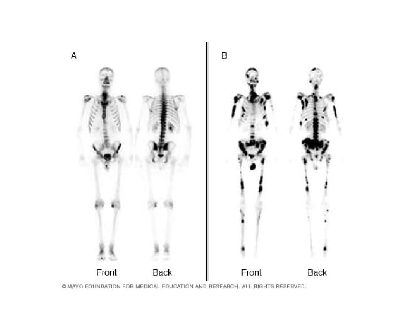
- What is a bone scan and how does it work?
- How is the emitted radiation detected in radionuclide imaging?
- What radioisotope-ligand pairs are used in nuclear imaging?
- What is the half-life and excretion route of technetium-99m, and how is it produced?
- What are the three phases of a bone scan?
- What is SPECT?
- What is PET-CT and how does it work?
- What do the note values T1 63% and T2 37% refer to?
Q1.What is a bone scan and how does it work?
- A type of radionucleotide imaging
- Use of a radioisotope attached to a ligand with affinity to a specific area of interest (e.g. bone), which emits radiation (gamma rays) when it disintegrates
- Technetium-99m bound to phosphate gives a map of blood flow and osteoblast activity
Q2.How is the emitted radiation detected in radionuclide imaging?
- Detected by a scintillation camera with Na iodide
Q3.What radioisotope-ligand pairs are used in nuclear imaging?
- Technetium-99m -> methyl diphosphonate
- Gallium -> transferrin
- Indium-111 -> WCC
Q4.What is the half-life and excretion route of technetium-99m, and how is it produced?
- Molybdenum-99 decays into technetium-99m
- Short half-life of 6 hrs
- Excreted via the kidney
Q5.What are the three phases of a bone scan?
- Vascular phase (1-2 min) - arterial flow and hyperperfusion
- Blood pool phase (3-5 min) - bone and soft tissue hyperaemia
- Static phase (4 hrs) - bone activity
Q6.What is SPECT?
- SPECT = single photon emission CT
- Gamma camera with CT component - multiplanar imaging increases resolution, decreases noise and increases localisation
Q7.What is PET-CT and how does it work?
- PET-CT = positron emission tomography CT
- Exploits the increased metabolic rate of tumours, i.e. glucose consumption
- Deoxyglucose labelled 18-fluorine (half-life 112 mins)
Q8.What do the note values T1 63% and T2 37% refer to?
- T1 63%: recovery of the vertical (longitudinal) axis
- T2 37%: loss of the horizontal (transverse) axis
T2 relaxation is a 37% loss of the horizontal (transverse) magnetization — incorrect — After one T2 time constant the transverse magnetization has decayed TO 37% of its original value, i.e. it has lost 63%; T1 is the time for 63% recovery of longitudinal magnetization — source
▸ Slide 71 · osteoporosisArthritis · 7 questions expand
Q1-Q77 questions — tap to reveal all answerslist
- What is the WHO definition of osteoporosis?
- What is the pathophysiology of osteoporosis?
- What age-related and molecular mechanisms contribute to osteoporosis?
- What is DEXA and how is the patient positioned?
- What causes a false negative DEXA result?
- What causes a false positive DEXA result?
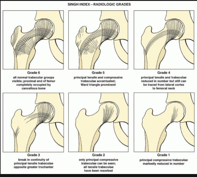
- What is the Singh index?
Q1.What is the WHO definition of osteoporosis?
- Global 3-dimensional reduction in bone mineral density
- T score <-2.5
- Leading to increased susceptibility to fracture
Q2.What is the pathophysiology of osteoporosis?
- Imbalance/uncoupling of bone formation and resorption
- Due to interplay of genetic, intrinsic, exogenous and lifestyle factors
Q3.What age-related and molecular mechanisms contribute to osteoporosis?
- Aging -> loss of gonadal function
- Molecular level: Wnt-catenin signalling pathway (key role in the fate of MSC) -> promotes survival of the osteoblast lineage of cells
Q4.What is DEXA and how is the patient positioned?
- DEXA = dual energy X-ray absorptiometry
- Hip: supine, hip IR 15 degrees
- Spine: supine, hip flexion 90 degrees
Q5.What causes a false negative DEXA result?
- Sclerotic metastases
- Previous surgery with implant
Q6.What causes a false positive DEXA result?
- Osteolytic metastases
- Laminectomy
- Collapse fracture
Q7.What is the Singh index?
- Grade according to the trabecular line
▸ Slide 72 · FRAX scoreArthritis · 3 questions expand
Q1-Q33 questions — tap to reveal all answerslist
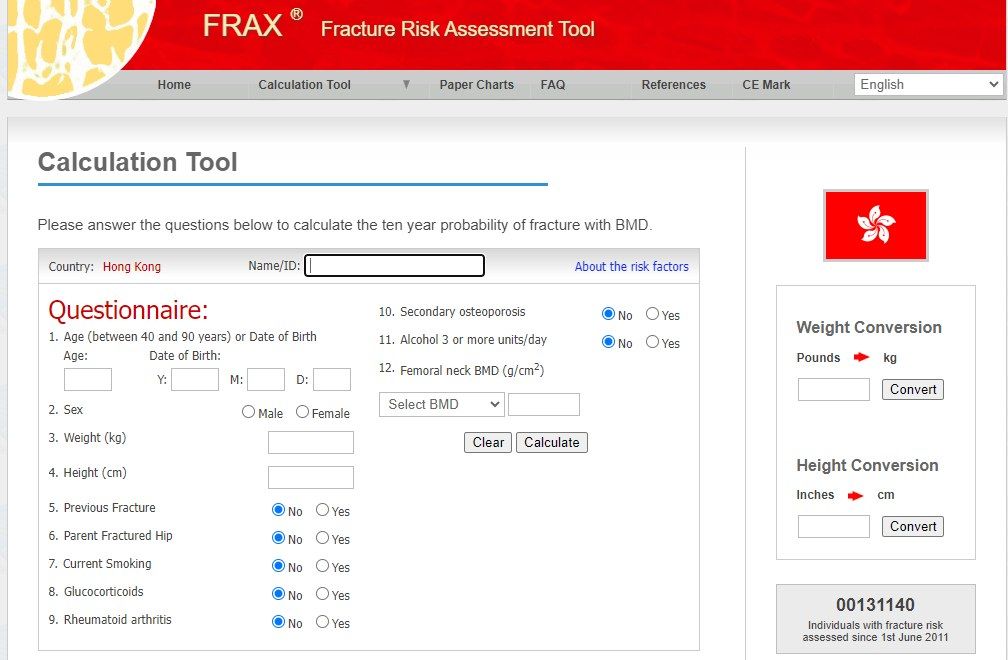
- What is the FRAX score and what is its limitation?
- Which version of the FRAX score is used in this lecture?
- List the risk factors used in FRAX.
Q1.What is the FRAX score and what is its limitation?
- Fracture risk assessment score
- Limitation: not comprehensive
Q2.Which version of the FRAX score is used in this lecture?
- The Hong Kong version
Q3.List the risk factors used in FRAX.
- Age, sex, weight
- Previous fracture; parent fractured hip
- Current smoking; glucocorticoids
- Rheumatoid arthritis; secondary osteoporosis
- Alcohol 3 or more units/day
- Bone mineral density (BMD)