Soft Tissue
Muscle structure and contractile mechanics
Muscle organisation, sarcomere architecture and optimal resting sarcomere length
16 questions
3 source pages
1 images
Images appear with the first question taken from each source page — tap a question to open it.
Q1Starting from the sarcomere, list the levels of organisation of a skeletal muscle.📷▸
- Sacromere > myofibril > muscle fibre (myofibril + SR, endomysium) > muscle fascicle (perimysium) > single muscle (epimysium)
- At the musculotendinous junction fibres run from tendon endotenon to muscle perimysium
- Muscle involvement increases surface area to distribute load transfer
Q2Classify muscles by shape and fascicular structure.▸
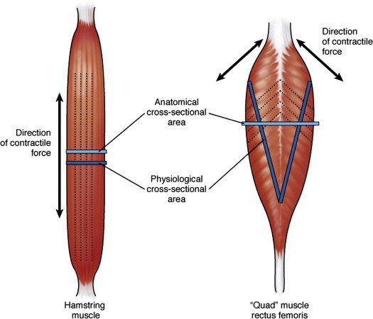
- Parallel: more ROM due to length of fibres, but less total force due to less physiological cross sectional area - strap (sartorius), fan (pec major), fusiform (biceps)
- Pennate: more fibres per cross-sectional area, more power but less ROM - unipennate (EDL), bipennate (rectus femoris), multipennate (deltoid)
- The horizontal component makes the pennate muscle belly solid and compact
Q3What are the types of muscle contraction?▸
- Isometric: force generated but no change in length; adv co-contraction with no shearing, protects joint; disadv limited performance gain
- Isotonic: constant tension with length change - concentric shorten, eccentric lengthen; adv allows joint motion; disadv speed not well controlled, resistance depends on gravity
- Isokinetic: speed constant through full ROM while load changes to maintain velocity; adv maximises strength
Q4Compare open and closed chain exercises.▸
- Closed chain: most distal segment stabilised and not moving; allows muscle co-contraction, generates compression force, more functional
- Open chain: extremity free to move; generates shear force; for training a particular muscle/muscle group
Q5Compare type 1 and type 2 muscle fibres.▸
- Type 1: red, aerobic (Kreb cycle - glycogen + fatty acids -> oxidative phosphorylation -> ATP), less strong, slower, endurance, first to atrophy in deconditioning [slow red ox]
- Type 2: white, anaerobic (ATP-CP, glycolysis producing lactic acid), stronger, fast acting
Q6What is the response of muscle to injury?▸
- Mild: DOMS - oedema and inflammation after unaccustomed eccentric exercise (24-48hrs, increased CK); strain - inflammation at MTJ from eccentric contraction, complication fibrosis
- Atrophy: disuse/neuropathy, affects monoarticular more than biarticular, increased fatigability, fatty infiltration on histology
- Laceration: limited recovery; distal stump denervation -> atrophy -> soft tissue bridge between tendon and muscle belly
- Repair phases: inflammatory (1/7), proliferative (10/7) (satellite cell -> myoblast, fibrin -> scar), remodel (90 days)
- Repair needs blood supply and innervation
Q7How do skeletal, cardiac and smooth muscle differ in nuclei?▸
- Skeletal muscle: multinucleated
- Cardiac: single central nucleus
- Smooth muscle: single central nucleus
Q8According to which criteria are muscles classified?▸
- Shape and fascicular structure
- Myoglobin content
- Bi- or mono-articular
- Orientation of line of pull relative to the joint surface (flexor, extensor, adductor, abductor)
- Type of contractile activity
Q9Describe the bands of the sarcomere.▸
- A band: where actin and myosin overlap (double refractive)
- I band (light): just actin (single refractive)
- H zone: just myosin
- H and I bands decrease with contraction
- M line anchors myosin in a hexagonal lattice (1M=6A)
- Z line anchors actin in a tetragonal lattice
Q10Describe the structure of actin and myosin.▸
- Actin: two chains of beads in a double helix
- Myosin: golf clubs with two heads
Q11Describe the Huxley sliding theory of muscle contraction.▸
- Tropomyosin surrounds actin
- Ca influx from the SR after AP arrival at the T tubules binds troponin C (I inhibitory, T binds troponin to tropomyosin), causing conformational change of troponin-tropomyosin complex
- Exposing the myosin binding site; myosin head attaches forming a cross bridge
- ATP hydrolysis causes S1 myosin head rotation -> power stroke; filaments page (I and H band will decrease)
- ATP binding will break cross bridge and reset myosin heads
Q12Where does the ATP for contraction come from?▸
- Kreb cycle - aerobic: glycogen + fatty acids -> oxidative phosphorylation -> ATP
- ATP-CP - anaerobic, glycolysis which produces lactic acid
Q13What is meant by optimal length of a muscle?▸
- Length allowing the maximal number of cross bridge formation
- No overlap of thin filaments
Q14What is the working range of muscle around optimal length?▸
- 70-130% of optimal length
Q15Give the clinical example of muscle working length given in the lecture.▸
- Abductor tension during THR (total hip replacement)
Q16Which cardiac law is related to the concept of optimal length?▸
- The Frank-Starling Law